IDENTIFICATION OF HOST AND VIRAL FACTORS OF ARTHRITIC ALPHAVIRUS PATHOGENESIS: THE ROLE OF MANNOSE BINDING LECTIN
AND THE VIRAL N-LINKED GLYCANS
Bronwyn Mei Gunn
A dissertation submitted to the faculty of the University of North Carolina at Chapel Hill in partial fulfillment of the requirements for the degree of Doctor of Philosophy in
Department of Microbiology and Immunology.
Chapel Hill 2013
Approved by:
Mark T. Heise, Ph.D. Aravinda deSilva, Ph.D. Ralph S. Baric, Ph.D. Dirk P. Dittmer, Ph.D.
ABSTRACT
BRONWYN MEI GUNN: Identification of host and viral factors of arthritic alphavirus pathogenesis: the role of mannose binding lectin and the viral N-linked glycans
(Under the direction of Mark T. Heise)
To my parents, Michael and Bee Fong,
who have always supported me
with unconditional love and constant encouragement,
even from the other side of the world.
You have given me every opportunity possible,
and I am so proud to be your daughter.
And to my brother Jonathan,
ACKNOWLEDGEMENTS
There are so many people to thank for shaping me into the scientist that I am today. First, I would like to thank my thesis advisor Mark Heise, who has been a truly wonderful mentor. He has taught me so much about virology, pathogenesis, and immunology, but most importantly, he has taught me how to be a rigorous, methodical, independent, and creative scientist. He has always supported and encouraged me, and I cannot thank him enough for his patience, kindness, advice, and guidance throughout my graduate career. I am very proud to be a Heise Lab trainee, and I look forward to many more years of continued mentorship.
Andrew Morgan, and Lance Blevins for their assistance with experiments, ideas, and discussions in lab.
TABLE OF CONTENTS
LIST OF TABLES ... xii
LIST OF FIGURES ... xiii
LIST OF ABBREVIATIONS ... xv
CHAPTER ONE: INTRODUCTION ... 1
1.1. Alphaviruses. ... 1
Alphavirus classification. ... 1
Geographic distribution and epidemics. ... 3
Transmission of alphaviruses. ... 4
Clinical disease and pathology of arthritic alphaviruses: Ross River virus. ... 5
Clinical disease and pathology of arthritic alphaviruses: Chikungunya virus. ... 8
1.2. Molecular biology of alphaviruses. ... 10
Genome organization and structure of alphaviruses. ... 10
Entry of alphaviruses into host cells. ... 12
Roles of the alphavirus nonstructural proteins. ... 14
Synthesis of the alphavirus glycoproteins and viral budding. ... 16
1.3. Alphavirus-induced disease. ... 17
Alphavirus pathogenesis models. ... 17
Encephalitic alphaviruses. ... 18
Arthritic alphavirus pathogenesis: chikungunya virus. ... 21
1.4. The host complement system. ... 22
The role of complement in alphavirus pathogenesis. ... 24
Complement activation pathways. ... 28
The lectin activation pathway of complement. ... 29
Mannose binding lectin (MBL): structure and ligands. ... 30
MBL polymorphisms. ... 32
Role of MBL in sterile inflammatory diseases. ... 33
Role of MBL in viral infection: Flaviviruses. ... 34
Role of MBL in viral infection: other viruses. ... 35
1.5. The viral N-linked glycans. ... 37
Glycosylation is an important post-translational modification. ... 37
N-linked glycosylation. ... 38
Innate immune recognition of N-linked glycans. ... 40
The alphavirus N-linked glycans. ... 41
Role of alphavirus glycans in pathogenesis. ... 43
1.6. Dissertation Objectives. ... 45
CHAPTER TWO: MANNOSE BINDING LECTIN IS REQUIRED FOR ALPHAVIRUS- INDUCED ARTHRITIS/MYOSITIS ... 53
2.1 Summary ... 53
2.2 Introduction ... 54
2.3 Materials and Methods ... 58
2.5 Discussion ... 77
2.6 Acknowledgements ... 81
CHAPTER THREE: ROSS RIVER VIRUS ENVELOPE N-LINKED GLYCANS CONTRIBUTE TO ALPHAVIRUS-INDUCED ARTHRITIS AND MYOSITIS THROUGH ACTIVATION OF THE HOST COMPLEMENT SYSTEM. ... 105
3.1 Summary ... 105
3.2 Introduction ... 106
3.3 Materials and Methods ... 108
3.4 Results ... 115
3.5 Discussion ... 126
3.6 Acknowledgements ... 130
CHAPTER FOUR: DISCUSSION AND FUTURE DIRECTIONS ... 155
4.1 MBL and the RRV E2 N-linked glycans contribute to RRV-induced disease through activation of the host complement system. ... 155
MBL contributes to development of severe RRV-induced disease ... 155
The role of MBL polymorphisms in severity of RRV-induced disease. ... 157
The RRV E2 N-linked glycans contribute to severe RRV-induced disease through activation of the host complement system. ... 160
Glycans on infected cells versus on the virion. ... 162
Other possible ligands that could induce MBL-dependent complement activation following RRV infection. ... 163
4.2 Future Directions ... 164
Role of the complement system in pathogenesis of other arthritic alphaviruses. ... 166
4.3 Conclusions and working model ... 167
APPENDIX: MECHANISMS OF TYPE I IFN INDUCTION IN MYELOID DENDRITIC CELLS BY MAMMALIAN CELL-DERIVED ROSS RIVER VIRUS ... 172
A1.1 Introduction ... 172
A1.2 Materials and Methods ... 175
A1.3 Results ... 178
A1.4 Discussion and Future Directions ... 181
A1.5 Acknowledgements... 185
LIST OF TABLES
LIST OF FIGURES
Figure 1.1: Life cycle of alphaviruses. ... 48 Figure 1.2: The host complement system. ... 50 Figure 2.1: MBL is required for development of severe RRV-induced
disease and tissue damage. ... 82 Figure 2.2: MBL is required for development of severe RRV-induced
pathology and tissue damage within skeletal muscle. ... 84 Figure 2.3: RRV infection induces MBL deposition onto cells ... 86 Figure 2.4: MBL does not bind or neutralize RRV virions. ... 88 Figure 2.5: Complement activation within quadriceps muscle is largely
dependent on MBL. ... 90 Figure 2.6: MBL is required for C3 deposition following RRV-infection. ... 92 Figure 2.7: MBL deficiency does not affect viral replication or tropism
within infected tissues. ... 94 Figure 2.8: MBL deficiency does not affect inflammatory cell recruitment. ... 97 Figure 2.9: MBL deficiency alters expression of inflammatory mediators
within the RRV infected muscle. ... 99 Figure 2.10: Levels of MBL are elevated in RRV patients. ... 101 Figure S2.1: Representative flow cytometry plots and gating scheme
used to characterize inflammatory infiltrates. ... 103 Figure 3.1: The RRV E2 N-linked glycans contribute to MBL deposition
onto infected cells. ... 131 Figure 3.2: Each E2 glycan contributes to MBL deposition onto infected
cells ... 134 Figure 3.3: RRV E2 glycans contribute to MBL deposition onto infected
and tissue damage. ... 141 Figure 3.6: RRV lacking E2 N-linked glycans retain ability to replicate
in vivo ... 143 Figure 3.7: Amounts of RRV genomes in quadriceps muscle are equivalent
at 3 dpi but not 5 dpi between RRV WT and E2 DM. ... 145 Figure 3.8: The RRV E2 glycan mutants and WT RRV induce equivalent
inflammatory responses. ... 147 Figure 3.8: The RRV E2 glycan mutants and WT RRV induce equivalent
inflammatory responses. ... 148 Figure 3.9: The RRV E2 N-linked glycans contribute to MBL deposition
and complement activation in quadriceps muscle. ... 149 Figure 3.10: The RRV E2 glycans contribute to complement deposition
in quadriceps muscle ... 151 Figure 3.11: RRV E2 glycans contribute to expression of complement
dependent pro-inflammatory genes ... 153 Figure 4.1: MBL levels vary between Collaborative Cross founder strains. ... 169 Figure 4.2: Current model of how viral N-linked glycans and MBL
contribute to RRV-induced disease. ... 170 Figure A1.1: Endocytosis of virus is required for type I IFN induction
from BMDCs following mamRRV infection. ... 186 Figure A1.2: TLR2 and TLR4 do not contribute to type I IFN induction
from mDCs in response to mam-RRV. ... 187 Figure A1.3: DC-SIGN and SIGN-R3 are not required for type I IFN
LIST OF ABBREVIATIONS BBB blood brain barrier
BHK baby hamster kidney
BMDC bone marrow derived dendritic cell BSL-3 biosafety level three
CC Collaborative Cross CHIKV Chikungunya virus CL-11 collectin-11
CLR C-type lectin receptor CNS central nervous system CPE cytopathic effect
CRD carbohydrate recognition domain CR3 complement receptor 3
CR4 complement receptor 4 DCs dendritic cells
DENV dengue virus
DF dengue fever
DHF dengue hemorrhagic fever DKO double knockout
DM double mutant dpi days post infection EBD Evans Blue dye
ELISA enzyme linked immunosorbent assay
EM electron microscopy
EMCV encephalomyocarditis virus ER endoplasmic reticulum
fB factor B
FITC fluorescein isothiocyanate GFP green fluorescent protein GlcNAc N-acetylglucosamine
HIV human immunodeficiency virus HS heparan sulfate
HSV-2 herpes simplex virus-2 H&E hemotoxylin and eosin IFN interferon
IHC immunohistochemistry IRI ischemic reperfusion injury kDa kilo-dalton
LCA leukocyte common antigen MAC membrane attack complex Mam-RRV mammalian cell-derived RRV MASPs MBL-associated serine proteases MBL mannose binding lectin
MEFs mouse embryonic fibroblasts
MIF macrophage migration inhibitory factor MOI multiplicity of infection
Mos-RRV mosquito cell-derived RRV NK natural killer
NLR NOD-like receptor
NRAMP natural resistance associated macrophage protein NSAID nonsteroidal anti-inflammatory
NTPase nucleoside triphosphatase ONNV O’nyong nyong virus PFA paraformaldehyde PFU plaque forming units PRR pattern recognition receptor RA rheumatoid arthritis
RLR RIG-I like receptor RRV Ross River virus
RT-PCR real time polymerase chain reaction
SARS-CoV severe acute respiratory syndrome coronavirus SINV Sindbis virus
VEEV Venezuelan equine encephalitis virus WEEV Western equine encephalitis virus WNV West Nile virus
CHAPTER ONE: INTRODUCTION
1.1. Alphaviruses.
Alphavirus classification.
The alphavirus genus is part of the Togaviridae family of enveloped viruses that have a single stranded positive sense RNA genome. The Togavirus family is divided into two groups, the alphaviruses and the rubiviruses, and the family was initially named for their members’ cloak-like appearance under the electron microscope; the word “Toga” is Latin for “cloak”. To date, over forty alphaviruses have been identified, many of which have been associated with human disease, and can be found on all continents of the globe and in a wide range of hosts (78). Rubella virus is the only rubivirus to date, and is an important childhood human pathogen. The two genus of Togaviridae are further defined by their similar genomic organization with the four nonstructural genes at the 5’ end of the genome followed by the structural genes whose expression is driven from a
Of the forty alphaviruses that have been described, twenty-nine cause human disease and are classified into seven distinct antigenic and genetic groups (reviewed in (78, 242)). These groups have been classified based on sequence similarity within the E1 glycoprotein as well as cross-reactivity of sera and members of each complex generally share similar disease characteristics (175). The Semliki Forest (SF) complex includes many of the arthritic alphaviruses such as the namesake Semliki Forest virus (SFV), Ross River virus (RRV), chikungunya virus (CHIKV), and O’nyong nyong virus (ONNV). The VEE and EEE complexes encompasses encephalitic viruses such as Venezuelan equine encephalitis virus (VEEV), eastern equine encephalitis virus (EEEV), and the WEE antigenic complex includes viruses that are associated with both arthritic and encephalitic viruses such as Sindbis virus (SINV), and western equine encephalitis virus (WEEV). Many aspects of alphavirus biology have been elucidated using two prototype alphaviruses: SINV of the WEE antigenic complex and SFV of the SF antigenic complex. While there are some differences between these viruses and the other alphaviruses such as RRV and CHIKV, the basic biology of replication and structure learned in SINV and SFV studies applies to the replication all alphaviruses.
Of note, alphaviruses were originally categorized with the Flaviviridae family of viruses as Group A and B arboviruses, respectively, based upon similarities in
Geographic distribution and epidemics.
Alphaviruses have a global distribution and can be found on all continents. While not all alphaviruses are associated with disease pathology, the ones that do cause disease are roughly separated into two general groups: the Old World and the New World alphaviruses. This grouping is based both on the type of disease associated with each virus and geographic location. The Old World alphaviruses are found mostly in Africa, Asia, and Oceania and tend to cause polyarthritis and arthralgia in humans. New World alphaviruses are found in the Americas and are more closely associated with encephalitis.
The New World viruses include EEEV, WEEV, and VEEV, and cause
encephalitis in humans and horses in various regions around the Americas. EEEV has been found in parts of eastern North America, as well as parts of Central America and is particularly virulent in humans, causing up to 70% mortality in symptomatic cases, and the horse fatality rate is 80-90% (42, 206). WEEV is found in western parts of the North America and South America, and is reportedly aerosol transmitted as well as mosquito-borne and has a 10% human fatality rate (78, 180). VEEV has been identified in the tropical regions of South America and epizootic outbreaks have involved over 75, 000 people (185).
Africa and caused an epidemic of rash and fever involving 2 million people in 1959 (247). RRV is found in Oceania, and is endemic within many areas of Australia and throughout Papua New Guinea and the surrounding islands (86). Outbreaks of RRV-induced disease have occurred on coastal areas of Western Australia, in the southern states of New South Wales and Victoria, as well as within Papua New Guinea, Fiji, and the Cook and
Solomon Islands affecting over 50, 000 people. SINV exhibits a broad geographic distribution and can be found in Northern Europe, Africa, Southeast Asia, and Oceania (78).
The worldwide distribution of alphaviruses that are associated with human disease and the ability to cause periodic and explosive outbreaks involving millions of people highlight the importance of understanding how these viruses cause disease in order to design a vaccine, develop therapeutics, as well as develop appropriate measures to prevent infection and future epidemics.
Transmission of alphaviruses.
Alphaviruses are generally maintained in an enzootic cycle where the viruses transmit between their arthropod vectors and a reservoir host organism, usually birds, small mammals, or primates. The reservoir hosts vary for the different alphaviruses, but generally support high levels of viral replication and viremia that allow for infection of the competent arthropod species. The arthropods, typically mosquitoes, become
and Ae. albopictus, as well as the Anopheles mosquitoes can act as additional vectors during epidemics (reviewed in (242)).
Human disease typically occurs through a spillover effect when humans come into contact with the infected mosquito or arthropod. Seasonal outbreaks of alphavirus-induced disease have been noted and are well documented with RRV in Australia, and alphaviruses can also enter into an urban epidemic cycle where viruses can transmit between humans and the mosquitoes without a reservoir host. This is thought to have occurred in nearly every large-scale epidemic of alphavirus-induced disease.
The factors that affect the emergence of disease to bring on an epidemic can be both environmental and genetic. Climatic events, such as unusually heavy rainfall, can alter the mosquito vector host range and bring epidemic mosquito vectors into endemic regions (240). Genetic changes within the virus genome also play a role in the epidemic potential of the virus. The evolution rate of alphaviruses is approximately 1 x 10⁴
bases/year, which is slower than some other RNA viruses due to the fact that alphaviruses are maintained in two disparate hosts (241). Alphaviruses can acquire changes in the genome that confer an ability to replicate within an additional mosquito vector or allow for more efficient replication within a host organism. Such a change was observed during the most recent CHIKV epidemic where a mutation in the E1 glycoprotein allowed for spread of the virus from an additional mosquito vector, Ae. albopictus (227).
Clinical disease and pathology of arthritic alphaviruses: Ross River virus.
There had been additional epidemics noted prior to 1959 throughout the Northern Territory, Queensland, and Papua New Guinea during the Second World War, but it was not until an epidemic in Murray Valley in Southeast Australia in 1956 that researchers began to suspect that the disease was caused by an arbovirus. CHIKV had recently been associated with an outbreak of acute virus polyarthritis in Tanganyika (Tanzania) in Africa that shared some similarities with the Australian epidemic. The Australian patient sera were assayed for cross-reactivity to other group A arbovirus including SFV and CHIKV. While some cross-reactivity was observed, Shope and Anderson concluding that the outbreak in Australia was due to a yet undescribed group A arbovirus (214). Doherty et al then isolated a virus from a pool of trapped Aedes vigilax mosquitoes by the Ross
River in Northern Queensland, which reacted strongly with patient sera (46). The virus isolated was formally named Ross River virus, and the strain isolated was dubbed T48 (Townsville and mosquito pool 48). T48 is the type strain of RRV and is the strain used in the laboratory to study RRV.
The largest outbreak of RRV occurred in 1979-1980 when RRV spread to islands in the South Pacific, including Fiji, Samoa, and the Cook Islands. An estimated 50, 000 people throughout the region were infected and presented with arthritis and/or arthralgia. RRV was isolated from several patients by several different research groups (1, 191, 225) confirming that the epidemic was associated with RRV. The sudden outbreak of infection in the region is thought to be due to high levels of viremia in many of the affected
Annual seasonal outbreaks of RRV-induced disease occur in Australia, most notably in Queensland, resulting in about 5000 reported cases of RRV-associated polyarthritis each year (86). Incidence of infection correlates with the presence of the mosquito vector and most of the infections reported occur in the late summer-early fall. The reservoir hosts for RRV are thought to be marsupials such as wallabies and
kangaroos as well as fruit bats (86). RRV has been isolated from about 42 different types of mosquito but is transmitted to humans primarily Ae. vigilax and Ae. camptorhynchus on the coast, and Culex annulirostris in the inland areas (194).
The incubation period for RRV in humans is thought to be about 7 to 9 days following infection by mosquito bite (56). Joint pain and arthritis affecting the ankles, wrist, fingers, and knees are the most common symptom in RRV-induced disease in humans, and patients have also reported pain in the back, neck, and elbows (86). Myalgia is another common symptom reported in patients, and about half of infected patients develop a macropapular rash that can cover the torso and limbs. Many patients develop a fever, and experience fatigue and malaise that can last for several weeks to months. Less common symptoms of RRV-disease include headache, splenomegaly, hematuria,
photophobia, and sore throat. The disease symptoms can generally last up to three weeks, with the joint pain and swelling most severe in the first week, and many patients are incapacitated by the debilitating pain and are unable to work during that time.
Furthermore, many patients continue to have chronic joint pain and arthritic symptoms for several months after initial infection (86).
natural killer (NK) cells were found in the synovial fluid, and the NK cells had equivalent cytotoxic activity as those derived from the periphery (91). Activated macrophages within the synovium were observed (32, 57), and subsequent studies in mouse models of RRV-induced disease demonstrate that the activated macrophages play a critical role in development of clinical disease signs (135). In contrast to other arthritic diseases where disease is driven in part by immune complexes with antigen, the presence of immune complexes was not observed within the synovium of affected joints from RRV-infected patients (57, 58).
Clinical disease and pathology of arthritic alphaviruses: Chikungunya virus.
Chikungunya virus (CHIKV) was first isolated from a patient in Tanganyika, which is present day Tanzania, in the early 1950s (188), however it is very likely that the epidemics of disease had occurred prior to its identification, and were mistakenly
attributed to dengue virus (29). The word “chikungunya” means “that which bends up” in the Makonde language, and is thought to describe the debilitating symptoms associated with virus infection (188).
events occurred in the areas surrounding the Indian Ocean such as India, Sri Lanka, and many countries in Southeast Asia, the virus was detected in patients in over 40 countries around the world, including the United States and Australia (27). Importantly, sustained local transmission of CHIKV between residents who had not visited affected areas occurred in regions of Italy and France that were previously free of the virus (181),
raising fears that the virus may be able to spread to other countries such as United States. A major factor in the ability of CHIKV to re-emerge in such an explosive manner is thought to be due to the presence of a single mutation in the E1 glycoprotein at position 226 (227). This mutation, a change from an alanine to a valine, allowed for more efficient entry and replication within an additional mosquito species, Aedes albopictus (227). Interestingly, a change at this same amino acid in SFV that confers cholesterol
independence has been associated with enhanced viral fusion with mosquito cells leading to more efficient replication within the mosquito (4, 230). The Ae. albopictus mosquito is an aggressive species that lives in urban areas in close proximity to humans and has a worldwide distribution. Thus, a natural mutation at E1 226 that allows for efficient replication within Ae. albopictus has allowed CHIKV to spread rapidly throughout the regions where the mosquito vector was present.
The onset of CHIKV-induced disease can occur immediately following the incubation period that lasts on average 2-4 days. The disease is characterized by polyarthritis along with severe arthralgia and myalgia affecting multiple joints, and is often accompanied by a high fever (19, 27, 188). Other common symptoms include photophobia, headaches, and a rash. The polyarthritis is a prominent feature of the
(19). While many of the main symptoms subside about 1-2 weeks following onset, a subset of patients may experience chronic arthralgia for months to years (18). The basis for the chronic arthralgia is has been proposed to be due to viral persistence. Indeed, CHIKV antigen has been detected in muscle biopsies from patients up to 18 months post infection (100), and viral RNA is detectable within joints of mice in a mouse model of CHIKV infection up to 3 weeks post infection (159). Similar to RRV, the pathology of CHIKV is associated with inflammation into the joints and muscle. Monocytes,
macrophages, CD4+ T cells, and high levels of NK cells are found within synovial aspirates from CHIKV patients (100), and elevated levels of pro-inflammatory cytokines such as IL-1β and IL-6 have been associated with severity of clinical disease (166).
In general, the current therapies for CHIKV and RRV-induced disease as well as other arthritic alphavirus diseases are merely palliative. Most patients are simply
administered nonsteroidal anti-inflammatory drugs (NSAIDs), analgesics, and aspirin and are advised to rest and engage in physical therapy (86, 221). Given the potential long-term treatment due to the prolonged nature of the disease, long-long-term use of NSAIDs is not ideal, and development of alternative therapeutics is needed.
1.2. Molecular biology of alphaviruses.
Genome organization and structure of alphaviruses.
at the 5’ end of the genome, and once translated, make up the replicase complex that allows for both positive and minus strand genome synthesis. The structural proteins (capsid, E3, E2, 6K, and E1) that compose the virion particle are encoded on a subgenomic mRNA that is transcribed and translated from the minus strand genomic template. The alphavirus genome also has a 5’ UTR and a 3’ UTR that contain virulence determinants and are required for efficient genome replication and translation (59, 84, 245).
The structure of the alphavirus virion has been determined through cryo-electron microscopy (EM) and X-ray reconstructions of SFV, SINV, and CHIKV (87, 88, 133, 162, 174, 234-236). The virion is approximately 70nm in diameter, and is composed of a nucleocapsid core that contains a single copy of genomic RNA, and an outer layer that is comprised of a lipid bi-layer derived from the host membrane and an exterior
glycoprotein shell. The glycoprotein shell on each virion has 240 copies of E1 and E2 that are organized into of 80 trimeric E1-E2 heterodimers, with E1 lying parallel to the lipid bi-layer and E2 protruding to the surface to form spikes. The E2 glycoprotein is thought to mediate receptor interaction on the surface of host cells and the E1
glycoprotein mediates fusion of the virus to the host cell (113).
Overview of the lifecycle of alphaviruses.
is a positive sense, capped RNA, it can be immediately translated by host translational machinery. The nonstructural polyprotein forms the viral replicase complex, which mediates minus-strand RNA synthesis. From the minus-strand template, additional copies of positive strand RNA are generated and either translated or packaged into virions. Synthesis of the viral structural proteins from the subgenomic RNA leads to synthesis of the capsid protein, which will go on to form the nucleocapsid, and the viral glycoproteins are translocated into the ER and then the Golgi, where they undergo post-translational modification including glycosylation. The glycoproteins are cleaved in the Golgi, and are transported to the plasma membrane. Interactions between the viral glycoproteins and the nucleocapsid drive viral budding and egress from the host cell plasma membrane to the extracellular milieu.
Entry of alphaviruses into host cells.
Entry of alphaviruses into host cells is thought to primarily occur through receptor-mediated endocytosis through engagement of the alphavirus protein E2 on the virion with receptors and attachment factors, although direct fusion of virus with host membranes has been proposed (49). While several putative receptors have been identified for some alphaviruses, there does not appear to be any one single receptor that mediates entry for all alphaviruses. Rather, since alphaviruses can infect a broad range of hosts and cell types, the prevailing trend is that alphaviruses can use a diverse set of receptors that vary according to cell type and host.
cells (140, 238). However, the laminin receptor does not appear to mediate entry into all types of host cells, such as the chick embryo fibroblasts (238), suggesting either that alphaviruses can use multiple receptors or that laminin acts more as an attachment receptor rather than a true receptor. Additional host factors have been identified as attachment factors that aid entry into particular cell types. Klimstra et al. showed that certain members of innate immune receptors called C-type lectins (CLRs) such as DC-SIGN and L-DC-SIGN, mediate entry of mosquito-derived SINV into dendritic cells (120).
The type IV α1β1 collagen receptor has been proposed to be a putative receptor for RRV
(128), as RRV bound specifically to α1β1-coated ELISA plates. However, RRV entry
into α1-knockout MEFs was not completely abolished, and further supports the overall
hypothesis that alphavirus can use multiple receptors and attachment factors to gain entry into cells. More recently, the ubiquitously expressed divalent metal ion transporter NRAMP was identified as a receptor for SINV into both insect (dNRAMP) and mammalian cells (NRAMP2) through an RNAi screen in Drosophila cells (190).
Interestingly, the usage of NRAMP2 was specific to SINV as RRV entry into mammalian cells was not dependent on NRAMP2. Thus, the alphavirus receptor is still elusive.
Of note, heparan sulfate (HS) is also thought to be an attachment receptor for alphaviruses, and mutations that confer binding to HS modulate virulence in some alphaviruses (67, 195). Passage of alphaviruses in cell culture have led to rapid
Receptor engagement induces a conformational change in E1 and E2 that allow for internalization into the host cell through clathrin-mediated endocytosis (43).
Subsequent acidification of the endosome destabilizes the E2 and E1 heterodimer and exposes the fusion peptide on the distal end of E1 (3, 130). The fusion peptide inserts into the host cell membrane and leads to E1 trimerization and subsequent formation of the fusion pore by bringing the viral membrane and host membranes together (reviewed in (125)). The nucleocapsid is released into the cytoplasm, where it disassociates and
releases the viral RNA. Since alphavirus genomic RNA is a capped positive strand RNA, it can be translated directly by the host translation machinery. The nonstructural proteins are synthesized as a single polyprotein either as nsP123 or nsP1234. The production of nsP1234 is a result of read-through of the opal stop codon at the end of nsP3 that occurs about 10-20% of the time (219). Many alphaviruses including SINV, RRV, VEEV, EEEV, and WEEV contain the opal codon (219). The polyprotein P123 and the viral RNA dependent RNA polymerase, nsP4, acts as a replication complex to initiate minus strand synthesis of the viral RNA. The polyprotein is then cleaved into mature nsP1, nsP2, and nsP3, and these proteins, along with nsP4 can act to generate many copies of the positive strand viral RNA for packaging and further replication (125).
Roles of the alphavirus nonstructural proteins.
While the formal roles of most of the alphavirus nonstructural proteins (nsPs) are involved in some aspect of genome replication, nearly all of nsPs are likely to be
The alphavirus nsP1 has multiple functions in viral replication. First, it mediates association of the viral replicase complex with cell membranes through palmitoylation groups on the protein (171). Furthermore, nsP1 acts as a methyltransferase and
guanylytransferase to cap the viral genomic and subgenomic RNAs (152, 153). Eukaryotic mRNAs contain a 5’ cap structure, which is a guanine nucleotide that has been methylated at the 7-position (m7G). The 5’ cap has multiple functions in mRNA stability and export from the nucleus, as well as promoting translation of the mRNA transcript. In order to be efficiently translated from host cells, most viruses also have a mechanism to cap their mRNA. Furthermore, cytoplasmic RNA innate immune sensors, such as RIG-I, recognize RNA motifs like 5’ triphosphates that are hidden from detection by the 5’ cap structure (139). Thus, the capping capacity of nsP1 acts to ensure efficient translation of the viral RNAs and serves to evade detection by cytoplasmic RNA sensors. Indeed, mutations that affect the capping ability of nsP1 in SINV and RRV induce more type I IFN compared to a wild-type virus in a RIG-I/MDA5 dependent manner (38).
The nsP2 protein is the largest of the alphavirus nonstructural proteins and also has multiple functions. The N-terminus of the protein is an RNA helicase that unwinds RNA, an NTPase, and a 5’ triphophatase, and the C-terminus of the protein is papain-like protease that cleaves the viral nonstructural polyprotein (45, 73, 85, 184, 218, 231). In old world alphaviruses, nsP2 is thought to mediate host translational shutoff as a mechanism to prevent activation of the type I IFN system (69, 70).
hypervariable and heavily phosphorylated (125). A recent study identified a putative role for the conserved SH3 domain within the C-terminus of nsP3 and appears to interact with amphiphysins and may act to regulate endocytosis and membrane trafficking (165). However, the overall role of the protein remains unknown.
Finally, the nsP4 protein is the RNA dependent RNA polymerase, and is the critical for replication. The nsP4 protein may interact with other nsPs through the N-terminus domain, but the functional consequences of these interactions are currently unknown (125, 213).
Synthesis of the alphavirus glycoproteins and viral budding.
processing occurs. E2 and E1 and then transported through the Golgi together as heterodimers, and finally to the plasma membrane by cytoplasmic vesicles (182, 258). Once at the plasma membrane, the mature E2-E1 heterodimers trimerize to form the glycoprotein spikes and subsequent interaction with the nucleocapsid to inititiate viral budding and egress from the host cell (125, 234, 235) The alphavirus glycoprotein spike is composed of three E1-E2 heterodimers and is easily visualized on the virion surface by cryo-EM.
Immediately following cleavage from pE2, the capsid protein begins to form the nucleocapsid. Capsid dimers form around the viral RNA and the protein interacts with the RNA through packaging signals in the viral RNA located within nsP1(244). To initiate budding, the nucleocapsid cores assemble and cluster at the plasma membrane. The mature glycoprotein E2-E1 trimer spikes are present on the surface of the plasma membrane and the interaction between the cytoplasmic tail of E2 and a hydrophobic pocket on capsid promotes formation of the virion and drives viral egress.
1.3. Alphavirus-induced disease. Alphavirus pathogenesis models.
The diseases associated with alphavirus infections generally fall into two categories: encephalitic and arthritic. New World alphaviruses are more commonly associated with encephalitis and Old World alphaviruses with arthritis. Details of
alphavirus pathogenesis have been elucidated primarily through the use of mouse models. Natural virus isolates associated with clinical disease have been identified through
outbreaks (78). Many of these isolates have been passaged through suckling mice to produce virus capable of efficient replication within the mouse. While both inbred and outbred strains of mice have been used to model alphavirus pathogenesis, the C57BL/6 line of inbred mouse is susceptible to alphavirus infection, and has been used extensively in many labs. Virus injected subcutaneously into the rear footpad is thought to first come into contact with Langerhans cells and dermal fibroblasts that are resident in the skin (141). Since infection of humans occurs by a mosquito bite, it is likely that some of the first cells that are infected are dendritic cells, and several host factors such as DC-SIGN and L-SIGN, as well as other unknown factors are thought to facilitate this interaction (120, 209). The virus replicates locally within the synovial fibroblasts in the ankle and is likely spread to the popliteal draining lymph node by infected dendritic cells (141). Once in the draining lymph node, the virus seeds a serum viremia and is able to spread
throughout the host, infecting the various target tissues that are specific for the different groups of viruses. Virus is cleared through several mechanisms involving both innate and adaptive arms of the immune system. Activation of the type I interferon (IFN) system is critical in early control of virus replication for most alphaviruses (197, 202, 245, 248), and there is evidence for T cells as well and B cells and antibody in clearance of virus within tissues (22, 131).
Encephalitic alphaviruses.
The pathogenesis of encephalitic alphaviruses such as VEEV, WEEV, EEEV have been studied using a combination of young and adult mouse models, and
While SINV infection in humans leads to arthralgia, SINV infection in mice causes an encephalitic disease that has been used to model acute encephalomyelitis, and studies from both SINV and VEEV have gained some insight into the mechanisms of disease following infection with neurovirulent viruses. Infection of adult mice with wild-type strains of VEEV and some strains of SINV causes a lethal neurologic disease (39, 93). Virus is thought to enter into the brain from the periphery through transient opening of the blood-brain barrier (BBB) (201). The opening of the BBB allows for viral infection of neurons and other cell types within the brain, leading to activation of pro-inflammatory cytokine response, and subsequent infiltration of inflammatory cells into the brain and CNS, leading to development of neurologic disease and eventually, death (203, 204).
Arthritic alphavirus pathogenesis: Ross River virus.
There have been several mouse models used to study arthritic alphavirus pathogenesis. Early studies of RRV pathogenesis used a young mouse model ranging from newborn mice to mice 10 days old where mice succumbed to viral infection (155). Non-lethal models of disease using older mice have been developed, and the most recent mouse model established to study RRV-induced disease was described by Morrison et al. (161), and is the mouse model used throughout this dissertation. Twenty-four day old C57BL/6 mice are subcutaneously infected with 1000 plaque forming units (PFU) of RRV T48 (RR64) in the left rear footpad, and go on to develop disease that is
characterized primarily by hind-limb dysfunction.
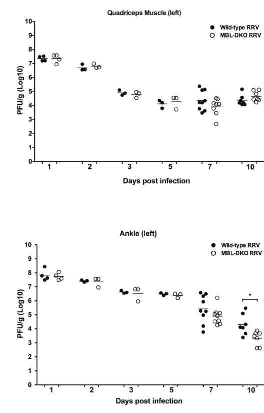
replicates to the highest titers in the skeletal muscle and ankle joints (161). Peak viral titer within these tissues occurs at 24-48 hours post infection, and steadily declines throughout the course of disease, and is virtually undetectable by plaque assay by 10 days post infection (dpi) (161). While peak viral titer occurs within the first 2 days of infection, infected mice do not start to exhibit clinical disease signs until about 5 dpi (161). Mice begin to show signs of hind limb weakness and altered gait at 5 dpi as determined by a grip test (161). By 7 dpi, infected mice begin to lose the ability to grip with their hind limbs and a subset of mice display the inability to right. Peak disease severity occurs from 10-12 dpi where most infected mice are dragging their hind limbs and are unable to right themselves. However, by about 14 dpi mice are beginning to recover and regain use of their hind limbs, and then by 21 dpi the infected mice have recovered and are
indistinguishable from uninfected mice (161).
Histopathological analyses of the target tissues within infected mice have shown that the disease signs observed in the mice correlate with the induction of the host
Monocyte chemotactic protein (MCP-1; CCL2) is involved in macrophage recruitment and chemotaxis of other immune cells, and inhibition of MCP-1 by administration of bindarit into mice abrogated RRV-induced disease by significantly reducing the numbers of CD11b+ cells recruited into the skeletal muscle (193). Another cytokine, macrophage migration inhibitory factor (MIF), which acts as a powerful pro-inflammatory cytokine that regulates multiple aspects of the host inflammatory response (28), has an important role in regulating the infiltration of immune cells into target tissues following RRV infection (94). MIF-/- mice show reduced disease and tissue damage following RRV infection compared to WT mice, and correlated with a reduction in inflammation in the skeletal muscle rather than altering viral titer (94). Finally, arginase I, which is produced by alternatively activated macrophages and neutrophils, thwarts clearance of RRV from skeletal muscle and ankle joints, and allows for sustained replication and disease at later time points (216).
All of these studies indicate a critical role for the host inflammatory response in mediating RRV-induced disease. However, there are gaps in our understanding regarding the underlying mechanisms of how the inflammatory response is initiated and/or
regulated following RRV infection. Furthermore, the specific RRV ligands that activate the inflammatory response have not been identified, thus many aspects of RRV
pathogenesis remain unknown and understudied.
Arthritic alphavirus pathogenesis: chikungunya virus.
Development of appropriate CHIKV mouse models have been hampered
of infection to replicate in distal tissues. However, several models using two different human isolates from the most recent CHIKV epidemic have been described. A young mouse model where 14 day old C57BL/6 mice and infected with a CHIKV isolate from Sri Lanka exhibits swelling, arthritis, tenosynovitis, and myositis in the injected foot, and shares many pathologic characteristics with the human CHIKV disease (159). Similar to RRV, CHIKV infection results in inflammation within the joints and skeletal muscle, although swelling of the joints occurs following CHIKV infection but is absent during the mouse model of RRV-induced disease. Pronounced swelling of the ankles occurs at 2-3 dpi and 6-7 dpi in the CHIKV mouse model. Analysis of the inflammatory cells
infiltrating into the leg reveals that NK cells and neutrophils are the predominant infiltrating cell types at 5 dpi, although a heterogenous mix of NK cells, neutrophils, monocytes/macrophages and CD8+/CD4+ T cells were observed at 7 dpi (159). An older mouse model, where both feet of mice are injected with a CHIKV isolate from La Reunion Island, demonstrate similar disease characteristics as the young mouse model, albeit less pronounced, but may be more amenable to vaccination and therapeutic studies (68). Studies using a neonatal mouse model demonstrated the importance of type I IFN in protection from lethal CHIKV infection (202), further highlighting the critical role of the innate immune system in protection from alphavirus infection.
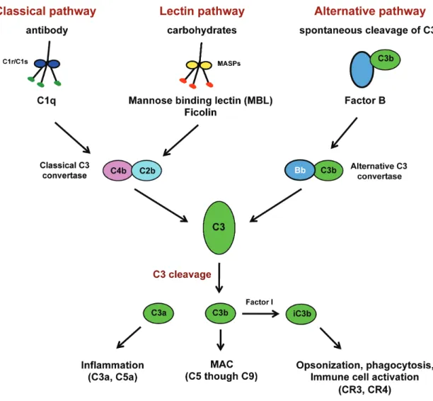
1.4. The host complement system.
recognition, signaling, initiation of inflammation, and direct pathogen lysis. The
complement system is truly a “system” as there are well over thirty extracellular and cell-associated proteins that help orchestrate the immune response to a given pathogen. A general schematic of the complement system is shown in Figure 1.2.
Activation of complement is thought to occur through three main pathways: classical, lectin, and alternative pathways. Regardless of which pathway activates complement, the critical step in complement activation is cleavage of the C3 protein, which is central to the complement system (reviewed in (183)). Cleavage of C3 requires formation of the C3 convertases that are generated on target pathogens or cells following activation through either the classical or the lectin pathway. Cleavage of C3 results first in the generation of C3a and C3b, and C3b becomes covalently attached to the target surface through recognition of the reactive thioester group that is exposed upon cleavage on C3b and carbohydrate groups on the target surface. Interestingly, the specificity of the C3 thioester group for carbohydrates, and in particular terminal sugars on
polysaccharides may have biological relevance since glycosylation on pathogens and stressed/infected cells can be different than healthy uninfected cells, and could be an additional safety guard to protect host cells from excessive complement activation (199). C3a is released and acts as a chemoattractant and anaphylatoxin to aid in activation of other arms of the immune response.
The C3b deposition on the target cell leads to formation of additional C3
inserts into the target membrane, allowing for C8 and then C9 to become part of the complex and generates a lytic pore called the membrane attack complex (MAC) that lyses the target cell. C5a is an additional anaphylatoxin that is released upon cleavage and acts on neutrophils and monocytes to induce rapid phagocytosis of opsonized targets.
In addition to the first C3 cleavage event that generates C3a and C3b, C3b is further cleaved by factor I in complex with CR1 that inactivates C3b, but generates iC3b and C3c, C3dg and eventually C3f and C3d, which all have biological functions in complement to aid in pathogen clearance (183). Perhaps the most effective cleavage product is iC3b. The iC3b cleavage product acts to opsonize the target cell and interacts with several complement receptors on phagocytic cells to promote phagocytosis and leukocyte signaling (183, 233, 251). Interactions with complement receptors such as CR3 and CR4 lead to enhanced phagocytosis of iC3b-opsonized cells and can trigger signaling downstream from both of the receptors (50, 183). CR3-mediated signaling within
phagocytic cells, such as monocytes/macrophages and neutrophils, can act to initiate and enhance cytotoxic effector functions of the cells (173, 232).
The role of complement in alphavirus pathogenesis.
The role of the complement system in alphavirus infection is either protective or pathologic role depending on the virus. Complement has a protective role in mouse models of SINV and VEEV infections, but is pathologic in RRV infection (23, 97, 99, 158).
consumes and depletes C3, had elevated levels of SINV in serum and in the brains
compared to untreated infected controls (97, 98). Interestingly, inflammation in the brains of the complement depleted mice was increased compared to controls, and correlated with prolonged disease signs, suggesting that complement may also regulate
inflammation (97). Analysis of the mechanisms of viral control by complement revealed that both the classical and alternative pathways were activated upon SINV infection even in the absence of virus-specific antibodies (99). Furthermore, mice genetically deficient in C5 displayed enhanced susceptibility to SINV infection, suggesting that formation of the MAC by complement is very important for control of serum viremia and infection within the tissues (96). Interestingly, despite the increase in viral titer with the CNS and serum, the C3 depleted mice did not show any enhanced mortality and actually had prolonged survival compared to undepleted mice (97), suggesting that there may be some pathology associated with presence of C3. The role of the lectin pathway has not been evaluated in the context of SINV infection.
directly into the brain showed no difference in mortality. Furthermore, C5-/- mice displayed wild-type kinetics and severity of disease, indicating that formation of the MAC was not required for limiting viral spread by complement. Instead, it is likely that other effector mechanisms involving C3 cleavage products have a key role in protection from VEEV infection.
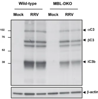
The pathology and disease associated with RRV infection in humans and mice is primarily mediated by the host inflammatory response. Studies by Morrison et al. demonstrated that the complement system is activated following RRV infection (158). RRV-infected mice exhibited elevated levels of circulating C3 in the serum compared to mock-infected mice, and also had increased amounts of C3 cleavage products in the quadriceps muscle and ankle joints (158). In addition, human patients with RRV-induced polyarthritis had elevated levels of C3a, indicative of complement activation, within the synovial fluid compared to patients with the non-inflammatory arthritis disease
osteoarthritis. Furthermore, the authors demonstrated that complement activation has a critical role in mediating damage and disease within target tissues. Mice deficient in C3, which is the central component of the complement system, developed a mild disease compared to wild-type mice. Interestingly, viral burden and localization of viral replication within the tissues was not affected by the presence or absence of C3, suggesting that complement regulated some aspect of inflammation. However, histopathologic analysis of both quadriceps muscle and ankle joints indicated the
C5-deficient mice did not differ significantly from WT mice, indicating that formation of MAC is not required for disease (Morrison TE and Heise MT, unpublished data).
Follow-up studies evaluating the role of other complement proteins in mediating RRV-induced disease demonstrated that complement receptor 3 (CR3, CD11b/CD18, Mac-1, amb2) contributes to disease (160). CR3 is present on multiple types of immune cells, including monocytes, macrophages, neutrophils, DCs, and T cells, and interacts with the C3 cleavage factor iC3b (259). Mice deficient in CR3/CD11b exhibited reduced RRV-induced disease compared to WT mice, and many aspects of the disease in
CR3/CD11b-/- were similar to the disease observed in C3-/- mice. Viral burden and the amount of inflammatory cells within the tissues were equivalent between CR3/CD11b -/-and WT mice, while disease -/-and tissue damage were reduced in CD11b-/- mice.
Ligation of CR3 by iC3b can induce signaling within CR3-bearing cells in a Syk-PI3K mediated pathway and can serve to activate pro-inflammatory effector programs within the cells (50, 132, 157). Interestingly, expression of a subset of pro-inflammatory genes was dependent on both CR3 and C3 following RRV infection (160). The
Taken together, these data indicate the complement system can play both a protective and a pathologic role following alphavirus infection. In SINV and VEEV infection, complement appears to be required to neutralize the viruses and help clear virus from the serum, thus limiting spread of the virus into additional tissues. In contrast, following RRV infection, the complement system does not neutralize the virus, but rather contributes to development of disease through activation of CR3-bearing inflammatory cells resulting in the inflammatory tissue destruction and disease. It is not currently clear why complement is protective in certain alphavirus infection but pathologic in another, but further exploration into the mechanisms of complement activation and identification of viral ligands may provide some insight into these processes.
Complement activation pathways.
C3 within the fB-C3 complex by Factor D generates the alternative C3 convertase, C3bBb, which cleaves C3 and activates the complement cascade.
The lectin activation pathway of complement.
The lectin activation pathway of complement is mediated through the serum proteins mannose binding lectin (MBL) and ficolins (L-, M-, and H-ficolin) that act as pattern recognition receptors (PRRs). Both MBL and the ficolins contain a collagen-like domain in the N-terminus that mediates oligomerization of the structural subunits to form higher order structures required for functional complement activation. MBL has a
carbohydrate recognition domain (CRD) in the C-terminus, whereas ficolins have a fibrinogen-like domain that acts in a similar manner to the CRD of MBL (63). The ever-expanding list of MBL ligands will be discussed in further detail in following sections, and the ficolins have been shown to bind to terminal N-acetylglucosamine (GlcNAc) on glycosylated proteins on bacteria as well as apoptotic cells (124, 147, 255). MBL, L-ficolin, and H-ficolin are produced in the liver and circulate within the serum, where as M-ficolin is produced by monocytes and granulocytes (63).
The MBL-associated serine proteases (MASPs) circulate throughout the body in association with the ficolins and MBL. There are three MASP proteins (MASP-1, -2, -3), however, only MASP-2 has been shown to be able to form the C3 convertase C4b2b to activate the complement cascade (192, 226). Interestingly, MASP-1 and MASP-3 have been shown to mediate cleavage of Factor D, which is required for activation of
be able to cleave C4 and C2 to generate the C3 convertase C4b2b, cleavage of C3 and complement activation.
Of note, collectin-11 (CL-11) was recently identified as an additional serum protein that circulates in association with MASP-1 and/or MASP-3 (83). CL-11 bound to several microbial and fungal species including Escherichia coli, Pseudomonas
aeruginosa, and Candida albicans. Furthermore, CL-11 bound to influenza A virus and
was able to partially inhibit the entry of virus into cells at high concentrations. The ability of CL-11 to activate complement has not yet been demonstrated, but given the microbial substrates, association with MASP-1 and/or MASP-3, and levels circulating in serum, it is likely that CL-11 will join the complement system as a PRR.
Mannose binding lectin (MBL): structure and ligands.
MBL was identified as a serum protein that could recognize and bind to carbohydrate structures (187), and was subsequently found to be able to activate the complement system (105). MBL is produced in primarily in the liver of several
The Mbl2 gene on chromosome 10 encodes the human MBL protein, and the functional protein is composed of multimers of the 32 kDa MBL polypeptide. Each polypeptide has a calcium-dependent carbohydrate recognition domain (CRD) at the C-terminus, and a collagenous region that forms into a triple helix with other two
polypeptides to make up one structural subunit of MBL. Each structural subunit
maintains a fixed 45Å distance between the three CRDs which allows for binding to the carbohydrate ligands (211). While each subunit can bind to the carbohydrate ligands, the affinity is relatively low, the oligomerized multimers of MBL are able to bind to larger arrays of carbohydrates thus increasing the affinity of binding and allows for activation of complement (106). The importance of 45Å spacing of the CRDs is to allow for specific binding to certain sugars, namely the hydroxyl groups in the hexose structure in N-acetylglucosamine, mannose, glucose, and fucose (102). These sugars are commonly the terminal carbohydrate residue on microbial glycoproteins and not commonly found on mammalian glycoproteins, which favor terminal galactose and sialic acid. The CRDs of MBL do not bind to the hexose structure in galactose or sialic acid therefore preventing binding and activation of complement, which further adds to the specificity of
recognition (243).
immune system to stress and possible infection. MBL is thought to bind to the pentose sugars present on free DNA and RNA from either apoptotic or necrotic cells to help facilitate phagocytic clearance (168). Similarly, binding of MBL N-acetylglucosamine on phospholipids may also serve to enhance recognition and clearance of apoptotic cells (119, 126). Thus, MBL has an important role in recognition of pathogens as well as damaged host cells that allows for activation of complement and the innate immune system.
MBL polymorphisms.
The levels of circulating MBL in healthy human adults varies substantially between individuals and across the population due to several polymorphisms and
promoter polymorphisms lead to extreme variation within the human populations and serum levels of MBL range from nearly undetectable to 10 µg/ml (47).
The importance of MBL in mediating host protection from a number of bacterial and viral pathogens has been demonstrated by several studies that have evaluated the susceptibility of patients who are functionally deficient in MBL to certain pathogens. Sumiya et al found that children with recurrent bacterial infections had genetically lower levels of MBL compared to healthy children (222), and several studies have shown enhanced susceptibility to certain viral infections including heptatitis B virus and HIV (25, 52).
Role of MBL in sterile inflammatory diseases.
Since MBL can bind to both self-antigens such as DNA and phospholipids as well as the glycosylated proteins of pathogens, it is not surprising that MBL, like complement, can be a double edged sword: required for protection from a number of different
pathogens, but can also lead to autoimmunity. Indeed, MBL has been associated with pathology in sterile inflammatory diseases such as ischemic reperfusion injury (IRI), and has been implicated in rheumatoid arthritis (RA).
Ischemic reperfusion injury occurs after blood flow is restored to tissues and organs after a period of deprivation. The pathology of disease is characterized by induction of a pro-inflammatory state within the affected tissue and subsequent inflammation that leads to eventual apoptosis, necrosis, and possible permanent tissue damage (76). The complement system has long been known to be involved in
and more recently, the lectin pathway has been shown to have a critical role in mediating complement activation following reperfusion (reviewed in (76)). Early in vitro studies demonstrated that the oxidative stress induced by IRI on endothelial cells initiated MBL and subsequent C3 deposition, likely through altered glycosylation of host proteins on the cells (34, 35). Subsequent studies in myocardial, gastrointestinal, and renal reperfusion injury animal models using either antibodies to deplete MBL, or MBL-A-/- MBL-C -/-(MBL-DKO) and MASP-2-/- mice further demonstrated that MBL and the lectin pathway regulated complement activation and subsequent inflammation and damage within the reperfused tissues (112, 205, 237).
Rheumatoid arthritis is an autoimmune disease that is characterized by chronic inflammation within the joints of affected individuals. Autoantibodies are thought to contribute to disease in part through activation of the complement system, but several studies have evaluated the lectin pathway in disease susceptibility and severity in cohorts of RA patients. However, the results from these studies are conflicting as different studies found a correlation of low MBL levels in RA patients with both protection and pathology (80, 109, 110, 198, 228). Thus, the role of MBL in inflammatory arthritis has not been established and remains unclear.
Role of MBL in viral infection: Flaviviruses.
Mice deficient in C3 are highly susceptible to WNV infection due to a defect in the development of a robust adaptive immune response. Neutralizing antibody responses as well as CD8+ T cell responses were reduced in C3-/- mice, leading to higher viral titers within the CNS of infected mice (149, 150). MBL deficient mice were also more
susceptible to WNV infection, and MBL deficient serum showed reduced neutralization of insect derived virus (61, 62).
MBL has also been reported to bind to and neutralize DENV, even in the absence of complement activation (61), and levels of MBL within human serum directly
correlated with neutralization of both mosquito and mammalian-derived DENV2 (7), suggesting that MBL has a protective role in DENV infection. However, several studies analyzing the role of MBL in determining disease severity in DENV-infected patients have yielded conflicting results. One study was unable to correlate low levels of MBL with patient progression from dengue fever (DF) to the more severe disease dengue hemorrhagic fever (DHF), yet other studies found that high levels of MBL were associated with DHF, and low levels of MBL protected DHF patients from
thrombocytopenia (2, 33, 164). From these studies, it is currently unclear the role of MBL in determining DENV severity, although experimental evidence suggests that MBL contributes to neutralization of DENV and is thus likely to have a protective role following DENV infection.
Role of MBL in viral infection: other viruses.
with N-linked glycans, and several groups have demonstrated that MBL can bind to both lab strains as well as primary isolates of HIV (54, 200). MBL binding to HIV is reported to be important in direct neutralization of the virus by preventing binding to host cells, although studies differ on the extent of neutralization (54, 254).
MBL has also been shown to contribute to host protection of several other viral infections such as herpes simplex virus-2 (HSV-2), Ebola virus, and SARS-coronavirus (65, 107, 111, 257). MBL was found to bind to HSV-2 virions in an ELISA assay, and MBL-DKO mice exhibited enhanced viral titer within the liver compared to WT mice (65). Furthermore, serum levels of MBL in asymptomatic HSV-2 patients were elevated compared to symptomatic patients and those with recurrent infections, suggesting that higher levels of MBL contribute to suppression of virus in human infection (65, 208).
lower levels of MBL compared to patients with genotypes leading to higher amounts of circulating MBL (107).
1.5. The viral N-linked glycans.
Glycosylation is an important post-translational modification.
Glycosylation and other forms of post-translational modifications on proteins have an important, yet understudied and perhaps underappreciated role in protein
structure, function, and regulation. On a broader scale, glycosylation of proteins is critical to development and function of many organisms, as mutations or deletions of the
glycosyltransferases required for proper glycosylation frequently result in deleterious phenotypes and the inability to glycosylate is lethal (reviewed in (229)). Furthermore, differential glycosylation of proteins can introduce a remarkable amount of variation and diversity to the form and function of any given protein, thus adding another layer of complexity to our already complex and beautiful world.
N-aceytlglucosamine (GlcNAc) core and branches of terminal mannose residues is convalently linked to the polyprotein, and is further processed by glycosidases and glycosyltransferases in the Golgi. O-linked glycosylation is commonly found on glycoproteins termed mucins and proteins that are secreted into the mucosa. O-linked glycosylation is characterized by the addition of GlcNAc to serine/threonine residues in the Golgi and does not appear to have any specific sequence that identifies putative sites.
The biological functions of glycans can be broadly categorized into the following roles: structural role, where glycans are required for proper folding of proteins; a role in mediating cell-intrinsic interactions where glycans mediate interactions within the cell organism with glycan-binding proteins; and cell-extrinsic roles which involve the
interactions between foreign organisms such as bacteria, viruses, and fungi. The presence of glycans can protect the protein from proteases, antibodies, or premature interactions during synthesis and trafficking. HIV is a notable example where the heavy glycosylation of gp120 can act as a glycan shield, protecting the virus from antibody-mediated
neutralization. The cell-extrinsic role of glycans involves recognition and interaction between the glycans and glycan-binding proteins on pathogens and other foreign
molecules. These interactions have a major impact on infectious disease, especially since these interactions are required for pathogen entry into a host cell, recognition of a
pathogen by the immune system, or affect some other aspect of disease.
N-linked glycosylation.
base glycan is a fourteen-sugar oligomannose composed of glucose (Glc3), mannose (Man9), and N-aceytlglucosamine (GlcNAc2). As the protein moves through the ER and the Golgi, the glycan gets trimmed by glucosidases and mannosidases and then modified by various transferases that add different sugar groups (galactose, GlnNAc, sialic acid) onto the glycan to generate the final glycan structure (215). There are three main types of glycans that are produced: complex, high mannose, and hybrid. While there is a
staggering amount of diversity within the three types of glycans, there are defining characteristic of each type. Complex glycans are characterized by the presence of terminal sialic acid residues; high mannose (also can be called oligomannose) glycans have terminal mannose residues; and hybrid glycans have a combination of terminal mannose and sialic acid residues.
Complex glycans can have extensive branching that allows for additional glycan diversity, and the different branching and terminal carbohydrates modifications are dictated in a tissue-specific and cell-lineage dependent manner. The ability to produce complex glycans is due to the presence of specific glycosidases and transferases that are able to trim down the base glycan, and rebuild with different sugars.
High mannose glycans are more typically found on glycosylated proteins
produced from invertebrates. While the base glycan is the same glycan as the one found on proteins produced in vertebrate cells, invertebrates do not produce certain glycosidases and transferases and thus are unable to produce complex or hybrid glycans. Glycosylation in bacteria is quite distinct, but plays a similar role in bacterial protein function,
Innate immune recognition of N-linked glycans.
Given that bacteria, viruses, parasites, and fungi use glycosylation to modify their proteins either for proper folding or interaction with host proteins, it is not surprising that there are several different innate immune receptors that recognize non-self or altered glycans to activate the immune system. Several Toll-like receptors (TLRs) such as TLR4 and TLR2 recognize glycoproteins to activate TLR signaling pathways resulting in initiation of pro-inflammatory programs and activation of adaptive immunity. TLR4 recognizes lipopolysaccharide on bacterial cell walls, and has been shown to recognize glycoproteins from many different viruses as well, including respiratory syncytial virus and MMTV (reviewed in (117)). TLR2 recognizes components of viral glycoproteins as well to stimulate innate immunity (reviewed in (6, 117)). However, to date, viral N-linked glycans present on viral glycoproteins have not been shown to be directly engage TLRs, although given their location on the glycoprotein, it is plausible that they might.
to contribute to the pathogenesis of flaviviruses such as DENV and Japanese encephalitis virus through induction of pro-inflammatory cytokines and inflammasome activation (30, 31, 250). With regard to alphaviruses, DC-SIGN and L-SIGN have reported to be
attachment factors that mediate entry of mosquito-derived virus into dendritic cells (120). The role other members of the C-type lectin family in alphavirus pathogenesis have not been evaluated.
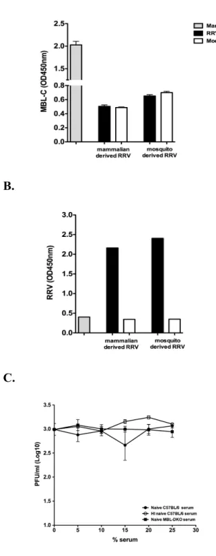
MBL has been shown to directly bind to the N-linked glycans of several viruses discussed in earlier sections. Mosquito-derived WNV and DENV are neutralized by MBL in part through recognition of the high mannose N-linked glycans on the viral E protein on virions leading to complement activation (61). The high mannose and complex glycans on HIV gp120 mediate binding and neutralization by MBL both on the virus and on the infected cell (90), and a single N-linked glycan on the tip of the SARS-CoV spike protein was required for the interaction between the virus and MBL (257). Importantly, all of the described interactions between viruses and MBL indicate that the interaction between viral N-linked glycans and MBL leads to a protective response following viral infection.
The alphavirus N-linked glycans.
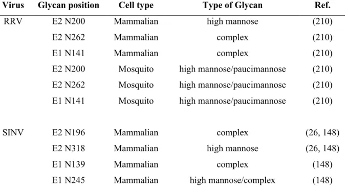
The positions of the SINV and RRV glycans have been mapped by cryo-EM analysis of the structure of the glycoprotein spike (174). Since E1 lies parallel to the virion surface, the E1 glycans are located roughly in between, but below, the E2 glycoprotein spike (174). The glycans at E2 N200 (RRV) and E2 N196 (SINV) are located at the tip of the E2 glycoprotein spike, with the SINV glycan on the top of the “petal” structure, and the RRV glycan on the bottom (174). The locations of the glycans at E2 N262 (RRV) and E2 N318 (SINV) differ slightly. In SINV, the glycan is closer to the lipid bilayer whereas the RRV glycan is in between the glycoprotein spikes. The positions of the E2 glycans on the glycoprotein spike, especially the E2 N200 (RRV) and E2 N196 (SINV) located on the tip of the spike, make the N-linked glycans attractive candidates to interact with host carbohydrate binding proteins.
The oligosaccharide content and type of glycan present at each of the viral glycosylation sites is dependent on the host cell. The glycan composition at each site has been determined for both RRV and SINV and is summarized in Table 1.1 (148, 210). EndoH and PNGase digestion of mutant viruses that lack the glycosylation sites revealed that the RRV E1 N141 and E2 N262 glycans are predominantly complex