IMMUNE RESPONSE TO ACUTE EXERCISE IN BREAST CANCER SURVIVORS: A COMPARISON TO HEALTHY WOMEN
Elizabeth Paige Harrell
A thesis submitted to the faculty at the University of North Carolina at Chapel Hill in partial fulfillment of the requirements for the degree of Master of Arts in the Department of Exercise
and Sport Science (Exercise Physiology) in the College of Arts and Sciences.
Chapel Hill 2019
© 2019
ABSTRACT
Elizabeth Paige Harrell: Immune Response to Acute Exercise in Breast Cancer Survivors: A Comparison to Healthy Women
(Under the direction of Erik Hanson)
To determine immune response to acute moderate-intensity aerobic exercise in breast cancer (BrCa) survivors compared with healthy controls. 15 sedentary females were age-matched to 21 BrCa survivors and performed 30 minutes of intermittent exercise at 60% wattage achieved at VO2peak.Hematological data was obtained, and immune cells were isolated from whole blood
TABLE OF CONTENTS
LIST OF TABLES ... vii
LIST OF FIGURES ... viii
LIST OF ABBREVIATIONS ... ix
CHAPTER I ... 1
INTRODUCTION... 1
Purpose ... 4
Research Questions ... 4
Research Hypotheses ... 5
Delimitations ... 5
Assumptions ... 6
Limitations ... 6
CHAPTER II... 7
REVIEW OF LITERATURE ... 7
Section I. Impact of Cancer Treatment on the Immune System ... 7
Section II. Exercise as Medicine for Cancer ... 8
Section III. Immune Response to Acute Exercise ... 9
Section IV. MAIT Cell Profile ... 14
Section VI. Justification of Methodology... 18
Summary... 22
CHAPTER III ... 23
METHODOLOGY ... 23
Participants... 23
Study Design ... 23
Body Composition Assessment ... 25
Familiarization Session ... 25
Graded Exercise Test ... 26
Blood Draws ... 26
Exercise Trial ... 27
Hematology Analysis... 28
Calculation of Plasma Volume Shifts ... 28
Peripheral Blood Mononuclear Cells (PBMC) Isolation and Immunofluorescence Labeling ... 28
Immune Function ... 29
Flow Cytometry ... 29
Statistical Analysis ... 30
CHAPTER IV ... 32
RESULTS ... 32
Participants... 32
Plasma Volume Shifts ... 36
MAIT Cell Assays... 36
CHAPTER V ... 39
DISCUSSION ... 39
LIST OF TABLES
Table 1 – T cell proliferation assays……….………....20 Table 2 – Participant characteristics………...33 Table 3 – Physiological response to VO2peak test and the exercise trial
consisting of 10 sets of cycling at 60% of peak wattage for 3
LIST OF FIGURES
Figure 1 – Schematic representation of calcineurin/NFAT signaling...17 Figure 2 – Schematic representation of PKC activation via PMA...18 Figure 3 – Gating strategy for MAIT cell and cytokine identification...30 Figure 4 – Average levels of physical activity (PA) per week prior to
LIST OF ABBREVIATIONS
BrCa – Breast Cancer
CAM – Complementary Alternative Medicine CD – Cluster Differentiation
DNAse – Deoxyribonuclease
DXA – Dual Energy X-Ray Absorptiometry EDTA – Ethylenediaminetetraacetic acid FMO – Fluorescence Minus One
IFN – Interferon IL - Interleukin
IPAQ – International Physical Activity Questionnaire MAIT- Mucosal Invariant T Cell
MHC – Major Histocompatibility Complex MR1 – MHC Related Protein-1
NFAT – Nuclear Factor of Activated T Cells NK – Natural Killer Cell
PAR-Q – Physical Activity Readiness Questionnaire PBMC – Peripheral Blood Mononuclear Cell
PBS – Phosphate Buffered Saline PKC – Protein Kinase C
TNF – Tumor Necrosis Factor
CHAPTER I INTRODUCTION
At this time, breast cancer (BrCa) is the leading form of cancer diagnosed in women worldwide (66). Over the past two decades, advances in detection and anti-cancer treatments have led to improved survival rates and a shift in focus to post-treatment care of these patients. Many major BrCa treatments damage a myriad of physiological systems and are often
accompanied by adverse side effects, including fatigue, reduced physical activity,
immunosuppression, and ultimately reduced quality of life (19, 48, 60). Diminished immune function is a significant post-treatment concern that often increases susceptibility to secondary malignancies and other co-morbidities, such as cardiovascular disease, in BrCa survivors (33, 34, 60).
proper tumor antigen and other pathogen surveillance, consequently leaving this population more vulnerable to viral and bacterial infections (19, 32, 38, 40). The most common treatments,
including chemotherapy, radiation, and surgical removal of tumors, have been shown to reduce total lymphocyte counts, T cell, B cell, and NK cell numbers, lessen cytolytic activity, impair antigen presentation, and decrease the vital functional capacity of these cells (4, 19, 31, 59, 61, 71). Subsequently, there is a need to identify potential interventions that may improve immune system function following such cancer treatments.
Mucosal associated invariant T (MAIT) cells are a unique subpopulation of pro-inflammatory leukocytes that display both innate and acquired immune system characteristics (26). These innate-like T cells express the semi-invariant T cell receptor (TCR; iVa7.2-Ja33) and develop in the thymus. Once these cells are selected by the major histocompatibility complex (MHC) class I-related protein (MR1) on hematopoietic cells, they are directed through peripheral circulation to mucosal tissue of the liver, spleen, gastrointestinal tract, and the lungs (27, 69). MAIT cells are able to recruit other immune cells and respond to a diverse set of antigens via cytokine secretion (8, 15). As a relatively novel cell subset, MAIT cells continue to emerge as noteworthy players in the immune system. Alterations in circulating MAIT cells have been linked to an increasing number of clinical disorders including forms of cancer (15, 43, 56, 75). Compared to other T cell populations, MAIT cells may be able to persist through chemotherapy and even recover beyond baseline values in the months following chemotherapy, as
demonstrated in BrCa survivors (15). If this is the case, this cell subpopulation may be
killing capacity to be effective. As such, acute exercise may be an adjunct therapy of interest for this population.
demonstrated that MAIT cells mobilize similarly to other T cell populations during an acute bout of exercise (28, 29). Furthermore, stimulated MAIT cells displayed increased TNF-α and IFN-γ
cytokine expression, indicating a potential greater responsiveness to stimulation following acute exercise (28). All of these findings advocate for acute exercise as a potential up-regulator of a natural immune protective mechanism against disease susceptibility (19, 73). Many immune studies assess cell counts and proportions, however, to better understand the cellular response to acute exercise, function needs to be assessed. MAIT cell response to acute exercise has never before been studied in cancer survivors. As BrCa survivors tend to be at a higher risk for developing co-morbidities, such as clinical depression and anxiety, diabetes, cardiovascular disease, and secondary cancers (22), following treatment care, examining MAIT cell function in BrCa survivors following acute exercise could potentially uncover clinically relevant
connections to exercise and immune health in this diseased population. Purpose
The purpose of this study was to investigate immune response to acute moderate intensity intermittent aerobic exercise in a BrCa survivor population compared with healthy but sedentary age-matched controls using hematological data.
Research Questions
1) Do leukocyte counts in the peripheral circulation change immediately or one-hour following a 30-minute bout of intermittent aerobic exercise at 60% peak wattage on a cycle ergometer? 2) How do leukocyte counts in the peripheral circulation compare between BrCa survivors and
3) Does MAIT cell function in the peripheral circulation change immediately or one-hour following a 30-minute bout of intermittent aerobic exercise at 60% peak wattage on a cycle ergometer?
4) How does MAIT cell function in the peripheral circulation compare between BrCa survivors and age-matched controls with no history of cancer diagnosis following a 30-minute bout of intermittent aerobic exercise at 60% peak wattage on a cycle ergometer?
Research Hypotheses
1) A 30-minute bout of intermittent aerobic exercise at 60% peak wattage on a cycle ergometer will increase leukocyte cell counts immediately post-exercise and return to baseline at one-hour following exercise.
2) Leukocyte counts in the peripheral blood circulation will be lower compared to healthy female controls, but counts will increase relatively similarly to the controls following a 30-minute bout of intermittent aerobic exercise at 60% peak wattage on a cycle ergometer. 3) A 30-minute bout of intermittent aerobic exercise at 60% peak wattage on a cycle ergometer
will increase MAIT cell function immediately post-exercise and return to baseline at one-hour following exercise.
4) MAIT cell function will increase relatively similarly to the controls following a 30-minute bout of intermittent aerobic exercise at 60% peak wattage on a cycle ergometer.
Delimitations
• All participants were females.
• All participants did not regularly participate in any exercise program 6 months prior to beginning the study, deeming them as sedentary.
• All participants were recruited from the central North Carolina area via flyer, email, face to face, and phone call.
• All participants were cleared by a physician for exercise participation prior to participating in the study.
Assumptions
• All participants followed the pre-assessment guidelines prior to testing sessions. • All participants gave their maximal effort during testing sessions.
• Participants did not partake in any other forms of exercise or change of diet while participating in the study.
• All participants honestly reported medical history and physical activity readiness. Limitations
• The results of this study may only apply to those whom are women, apparently healthy but sedentary or have a BrCa history diagnosis, and at least 35 years of age or post-menopausal. Results may not be applied to females of all ages, males, or those with a history of other cancer diagnoses.
• BrCa survivors included in this study all completed treatment within one year of participation but could be of any age. All healthy control participants were at least 35 years of age.
• It is possible that participants did not adhere to pre-assessment guidelines entirely as researchers were not with them during the hours prior to testing.
CHAPTER II
REVIEW OF LITERATURE
This review is divided into six sections: Section I. Impact of Cancer Treatment on the Immune System, Section II. Exercise as Medicine for Cancer, Section III. Immune Response to Acute Exercise, Section IV. MAIT Cell Profile, Section V. PKC Pathway Activation via PMA-Ionomycin, and Section VI. Justification of Methodology.
Section I. Impact of Cancer Treatment on the Immune System
At this time, BrCa is the leading form of cancer diagnosed in women worldwide. With 30% of new cancer diagnoses in women being attributed to BrCa, cases total approximately 3.1 million women in the United States with a history of BrCa diagnosis as of 2019 (66). Faster detection and anti-cancer treatment advances over the past three decades have led to increased longevity in oncology patients, especially in the BrCa population (60, 65). However, patients continue to face challenges into survivorship as many major cancer treatments diminish the function of physiological symptoms across the body and often result in a number of deleterious treatment-related side effects associated with reduced quality of life. Negative influences on physical characteristics such as reduced cardiac function and muscular strength, increased fat mass and reduced muscle mass, and chronic systemic inflammation and immunosuppression are among common treatment side effects that diminish overall quality of life and increase disease susceptibility in cancer patients (60). Thus, there is a need for interventions to effectively
As the primary protector against illness, the immune system plays a vital role in secondary cancer prevention and the development of other common comorbidities, such as cardiovascular disease, during survivorship. However, typical cancer treatments, including chemotherapy, radiation, and surgical removal of tumors have been linked to short- and long-term weakened immunity. Specifically, decreased total lymphocyte counts, reduced T cells (CD4+, CD8+), B cells (CD19+), and NK cells (CD3-CD16+CD56+), and impaired T, B, and NK cell functional response has been shown in BrCa survivors who received chemotherapy (19, 31, 59, 61). Radiation is another form of treatment that reduces total lymphocyte number, NK cell cytolytic activity, T cell ratios (CD4+/CD8+), and T and B cell proliferation responses to mitogenic stimulation (4, 19). Studies have also indicated that surgical interventions such as mastectomies, lumpectomies, and lymph node dissections can impair monocyte phagocytosis, antigen presentation, B cell immunoglobulin production, T cell response to mitogen stimulation, and decrease NK cell cytolytic activity (19, 71). Additionally, evidence suggests increases in proinflammatory cytokines and decreases in anti-inflammatory cytokines may be linked to concerning treatment related-side effects involved with systemic inflammation that has been correlated to changes in body composition and immunity (40). These deficits in baseline immune function may persist for weeks to years following the completion of treatment (9, 65). Although further research is needed linking changes in immune response directly to anticancer therapies, sufficient evidence suggests that such treatment related reductions in immune system function may put BrCa survivors at greater risk for cancer reoccurrence and additional comorbidities. Section II. Exercise as Medicine for Cancer
During the past three decades, exercise as a Complementary Alternative Medicine
diagnosed with BrCa has shown that when administered prior, during, and post cancer treatment, exercise interventions decrease fatigue, improve strength and endurance, conserve muscle mass, improve metabolism, improve stress management, improve heart rate coherence and heart rate variability, and generally help patients feel better physically and emotionally (2). Exact
mechanisms underlying the impact of exercise on the immune system in cancer patients still requires further study, nonetheless exercise has demonstrated its abilities to enhance immune surveillance and function through cellular mobilization, cytotoxic immune cell activation, and regulation of inflammatory signaling pathways (19, 32, 40). As a result of possible inflammation control, it is suggested that exercise may play a role in protecting against chronic inflammation-associated diseases, including tumor growth (36, 40, 72). Exercise is a cost effective and natural strengthener to multiple physiological systems that aid in preventing secondary cancers and other co-morbidities (60). Whereas other adjuvant therapies tend to cause adverse side effects, are often expensive, and sometimes fail to control micro metastases (19).
Although studies examining the effects of exercise in BrCa patients continue to rise, many fundamental questions remain with regard to optimal exercise prescriptions that include variation in exercise training type, intensity, duration, and frequency in addition to individualized care. To develop precise exercise guidelines specific for BrCa patients, the exercise oncology field is in critical need of studies with more detailed descriptions of testing and exercise prescriptions and studies that investigate specific physiological system responses in BrCa patients following exercise.
Section III. Immune Response to Acute Exercise
system forms a physiological response. The immune system also does this in response to
physical stressors, such as exercise. In response to acute exercise stimuli, leukocyte populations, including NK cells, helper T lymphocytes (CD4+), cytotoxic T lymphocytes (CD8+), MAIT cells, and B lymphocytes (CD19+) tend to demonstrate a biphasic cell count response, where counts increase during exercise and decrease during recovery following exercise (6, 25, 29, 54) In regard to leukocyte subpopulation function in response to acute exercise, results vary. Some populations, such as neutrophil and lymphocyte apoptosis increase function during exercise and recovery, whereas others, such as lymphocyte proliferation in response to mitogens decrease function during exercise and recovery (54). Alternatively, NK cells increase function during exercise and decrease function during recovery (54). Duration and intensity of exercise impacts the magnitude of noted cellular immune responses, where lower intensities and shorter durations may not elicit as large of a response in comparison to greater intensities and longer durations (51, 54, 73).
When considering the relationship between exercise intensity and duration in cancer populations, the field of exercise immunology has developed the “J-shaped” hypothesis, which proposes an inverse relationship between exercise and illness susceptibility (19). Briefly, the hypothesis states that moderate intensity exercise may lead to enhanced immune function and consequently decreased disease risk, though exhaustive exercise and overtraining may lead to weakened immune function and therefore increased disease risk (19). However, no
during exercise recovery may be important for immune regulation and surveillance as opposed to being viewed as a state of immune vulnerability for the body (6).
While a heap of immune-oncology evidence exists evaluating the effects of exercise training, few studies have examined the influence of specifically acute exercise on cellular immune responses in cancer populations. Six noteworthy studies have measured acute effects in pediatric, leukemia, prostate, and BrCa patients. Shore and Shepard (63) assessed changes in lymphocyte counts, cytolytic activity, and mitogen-induced lymphocyte proliferation in response to 30 minutes of exercise at ventilatory threshold in six children aged 10 to 14 years old, all who had been successfully treated for acute lymphoblastic leukemia. The six patients were all
currently receiving chemotherapy or had completed chemotherapy treatment, and findings were compared to those of 11 healthy children. Similarly, Ladha et al. (41) investigated the effects of acute exercise on neutrophil count and function in four acute lymphoblastic leukemia patients between the ages of 7 to 18 years in comparison to six healthy matched controls. All participants completed a 30-minute exercise session consisting of intermittent run-walk intervals on a
treadmill at 70%-85% of VO2peak (maximal oxygen consumption). Blood samples were collected
for both studies at pre-exercise, post-exercise, and during recovery bouts 30-minutes to 2 hours post-exercise. Shore and Shepard (63) reported leukocytosis and lymphocytosis in both patients and controls. However, cell counts were lower in patients still receiving chemotherapy treatment, and T cell counts (CD3+, CD4+, CD8+) did not demonstrate changes from pre to post-exercise. The CD4+/CD8+ ratio was also lower pre-exercise when compared to post-exercise in the healthy group. Likewise, NK cell count (CD56+), cytolytic activity, and lymphocyte
neutrophil count from pre-exercise to immediately post-exercise (P = 0.011), significant decreases at 1-hour post-exercise (P = 0.045), and significant increases again from 1-hour to 2-hours post-exercise. Similar changes in other measured immune components were observed, but no significant differences were observed between groups.
exercise at week 20. At week 10, total leukocyte (P = 0.035) and neutrophil (P = 0.029) counts significantly increased following acute hydraulic exercise, whereas hemoglobin and all leukocyte counts (P < 0.05) increased following acute isotonic exercise at 20 weeks.
The impact of acute exercise on NK cell response was studied in BrCa survivors by Evans et al. (17). Nine women with stage I to stage III BrCa who were 3-6 months post-treatment were assessed in comparison to nine sedentary control women. All participants completed an acute bout of aerobic exercise on a cycle ergometer, which consisted of 10 three-minute intervals at 60% of VO2peak. Blood samples were collected pre-exercise, immediately
post-exercise, 2 hours post-exercise, and 24 hours post-exercise. NK cell counts significantly increased immediately following exercise in both groups (P = 0.004-0.008) and returned close to baseline values during recovery (P = 0.129-0.547). NK cell counts were significantly lower in the BrCa survivors when compared to the control women (P = 0.46).
Finally, Zimmer et. al., investigated the short-term effects of a half marathon on T cells and pro-inflammatory cytokines in nine BrCa survivors compared to nine healthy age matched female controls (74). Blood samples were collected before, 15 minutes after, and 24 hours following the completion of a half marathon. Immune cell subsets showed similar mobilization behavior in the two groups revealing that NK cells and cytotoxic T cells demonstrated decreased proportions immediately following exercise with NK cells remaining below baseline levels and cytotoxic T cells recovering 24 hours post-exercise. However, helper T cell proportions (P = 0.001) of the cancer survivors were significantly lower and cytotoxic T cell proportions (P < 0.001) were significantly higher in comparison to the healthy controls.
history of cancer diagnosis or treatment. Absolute cell counts may be lower in the cancer populations potentially due to immune damage induced by the cancer and/or cancer treatment therapies, but overall responses have shown to be comparable to controls. Collectively this supports that acute exercise does not disrupt normal immune responses in cancer people who indeed may have weakened immune systems. All of these studies discuss the importance of further research in this field in order to better understand immune responses in exercise oncology and work towards optimizing exercise prescription for this clinical population.
Section IV. MAIT Cell Profile
while also promoting innate immune responses, and in turn, enhance protection against infection (26).
MAIT cells express a functional ABCB1 transporter that enables them to rapidly efflux drugs (14), with this characteristic potentially allowing MAIT cells to persist through
chemotherapy unlike many other T cell populations that are damaged during this cancer treatment. To determine if this ABCB1 expression in MAIT cells could be correlated with cytotoxic drugs, Dusseaux et al. (14) collected blood samples from BrCa patients before, during, and after six cycles of anthracycline treatment. MAIT cell number decreased slightly after the third cycle of treatment and remained stable one-month after the sixth cycle. In comparison, CD4+ and CD8+ T cell number significantly decreased during chemotherapy and slightly recovered following treatment but remained significantly different than MAIT cell numbers. Perhaps this evidence suggests MAIT cells may be more resilient to chemotherapy than other immune cell populations and consequently beneficial to immune regulation for cancer survivors. It is important however to consider immune cell mobilization and function for effective
immunity, which was not included in the analyses of the above study.
As previously discussed, acute exercise is known to mobilize the immune system (54, 58, 73). As such, maximal and submaximal exercise has shown to increase MAIT cell frequency and number in healthy young males (28, 29). The primary mechanism behind lymphocytosis during exercise is due to the increase in circulating catecholamines, and more specifically, epinephrine. When epinephrine is released from the adrenal glands into circulation during exercise, 2
leukocytosis, and the underlying mechanism is most likely due to the offset of these binding proteins. Additionally, increased shear stress may be a component in mobilizing lymphocytes. Increased blood pressure during exercise increases shear mechanical forces on the endothelial wall, which in turn may release cells into systemic circulation via alterations in binding proteins (64). MAIT cells are an understudied population in exercise immunology, but the few studies that have successfully researched this particular cell type have reasonable evidence to support acute exercise as a feasible and manipulatable stimulus for MAIT cell mobilization.
While maximal exercise is not always feasible for some clinical populations, such as cancer survivors, submaximal exercise is an attractive alternative. Hanson and colleagues (28) demonstrated that MAIT cell numbers follow a partial biphasic response following submaximal exercise. Stimulated MAIT cells significantly increased TNF-α and demonstrated a trend for increases in IFN-γ expression, together indicating enhanced pathogen responsiveness following acute exercise (28). To date, MAIT cell response to acute exercise has never been studied in cancer. As such, this research could reveal important connections to acute exercise and immune health in this clinical population.
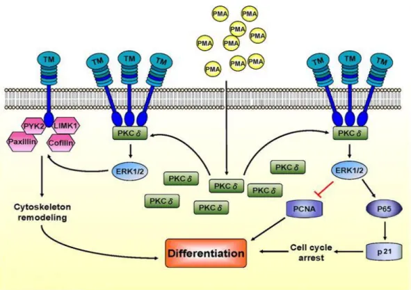
Section V. PKC Pathway Activation via PMA-Ionomycin
may be altered, so cancer cells that have been affected by treatments, such as chemotherapy or radiation, may not demonstrate normal activation responses (57).
Figure 1. Schematic representation of calcineurin/NFAT signaling (67).
synergistically with PMA to trigger intracellular calcium release. Collectively, these two
chemicals bypass the biological signaling cascade and directly activate the PKC pathway needed for nuclear factor of activated T cell (NFAT) signaling and cytokine production, a cascade that is typically initiated by ligand-surface receptor binding (1, 7, 11).
Figure 2. Schematic representation of PKC activation via PMA (70).
Section VI. Justification of Methodology
A brief explanation of certain methodology is provided as they were designed based off previous studies conducted by our authors.
Systemic epinephrine is known to stimulate androgenic leukocytosis around 70% of maximal oxygen uptake in healthy individuals (39). Evans et al. (17) conducted a study
ergometer at 60% of VO2peak, which resulted in significant increases in NK cell counts
immediately following exercise (17). It is important to note that ventilatory threshold is known to lie at exercise intensities between 50-75% VO2max (5). Hanson et al. (28) observed significant
increases in MAIT cell counts and cytokine expression following submaximal acute exercise in healthy young men who cycled at 90% of ventilatory threshold, an intensity equivalent to 45-68% of VO2max. Additionally, Hanson and colleagues (30) assessed stress hormone response to
acute exercise in prostate cancer patients in comparison to controls. The participants
intermittently exercised at 60% of the peak power output achieved during their VO2peak test. On
average, control participants cycled at 77.6% (+/-6.4), prostate patients without androgen therapy cycled at 80.5% 9.0), and prostate cancer patients on androgen therapy cycled at 84.8% (+/-8.1) of their VO2peak (30). The studies described above were successfully conducted with similar
clinical populations to that of the current study’s interest. This study utilized an acute,
intermittent exercise bout on a cycle ergometer for immune stimulation. All participants cycled at 60% of VO2peak wattage; an intensity roughly equivalent to 70% VO2max, and about 10%
greater than the intensity employed by Evans et al. (17).
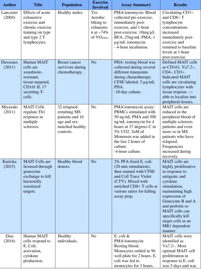
Table 1.T Cell proliferation assays
Author Title Population Exercise
Involved Assay Summary Results
Lancaster (2004)
Effects of acute exhaustive exercise and chronic exercise training on type and type 2 T lymphocytes.
Healthy males. Yes Aerobic biking to exhaustio n at ~74% of VO2max.
PMA/ionomycin: Blood collected pre-exercise, immediately post-exercise, and 1-hour post-exercise. 10mg/µL BFA, 25ng/mL PMA, 1 µg/mL ionomycin. - 4-hour incubation.
Circulating CD3+ and CD8+ T lymphocyte concentrations increased immediately post-exercise and returned to baseline levels at 1-hour post-exercise. Dusseaux (2011) Human MAIT cells are xenobiotic-resistant, tissue-targeted, CD161 IL 17 secreting T- cells.
Breast cancer survivors during chemotherapy.
No PHA: resting blood was collected during several different timepoints during chemotherapy. CFSE labeled. 5 µg/mL PHA.
-10-day culture.
Defined MAIT cells as CD161, Va7.2+, CD4-, CD3+. Indicated MAIT cells are circulating lymphocytes with tissue tropism --> able to localize into peripheral tissues. Miyazaki (2011) MAIT Cells regulate Th1 response in multiple sclerosis. 32 relapsed-remitting MS patients and 16 age and sex matched healthy controls.
No PMA/ionomycin assay: PBMCs stimulated with 50 ng/mL PMA and 500 ng/mL ionomycin for 4 hours at 37 degrees C in 5% CO2. 2uM of Monensin was added in the last 2 hours of culture.
-4-hour culture.
MAIT cells are reduced in the peripheral blood of multiple sclerosis patients and even more so in MS patients who have relapsed. Frequencies increased during recovery. Kurioka (2015)
MAIT Cells are licensed through granzyme exchange to kill bacterially sensitized targets.
Healthy blood donors.
No 2% PFA-fixed E. coli: (20 min stimulation), then stained with CFSE and Cell Trace Violet (CTV). Mixed with enriched CD8+ T cells at various ratios for killing assay prep.
MAIT cells are highly proliferative in response to antigenic and cytokine stimulation, maintaining high expression of Granzyme B and A and perforin so MAIT cells can specifically kill target cells in an MR1 dependent manner. Dias
(2016)
Human MAIT cells respond to E. Coli: activation, cytokine production, Healthy individuals.
No E. coli &
PMA/ionomycin: Resting blood.
Monocytes settled in 96 well plate for 2 hours. E. coli was fed to
monocytes for 3 hours.
proliferation, and cytotoxicity.
Isolated Va7.2+ was added and stimulation time was dependent on concentration of anti-CD28. Monensin and BFA added along with PMA cocktail to for 6 hours. Va7.2+ cells were stained with CTV and left to culture for several days.
~11 hours then 3 vs 5 vs 7 days predominately MR1 dependent. Gheradin (2018) Enumeration, functional responses and cytotoxic capacity of MAIT cells in newly diagnosed and relapsed multiple myeloma. Healthy individuals, and multiple myeloma patients.
No PMA assay: PBMCs co-cultured for 20 hours in human RF10 complete media in the presence of PFA-fixed DH5a. MAIT cell killing assay: rested overnight in RF10 complete media. Washed then stained for 20 min with mAbs directed against CD2, CD161, TRAV1-2 as well as 7AAD After culture, cells. Killing 20 hours. PMA 4 hours. E. coli 24 hours.
Frequency and function of MAIT in mm pt. b/c role in tumor immunity unknown.
Frequency in blood decreased from healthy adults (but similar to elderly). Frequency decreases as a function of age. No significant difference in percentage of MAIT between
mm/healthy, but significant difference in mm absolute number vs. healthy. Seth (2018) Primary sclerosing cholangitis leads to dysfunction and loss of MAIT cells.
Healthy
individuals, IBD patient, PBS pt., and PSC patient with and without IBD
No E. coli assay: PBMC's stimulated for 24 h with E. coli or combo with IL-12 or IL-18 -24-hour culture
Summary
CHAPTER III METHODOLOGY Participants
15 middle aged females with no history of cancer diagnosis were recruited to complete this study. Participants were sedentary but otherwise healthy, as defined as participating in two or less days of physical activity per week within six months prior to participation. All
participants were age-matched to the University of North Carolina at Chapel Hill’s ongoing Get REAL and HEEL BrCa survivor clinical trial participants. All BrCa survivor participants (n=21) were females who had histologically confirmed early stage (non-metastatic) BrCa, completed primary cancer treatment (chemotherapy, radiation, and/or surgery but may continue to be on endocrine therapy) within one year of taking part in the study, and were oncologist-approved to participate in exercise, but had no previous participation in UNC’s Get REAL and HEEL
exercise program. Exclusion criteria for the healthy controls included: a medical history of BrCa diagnosis or cancer treatment (chemotherapy, radiation, surgery, or hormonal therapy),
exercising more than two times per week for at least six months prior to beginning the study, any contraindications to participating in exercise, and pregnant women. The study was approved by the University of North Carolina at Chapel Hill’s institutional review board. Subjects signed an informed consent form prior to participation in the study.
Study Design
Physical Activity Questionnaire (IPAQ) was completed to determine baseline physical activity levels for the purposes of matching. Baseline assessments took place over three days of testing.
On day one of testing, pre-assessment guidelines were reviewed with the participants, including confirmation of overnight fast, no alcohol consumption during the previous 24 hours, and no caffeine prior to testing, then anthropometric data, including height, weight, blood pressure, and resting heart rate were collected. Body composition was measured via dual energy X-ray absorptiometry (DXA) (Hologic Inc., Bedford, MA, USA; Apex Software Version 3.3), a 12-lead resting electrocardiogram (EKG) (GE CASE Cardiosoft V. 6.6 ECG diagnostic system; General Electric, Palatine, IL, USA) was obtained, and familiarization for the cardiopulmonary exercise test (CPET) protocol was completed.
Participants reported back to the oncology lab approximately 3-7 days after the initial visit for day two of testing. Participants completed a cardiopulmonary graded exercise test on a cycle ergometer to determine maximal cycling power output (wattage) and VO2peak.
During visit three, blood samples were collected from participants at three separate time points via intravenous arm cannulation to evaluate blood biomarker responses to an acute exercise bout. First, resting blood samples were collected. Then participants completed a moderate intensity interval exercise trial. The acute exercise bout consisted of 10 3-minute intervals cycling on the cycle ergometer at 60% of maximal wattage achieved during VO2peak
testing with 1.5 minutes of stationary rest between each interval. Additional blood draws were performed immediately post exercise and one-hour post exercise trial.
Peripheral blood mononuclear cells (PBMC) were isolated using density gradient
time for analysis, PBMCs were thawed, treated and labeled with immunofluorescent antibody cocktail to distinguish specific cell type, and analyzed via flow cytometry (UNC Flow Cytometry CORE Lab) to determine Mucosal Associated Invariant T (MAIT) cell count and activation levels.
Body Composition Assessment
Participants reported to the exercise oncology research lab and researchers confirmed that pre-assessment guidelines were followed. Participant height (Perspectives Enterprises, Portage, MI, USA) and body mass (Tanita Corp, Tokyo, Japan) were measured and recorded. Control participants who indicated they were not post-menopausal on the medical history questionnaire provided a urine sample for a pregnancy test. Upon a negative pregnancy test result, participants moved forward with testing and completed the body composition assessment. Participants were centered on the DXA table in the supine position. All metal or jewelry was removed prior to the scan. A full body DXA scan was completed for each participant using standard DXA screening procedures to record total body mass, compositional aspects of lean body mass (LBM), fat tissue mass (FM), and percentage body fat (% BF). Body mass was determined using a high-grade analytical balance-scale (accuracy 10 grams).
Familiarization Session
continually at 15 watts per minute. Participants cycled until they reached 75% of heart rate reserve which allowed them to experience the general protocol and associated equipment without reaching maximal exhaustion.
Graded Exercise Test
Participant returned to the laboratory and researchers confirmed that pre-assessment guidelines were followed. All participants completed the CPET in the same manner as the familiarization session but continued to maximal exertion. Oxygen uptake and pulmonary ventilation was continuously recorded by a Parvo Medics’ TrueOne 2400 metabolic cart (Parvo Medics, Sandy, UT, USA), and breath by breath metabolic analysis was used to monitor whole body oxygen consumption (VO2). HR and RPE (6-20) were continuously monitored and
recorded throughout the testing. Termination of the test was determined by either subject reaching volitional exhaustion and signaling to stop the test, VO2 plateau or decrease with
increase in exercise intensity, or an abnormal subject response to the test was observed and therefore the research team terminated the test.
Blood Draws
Immediately post-exercise, the participants remained seated on the cycle ergometer and the second time point of blood was collected. Participants then cooled down while cycling at a light load (~10 watts) until heart rate and blood pressure approached baseline before moving to a chair for a 1-hour stationary recovery. During the recovery period, participants were given ad libitum access to water, but no other food or drink was consumed during this time before the final blood draw. At 1-hour post-exercise, a third blood draw was collected, and the cannula was removed.
Approximately 36 mL of blood were drawn into blood collection tubes containing EDTA and serum separators (BD Bioscience, Franklin Lanes, NJ, USA). The blood collection tubes were inverted several times and placed on ice for the duration of the visit until blood analysis. Exercise Trial
Participants completed an acute, intermittent standardized exercise protocol made up by 10 intervals. Each interval consisted of 3 minutes of cycling at 60% of the peak wattage obtained during the CPET on day two of testing and was followed by 1.5 minutes of passive recovery without pedalling. A total of 30 minutes of exercise in a 45-minute session was used to stimulate the immune system, as used previously (30). Participants were fitted with a heart rate monitor and began the exercise trial with a 1-minute warm up period on the cycle ergometer at zero resistance. Following the warmup, resistance increased to half of the calculated 60% peak wattage for 1-minute, then increased to the full 60% peak wattage for each 3-minute stage. HR and RPE (6-20) were recorded in the last 30 seconds of all stages.
Hematology Analysis
Immediately following each blood collection, complete blood counts (CBC) were run in duplicate with a maximal white blood cell difference of 0.1 cells/µL for each time point using a hematology analyzer (XP-300, Sysmex Corporation, Lincolnshire, IL, USA).
Calculation of Plasma Volume Shifts
Exercise induced plasma volume shifts were calculated according to the equation given by Dill and Costill (12). The hematocrit and hemoglobin values that were used in these
calculations were obtained from the complete blood count data pre-exercise, immediately post-exercise, and 1-hour post-exercise. Plasma volume shifts were reported to estimate the effect that exercise had on fluid shifts, which may affect concentrations of leukocytes.
Peripheral Blood Mononuclear Cells (PBMC) Isolation and Immunofluorescence Labeling Whole blood was diluted in a 1:1 ratio with phosphate buffered saline, and peripheral blood mononuclear cells (PBMCs) were isolated using SepMate tubes following manufacturer instructions (Stemcell, Vancouver, BC Canada). Cells were washed, counted via hemocytometer, and aliquoted for cryopreservation in 90% fetal bovine serum (FBA; VWR, Atlanta, GA, USA) and 10% DMSO (VWR, Atlanta, GA, USA), then stored at -80°C for future analysis. PBMCs were then thawed and used for immune cell phenotyping to conduct MAIT cell profile analysis.
Immune Function
Cryopreserved PBMCs were available for all participants. PBMCs were thawed, washed with complete media (90% RPMI, 10% FBS, 1.0% penicillin-streptomycin; (Sigma Aldrich, St. Louis, MO, USA), and rested overnight at 37°C and 5% CO2. PBMCs were then stimulated for 4 hours with 2 ng/mL of phorbol 12-myristate 13-acetate (PMA) and 1 µg/mL ionomycin (Sigma Aldrich, St. Louis, MO, USA) at 37°C and 5% CO2. Following the stimulation, cells were washed, and surface markers were labeled as described above. Cells were washed in 1x
phosphate buffered saline and treated with a fixation and permeabilization kit (BD Biosciences Cytofix/Cytoperm, San Jose, CA, USA) following manufacturer instructions. To quantify cytokine production, intracellular staining was performed using mouse anti-human monoclonal antibodies in 100 µL of permeabilization buffer [IFN-γ (APC); TNF-α (BV650), Biolegend, San Diego, CA, USA)] for 30 minutes at 4°C in the dark. Cells were washed to remove excess antibody prior to being suspended in 200 µL of cell staining buffer for flow cytometry analysis. Flow Cytometry
were calculated. All gating analyses were performed using FlowJo (Ashland, OR, USA). Circulating cell number was determined by multiplying the percentage of lymphocytes expressing the markers of interest with the hematology total lymphocyte count for each population.
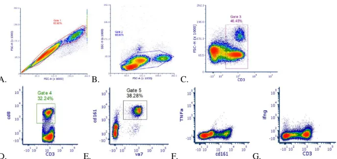
A. B. C.
D. E. F. G.
Figure 3.Gating strategy for MAIT cell and cytokine identification. A. Single cells. B. Side Scatter-Forward Scatter plot used to select lymphocytes. C. CD3+ T cells D. CD3+ and CD8+ Cytotoxic T cells E. MAIT cells were identified as Vα7.2+CD161+ cells. F. TNF-α expression in MAIT cells. G. IFN-γ expression in MAIT cells.
Statistical Analysis
Data collected was analyzed with Jamovi Statistics Version 0.9 (Jamovi Project
Computer Software, 2018). The α level was set a priori for all statistical procedures at α = 0.05. Descriptive statistics were used to summarize subject characteristics and values were reported as
mean ± SD. One-way ANOVAs were used to compare between group analyses for participant
characteristics and physiological responses to the VO2peak test and exercise trial. One-sample
t-tests were used to analyze plasma volume shifts from baseline to immediately post-exercise and
used to compare hematological data at three separate time points: pre-exercise, immediately
post-exercise, and one-hour after exercise between two separate groups: BrCa survivors and
CHAPTER IV RESULTS Participants
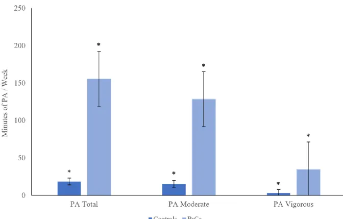
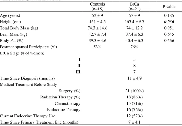
15 sedentary controls and 21 BrCa survivors completed the study. Physical characteristics for the 36 participants are presented in Table 2. The two groups were age-matched (P = 0.185). Although self-reported physical activity prior to participating in the study was different between groups (Figure 4), cardiorespiratory fitness, reported as relative VO2peak, was not different
between groups (P = 0.865) (Table 3).
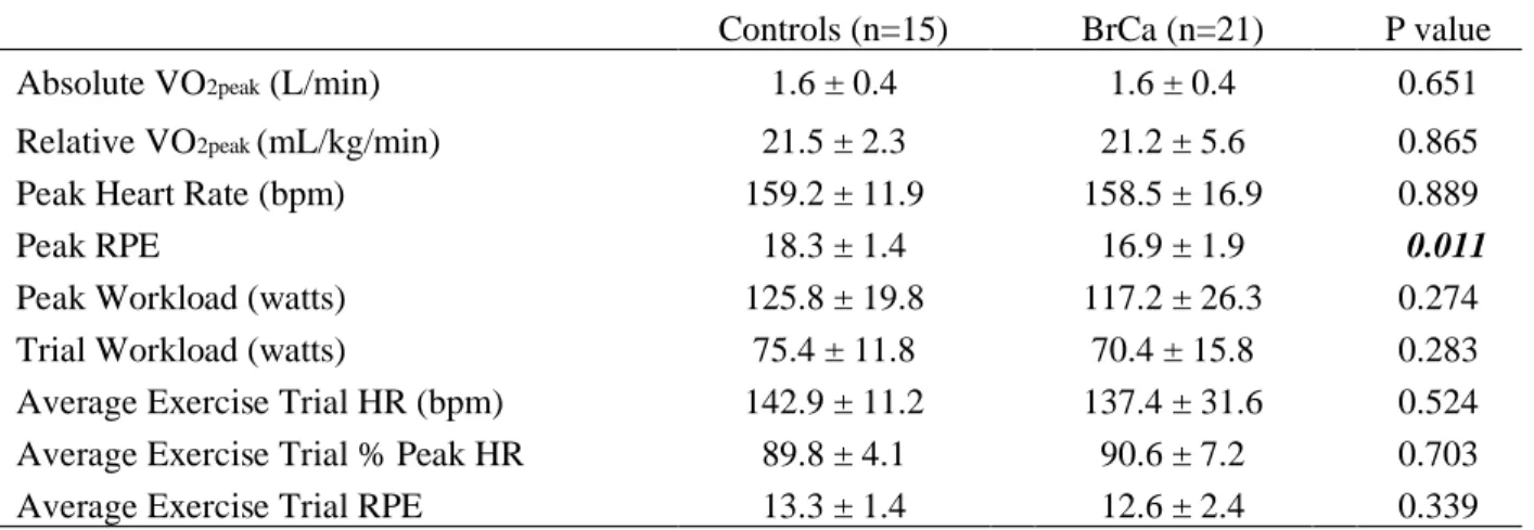
HR, RPE, workload, and metabolic responses to VO2peak test and the 45-minute acute
exercise trial are presented in Table 3. Relative VO2peak (P = 0.865), absolute VO2peak (P =
0.651), and peak heart rate (P = 0.889) achieved were similar between groups. All exercise trials were completed at 60% of VO2peak wattage, with an average workload of 75.4 watts in controls
and 70.4 watts in BrCa survivors. Additionally, exercise trial average HR (P = 0.524), percent of peak HR (P = 0.703), and RPE (P = 0.339) were all similar between groups. The exercise trial was reported as “very hard,” based on an average RPE of 18.3 (controls) and 16.9 (BrCa survivors).
Table 2. Participant characteristics
Controls
(n=15)
BrCa
(n=21) P value
Age (years) 52 ± 9 57 ± 9 0.185
Height (cm) 161 ± 4.5 165.4 ± 6.7 0.036
Total Body Mass (kg) 74.3 ± 14.6 74 ± 12.2 0.951
Lean Mass (kg) 42.7 ± 7.4 37.4 ± 6.3 0.645
Body Fat (%) 39.3 ± 4.6 40.4 ± 6.3 0.566
Postmenopausal Participants (%) 53% 76%
BrCa Stage (# of women)
I 5
II 8
III 7
Time Since Diagnosis (months) 11 ± 4.9
Medical Treatment Before Study
Surgery (%) 21 (100%)
Radiation Therapy (%) 18 (86%)
Chemotherapy 15 (71%)
Endocrine Therapy 16 (76%)
Current Endocrine Therapy Use 12 (57%)
Time Since Primary Treatment End (months) 7 ± 4.1
Table 3. Physiological response to VO2peak test and the exercise trial consisting of 10 sets of
cycling at 60% of peak wattage for 3 minutes followed by 1.5 minutes of passive recovery.
Controls (n=15) BrCa (n=21) P value
Absolute VO2peak (L/min) 1.6 ± 0.4 1.6 ± 0.4 0.651
Relative VO2peak (mL/kg/min) 21.5 ± 2.3 21.2 ± 5.6 0.865
Peak Heart Rate (bpm) 159.2 ± 11.9 158.5 ± 16.9 0.889
Peak RPE 18.3 ± 1.4 16.9 ± 1.9 0.011
Peak Workload (watts) 125.8 ± 19.8 117.2 ± 26.3 0.274
Trial Workload (watts) 75.4 ± 11.8 70.4 ± 15.8 0.283
Average Exercise Trial HR (bpm) 142.9 ± 11.2 137.4 ± 31.6 0.524
Average Exercise Trial % Peak HR 89.8 ± 4.1 90.6 ± 7.2 0.703
Average Exercise Trial RPE 13.3 ± 1.4 12.6 ± 2.4 0.339
Abbreviations: BrCa, Breast Cancer Survivor; VO2peak, peak oxygen uptake; HR, heart rate; RPE, Rate of Perceived Exertion.
Reported as mean ± standard deviation.
Changes in Leukocyte Counts
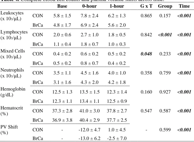
Cell counts for total leukocytes, lymphocytes, mixed cells, and neutrophils, as well as plasma volume shifts at each time point are presented in Table 4. Leukocyte counts were not available 1-hour post-exercise for 2 BrCa survivor participants and 1 control participant due to difficulties in cannulation. These missing samples accounted for a small percentage, and accepted statistical procedures allowed for appropriate analysis despite the missing data points. All leukocyte populations significantly increased from pre-exercise to immediately post-exercise (P < 0.001) and returned to baseline values 1-hour following the exercise bout (P < 0.001). Exercise and recovery patterns from leukocytes and leukocyte subsets were largely similar between controls and BrCa survivors, with the exception of lymphocyte counts that were significantly different between groups (P < 0.001). Main effects for time differences were analyzed for each group. At 0-hour, controls increased lymphocyte number by 35% (0.7 x 10³
From 0-hour to 1-hour following the exercise bout, controls decreased by 33.3% (-0.9 x 10³, (P <
0.001), and BrCa by 44.4% (-0.8 x 10³, (P < 0.001). A significant group x time interaction was
present for mixed cell counts (P = 0.048). Immediately post-exercise, controls increased by 50% (0.2 x 10³, P < 0.001), and BrCa increased by 60% (0.3 x 10³, P < 0.001). From 0-hour to 1-hour
following the exercise bout, controls decreased by 16.7% (-0.1 x 10³, P < 0.001), and BrCa by
50% (-0.4 x 10³, P < 0.001). Lymphocyte counts were statistically different between BrCa and
controls (P < 0.001).
Table 4. Complete blood cell counts and plasma volume shifts across time.
Base 0-hour 1-hour G x T Group Time
Leukocytes
(x 103/µL) CON 5.8 ± 1.5 7.8 ± 2.4 6.2 ± 1.3 0.865 0.157 <0.001
BrCa 4.8 ± 1.7 6.9 ± 2.4 5.6 ± 2.0
Lymphocytes
(x 103/µL) CON 2.0 ± 0.6 2.7 ± 1.0 1.8 ± 0.5 0.842 <0.001 <0.001
BrCa 1.1 ± 0.4 1.8 ± 0.7 1.0 ± 0.3
Mixed Cells
(x 103/µL) CON 0.4 ± 0.2 0.6 ± 0.2 0.5 ± 0.2 0.048 0.233 <0.001
BrCa 0.5 ± 0.2 0.8 ± 0.7 0.4 ± 0.2
Neutrophils
(x 103/µL) CON 3.5 ± 1.1 4.5 ± 1.6 4.0 ± 1.0 0.358 0.759 <0.001
BrCa 3.1 ± 1.6 4.3 ± 2.0 4.2 ± 1.8
Hemoglobin
(g/dL) CON 12.5 ± 1.3 13.5 ± 1.5 12.3 ± 1.4 0.160 0.927 <0.001
BrCa 12.3 ± 1.1 13.4 ± 1.1 12.5 ± 0.9
Hematocrit
(%) CON 37.3 ± 2.8 41.0 ± 3.0 37.8 ± 2.7 0.547 0.587 <0.001
BrCa 36.9 ± 3.8 40.4 ± 2.9 37.7 ± 2.5
PV Shift
(%) CON - -12.0 ± 4.7 1.0 ± 4.5 - 0.599 <0.001
BrCa - -13.0 ± 6.2 -2.5 ± 7.0
Abbreviations: BrCa, Breast Cancer Survivor; B, Baseline pre-exercise; 0h, immediately post-exercise; 1h, 1-hour post-exercise; PV, plasma volume.
Plasma Volume Shifts
Plasma volume shifts decreased by -12.0 ± 4.7 in controls and -13.0 ± 6.2 (both P <0.001, Table 4) in BrCa immediately following exercise before returning to baseline levels by 1-hour exercise. There were no differences between groups at 0-hour (P = 0.599) or 1-hour post-exercise (P = 0.089).
MAIT Cell Assays
Concurrent issues involving cell clumping following the thaws occurred, which lead to significant cell loss, so 10 µL deoxyribonuclease (DNAse) was added to the wash media used for the first centrifuge following thaw. Cells continued to clump, so a “rest-time following thaw” experiment that utilized DNAse in RPMI complete media for the initial wash and
A. B. C.
D. E.
F. G. H.
I. J.
Figure 5. TNF-α and IFN-γ cytokine expression in PMA Dose Experiment. A. TNF-α at 2ng PMA. B. TNF-α at 10 ng PMA. C. TNF-α
at 50ng PMA. D. TNF-α at 100ng PMA. E. TNF-α at 1000ng PMA. F. IFN-γ at 2ng PMA. G. IFN-γ at 10 ng PMA. H. IFN-γ at 50ng
PMA. I. IFN-γ at 100ng PMA. J. IFN-γ at 1000ng PMA.
CHAPTER V DISCUSSION
This aim of this study was to examine immune response to an acute bout of moderate intensity intermittent aerobic exercise in a BrCa survivor population compared with healthy but sedentary age-matched controls. In support of our hypotheses, leukocyte counts significantly increased immediately following the exercise trial and returned to baseline 1-hour after exercise similarly in BrCa survivors and controls. Although MAIT cell stimulations were unsuccessful, progress was made towards optimizing protocol for future functional analyses. It is important to note that the control group was closely age matched, displayed nearly identical body composition and cardiorespiratory fitness profiles to the BrCa survivors, and the physical stress of the
exercise trial, reported as trial workload, average HR, RPE, and percent of peak HR, was similar between groups. This is key as immune response tends to be directly proportional to exercise intensity (51, 54). However, because this acute exercise bout was intermittent, results may differ from responses to continuous exercise. Nonetheless, utilizing a standardized exercise intensity workload of 60% peak wattage for all participants allowed for valid interpretation of our study results.
immune response patterns of the control group, but were overall consistently lower in comparison, particularly in lymphocytes. Immune cell count deficiencies are a hallmark
characteristic in cancer patients post-treatment, so this suppressed immune response seen in the BrCa group may be due to the various cancer treatments they experienced prior to study
participation (4, 19, 31, 59, 61, 71). The small number of studies that have examined immune response to acute exercise in cancer populations produced similar findings to this study. Shore and Shepard (63) and Ladha et al. (41) assessed changes in leukocyte counts in response to 30 minutes of aerobic exercise in pediatric lymphoblastic leukemia patients. Shore and Shepard (63) participants exercised at VT whereas Ladha (41) participants exercised intermittently at 70-85% of VO2peak. Both studies reported significant leukocytosis immediately post-exercise and a return
to baseline values during exercise recovery with cell counts lower in cancer patients but not significantly different than healthy matched controls. Additionally, Evans et al. (17) exhibited biphasic exercise and recovery patterns of leukocyte counts and plasma volume shifts, which were largely similar between BrCa survivors and a control group following 30 minutes of moderate intensity intermittent aerobic exercise on a cycle ergometer. It is worth noting that immediately post-exercise leukocyte counts were greater in the current study’s participants compared to Evans et al. (17) whom analyzed similar BrCa and healthy control populations. This was most likely due to our participants exercising at 60% of their VO2peak wattage whereas
Evans’ participants exercised at 60% VO2peak.60% peak wattage is roughly equivalent to 70%
VO2peak, and thus this 10% increase in intensity may have provoked greater leukocytosis. BrCa
considered studies, and in turn, are encouraging as they suggest this moderate-intensity and duration form of exercise is both tolerable and practical for recently treated BrCa survivors.
The purpose of this study did not involve direct exploration of potential physiological mediators for leukocyte response to the acute moderate-intensity intermittent aerobic exercise bout. However, it is suggested that leukocytosis during exercise is due to a synergistic effect of increases in circulating catecholamines that occurs with increased cardiac output and
hemodynamics of shear mechanical forces that downregulate vascular endothelial-leukocyte adhesion (13, 64, 72). Although not measured in this study, similar protocols in BrCa (18) and prostate cancer survivors (30) have shown increases in catecholamines following acute exercise. The decrease in cell counts during recovery time may also be related to exercise-induced
endocrine and immune system interactions.
trial assays. Different phases of the menstrual cycle can illicit alterations in immune function (24). Both of these physiological variables may explain the perplexing results of the pilot experiments.
Protocol optimization included PMA dose responses, rest and stimulation incubation times, and cell clumping experiments. Previous studies had successful stimulation of T cells and specifically MAIT cells using PMA and ionomycin (Table 1). A methodology optimization study stimulating T cells in rats reported that 25 ng/mL of PMA combined with 1 µg/mL of ionomycin incubated for 6 hours was the best TNF-α, IFN-γ, and IL-2 cytokine production method (1). Lancaster and colleagues (Lancaster et al. 2004) used 25 ng/mL of PMA with 1 µg/mL
ionomycin in a 4-hour incubation to stimulate T lymphocytes to analyze IFN-γ cytokine response to exercise. Miyazaki et al. (Miyazaki et al. 2011) cultured MAIT cells from multiple sclerosis patients for 4 hours in 50 ng/mL PMA with 500 ng/mL ionomycin to examine intracellular cytokine expression, resulting in about 84% IFN-γ expression, and Hanson et al. (28) stimulated MAIT cells from healthy young males in response to acute submaximal aerobic exercise on a cycle ergometer for 4 hours in 2 ng/mL with 1 µg/mL ionomycin to produce about 70% TNF-α and 60% IFN-γ cytokine expression at baseline. The highest cytokine activation we
preliminary findings, protocol optimization was moving in the right direction for future analyses in MAIT cell counts, frequencies, and function between BrCa survivors and healthy controls.
This study is one of few to describe immune response to acute exercise in a cancer population. As with any study, there are limitations to the present investigation. First, there was variation in BrCa stage, forms of cancer treatment, and study enrollment time since completion of treatment. All cancer participants had been diagnosed and treated for stage I to III non-malignant BrCa, treated with chemotherapy, radiation, surgery, and/or endocrine therapy, and enrolled in this study within one year of completing treatment. However, physiological responses to exercise participation may differ for those with different cancer stages, forms of treatment, and longer recovery times since treatment therapies. Second, are the differences in physical activity between groups. A sedentary population was recruited as the control group because literature reports that cancer survivors tend to be less physically active than healthy persons. Contrarily, BrCa survivors in this study reported significantly higher physical activity habits prior to study participation than the age-matched controls, as illustrated in Figure 4. It is important to note that the IPAQ, a self-report physical activity questionnaire, was used to collect physical activity data, and self-report has its limitations as it is dependent on participant integrity. Although the IPAQ is a widely accepted and useful tool for measuring subjective physical activity, studies have shown that self-report questionnaires tend to report greater physical activity time than objective
additional limitation was the employment of a discontinuous exercise trial protocol, which may or may not have produced results consistent with that of a continuous protocol. Though, all participants were able to maintain 60% of their VO2peak wattage for the entirety of the exercise
trial. Finally, all cellular assays were performed ex vivo. Although these assays have been successfully practiced in many studies, the methods used may not be able to mimic the in vivo biological environment. For cytokines detection in PBMC’s, the isolation and cell culture procedures may wash out important immune molecules, including cells themselves, and disrupt molecular interactions (1, 35). This is particularly important to consider in cancer patient samples as cellular processes may have been altered during cancer prognosis and treatment.
It is well established that exercise has the ability to mobilize the immune system and activate cytotoxic immune cells that assist in immune regulation and pathogen defense in both healthy and oncology populations. The results of this study provide insightful information about cardiopulmonary and immune responses to exercise in individuals who have low
cardiorespiratory fitness levels due to being sedentary and/or undergoing cancer treatment. As previously discussed, most major cancer treatments compromise the function of many
In summary, 30 minutes of moderate-intensity intermittent aerobic exercise on a cycle ergometer elicited similar leukocyte responses in BrCa survivors to that of physically similar sedentary women without a history of cancer diagnosis. The present study was not able to assess MAIT cell function in BrCa survivors or the control group, though progress was made in
optimizing the stimulation assay protocol for future analyses. Immunosuppression post-cancer treatment is a serious concern associated with secondary malignancies, increased illness
REFERENCES
1. Ai W, Li H, Song N, Li L, Chen H. Optimal method to stimulate cytokine production and its use in immunotoxicity assessment. Int J Environ Res Public Health 10: 3834–42, 2013.
2. Battaglini CL, Mills RC, Phillips BL, Lee JT, Story CE, Nascimento MG, Hackney AC. Twenty-five years of research on the effects of exercise training in breast cancer survivors: A systematic review of the literature. World J Clin Oncol 5: 177–90, 2014. 3. Billerbeck E, Kang Y-H, Walker L, Lockstone H, Grafmueller S, Fleming V, Flint J,
Willberg CB, Bengsch B, Seigel B, Ramamurthy N, Zitzmann N, Barnes EJ, Thevanayagam J, Bhagwanani A, Leslie A, Oo YH, Kollnberger S, Bowness P, Drognitz O, Adams DH, Blum HE, Thimme R, Klenerman P. Analysis of CD161 expression on human CD8+ T cells defines a distinct functional subset with tissue-homing properties. Proc Natl Acad Sci 107: 3006–3011, 2010.
4. Blomgren H, Baral E, Edsmyr F, Strender LE, Petrini B, Wasserman J. Natural Killer Activity in Peripheral Lymphocyte Population Following Local Radiation Therapy. Acta Radiol Oncol 19: 139–143, 1980.
5. Brown SP. Exercise Physiology: Basis of Human Movement in Health and Disease [Online]. Lippincott Williams & Wilkins.
https://books.google.com/books/about/Exercise_Physiology.html?id=1b0iwv8-jGcC [6 Mar. 2019].
6. Campbell JP, Turner JE. Debunking the Myth of Exercise-Induced Immune Suppression: Redefining the Impact of Exercise on Immunological Health Across the Lifespan. Front Immunol 9: 648, 2018.
7. Chatila,’ T, Silverman L, Miller,’ And R, Geha R. MECHANISMS OF T CELL ACTIVATION BY IONOMYCIN’ THE CALCIUM IONOPHORE [Online]. http://www.jimmunol.org/ [17 Apr. 2019].
8. Chen Z, Wang H, D’Souza C, Sun S, Kostenko L, Eckle SBG, Meehan BS, Jackson DC, Strugnell RA, Cao H, Wang N, Fairlie DP, Liu L, Godfrey DI, Rossjohn J, McCluskey J, Corbett AJ. Mucosal-associated invariant T-cell activation and accumulation after in vivo infection depends on microbial riboflavin synthesis and co-stimulatory signals. Mucosal Immunol 10: 58–68, 2017.
10. Cosgrove C, Ussher JE, Rauch A, Gartner K, Kurioka A, Huhn MH, Adelmann K, Kang Y-H, Fergusson JR, Simmonds P, Goulder P, Hansen TH, Fox J, Gunthard HF, Khanna N, Powrie F, Steel A, Gazzard B, Phillips RE, Frater J, Uhlig H, Klenerman P. Early and nonreversible decrease of CD161++/MAIT cells in HIV infection. Blood 121: 951–961, 2013.
11. Crawford TQ, Jalbert E, Ndhlovu LC, Barbour JD. Concomitant evaluation of PMA+ionomycin-induced kinase phosphorylation and cytokine production in T cell subsets by flow cytometry. Cytom Part A 85: 268–276, 2014.
12. Dill DB, Costill DL. Calculation of percentage changes in volumes of blood, plasma, and red cells in dehydration. J Appl Physiol 37: 247–248, 1974.
13. Dimitrov S, Lange T, Born J. Selective mobilization of cytotoxic leukocytes by epinephrine. J Immunol 184: 503–11, 2010.
14. Dusseaux M, Martin E, Serriari N, Guillet IP, Premel V, Louis D, Milder M, Le Bourhis L, Soudais C, Treiner E, Lantz O. Human MAIT cells are xenobiotic-resistant, tissue-targeted, CD161 hi IL-17–secreting T cells. 117: 1250–1259, 2011.
15. Dusseaux M, Martin E, Serriari N, Péguillet I, Premel V, Louis D, Milder M, Le Bourhis L, Soudais C, Treiner E, Lantz O. Human MAIT cells are xenobiotic-resistant, tissue-targeted, CD161hi IL-17-secreting T cells. Blood 117: 1250–9, 2011.
16. Dyrstad SM, Hansen BH, Holme IM, Anderssen SA. Comparison of self-reported versus accelerometer-measured physical activity. Med Sci Sports Exerc 46: 99–106, 2014. 17. Evans ES, Hackney AC, McMurray RG, Randell SH, Muss HB, Deal AM, Battaglini
CL. Impact of Acute Intermittent Exercise on Natural Killer Cells in Breast Cancer Survivors. Integr Cancer Ther 14: 436–445, 2015.
18. Evans ES, Hackney AC, McMurray RG, Randell SH, Muss HB, Deal AM, Battaglini CL. Impact of Acute Intermittent Exercise on Natural Killer Cells in Breast Cancer Survivors. Integr Cancer Ther 14: 436–445, 2015.
19. Fairey AS, Courneya KS, Field CJ, Mackey JR. Physical exercise and immune system function in cancer survivors. Cancer 94: 539–551, 2002.
20. Fearon DT. Innate immunity and the biological relevance of the acquired immune response. [Online]. QJM 92: 235–7, 1999.
http://www.ncbi.nlm.nih.gov/pubmed/10615477 [23 Feb. 2019].
Kent SJ. MAIT cells are depleted early but retain functional cytokine expression in HIV infection. Immunol Cell Biol 93: 177–188, 2015.
22. Fu MR, Axelrod D, Guth AA, Cleland CM, Ryan CE, Weaver KR, Qiu JM,
Kleinman R, Scagliola J, Palamar JJ, Melkus GD. Comorbidities and Quality of Life among Breast Cancer Survivors: A Prospective Study. J Pers Med 5: 229–42, 2015. 23. Galvão DA, Nosaka K, Taaffe DR, Peake J, Spry N, Suzuki K, Yamaya K,
McGuigan MR, Kristjanson LJ, Newton RU. Endocrine and immune responses to resistance training in prostate cancer patients. Prostate Cancer Prostatic Dis 11: 160–165, 2008.
24. Gillum TL, Kuennen MR, Schneider S, Moseley P. A Review of Sex Differences in Immune Function after Aerobic Exercise. [date unknown].
25. Gleeson M, Bishop NC. The T cell and NK cell immune response to exercise. [Online]. Ann Transplant 10: 43–8, 2005. http://www.ncbi.nlm.nih.gov/pubmed/17037088 [31 Jan. 2019].
26. Godfrey DI, Uldrich AP, Mccluskey J, Rossjohn J, Moody DB. The burgeoning family of unconventional T cells. Nat. Immunol.: 2015.
27. Gold MC, McLaren JE, Reistetter JA, Smyk-Pearson S, Ladell K, Swarbrick GM, Yu YYL, Hansen TH, Lund O, Nielsen M, Gerritsen B, Kesmir C, Miles JJ,
Lewinsohn DA, Price DA, Lewinsohn DM. MR1-restricted MAIT cells display ligand discrimination and pathogen selectivity through distinct T cell receptor usage. J Exp Med 211: 1601–10, 2014.
28. Hanson ED, Danson E, Evans WS, Wood WA, Battaglini CL, Sakkal S. Exercise Increases Mucosal-associated Invariant T Cell Cytokine Expression but Not Activation or Homing Markers. Med Sci Sports Exerc 51: 379–388, 2019.
29. Hanson ED, Danson E, Nguyen-Robertson C V., Fyfe JJ, Stepto NK, Bartlett DB, Sakkal S. Maximal exercise increases mucosal associated invariant T cell frequency and number in healthy young men. Eur J Appl Physiol 117: 2159–2169, 2017.
30. Hanson ED, Sakkal S, Evans WS, Violet JA, Battaglini CL, McConell GK, Hayes A. Altered stress hormone response following acute exercise during prostate cancer
treatment. Scand J Med Sci Sports 28: 1925–1933, 2018.
31. Head JF, Elliott RL, McCoy JL. Evaluation of lymphocyte immunity in breast cancer patients. [Online]. Breast Cancer Res Treat 26: 77–88, 1993.
32. Hojman P. Exercise protects from cancer through regulation of immune function and inflammation. Biochem Soc Trans 45: 905–911, 2017.
33. Hojman P, Gehl J, Christensen JF, Pedersen BK. Molecular Mechanisms Linking Exercise to Cancer Prevention and Treatment. Cell Metab 27: 10–21, 2018.
34. Holmes MD, Chen WY, Feskanich D, Kroenke CH, Colditz GA. Physical Activity and Survival After Breast Cancer Diagnosis. JAMA 293: 2479, 2005.
35. House R V. Theory and Practice of Cytokine Assessment in Immunotoxicology. Methods 19: 17–27, 1999.
36. Jakóbisiak M, Lasek W, Gołab J. Natural mechanisms protecting against cancer. [Online]. Immunol Lett 90: 103–22, 2003.
http://www.ncbi.nlm.nih.gov/pubmed/14687712 [24 Feb. 2019].
37. Jönsson S, Olsson B, Jacobsson S, Palmqvist L, Ricksten A, Ekeland-Sjöberg K, Wadenvik H. BCR-ABL1 transcript levels increase in peripheral blood but not in granulocytes after physical exercise in patients with chronic myeloid leukemia. Scand J Clin Lab Invest 71: 7–11, 2011.
38. Koelwyn GJ, Wennerberg E, Demaria S, Jones LW. Exercise in Regulation of Inflammation-Immune Axis Function in Cancer Initiation and Progression. [Online]. Oncology (Williston Park) 29: 908–20, 922, 2015.
http://www.ncbi.nlm.nih.gov/pubmed/26676894 [31 Jan. 2019].
39. Kotchen TA, Hartley LH, Rice TW, Mougey EH, Jones LG, Mason JW. Renin, norepinephrine, and epinephrine responses to graded exercise. J Appl Physiol 31: 178–84, 1971.
40. Kruijsen-Jaarsma M, Révész D, Bierings MB, Buffart LM, Takken T. Effects of exercise on immune function in patients with cancer: a systematic review. [Online]. Exerc Immunol Rev 19: 120–43, 2013. http://www.ncbi.nlm.nih.gov/pubmed/23977724 [23 Feb. 2019].
41. Ladha AB, Courneya KS, Bell GJ, Field CJ, Grundy P. Effects of Acute Exercise on Neutrophils in Pediatric Acute Lymphoblastic Leukemia Survivors: A Pilot Study. J Pediatr Hematol Oncol 28: 671–677, 2006.