S T U D Y P R O T O C O L
Open Access
Colchicine effectiveness in symptom and
inflammation modification in knee osteoarthritis
(COLKOA): study protocol for a randomized
controlled trial
Ying-Ying Leung
1,2*, Julian Thumboo
1,2, Bak Siew Wong
3, Ben Haaland
1, Balram Chowbay
1, Bibhas Chakraborty
1,
Mann Hong Tan
4and Virginia B Kraus
5Abstract
Background:Despite the high prevalence and global impact of knee osteoarthritis (KOA), current treatments are palliative. No disease modifying anti-osteoarthritic drug (DMOAD) has been approved. We recently demonstrated significant involvement of uric acid and activation of the innate immune response in osteoarthritis (OA) pathology and progression, suggesting that traditional gout therapy may be beneficial for OA. We therefore assess colchicine, an existing commercially available agent for gout, for a new therapeutic application in KOA.
Methods/Design:COLKOA is a double-blind, placebo-controlled, randomized trial comparing a 16-week treatment
with standard daily dose oral colchicine to placebo for KOA. A total of 120 participants with symptomatic KOA will be recruited from a single center in Singapore. The primary end point is 30% improvement in total Western Ontario and McMaster Universities Osteoarthritis Index (WOMAC) score at week 16. Secondary end points include improvement in pain, physical function, and quality of life and change in serum, urine and synovial fluid biomarkers of cartilage metabolism and inflammation. A magnetic resonance imaging (MRI) substudy will be conducted in 20 participants to evaluate change in synovitis. Logistic regression will be used to compare changes between groups in an intention-to-treat analysis.
Discussion:The COLKOA trial is designed to evaluate whether commercially available colchicine is effective for improving signs and symptoms of KOA, and reducing synovial fluid, serum and urine inflammatory and biochemical joint degradation biomarkers. These biomarkers should provide insights into the underlying mechanism of
therapeutic response. This trial will potentially provide data to support a new treatment option for KOA. Trial registration:The trial has been registered at clinicaltrials.gov as NCT02176460. Date of registration: 26 June 2014.
Keywords:Knee osteoarthritis, colchicine, biomarkers, randomized controlled trial, double-blind, placebo-controlled, randomized
* Correspondence:[email protected]
1Duke NUS Graduate Medical School, Singapore, Singapore 2
Department of Rheumatology & Immunology, Singapore General Hospital, Singapore, Singapore
Full list of author information is available at the end of the article
Background
Knee osteoarthritis (KOA) is one of the five leading causes of disability among noninstitutionalized adults [1]. The risk of mobility disability (defined as needing help walking or climbing stairs) attributable to OA alone is greater than any other medical condition in people aged 65 and over [2]. In the World Health Organization Global Burden of Disease Study of 21 epidemiological regions across the world, from 1990 to 2010 there was a 26.8% increase in the burden of KOA as measured by years lived with disability per 100,000 persons [3]. The global prevalence of KOA will continue to rise in tan-dem with increases in the prevalence of obesity and lon-gevity worldwide. Despite the high prevalence and global impact of KOA, current treatments are limited to pallia-tive measures broadly focused on analgesia, and when this fails, surgical knee replacement. Given its frequency, associated disability and societal cost, patients with KOA are in urgent need of effective treatment that reduces pain and symptoms, and slows progression of OA. There are a number of agents, like glucosamine sulfate, chon-droitin sulphate, diacerin, doxycycline and licofelone that have demonstrated structural modifying capabilities in clinical trials [4]. Overall, these trials did not report a favorable profile with respect to side effects, no con-comitant symptomatic relief or failed to demonstrate the structural modifying endpoints as required by the Food and Drug Administration (FDA) [5].
In a KOA cohort without clinical evidence or self-report of gout, we found a significant correlation of syn-ovial fluid uric acid with radiographic and scintigraphic measures of OA severity [6]. We also observed strong correlations of radiological OA severity and synovial fluid uric acid with synovial fluid interleukin (IL)-18 and IL-1β; these two cytokines are classically produced dur-ing gout attacks by innate immune system activation mediated by uric acid crystal-induced inflammasome as-sembly in macrophages. These results strongly support the involvement of uric acid and the innate immune sys-tem in OA pathology and progression. The pain and symptom relieving effects of colchicine for KOA have been demonstrated in three small, but well-performed human randomized controlled trials (RCTs) [7-9]; however, the mechanism of action of colchicine in KOA has never been evaluated. We hypothesize that colchi-cine will block inflammasome-mediated inflammation, thereby improve the signs and symptoms of OA, and re-duce synovial fluid serum and urine inflammatory and biochemical joint degradation biomarkers. We propose a randomized controlled trial (RCT) of colchicine, an agent traditionally used for the treatment of gout, to examine the effects on signs and symptoms of KOA and to evaluate the mechanism of action of this therapy in OA through analysis of synovial fluid and systemic biomarker
profiles. The protocol of the COLKOA trial is outlined in this paper.
Methods/Design Trial objectives
The trial is designed to address the following objectives:
1. To determine whether daily oral colchicine at standard clinical doses (0.5 mg two times daily), compared to placebo, can decrease the pain of symptomatic KOA and improve function when used in addition to the patient’s current stable analgesic regimen.
2. To evaluate the mechanism of action of colchicine for reducing KOA signs and symptoms through analyses of synovial fluid, serum, and urine biomarker profiles; characterizing the state of joint tissue metabolism (through joint degradation and synthesis markers), inflammatory mediators of the innate immune system and the NACHT-LRR-PYD-containing protein-3 (NALP3) inflammasome before and after colchicine treatment compared to placebo.
Study design
COLKOA is a single center, double-blind randomized placebo-controlled clinical trial, conducted at Singapore General Hospital, comparing colchicine with placebo in patients with primary KOA. The study protocol was ap-proved by the SingHealth Centralized Institutional Re-view Board (ref. CIRB2012/659/E), and informed consent will be obtained from all participants before the start of the study. A clinical trial certificate (CTC1300061) was obtained from the Singapore Health Sciences Authority.
Participant selection and eligibility criteria
Adults aged 21 to 79 with symptomatic KOA will be recruited through advertisements, referrals from primary health care, orthopedic and internal medicine specialists. Study inclusion requires the following: symptomatic KOA meeting American College of Rheumatology (ACR) cri-teria [10]; radiographic cricri-teria for KOA with
Kellgren-Lawrence (KL) stage of ≥2 in at least one knee at
screening [11]; symptomatic KOA defined as a positive
response to the question,“Do you have pain, aching or
stiffness of the knee on most days of the past month;
and a score of ≥40 out of 100 on a visual analog scale
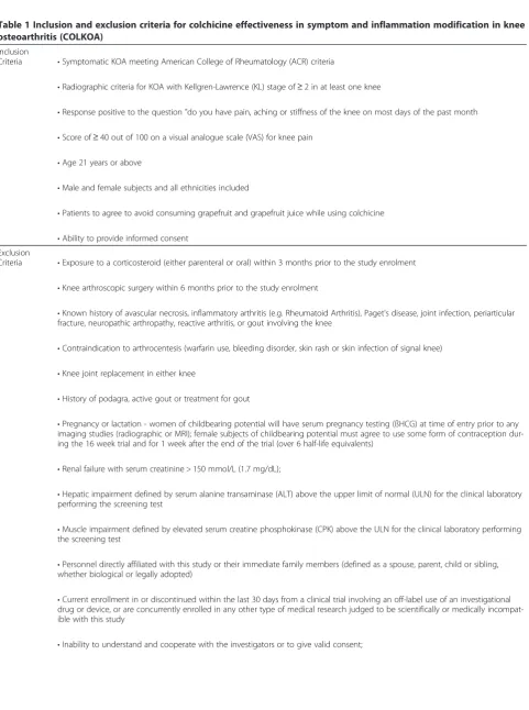
Table 1 Inclusion and exclusion criteria for colchicine effectiveness in symptom and inflammation modification in knee osteoarthritis (COLKOA)
Inclusion
Criteria •Symptomatic KOA meeting American College of Rheumatology (ACR) criteria
•Radiographic criteria for KOA with Kellgren-Lawrence (KL) stage of≥2 in at least one knee
•Response positive to the question "do you have pain, aching or stiffness of the knee on most days of the past month
•Score of≥40 out of 100 on a visual analogue scale (VAS) for knee pain
•Age 21 years or above
•Male and female subjects and all ethnicities included
•Patients to agree to avoid consuming grapefruit and grapefruit juice while using colchicine
•Ability to provide informed consent Exclusion
Criteria •Exposure to a corticosteroid (either parenteral or oral) within 3 months prior to the study enrolment
•Knee arthroscopic surgery within 6 months prior to the study enrolment
•Known history of avascular necrosis, inflammatory arthritis (e.g. Rheumatoid Arthritis), Paget's disease, joint infection, periarticular fracture, neuropathic arthropathy, reactive arthritis, or gout involving the knee
•Contraindication to arthrocentesis (warfarin use, bleeding disorder, skin rash or skin infection of signal knee)
•Knee joint replacement in either knee
•History of podagra, active gout or treatment for gout
•Pregnancy or lactation - women of childbearing potential will have serum pregnancy testing (ßHCG) at time of entry prior to any imaging studies (radiographic or MRI); female subjects of childbearing potential must agree to use some form of contraception dur-ing the 16 week trial and for 1 week after the end of the trial (over 6 half-life equivalents)
•Renal failure with serum creatinine > 150 mmol/L (1.7 mg/dL);
•Hepatic impairment defined by serum alanine transaminase (ALT) above the upper limit of normal (ULN) for the clinical laboratory performing the screening test
•Muscle impairment defined by elevated serum creatine phosphokinase (CPK) above the ULN for the clinical laboratory performing the screening test
•Personnel directly affiliated with this study or their immediate family members (defined as a spouse, parent, child or sibling, whether biological or legally adopted)
•Current enrollment in or discontinued within the last 30 days from a clinical trial involving an off-label use of an investigational drug or device, or are concurrently enrolled in any other type of medical research judged to be scientifically or medically incompat-ible with this study
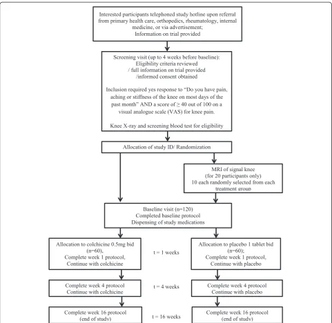
and no future treatment anticipated. As grapefruit is a moderate CYP3A4 inhibitor, and colchicine toxicity has been reported in a patient consuming it concomitantly, participants in this study have to agree to avoid con-suming grapefruit and grapefruit juice during the course of the RCT. The study design and schedule are summa-rized in Figure 1 and Table 2.
Treatment assignment and allocation concealment
The US Food and Drug Administration (FDA) and Singapore Health Sciences Authority approved colchicine for the treatment of acute flare-ups of gout and for chronic treatment of Familial Mediterranean Fever (FMF). Colchicine is available as a generic medication in Singapore. Generic colchicine (SIN 12490P) are repackaged for use in this study. Identical placebo tab-lets are produced to ensure allocation concealment. Upon production, study medications are packaged and re-labeled into numbered bottles by designated pharmacists according to the randomization list prepared by the trial biostatistician. Participants are randomly assigned in blocks of ten to the intervention arm or the placebo arm in a 1:1 ratio.
Blinding
Investigators and participants will remain blinded throughout the trial. Emergency unblinding will be allowed in limited situations that are perceived to impact the safety of study participants. Code-break envelopes for the full randomization schedule will be maintained by the designated biostatistician.
Intervention
Participants with symptomatic KOA will be randomized to oral colchicine 0.5 mg or placebo twice daily for 16 weeks. Participants are permitted to remain on their baseline adjunctive therapy, including topical analgesics, supplements, nonsteroidal anti-inflammatory drugs (NSAIDs) without changes of drug and dosage for the duration of the study. They will also be allowed the use of paracetamol <2 g/day as rescue analgesia. Pill count-ing at the end of the study will be conducted to deter-mine the amount of study drug and rescue medicine
utilized over the course of the 16-week study. Participants are asked not to start any new pharmacological and nonpharmacological therapies for their KOA, including new topical medications, acupuncture, exercise class or physiotherapy, for the duration of the study. Detailed in-formation on existing or new medication use for knee symptoms is collected at weeks 1, 4 and 16.
Clinical variables
Clinical variables include age, sex, ethnic group, educa-tional level, history of major joint injuries, history of knee joint surgery, comorbidities, and current medica-tions and dosage, presence or absence of pain in other joints; severity of pain of the signal knee and non-signal knee at rest and on movement. Physical examination will include measurement of joint alignment [13] and range of motion of the signal knee. The signal knee is defined as the one meeting entry criteria; if both knees meet entry criteria, the more symptomatic knee at base-line will be considered the signal knee; if symptoms are equal on both sides, the dominant knee will be designated the signal knee. Patient reported outcome measures include Western Ontario and McMaster Universities Osteoarthritis Index (WOMAC) [14], Health Assessment Questionnaire (HAQ) [15] and Outcome survey short-form 36 version 2 (SF36v2) [16]. All clinical variables are obtained at baseline and week 16, the end of the study.
Biospecimen collection
Prior to randomization, participants will have simultan-eous blood and urine collection and joint fluid aspiration from both knees by an experienced rheumatologist. Blood samples are obtained at least 2 hours post-prandial, and urine samples are obtained from the second void of the morning. The blood and urine sample collection is re-peated at week 1, week 4 and the end of study (week 16). Synovial fluid collection is repeated at the end of study. Synovial fluid is aspirated directly from each knee by a 20-gauge needle. If no fluid is obtained, 10 ml of sterile saline is injected into the joint and the fluid aspirated after gentle compression of the knee. The dilution factor of the lavage fluid (that obtained by saline Table 1 Inclusion and exclusion criteria for colchicine effectiveness in symptom and inflammation modification in knee osteoarthritis (COLKOA)(Continued)
•Contraindication for magnetic resonance imaging (MRI) - this is exclusion only for the subset of individuals selected for this im-aging procedure
•Anticipation of need for joint replacement within 4 months of the start of the intervention
injection) is determined according to an established urea method [17].
Soluble biomarker variables
In addition to uric acid, four types of soluble bio-markers will be assessed: inflammasome associated biomarkers, biomarkers of oxidant stress, other inflam-matory biomarkers, and cartilage degradation bio-markers. The serum (s) and synovial fluid (sf ) markers to be measured by category are the following: s/sf uric
acid; inflammasome associated (s/sf IL-1 ß , IL-18, tumor
necrosis factor (TNF)α, caspase-1, Toll-like receptor
[image:5.595.57.539.87.555.2]In addition, s/sf urea will be measured to correct sf biomarker concentrations for any dilution introduced by lavage.
Magnetic resonance imaging variables
The specific subjects selected for MRI were pre-specified, without knowledge of their clinical data, as the 6th to 25th participants being randomized to colchicine and placebo
[image:6.595.59.539.102.569.2]respectively. This choice ensured, based on block randomization, that half (n = 10) would be colchicine-treated and half (n = 10) placebo-colchicine-treated. The MRI exami-nations consist of a standardized set of acquisitions with specific positioning and localization requirements. The MRI imaging will be performed using a 3 Tesla Siemens Skyra (2012, Simens, Erlangen, Munich, Germany) with the signal knee immobilized in a 15 Channel Transmit/ Table 2 Schedule of assessment and data collection
Screening visit Allocation Post allocation
Time point Up to 4 weeks before
BL
0 Baseline visit (BL)
Week 1
Week 4
Week 16 (end)
Enrolment:
Eligibility screening x
Informed consent x
Screening knee X-ray x
Screening blood test * x
Physical Examination x x x x x
Interventions:
Colchicine 0.5 mg bid Start here End here
Placebo 1 tablet bid Start here End here
Assessments:
Physical Examination x x x x x
Full blood count x x
Renal function test x x x x
Liver function test x x x x
Uric acid x x x x
CPK x x x x
βHCG (for women with child bearing potential) x
Blood biomarkers collection x x x x
Urine biomarkers collection x x x x
Blood for pharmcogenomic x
Colchicine level x x x x
Both knees aspiration x x
Knee X-ray (both sides) x
MRI of signal kneeǂ(for 20 subjects only) x
Adverse event enquiries x x x x
Enquiries to compliance to usual medications for knee
x x x x
Clinical data collection x x
Pain score enquiries (signal knee) x x x x
Demographic data collection x
WOMAC x x
HAQ x x
SF36v2 x x
Receive Knee coil. The intravenous contrast medium used will be Dotarem, 0.5 mmol/ml (Gadoteric Acid, Guerbet, France) with a maximum administered volume of 10 ml. The whole knee joint will be scored by an expe-rienced musculoskeletal radiologist blinded to clinical details of participants, using the Boston Leeds Osteo-arthritis Knee Score (BLOKS) [18]. At the time of the screening evaluation, knee radiography of all patients will be performed with the SynaFlexor platform and variable beam angle as previously described [19] to optimize images for serial joint space width measure-ments in the event of any future long-term follow-up of the participants in this trial.
Safety evaluation
Safety and tolerability of treatment will be assessed at weeks 1, 4 and 16. Safety is assessed by identifying ad-verse events (AEs) during treatment using open-ended questions and a checklist that includes diarrhea, myalgia and muscle weakness, and physical examination of muscle tenderness and muscle strength. Laboratory assessments include renal and liver function tests and creatine phos-phokinase (CPK). All patients are given a 24-hour emer-gency contact number for enquiries should there be adverse events. Patients who are noted to have significant adverse events at week 4, such as myalgia or an increase in arthralgia will be contacted by the study team via tele-phone and arrangements will be made for extra outpatient visits as clinically indicated. Adverse events are recorded at each visit and up to 16 weeks and are analyzed for their seriousness, intensity and causal relationship with treat-ment and outcome according to the National Institutes of Health standardized Common Terminology Criteria [20]. A Data Safety and Monitoring Board consisting of two in-dependent rheumatologists and one biostatistician will monitor the study biannually and review reportable ad-verse events and be responsible for the decision to con-tinue the study in the event of a drug related serious adverse effect.
Outcomes
The primary endpoint will be 30% improvement in total WOMAC score of the signal knee. Secondary endpoints include change in WOMAC pain score and physical function score, HAQ, SF36v2 and the quantity of rescue medication used. Other secondary endpoints include treatment response defined by the Outcome Measures in Rheumatology Clinical Trials and Osteoarthritis Re-search Society International (OMERACT-OARSI) 2004 criteria [21], wherein participants are classified as re-sponders if the pain or physical function score is
de-creased by ≥30% and by at least 20 mm on the VAS.
Other endpoints include statistically significant (P<0.05) treatment related change in MRI detected synovitis and
cartilage morphology and decline in synovial fluid IL-18,
IL-1β, or TNF-α. We will also explore the
polymor-phisms of multidrug resistance gene-1 (MDR-1) genes (1236C > T, 2677G > A/T, 3435C > T) that encodes P-gp and cytochrome CYP3A4*1B, CYP3A5*3 and CYP3A5*6; corresponding to treatment response to colchicine and drug levels in serum and synovial fluid.
Sample size estimation
Based on the results of an existing RCT [8], we expect to achieve a 30% improvement rate in WOMAC in 57% and 23% of the subjects in the colchicine and placebo groups, respectively. To detect this difference at a sig-nificance level of 5% (P<0.05) and 80% power, 32 partic-ipants per group (64 total) will be required; for 90%, 42 participants per group (84 total) will be required. We therefore plan to enroll 120 participants for this trial to insure 90% power even with a dropout rate of up to 30%.
Statistical analysis
Analyses will be conducted using R 3.1.1 (R Core Team,
Vienna, Austria). Throughout, P values <0.05 will be
considered statistically significant. Analyses will be per-formed from an intention-to-treat perspective. The pro-portion of participants achieving the primary endpoint, 30% improvement in total WOMAC at week 16, will be compared between the colchicine and placebo groups using a chi-squared test. Participants who do not have a
follow-up WOMAC will be assumed to not have a≥30%
aPvalue <0.05 entry criterion. Secondary endpoints will be analyzed in the context of GEE models, depending on whether the endpoint is categorical or continuous, with a fixed effect for treatment. Secondary endpoints that are measured on both of a patient’s knees will add-itionally include fixed effects for KOA and KOA by treatment interaction. Secondary analyses will also be supplemented by covariate adjusted tests, where covari-ates will once again be selected using stepwise variable selection with AIC. To evaluate the mechanism of ac-tion of colchicine for reducing knee OA signs and symptoms, the change from baseline to 16 weeks (at study end) in synovial fluid, serum, and urine biomarker profiles will be compared between oral colchicine and placebo treatment. The relationship between response
(≥30% improvement in WOMAC) and change from
baseline to 16 weeks (at study end) in synovial fluid, serum, and urine biomarker profiles will be assessed in the context of a logistic regression model. Various ex-ploratory statistical models and analyses such as struc-tural equation modelling and factor analysis will be performed to further elucidate the mechanisms of ac-tion of colchicine.
Discussion
This RCT is proposed at a time when effective treat-ments for OA are urgently needed. It is well grounded in a strong biological rationale and a strong clinical ra-tionale with the several small preceding RCTs of colchi-cine for KOA. This proposed RCT is two- to threefold larger than any of the existing trials allowing greater power to test and validate the clinical effectiveness of colchicine for knee OA signs and symptoms. This RCT will break new ground by exploring the mechanism of action of colchicine in knee OA through testing the ability of colchicine to inhibit inflammasome activation in OA. Finally and importantly, this trial can serve as a valuable paradigm for a subsequent larger longer-term multicentered RCT with long-term radiographic and MRI follow-up to evaluate the effects of colchicine to slow or halt progression of OA structural deterioration in addition to evaluating its long-term effects on OA symptoms and function.
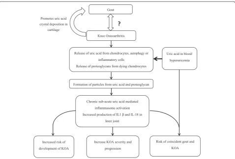
The association of uric acid and OA has long been ob-served [23], although not statistically significant after controlling for body mass index in cohort studies [24-26]. One study noted that acute attacks of gout co-incided with the presence of clinical OA in the same joint suggesting that OA may facilitate the localized de-position of monosodium urate (MSU) crystals [27]. In a recent study of cadaveric ankles (talus surface only), crystals were strongly associated with cartilage lesions -overall 92% of cartilage lesion cases were associated with crystal deposition of which 67% and 33% were
MSU and calcium pyrophosphate dihydrate (CPPD) crystals, respectively [28]. Apart from the morphological changes, crystal deposition in joints was also associated with immunohistochemical changes including superfi-cial zone protein and collagen X that is associated with cartilage degeneration and repair. A study of human chondrocyte explants also demonstrated that MSU crys-tals inhibit chondrocyte viability and function [29]. In a study among OA subjects without clinical gout, we re-cently demonstrated a strong positive association
be-tween synovial fluid uric level, IL18 and IL1β and OA
severity as measured by imaging [6]. This suggested that MSU crystals may contribute both to the initiation and propagation of cartilage degradation. We hypothesize in OA that the degradation of cartilage extracellular matrix components may facilitate the nucleation of MSU crystals, forming microparticles that trigger the innate immune response via activation of dendritic cells and macrophages with subsequent priming of T cells (Figure 2).
This new conception of the pathogenesis of OA has very important treatment implications; it suggests that existing therapies for gout may be of benefit in OA. One such long-standing therapy for gout, colchicine, effect-ively suppresses the innate immune response at various levels; this makes colchicine a strong candidate as an effective treatment. At micromolar concentrations (from 0.5μM to 5μM) colchicine has several anti-inflammatory mechanisms of action: it suppresses activation of the MSU crystal-induced NALP3 inflammasome, and it suppresses IL-1βprocessing and release and neutrophil recruitment. It is important to realize that colchicine preferentially ac-cumulates in leukocytes. The concentration in neutrophils may be more than 16 times the peak concentration in plasma. It is possible that a continuous low prophylactic dose of colchicine can achieve an intracellular concentra-tion in macrophages high enough to inhibit NALP3 inflammasome activation that is triggered by danger sig-nals formed after release of uric acid and cartilage extra-cellular matrix fragments from degrading cartilage in OA.
At much lower (nanomolar) concentrations, colchicine has additional mechanisms of action: it blocks the re-lease of a crystal-derived chemotactic factor from neu-trophil lysosomes; and it blocks neuneu-trophil adhesion to endothelium. In anin vivostudy colchicine inhibits MSU crystal-induced production of superoxide anions from mouse neutrophils and macrophages at doses 100 times
lower (given by oral gavage at doses of 0.05 μmol/kg,
4 hours prior to stimulation) than those required for in-hibition of neutrophil infiltration (oral gavage at
5 μmol/kg) [30]. This suggests that colchicine at the
has been demonstrated in preceding small RCTs. One of these RCT was done in subjects with signs of knee inflam-mation, while the other two were among subjects with pri-mary KOA [7-9]. The main limitations in these RCTs include single-centered studies with small sample sizes. One study was in a subgroup of patients who had signifi-cant knee effusions who received intra-articular cortico-steroids, thus limiting its generalizability [7]. Two studies did not use standardized outcome measures [7,9], and in all studies, the underlying mechanism of colchicine on KOA were not evaluated. The long-term safety profile of colchicine is well known based on its traditional use as a treatment for gout, its evaluation in clinical trials used for marketing approval of colchicine for gout [31], and its use as the standard first line and long-term treatment for FMF [32-34]. An additional recent RCT, evaluating colchicine at 0.5 mg/day for 3 years in addition to statins and stand-ard therapies, appeared effective for the prevention of car-diovascular events in participants with stable coronary disease and also demonstrated an excellent safety profile [35]. Apart from the US, another distinct advantage of col-chicine is its availability as generic medication. If this
proof of concept study is successful, it may potentially benefit a large OA patient population at an affordable price.
[image:9.595.59.538.87.411.2]groups due to regression to the mean. However, patients with higher pain scores constitute the population of inter-est and randomization ensures that there will be no sys-tematic difference between the arms other than treatment versus placebo.
In summary, KOA is disabling and is increasingly prevalent. A DMOAD treatment to relieve symptoms and progression is urgently needed. Colchicine has the-oretical and some clinical evidence of benefit in subjects with KOA. It has a good safety profile and is practical for long term use. The COLKOA trial will stringently test its clinical efficacy and underlying mechanism of ac-tion. This will potentially provide a new and affordable treatment for KOA.
Trial status
ClinicalTrials.gov Identifier: NCT02176460. Recruitment for this trial started in October 2013. As of December 2014, 83 participants have been recruited and 61 participants have completed the trial protocol. At the time of manuscript submission, recruitment and follow-up of patients is in progress.
Additional file
Additional file 1: Table S1.Drugs that interact negatively with colchicine [12].
Abbreviations
ACR:American College of Rheumatology; AEs: adverse events; AIC: Akaike information criterion; ALT: alanine transaminase; CPK: creatine phosphokinase; CPPD: calcium pyrophosphate dehydrate; Cmax: mean peak concentration; CTXII: C-terminal crosslinked telopeptide type II collagen; CYP3A4: Cytochrome 450 3A4; DMOAD: disease modifying anti-osteoarthritis drug; FDA: Food and Drug Administration; FMF: familial Mediterranean Fever; HAQ: Health Assessment Questionnaire; Hs-CRP: high sensitivity C-reactive protein; IL: interleukin; KL: Kellgren-Lawrence staging of radiographic OA; KOA: knee osteoarthritis; MAR: missing at random; MDR-1: multidrug resistance gene-1; MMP: matrix metalloproteinase; MRI: magnetic resonance imaging; MSU: monosodium urate; NALP3: NACHT-LRR-PYD-containing protein-3 NSAIDs, nonsteroidal anti-inflammatory drugs; OMERACT-OARSI: Outcome Measures in Rheumatology Clinical Trials and Osteoarthritis Research Society International criteria; P-gp: P-glycoprotein; PPi: inorganic pyrophosphate; RCT: randomized controlled trial; SF36v2: Outcome Survey Short-form 36 version 2; S: Serum; Sf: synovial fluid; TLR: toll-like receptor; TNF: tumor necrosis factor; ULN: upper limit of normal; VAS: visual analog scale; WOMAC: Western Ontario and McMaster Universities Osteoarthritis Index.
Competing interests
All authors declare that they have no competing interests.
Authors’contributions
YYL conceived the study, obtained funding support, participated in study design, and drafted the manuscript. JT participated in study design. BSW participated in study design, and participated in drafting the manuscript. BH participated in study design, and assisted with drafting the manuscript. BalC participated in study design, and assisted with drafting the manuscript. MHT participated in study design. VBK conceived the study, obtained funding support, participated in study design, and assisted with drafting the manuscript. All authors read and approved the final version for submission.
Acknowledgments
We wish to acknowledge the Duke-NUS Governing Board for funding support to VBK and YYL for this Duke/Duke-NUS Collaborative Project. We also wish to acknowledge the assistance of the Duke Image Analysis Laboratory, directed by Dr. Cecil Charles and assisted by Ms Maureen Ainslie, for expert assistance in developing a training manual for the knee radiography protocol.
Author details 1
Duke NUS Graduate Medical School, Singapore, Singapore.2Department of Rheumatology & Immunology, Singapore General Hospital, Singapore, Singapore.3Department of Diagnostic Radiology, Singapore General Hospital, Singapore, Singapore.4Department of Orthopaedic Surgery, Singapore
General Hospital, Singapore, Singapore.5The Duke Molecular Physiology Institute and Division of Rheumatology, Department of Medicine, Duke University School of Medicine, Durham, NC, USA.
Received: 23 January 2015 Accepted: 20 April 2015
References
1. Murray C, Lopez A. The global burden of disease. 1996.
2. Guccione AA, Felson DT, Anderson JJ, Anthony JM, Zhang Y, Wilson PW, et al. The effects of specific medical conditions on the functional limitations of elders in the Framingham Study. Am J Public Health. 1994;84:351–8. 3. Vos T, Flaxman AD, Naghavi M, Lozano R, Michaud C, Ezzati M, et al. Years
lived with disability (YLDs) for 1160 sequelae of 289 diseases and injuries 1990–2010: a systematic analysis for the Global Burden of Disease Study 2010. Lancet. 2012;380:2163–96.
4. Martel-Pelletier J, Wildi LM, Pelletier JP. Future therapeutics for osteoarthritis. Bone. 2012;51:297–311.
5. Conaghan PG, Hunter DJ, Maillefert JF, Reichmann WM, Losina E. Summary and recommendations of the OARSI FDA osteoarthritis Assessment of Structural Change Working Group. Osteoarthritis Cartilage. 2011;19:606–10. 6. Denoble AE, Huffman KM, Stabler TV, Kelly SJ, Hershfield MS, McDaniel GE, et al. Uric acid is a danger signal of increasing risk for osteoarthritis through inflammasome activation. Proc Natl Acad Sci U S A. 2011;108:2088–93. 7. Das SK, Mishra K, Ramakrishnan S, Srivastava R, Agarwal GG, Singh R, et al. A
randomized controlled trial to evaluate the slow-acting symptom modifying effects of a regimen containing colchicine in a subset of patients with osteoarthritis of the knee. Osteoarthritis Cartilage. 2002;10:247–52. 8. Das SK, Ramakrishnan S, Mishra K, Srivastava R, Agarwal GG, Singh R, et al. A
randomized controlled trial to evaluate the slow-acting symptom-modifying effects of colchicine in osteoarthritis of the knee: a preliminary report. Arthritis Rheum. 2002;47:280–4.
9. Aran S, Malekzadeh S, Seifirad S. A double-blind randomized controlled trial appraising the symptom-modifying effects of colchicine on osteoarthritis of the knee. Clin Exp Rheumatol. 2011;29:513–8.
10. Altman R, Alarcón G, Appelrouth D, Bloch D, Borenstein D, Brandt K, et al. The American College of Rheumatology criteria for the classification and reporting of osteoarthritis of the hip. Arthritis Rheum. 1991;34:505–14. 11. KELLGREN JH, LAWRENCE JS. Radiological assessment of osteo-arthrosis.
Ann Rheum Dis. 1957;16:494–502.
12. FDA. Online, available at: http://www.fda.gov/Drugs/DrugSafety/ PostmarketDrugSafetyInformationforPatientsandProviders/ DrugSafetyInformationforHeathcareProfessionals/ucm174315.htm. 13. Kraus VB, Vail TP, Worrell T, McDaniel G. A comparative assessment of
alignment angle of the knee by radiographic and physical examination methods. Arthritis Rheum. 2005;52:1730–5.
14. Bellamy N, Buchanan WW, Goldsmith CH, Campbell J, Stitt LW. Validation study of WOMAC: a health status instrument for measuring clinically important participant relevant outcomes to antirheumatic drug therapy in participants with osteoarthritis of the hip or knee. J Rheumatol. 1998;15:1833–40.
15. Fries JF, Spitz PW, Young DY. The dimensions of health outcomes: the health assessment questionnaire, disability and pain scales. J Rheumatol. 1982;9:789–93.
16. Ware JE, Sherbourne CD. The MOS 36-item short-form health survey (SF-36). I. Conceptual framework and item selection. Med Care. 1992;30:473–83. 17. Kraus VB, Stabler TV, Kong SY, Varju G, McDaniel G. Measurement of synovial
18. Hunter DJ, Lo GH, Gale D, Grainger AJ, Guermazi A, Conaghan PG. The reliability of a new scoring system for knee osteoarthritis MRI and the validity of bone marrow lesion assessment: BLOKS (Boston Leeds Osteoarthritis Knee Score). Ann Rheum Dis. 2008;67:206–11. 19. Charles HC, Kraus VB, Ainslie M, Hellio Le Graverand-Gastineau MP.
Optimization of the fixed-flexion knee radiograph. Osteoarthritis Cartilage. 2007;15:1221–4.
20. Common Terminology Criteria for Adverse Events (CTCAE) Version 4. 2009 http://evs.nci.nih.gov/ftp1/CTCAE/About.html. Accessed 28 Apr 2015.21. 21. Pham T, van der Heijde D, Altman RD, Anderson JJ, Bellamy N, Hochberg M,
et al. OMERACT-OARSI initiative: Osteoarthritis Research Society International set of responder criteria for osteoarthritis clinical trials revisited.
Osteoarthritis Cartilage. 2004; 12:389–99.
22. Roderick J A Little, Rubin DB. Statistical analysis with missing data. Wiley-Interscience, New Jersey; 2002
23. Acheson RM, Collart AB. New Haven survey of joint diseases. XVII. Relationship between some systemic characteristics and osteoarthrosis in a general population. Ann Rheum Dis. 1975; 34:379–87
24. Anderson JJ, Felson DT. Factors associated with osteoarthritis of the knee in the first national Health and Nutrition Examination Survey (HANES I). Evidence for an association with overweight, race, and physical demands of work. Am J Epidemiol. 1988;128:179–89.
25. Felson DT, Anderson JJ, Naimark A, Walker AM, Meenan RF. Obesity and knee osteoarthritis. The Framingham Study. Ann Intern Med. 1988;109:18–24.
26. Schouten JS, van den Ouweland FA, Valkenburg HA. A 12 year follow up study in the general population on prognostic factors of cartilage loss in osteoarthritis of the knee. Ann Rheum Dis. 1992;51:932–7.
27. Roddy E, Zhang W, Doherty M. Are joints affected by gout also affected by osteoarthritis? Ann Rheum Dis. 2007;66:1374–7.
28. Muehleman C, Li J, Aigner T, Rappoport L, Mattson E, Hirschmugl C, et al. Association Between Crystals and Cartilage Degeneration in the Ankle. J Rheumatol. 2008;35:1108–17.
29. Chhana A, Callon KE, Pool B, Naot D, Gamble GD, Dray M, et al. The Effects of Monosodium Urate Monohydrate Crystals on Chondrocyte Viability and Function: Implications for Development of Cartilage Damage in Gout. J Rheumatol. 2013;40:2067–74.
30. Chia EW, Grainger R, Harper JL. Colchicine suppresses neutrophil superoxide production in a murine model of gouty arthritis: a rationale for use of low-dose colchicine. Br J Pharmacol. 2008;153:1288–95.
31. Terkeltaub RA, Furst DE, Bennett K, Kook KA, Crockett RS, Davis MW. High versus low dosing of oral colchicine for early acute gout flare: Twenty-four-hour outcome of the first multicenter, randomized, double-blind, placebo-controlled, parallel-group, dose-comparison colchicine study. Arthritis Rheum.
2010;62:1060–8.
32. Dinarello CA, Wolff SM, Goldfinger SE, Dale DC, Alling DW. Colchicine therapy for familial Mediterranean fever. A double-blind trial. N Engl J Med. 1974;291:934–7.
33. Ben-Chetrit E, Levy M. Colchicine prophylaxis in familial Mediterranean fever: reappraisal after 15 years. Semin Arthritis Rheum. 1991;20:241–6.
34. Kallinich T, Haffner D, Niehues T, Huss K, Lainka E, Neudorf U, et al. Colchicine use in children and adolescents with familial Mediterranean fever: literature review and consensus statement. Pediatrics. 2007;119:e474–83.
35. Nidorf SM, Eikelboom JW, Budgeon CA, Thompson PL. Low-dose colchicine for secondary prevention of cardiovascular disease. J Am Coll Cardiol. 2013;61:404–10.
36. Food and Drug Administration. Guidance for industry Clinical development programs for drugs devices and biological products intended for the treatment of osteoarthritis FDA document 1999, 7. 1999.
Submit your next manuscript to BioMed Central and take full advantage of:
• Convenient online submission
• Thorough peer review
• No space constraints or color figure charges
• Immediate publication on acceptance
• Inclusion in PubMed, CAS, Scopus and Google Scholar
• Research which is freely available for redistribution