Journal of Chemical and Pharmaceutical Research, 2016, 8(4):853-857
Research Article
CODEN(USA) : JCPRC5
ISSN : 0975-7384
Antimicrobial efficacy of ethyl acetate extract of Mesua ferrea
R. Lakshmi Sundaram
V ClinBio labs, Central Research Facility, Sri Ramachandra University, Porur, Chennai-116,Tamil Nadu, India _____________________________________________________________________________________________
ABSTRACT
India is a varietal emporium of medicinal plants and is one of the richest countries in the world in regard to genetic resources of medicinal plants. It exhibits a wide range in topography and climate, which has a bearing on its vegetation and floristic composition. Moreover, the agro-climatic conditions are conducive for introducing and domesticating new exotic plant varieties . In recent years, secondary plant metabolites, previously with unknown pharmacological activities, have been extensively investigated as a source of medicinal agents. Thus, it is anticipated that phytochemicals with adequate antibacterial efficacy were used for the treatment of bacterial infections. Since time immemorial, man has used various parts of plants in the treatment and prevention of various ailments. Therefore, this study was undertaken to focus on the invitro antimicrobial effects of Mesua ferrea were tested for their antibacterial activities on selected strains of bacteria namely, Bacillus cereus, Shigella flexneri and Pseudomonas aeruginosa. These activities were compared with standard antibiotics, namely, Streptomycin and Ampicillin. Antimicrobial activity was measured using the standard method of diffusion disc plates on agar and the MIC was calculated using dilution method. Our results clearly indicate that the plants have the antimicrobial properties.
Keywords: Antimicrobial, Pseudomonas aeruginosa, Shigella flexneri, Resistance,Antibiotics.
_____________________________________________________________________________________________
INTRODUCTION
ovoid and the seeds are angular, smooth and chest-nut brown[8]. Therefore it's of interest to investigate the antibacterial Activis of Mesua Ferrea.
EXPERIMENTAL SECTION
Plant Materials
Aerial parts of the material was collected from their authorized Ayurvedic store and the plants were identified and authenticated by the botanist.
Extraction from plants
The plant materials were dried in the shade and powdered in a mechanical grinder. The powder of the plant materials were initially de-fatted with petroleum Benzene (600C-80°C), followed by extraction with 1000 ml of ethyl acetate by using a Soxhlet extractor for 72 hrs at a temperature not exceeding the boiling point of the solvent .The extract was filtered using Whattman filter paper (No.1) and then was concentrated and dried at 45°C for ethanol elimination, and the extracts were kept in sterile bottles, under refrigerated conditions until further use. The dry weight of the plant extracts was obtained by solvent evaporation and used to determine concentration in mg/ml. The extract thus obtained was directly used in the assay of antimicrobial activity.
Antibiotics
Broad spectrum antibiotics, Streptomycin and Ampicillin were used as control drugs.
Bacterial Strains
The strains of microorganisms (Bacillus cereus ATCC33019, Shigella flexneric700930 and Pseudomonas aeruginosa ATCC27853 were used.
Determination of Antimicrobial Activity
Antimicrobial activity was measured using the standard method of diffusion disc plates on agar and the MIC was calculated using dilution method (Kirby- Bauer method).
Dilution Methods
Dilution susceptibility testing methods were used to determine the minimal concentration of antimicrobial to inhibit or kill the microorganisms. This was achieved by dilution of antimicrobial in either agar or broth media. Antimicrobials are generally tested in log2 serial dilutions.
Broth Dilution Method
The Broth Dilution Method is a simple procedure for testing a small number of isolates, even single isolates.
Preparation of microorganisms for experiment
The pure cultures of organisms (Bacillus cereus, Pseudomonas aeruginosa and Shigella flexneri) were sub-cultured in nutrient broth. They were inoculated, separately, into nutrient broth and kept at 37°C for 24 hours. Then, they were kept at 4°C until use.
Growth Method
At least three to five well-isolated colonies, of the same morphological type, were selected from an agar plate culture of a particular microorganism. The top of each colony was touched with a loop, and the growth was transferred into a tube, containing 4 to 5 ml of Nutrient broth medium. The broth culture was incubated at 35°C for 8 hours. After the incubation period broth culture became turbid.
Disc Diffusion Method:
(a) Mueller-Hinton Agar Medium
Mueller-Hinton Agar is considered to be the best for routine susceptibility testing of non-fastidious bacteria for the following reasons; It shows an acceptable batch-to-batch reproducibility for susceptibility testing. Medium is transparent, so that the inhibition zone can be visualized clearly. It gives satisfactory growth of most non fastidious pathogens. A large body of data and experience has been collected concerning susceptibility tests Performed with this medium.
Preparation of Mueller-Hinton Agar
a uniform depth of approximately 4 mm. This corresponds to 60 to 70 ml of medium for plates with diameters of 150 mm and 25 to 30 ml for plates with a diameter of 100 mm. The agar medium was allowed to cool to room temperature and unless the plate is used the same day or stored in a refrigerator. Plates were used within seven days after preparation unless adequate precautions, such as wrapping in plastic, have been taken to minimize drying of the agar. A representative sample of each batch of plates was examined for sterility by incubating at 30 to 35°C for 24 hrs or longer.
(a)Preparation of antibiotic stock solutions
Powders of the two antibiotics (Streptomycin and Ampicilline) were brought from authorized medical shop. They were accurately weighed and dissolved in sterile distilled water in appropriate dilutions to yield the required concentrations. The stocks were kept in aliquots of 5 ml volumes and frozen at -20°C.
Preparation of plant extract solutions for the experiment
The dried plant extracts were weighed and dissolved in sterile distilled water to prepare an appropriate dilution to get required concentrations (1.0mg/ ml, 1.5mg/ ml, 2.0 mg/ ml, and 2.5 mg/ ml). They are kept under refrigeration.
(b)Preparation of dried filter paper discs
Whattman filter paper (No.1) was used to prepare discs approximately 6 mm in diameter, which are placed in a Petri dish and sterilized in a hot air oven. After the sterilization, the discs were poured into the different concentration of broad spectrum antibiotics and into the prepared plant extract solutions and again kept under refrigeration for 24 hrs.
RESULTS AND DISCUSSION
Reading of Minimum Inhibition Concentration
Minimum inhibition concentration was expressed as the lowest dilution which inhibited growth judged by the lack of turbidity in the tube, because very faint turbidity might be given by the inoculums itself. The inoculate tube was kept in the refrigerator overnight and was used as the standard for the determination of complete inhibition.The plant extracts were found to be effective against the three selected bacterial species [9].
Reading Zone of Inhibition and Interpreting Results
After 16 to 18 hrs of incubation, each plate was examined. Once the resulting zones of inhibition came uniformly circular and in a confluent lawn of growth, the diameters of the zone of complete inhibition (as judged by the unaided eye) are measured, including the diameter of the disc. Zones are measured to the nearest mm using a ruler, which was held on the back of the inverted Petri plate. Clear inhibition zones indicated the presence of antimicrobial activity, The use of medicinal plants in the world and especially in India, contribute significantly to primary health care. The antimicrobial medicinal plants are well documented [10]. The results of different studies provide evidence that some medicinal plants might indeed be potential sources of new antibacterial agent even against some antibiotic resistant strains [11].
Table 1 shows that the MIC value of antibiotics such as Streptomycin and Ampicilin
Microorganisms Streptomycin Ampicilin Mesua ferrea
Bacillus cereus 0.260 0.260 0.260
Pseudomonas aeruginosa 0.400 0.400 0.400
Shigella flexneri 0.220 0.310 0.160
[image:3.595.111.502.615.746.2]
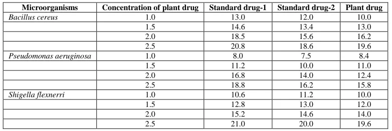
Table 2 shows the minimum inhibitory concentrations of control and plant drug.
Microorganisms Concentration of plant drug Standard drug-1 Standard drug-2 Plant drug
Bacillus cereus 1.0 13.0 12.0 10.0
1.5 14.6 13.4 13.0
2.0 18.5 15.6 16.2
2.5 20.8 18.6 19.6
Pseudomonas aeruginosa 1.0 8.0 7.5 8.4
1.5 11.2 10.0 11.0
2.0 16.8 14.0 12.4
2.5 18.8 16.2 15.8
Shigella flexnerri 1.0 10.6 11.2 10.0
1.5 12.8 13.0 12.0
2.0 15.2 14.6 14.0
In the present study the results show that the extract from Mesua ferrea possess antimicrobial activities against Bacillus cereus, Shigella flexneri and Pseudomonas aeruginosa. The extract compared favorably with the standard antibiotics Streptomycin and Ampicillin. The plant extract showed more activity like that of broad spectrum antibiotic activities. The MIC of Mesua ferrea were shown in table-2. The standard Streptomycin and Ampicillin had MIC values varying between 0.244 mg/ml and 0.488 mg/ml. The results indicated that the extract of the plant has stronger activity than that of standard antibiotics (Table-1). Since ancient times, herbs and /or their essential oils have been known for their varying degrees of antimicrobial activities [12,13,14&15]. More recently medicinal plant extracts were developed and proposed for use in food as natural antimicrobials [16&17] Antimicrobials are powerful but controversial tools. Food animals are often exposed to antimicrobial compounds to treat or prevent infectious diseases and/or to promote growth. The early history of supplementing animal feeds with antimicrobials parallels the isolation, identification and characterization of Vitamin B [18]. In 1948. Further research in this area showed that several feed ingredients, including dried mycelia of certain fungi, were more potent as growth promoters in the diet of chicks than was vitamin B alone. On the basis of the results obtained in the present study, we conclude that the extract has significant antimicrobial activity. Further studies are on to isolate the active principles responsible for their antimicrobial activities [19].
Except for Schigella flexineri, Bacillus anthracis and Pseudomonas aeruginosa, Enterococci may be resistant to Ampicilline and Streptomycin because of production of low affinity Streptomycin binding protein (PBPs) or less commonly because of the production of β Lactamase [20]. The disc diffusion test can accurately detect isolates with altered Streptomycin binding proteins, but it will not reliably detect β-lactamase producing strains. The rare β
lactamase producing stains are best detected by using a direct, nitrocefin based β-lactamase test. Certain Streptomycin, ampicillin resistance Enterococci may possess a high level of resistant (Streptomycin-MIC>µ g/ml or Ampicilline>64 µg/ml). The disc diffusion test can not differentiate those with normal resistance from this high level resistance [21] . For, Enterococcus recovered from blood and CFS, the laboratory should consider determining the actual MIC from Streptomycin or Ampicilline since E . faceium strains with normal lower level resistance (Streptomycin MIC<64 µg/ml and Ampicilline <32 µg/ml) should be considered potentially susceptible to synergy with an ammioglycoside (in the absence of high level amminoglycoside resistance) whereas strains with higher level resistance may be resistant to such synergy [22]. Our results clearly indicate that the antimicrobial potential of Mesua ferrea
DISCUSSION
Our results clearly indicate that the medicinal plant Mesua ferrea possess the strong antimicrobial activities against the pathogenic bacterial strains such as Bacillus cereus, Pseudomonas aeruginosa and Shigella flexneri as compared to commercial antibiotics such as streptomycin and ampicillin.
REFERENCES
[1] H Westh; CS Zinn; Rosdahl VT, Microb Drug Resist., 2004. 10, 169-176.
[2] JE Bandow; H Brotz; Leichert Lio,Antimicrob Agents Chemother., 2003 47, 948-955. [3] R Rojas; Bustamante B, J Bauer. Ethnopharmaco., 2003 , 88, 199- 204.
[4] N Benkeblia, Lebensm-Wiss u-Technol., 2004, 37, 263-268. [5] ML Colombo; E Bosisio, Pharmacol Re., 1996,33, 127-134.
[6] MW Iwu, AR Duncan, CO Okunji, Alexandria, VA: ASHS Press. Pp,1999, 457-462.
[7] FB Holetz, GL; N Pessini; DAG Sanches; CV Cortez; Nakamura ; BPD Filho, Mem. Inst. Oswaldo Cruz.,
2002, 97, 1027-1031.
[8] Y Rukayadi; JS Shim; JK Hwang; Mycoses., 2008, 51(4), 308-312.
[9] A Anandakumar; M Balasubramaniam; R Muralidharan Nagakesara. Ancient Sci Life.,1986,5, 263-268. [10] D Buckles, Economic Bot,1995,49,13-25.
[11]ME Daxenbichler; CH Van Etten; EA Hallinan; FR Earle; and SA Barclay,Journal of Medicinal Chemistry.,1971,14:463-465.
[12] EA Bell; DH Janzen, Nature., 1971, 229,136-137.
[13] CR Carson; KA Hammer; TV Riley,Microbios.,1995, 82(332),181-185.
[14]J Lin; AR Opoku; M Geheeb-Keller; AD Hutchings; SE Terblanche; AK Jager; J van Staden, J Ethnopharmacol.,1999, 68,267–74.
[15] M Valero; MC almeron, Int. J. Food Microbiol., 2003, 85, 73-81.
[16] WM Kone; KK Atindehou; C Terreaux; K Hostettmann; D Traore; M Dosso, J. Ethnopharmacol., 2004, 93, 43-49.
[19] J Del Campo; M J Amiot; C Nguyen-The, J. Food Prot., 2000, 63(10), 1359-1368. [20] M Oyaizu, Jpn J Nutr., 1986,44,307-314.
[21] TR Hatano; M Edamatsu; A Hiramatsu; Y Mori. Fujita, Chem. Pharm. Bull., 1989, 37: 2016-2021.