Journal of Chemical and Pharmaceutical Research, 2016, 8(3):258-263
Research Article
CODEN(USA) : JCPRC5
ISSN : 0975-7384
Development and validation of a RP-HPLC method for the determination of
Temozolomide in rats: Application to plasma pharmacokinetics and brain
distribution study
Bhavin K. Patel and Rajesh H. Parikh*
Ramanbhai Patel College of Pharmacy, Charotar University of Science and Technology, CHARUSAT Campus, Changa, Dist. Anand, 388421, Gujarat, India
_____________________________________________________________________________________________
ABSTRACT
A simple, sensitive and specific reverse phase - high performance liquid chromatography (RP-HPLC) method have been developed and validated for the estimation of Temozolomide (TMZ) in its plasma and brain homogenate of Rats. TMZ concentration in plasma and brain homogenate was determined on a C18 column after liquid-liquid extraction. Elution was applied with the mixture of 0.1% aq. Acetic Acid- Acetonitrile (90:10 V/V). To prevent chemical degradation of TMZ, plasma samples were acidified to pH < 3. The flow rate was 1 ml/min and separation was monitored by UV detection at 316 nm. Chromatogram showed peak at a retention time of 5.643± 0.0811 and 5.781±0.0545 for the plasma and brain respectively. All validation parameters met the acceptance criteria. Calibration curve, prepared using freshly spiked plasma and brain homogenate samples,was linear within the range of 1µg/ml to 100 µg/ml. Validation of the method for linearity and range, intra-day precision, selectivity, specificity and limits of detection and quantification were obtained. The method was found to be sufficiently accurate and precise over the studied range of concentrations. The developed method is suitable for pharmacokinetic studies in rats.
_____________________________________________________________________________________________
INTRODUCTION
Temozolomide (TMZ) is an imidazotetrazinone derivative with methylating properties, which is used in the treatment of malignant primary brain tumors, glioblastoma and astrocytoma[1-3]. TMZ undergoes chemical degradation to its active metabolite, i.e., methyl-triazeno-imidazolecarboxamide (MTIC). Thereafter, MTIC rapidly destroys to the inactive derivative n-5-aminoimidazole-4-carboxamide (AIC) and methyldiazonium cation. This process is irreversible and depends on the pH [4]. Kim et al. demonstrated that TMZ was not very stable in vitro at 37 ˚C in plasma, however, it was stable in human plasma acidified to pH < 4 [5]. The stability of TMZ in human plasma can be attained using phosphoric acid or 1 M hydrochloric acid. Literature survey reveals that certain Chromatographic and Spectrophotometric methods were reported for estimation of Temozolomide. The most frequently used techniques for the analysis of TMZ are high-performance liquid chromatography with UV detection (HPLC-UV) and liquid chromatography coupled to mass spectroscopy (LC/MS/MS). TMZ has been extracted from biological samples using liquid-liquid extraction or solid-phase extraction [5-13]. The main aim of the existing study was to validate the HPLC-UV method for the determination of temozolomide in human plasma and brain homogenatein order to allow plasma pharmacokinetic and brain distribution studiesof TMZ in rats.
EXPERIMENTAL SECTION
Materials
TMZ was received as a gift from Naproad Life Science, Mumbai, India. Acetonitrile, phosphoric acid and glacial acetic acid were of HPLC grade. All other chemicals used were of analytical reagent grade.
Methods
Preparation of Mobile Phase
The mobile phase contained 10% V/V acetonitrile and 90% V/V aqueous phase. The aqueous phase contained 0.1 % solution of acetic acid. The mobile phase was run in binary mode. The flow rate of the mobile phase was maintained at 1 ml/min. Injection volumes were 20 µl. The mobile phase was degassed by using bath sonicator and filtered prior to its use. The HPLC column was kept at ambient temperature.
Calibration Standards
Stock solutions of temozolomide were prepared by dissolving accurately weighed10 mg of Temozolomide in 20% aqueous methanol containing 0.1% glacial acetic acid in order to get stock solution of 100 µg/ml. The subsequent dilutions (1µg/ml, 10 µg/ml, 20µg/ml, 25 µg/ml, 50 µg/ml) of TMZ were prepared as calibration curve standards.
Preparation of plasma and brain Standard Solution
Standard solution of plasma and brain standard solution (5-100 µg/ml) was prepared by liquid-liquid extraction method. 0.25 ml of Temozolomide from the stock solutions were spiked in 0.25ml of plasma and 10% W/V brain homogenate and it was transferred in 10 ml screw capped bottle and 50 µl of H3PO4 (20% w/v) was added in order to stabilize it. Above prepared solution was deproteinized by addition of 5 ml Ethyl Acetate and vortexed for 10 minutes.Vortexed solution was centrifuged at 4500*g (9000 rpm) at 4 ºC for 10 min in order to separate out two phase. Organic layer was collected and evaporated in china dish to obtained solid residue. The obtained residues were dissolve in Mobile phase system and samples were injected into the chromatographic system.
HPLC system and operating conditions
Separation was accomplished with a Chromosil C18 column (250 X 4.6 mm, 5µ). The mobile phase comprised of
0.1% aq. Acetic Acid- Acetonitrile (90:10 V/V) with 0.5 ml of tri-ethyl amine (TEM) and the elution was in binary mode at ambient temperature with a flow rate of 1 ml/min. The UV detector was set at 316 nm for Temozolomide detection for 10 min run time and the peak areas were calculated using the data analysis program.
Table 1: HPLC method conditions for Bioanalytical method development of TMZ
Mobile phase 0.1% aq. Acetic Acid- Acetonitrile (90:10 V/V)
Pump mode Binary
Column Chromosil C18 column (250 X 4.6 mm, 5µ)
Column temp Ambient
Wavelength 316 nm
Injection volume 20 µl
Flow rate 1 ml/min
Run time 10 min
Suitability
The system suitability parameters were calculated for the standard solutions of Temozolomide. The values obtained demonstrated the suitability of the system for the analysis of Temozolomide in plasma and brain homogenate. The system suitability parameters should fall within acceptable standard deviation range during routine performance of the method.
Selectivity and Sensitivity
The selectivity of the method was assessed by comparing the chromatograms acquired from the samples containing Temozolomide with those obtained from blank samples. The sensitivity was determined in terms of lower limit of quantification where the response of lower limit of quantification.
Linearity Range
Application of Assay
The above method was successfully applied for the pharmacokinetic studies of Temzolomide in rats. The Institutional Animal Ethics Committee (IAEC), registered with the Government of India approved the protocol (RPCP/IAEC/2013-2014/R-34) for the animal experiments. Albino rats (Wistar) of either sex were taken for the study. The rats were fasted overnight with free access to water before administration of TMZ. The drug solution at the dose of 150 mg/m2 was administered to rats through intravenous (I.V) route. The rats were anesthetised by intraperitoneal injection of ketamine (40mg/kg). At predefined time intervals, 0.5, 1, 2, 4, 6 and 8 h, 0.5 ml of blood samples were collected from rats by cardiac puncture. Plasma was collected and separate aliquots were removed for temozolomide analysis.
After each blood collection, the animals were sacrificed and the brain was isolated, weighed, homogenized with the help of tissue homogenizer in order to get 10% W/V of brain homogenate. The brain homogenate was centrifuged and supernatant was preserved in acidic media using saline and 0.3% H3PO4 (20% w/v). The final sample was stored at -80 ˚C until HPLC analyses. The pharmacokinetic parameters were calculated with a non-compartmental model and reported.
RESULTS AND DISCUSSION
The developed HPLC method was optimized for the analysis of Temozolomide in human plasma and brain homogenate. Different Mobile phase compositions were selected on the basis of trial & error methodand the optimum mobile phase was finalized to measure Temozolomide in brain and plasma. The developed method was validated for selectivity, linearity, limit of quantification, accuracy, precision and recovery as per the international guidelines (FDA guideline, 2001).
System suitability
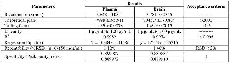
[image:3.595.108.506.432.542.2]It is essential for the assurance of the quality performance of chromatographic system. The system suitability parameters such as retention time, tailing factor, number of theoretical plate and precision of replicate injection were also be calculated. It was observed that all the values are within the limits. Table 2indicates the summary of all system suitability parameters.
Table 2: Summary of system suitability parameters for HPLC method of analysis for TMZ in brain and Plasma
Parameters Results Acceptance criteria
Plasma Brain
Retention time (min) 5.643± 0.0811 5.781±0.0545 ---
Theoretical plate 7898 ±195.911 8045.7 ±170.874 >2000
Tailing factor 1.39 ± 0.0078 1.49 ± 0.0015 <1.5
Linearity 1 µg/mL to 100 µg/mL 1 µg/mL to 100 µg/mL ---
R2 0.9982 0.9974 > 0.995
Regression Equation Y = 10384x + 34586 y = 12374x + 35315 ---
Repeatability (%RSD) (n=6) (50 mcg/ml) 1.12% 1.46% RSD < 2%
Specificity (Peak purity index) 0.899987 0.889887 1
0.889972 0.879910
Repeatability
[image:3.595.94.519.615.696.2]It was performed and %RSD values were found to be less than 2%. Results of intraday precision were shown in Table 21.
Table 3: Repeatability data for TMZ
Conc. (µg/ml) Peak Area Mean area ±SD Mean conc ±SD %RSD
Plasma Brain Plasma Brain Plasma Brain Plasma Brain
50
549217 647522
557811±7117.15 654933±9619.80 50.37± 0.65 50.07±0.77 1.275 1.468 550406 657080
559393 672780 560306 652347 568504 646193 559040 653676
Selectivity
of analysis. Both the peaks of temozolomide of plasma and brain homogenate did not interfere with any endogenous components of plasma and brain. A good resolution has been observed for both the peaks of Temozolomide (Fig. 1 and 2).
[image:4.595.100.508.502.754.2]Fig. 1: HPLC Chromatogram of 50 µg/ml TMZ Spiked in Plasma
Fig. 2: HPLC Chromatogram of 25 µg/ml TMZ Spiked in Brain Homogenate
.
Linearity
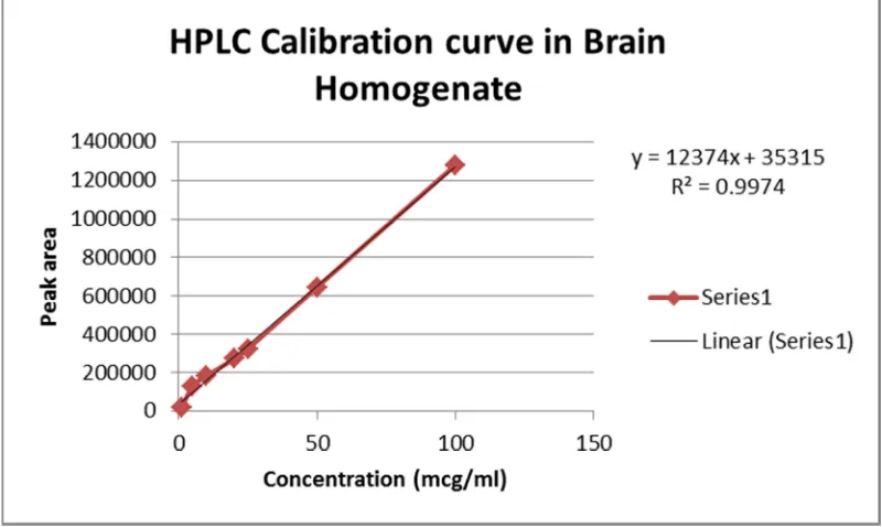
The linearity was determined from the constructed standard calibration curve. The upper limit of the range was taken as 100 µg/ml since the plasma and brain concentration of temozolomide is lower than this value following intravenous administration. Seven non-zero samples (1 µ g/ml to 100 µg/ml) were used to draw the standard calibration curve (n = 6). The calibration curves for plasma (Fig. 3) and brain homogenate (Fig. 4) were obtained by plotting the chromatographic peak area versus concentration of temozolomide. Samples were prepared and injected same day. The linear regression data for the calibration curves (n=6) shows a good linear relationship over the concentration range of 1 to 100 µg/mL for Temozolomide with respect to peak area. The calibration curve was linear at the given range in both Plasma (R2 = 0.9951) as well as brain homogenate (R2 = 0.9951).
. Fig. 4: Calibration Curve of TMZ in Brain
Lower limit of quantification (LLOQ) and lower limit of detection (LLOD)
The limit of detection and limit of quantitation for the Temozolomide was calculated from the linearity data using relative standard deviation of the response and slope of the calibration curve. By the analysis of samples with known concentrations of analyte and establishing the minimum level at which the analyte can be reliably detected. The LOQ and LOD as per these criteria were found and indicated in table 2. This method was found to be sensitive enough to apply for the pharmacokinetic studies.
Table 4: LOD & LOQ data for TMZ Parameters Plasma Brain LOD (µg/ml) 2.261 2.56
LOQ (µg/ml) 6.853 7.771
In-vivo Pharmacokinetic Study
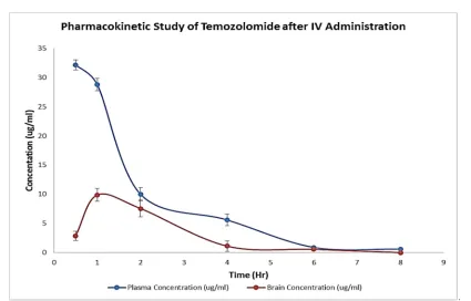
[image:5.595.105.505.186.425.2]Temozolomide concentration profiles in plasma and CSF following I.V. administration in rats are shown in Fig. 5. TMZ attained a high concentration of 32.142 ± 0.826 ng/ml in plasma at 30 min following I.V. administration, and then followed by an exponential decline was observed depending on the time. On the other side TMZ attained maximum plasma concentration of 9.860 ± 1.108 ng/ml was achieved at 1 hr.
Table 5: Pharmacokinetic Parameters for Plasma and Brain in after intravenous administration of TMZ in rats
Pharmacokinetic Parameters Plasma (Mean ± S.D.)
Brain (Mean ± S.D.)
C max (µg/ml) 32142 ± 826 9860 ± 1108
AUC (µg h/ml) 66241 ± 4870 23579 ± 5079
.
Fig.5:Pharmacokinetic Study of Temozolomide in Plasma and Brain Homogenate after Intravenous Administration
Table 5 indicates the pharmacokinetic parameters of plasma and brain following intravenous administration of TMZ in rats. This proves the application of developed bioanalytical method in brain and plasma pharmacokinetic studies.
CONCLUSION
In summary, HPLC method for the quantitation of Temozolomide in human plasma and brain was developed and fully validated as per regulatory guidelines. This method offers significant advantages in terms of sensitivity and selectivity, faster run time (10 min) and lower sample requirements. The current method has shown acceptable precision and adequate sensitivity for the quantification of Temozolomide. The method has also been successfully applied for the pharmacokinetics study of temozolomide in plasma and Brain following intravenous administration in rats and pharmacokinetic parameters have been derived. Hence, this method may be convenient for the estimation of Temozolomide in plasma and brain homogenate of rats.
Acknowledgment
The authors are thankful to Naproad Life Science, for providing the gift sample of Temozolomide. The authors are also thankful to Ramanbhai Patel College of Pharmacy and Charotar University of Science and Technology for all their financial assistance and support.
REFERENCES
[1]Friedman HS; Kerby T; Calvert H,Clin. Cancer Res., 2006, 6, 2585–2597.
[2]Stupp R; Gander M; Leyvraz S; Newlands E, Lancet Oncol.,2001, 2, 552–560.
[3]YungWK; PradosMD; Yaya-Tur RJ,Clin.Oncol.,1999,17, 2762–2771.
[4]Denny BJ; Wheelhouse RT; Steven FG,Biochemistry.,1994, 33, 9045–9051.
[5]Kim H; Likhari P; Parker D; Statkevich P, J. Pharm. Bio. Anal., 2001, 24, 461–468. [6]Estlin EJ.; Lashford L; Ablett S; Price L; Gowing R, Br. J. Cancer., 1998, 78, 652-655.
[7]Baruchel S; Diezi M; Hargrave D; Stempak D, Eur. J. Cancer, 2006, 42, 2335-2340.
[8]Diez BD; Statkevich P; Zhu Y; Abutarif MA, Cancer Chemother. Pharmacol.,2010, 65, 727-733.
[9]Meany HJ; Warren KE; Fos E; Cole DE, Cancer Chemother. Pharmacol.,2009, 65, 137-146.
[10]Aoki T; Nishikawa R; Mizutani T; Nojima K; Mishima K, Int. J. Clin. Oncol.,2007,12, 341-346.
[11]Reid JM; Stevens DC; Joseph R,Clin. Cancer Res., 1997, 3, 2393-2401.
[12]Portnow J; Badie B; Chen M; Liu A. Clin, Cancer Res., 2009, 15, 7092-7097.