http://dx.doi.org/10.4236/ojts.2015.51004
How to cite this paper: Hata, A., Sekine, Y., Koh, E. and Yamaguchi, N. (2015) Preoperative Exercise Testing Is a Better Pre-dictor of Postoperative Complications than Pulmonary Function Testing for Patients with Lung Cancer. Open Journal of Thoracic Surgery, 5, 15-20. http://dx.doi.org/10.4236/ojts.2015.51004
Preoperative Exercise Testing Is a Better
Predictor of Postoperative Complications
than Pulmonary Function Testing for
Patients with Lung Cancer
Atsushi Hata
1, Yasuo Sekine
1*, Eitetsu Koh
1, Nobuyuki Yamaguchi
21Department of Thoracic Surgery, Tokyo Women’s Medical University Yachiyo Medical Center, Yachiyo, Japan 2Division of Clinical Physiology, Tokyo Women’s Medical University Yachiyo Medical Center, Yachiyo, Japan
Email: *[email protected]
Received 23 February 2015; accepted 24 March 2015; published 27 March 2015
Copyright © 2015 by authors and Scientific Research Publishing Inc.
This work is licensed under the Creative Commons Attribution International License (CC BY). http://creativecommons.org/licenses/by/4.0/
Abstract
Objective: The aim of this preliminary study was to evaluate the feasibility of exercise testing (ET) for predicting postoperative complications in patients with impaired pulmonary function. Me-thods: Thirteen patients were prospectively enrolled. The enrollment criteria were FEV1.0% < 70%
and emphysema score > 8 by the Goddard classification or interstitial pneumonia on chest com-puted tomography. Patients underwent testing for pulmonary function, six-minute walking test (6MWT), and stair-climbing test (SCT). Postoperative cardiopulmonary complications (PCPCs) were recorded. Results: Four patients developed PCPCs. There were no significant differences between the patients with PCPCs (n = 4) and those without PCPCs (n = 9) for background data and PFT. The distances achieved in the 6MWT were 503 ± 72.7 m for patients without PCPCs and 369 ± 50.7 m for patients with PCPCs (p = 0.011). The SCT climbing heights were 20.4 ± 5.3 m for patients without PCPCs and 14.9 ± 4.0 m for patients with PCPCs (P = 0.187). Cut-off points, including a 6MFT distance of less than 400 m, SCT height lower than 15 m, and SCT climbing speed less than 8.5 m/min, were predictive of CPCP. Conclusions: Exercise testing is more feasible for predicting postoperative cardiopulmonary complications than stationary pulmonary function testing.
Keywords
Lung Cancer, Exercise Testing, Postoperative Complication, Pulmonary Function Testing
*
1. Introduction
Pulmonary resection is a standard treatment option for patients with stage I and II non-small cell lung cancer. Preoperative physiologic evaluation is mandatory for resection of one or more pulmonary lobes. The American College of Chest Physicians (ACCP) has proposed evidence-based clinical practice guidelines for preoperative physiologic evaluation of patients with lung cancer [1]. The guidelines present a risk assessment algorithm, which recommends that patients with abnormal results on pulmonary function testing (PFT) undergo additional testing, such as diffusing capacity of the lung for carbon monoxide (DLco) and cardiopulmonary exercise testing (CPET). However, sometimes the predicted postoperative (PPO) pulmonary function is underestimated and the actual postoperative physiologic capacity has no relationship to the predicted capacity [2].
Lung volume reduction surgery has been found to be beneficial for patients with heterogeneous emphysema, and lobectomy has also been shown to be beneficial for patients with a malignant tumor located in an upper emphysematous lobe [3]. These findings suggest that postoperative lung function and exercise capacity may differ based on the physiologic or anatomic pulmonary abnormalities and the volume of lung resected.
Criteria for safe lung resection have been reported, but they may over- or underestimate the risk of postopera-tive cardiopulmonary complications (PCPCs) in patients who undergo lung surgery [4], because PFT only esti-mates pulmonary function and does not evaluate cardiac function. Furthermore, since the estimation of PPO PFT is based on the number of segments to be resected, the risk for patients with emphysema could be overestimated if the lung resection benefits the patient because of the reduction in volume of emphysematous lung. On the oth-er hand, CPET can evaluate cardiopulmonary function and oxygen transport [5]. CPET can reveal soth-erious pa-thophysiologic abnormalities of the oxygen transport system and can predict the risk of postoperative complica-tions [4]. Therefore, PFT and CPET are both indicated for patients with impaired pulmonary function [6].
The aim of this preliminary study was to evaluate the feasibility of exercise testing for predicting postopera-tive complications in patients with impaired pulmonary function who undergo lung resection.
2. Materials and Methods
The study was approved by the institutional review board of our hospital. This study comprised 13 male patients with impaired pulmonary function who were candidates for lung resection. They were prospectively enrolled from August 2011 through August 2013 after providing informed consent. Impaired pulmonary function was defined as follows: Forced expiratory volume in one second (FEV1.0%)/Forced vital capacity (FVC) less than
70% and emphysema score higher than 8 points by the Goddard classification or interstitial pneumonia on chest computed tomography [7]. Preoperative functional evaluation consisted of spirometry, DLco, and exercise test-ing, as follows: six-minute walking test (6MWT) and stair-climbing test (SCT).
The following spirometric variables were considered: FEV1.0; predicted postoperative (ppo) FEV1.0 (ppo
FEV1.0 [preoperative FEV1.0 × number of postoperative functioning segments/number of preoperative segments]);
predictive postoperative DLco (ppoDLco) [(preoperative DLco × number of postoperative functioning segments/ number of preoperative segments]); and FEV1.0/FVC [2].
The 6MWT is a simple, practical test that requires a 100-ft hallway, but no exercise equipment or advanced training for technicians [8]. This test measures the distance that a patient can quickly walk on a hard, flat surface during a 6-min period. The SCT is also a practical test that measures the total height of stairs a patient can climb [4] [9]. Each stair step is 0.17-m high. The study patients were asked to climb, at their own pace, the maximum number of steps before needing to stop because of exhaustion, limiting dyspnea, leg fatigue, or chest pain. The total number of steps climbed and the time taken to complete the test were recorded for each patient. Each pa-tient’s climbing pace was calculated from the total height climbed per time taken to complete the test in minutes. Finger pulse oximetry was used to measure oxygen saturation and pulse rate continuously for both tests.
Each patient was accompanied by a physician during his tests and encouraged to complete the test. Moreover, the physician maintained continuous verbal interaction with the patient to assess dyspnea and the occurrence of other signs/symptoms. The 6MWT and SCT evaluate the global and integrated responses of all the systems in-volved during exercise, including the pulmonary and cardiovascular systems, systemic circulation, peripheral circulation, neuromuscular units, and muscle metabolism.
fibrillation, or cardiac failure. A patient with a concomitant cardiac disease was allowed to undertake the tests after undergoing cardiac evaluation and subsequently considered to be hemodynamically stable. Patients with orthopedic disorders such as herniated intervertebral disks and dysfunction of lower extremities were also ex-cluded from this study because they could not undergo exercise testing.
PCPCs were considered to be cardiopulmonary complications occurring within 30 days after the surgery or those occurring during a longer period if the patient was still in the hospital. According to other reports [10] [11] and for the sake of comparison, the following complications were recorded: respiratory failure requiring me-chanical ventilation for 48 h; pneumonia; atelectasis requiring bronchoscopy; pulmonary edema; pulmonary embolism; myocardial infarction; hemodynamically unstable arrhythmia requiring medical treatment; cardiac failure; and death.
3. Statistical Analysis
We compared the results of patients who developed PCPCs with those of the patients without PCPCs. Further-more, we evaluated cut-off values for 6MWT and SCT for use as predictors of postoperative complications. The Mann-Whitney U test was used to analyze continuous variables, and the Fisher exact test to analyze categorical variables. Continuous variables were calculated as mean ± SD and categorical variables as number of patients (percent). A P-Value < 0.05 was considered to be statistically significant. Analysis was performed using JMP11 software (SAS Institute Inc., NY, USA).
4. Results
Table 1 summarizes the characteristics of the 13 patients. Twelve patients had lung cancer and one patient had metastatic lung cancer. The patients underwent 7 lobectomies and 6 wedge/segment ectomies (limited resec-tions). There were no significant differences between patients with and without PCPCs. Four patients developed the following postoperative pulmonary complications: three patients developed pneumonia and one patient de-veloped pulmonary dysfunction that required home oxygen therapy (Table 2). Two patients with pulmonary complications died in the hospital.
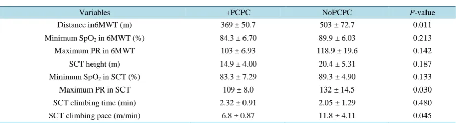
Table 3 shows the PFT and DLco data. There were no significant differences between the patients with and without PCPCs. Table 4 shows the 6MWT and SCT data. The patients who developed PCPCs had a signifi-cantly shorter mean preoperative 6MWT distance than the patients without PCPCs. The mean maximum SCT pulse rate and climbing pace of the patients with PCPCs were significantly lower than those of the patients without PCPCs.
We evaluated cut-off values for the CMWT and SCT based on previous reports that indicated that patients with a 6MWT less than 400 m or SCT height less than 15 m tended to have postoperative complications [4] [12].
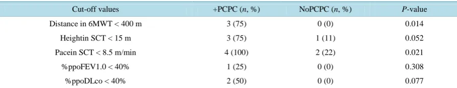
Table 5 shows that three patients who developed PCPCs walked less than 400 min 6 min and none of the pa-tients without PCPCs walked less than 400 m (P = 0.014). Three patients with PCPCs climbed less than 15 m in the SCT and one patient without PCPCs climbed less than 15 m (Table 5, P = 0.052), and four patients with PCPCs and two patients without PCPCs achieved a climbing paces lower than 8.5 m/min (P = 0.021). For the 6MWT, a 6-min walking distance shorter than 400 m had a sensitivity of 75%, specificity of 100%, positive predictive value of 100% and negative predictive value of 90% for the development of PCPC. For the SCT, a climbing pace shorter than 8.5 m/min had a sensitivity of 100%, specificity of 77.8%, positive predictive value of 66.7%, and negative predictive value of 100%. Conversely, there were no significant differences in the num-bers of patients with and without PCPCs who satisfied the traditional criteria of %ppoDLco less than 40% and %ppoFEV1 less than 40% (Table 5; P = 0.308, P = 0.077, respectively).
5. Discussion
We found that 6MWT and SCT were more valid assessments than conventional PFT for predicting the devel-opment of PCPCs in our study patients with impaired pulmonary function and emphysema. Although the PFT variables, which were regarded to be predictors of postoperative complications, did not predict PCPCs, the walking distance of the 6MWT and height and climbing pace of the SCT were predictive of PCPCs.
Table 1. Patient characteristics.
Variables +PCPC (n = 4) NoPCPC (n = 9) P-value
Age 78.5 ± 4.5 74.9 ± 7.7 0.407
BMI 18.7 ± 2.72 22.7 ± 3.23 0.087
Smoking Index (pack-year) 57.0 ± 12.9 40.6 ± 23.2 0.189
Cardiac Disease, n (%) 2 (50) 2 (22.2) 0.530
Lobectomy, n (%)/Limited resection 3 (75)/1 (25) 4 (44.4)/5 (55.6) 0.559
Blood loss (g) 1336 ± 1059 178 ± 237 0.036
VATS, n (%) 4 (100) 6 (66.7) 0.497
PCPC: postoperative cardiopulmonary complication; BMI: body mass index; VATS: video-assisted thoracic surgery.
Table 2. Type of postoperative complications.
Age (y) Procedure Stage-Type Preoperative disease Complication
84, Male Segmentectomy IA-SCC COPD Pneumonia, death
78, Male Lobectomy IB-SCC CPFE Acute respiratory distress syndrome
79, Male Lobectomy IIIA-Ad COPD Hepatic failure, pneumonia, death
73, Male Lobectomy IIA-SCC COPD Respiratory failure
SCC: squamous cell carcinoma; Ad: adenocarcinoma; COPD: chronic obstructive pulmonary disease; CPFE: combined pulmonary fibrosis and em-physema.
Table 3. Pulmonary function data.
Variables +PCPC NoPCPC P-value
FVC 2.50 ± 0.72 3.28 ± 0.71 0.189
%FVC 80.9 ± 23.7 101.7 ± 18.2 0.190
FEV1 1.70 ± 0.43 2.12 ± 0.67 0.247
%FEV1 85.0 ± 28.4 97.6 ± 26.8 0.488
% ppoFEV1.0 67.3 ± 27.0 85.9 ± 25.7 0.396
FEV1/FVC% 69.6 ± 13.5 65.5 ± 18.8 0.939
RV/TLC 45.9 ± 18.0 36.9 ± 8.60 0.671
%DLco 77.3 ± 38.7 80.2 ± 22.5 0.671
%DLco/VA 58.9 ± 28.5 70.0 ± 20.3 0.799
PaO2 83.7 ± 2.33 98.7 ± 31.5 0.552
PaCO2 38.2 ± 6.15 37.0 ± 6.23 1.000
CT evaluation
Goddard Score 9.75 ± 4.35 4.00 ± 7.16 0.0601
PF, n (%) 2 (50) 1 (11) 0.2028
PCPC: postoperative cardiopulmonary complication; ppo: predicted postoperative: DLco: diffusing capacity of the lung for carbon monoxide: VA: alveolar ventilation; PF: pulmonary fibrosis.
Table 4. 6MWT and SCT data.
Variables +PCPC NoPCPC P-value
Distance in6MWT (m) 369 ± 50.7 503 ± 72.7 0.011
Minimum SpO2 in 6MWT (%) 84.3 ± 6.70 89.9 ± 6.03 0.213
Maximum PR in 6MWT 103 ± 6.93 118.9 ± 19.6 0.142
SCT height (m) 14.9 ± 4.00 20.4 ± 5.31 0.187
Minimum SpO2 in SCT (%) 83.3 ± 7.29 89.3 ± 4.90 0.133
Maximum PR in SCT 109 ± 8.0 132 ± 14.5 0.030
SCT climbing time (min) 2.32 ± 0.91 2.05 ± 1.29 0.480
SCT climbing pace (m/min) 6.8 ± 0.87 11.8 ± 4.11 0.045
Table 5. Cut-off values of selected variables.
Cut-off values +PCPC (n, %) NoPCPC (n, %) P-value
Distance in 6MWT < 400 m 3 (75) 0 (0) 0.014
Heightin SCT < 15 m 3 (75) 1 (11) 0.052
Pacein SCT < 8.5 m/min 4 (100) 2 (22) 0.021
%ppoFEV1.0 < 40% 1 (25) 0 (0) 0.308
%ppoDLco < 40% 2 (50) 0 (0) 0.077
PCPC: postoperative cardiopulmonary complication; ppo: predicted postoperative value.
predicted FEV1.0 and percent predicted DLco > 40% are considered to be operable [12]. These standards are
meaningful but not always predictive of the development of PCPCs without the addition of other tests [13]. Various tests and scoring systems have been reported [14]-[18]. A modified Thoracoscore, which predicts postoperative outcomes after thoracic surgery, can be calculated based on the following variables: age, gender, priority of the procedure, malignancy, type of procedure, Zubrod score, ASA class, and number of co-morbidities [18]. How-ever, such a scoring system is difficult to use on a daily basis in clinical practice.
The 6MWT and SCT are global exercise tests that can evaluate cardiopulmonary function, particularly for pa-tients with chronic obstructive pulmonary disease (COPD) [8] [13]. These tests consist of the following va-riables: distance walked, height climbed, change in SpO2, maximum pulse rate, and SCT pace [4] [13].
There were no significant differences in the traditional PFT results for the patients with and without PCPCs. Three of four patients developing PCPCs had percentppoFEV1.0 > 40%. This finding indicates that the patient’s
condition may be underestimated if only traditional PFT data is used.
This prospective study has some limitations and biases. First, this was a small dataset from a single-center study. Therefore, there may be several confounding variables, and a large prospective study is needed to verify the findings. Second, there was no single surgical procedure, because the type of procedure used was based on lung cancer stage and the patient’s physical condition. Third, COPD should not be defined by functional limita-tion and radiologic evalualimita-tion only. Other clinical criteria should have been taken into consideralimita-tion [19]. It is possible that the use of other criteria for COPD could have produced different results.
6. Conclusion
For lung cancer patients with impaired pulmonary function, exercise testing is more feasible for predicting postoperative cardiopulmonary complications than stationary pulmonary function testing. 6MWT < 400 m, SCT height < 15 m, and SCT climbing pace < 8.5 m/min are useful values for predicting postoperative cardiopulmo-nary complications.
Disclosure Statements
All authors have no conflict of interest.
References
[1] Colice, G.L., Shafazand, S., Griffin, J.P., Keenan, R. and Bolliger, C.T., American College of Chest Physicians (2007) Physiologic Evaluation of the Patient with Lung Cancer Being Considered for Resectional Surgery: ACCP Evidenced- Based Clinical Practice Guidelines (2nd Edition). Chest, 132, 161S-177S.
[2] Sekine, Y., Iwata, T., Chiyo, M., Yasufuku, K., Motohashi, S., Yoshida, S., et al. (2003) Minimal Alteration of Pul-monary Function after Lobectomy in Lung Cancer Patients with COPD. The Annals of Thoracic Surgery, 76, 356-361.
http://dx.doi.org/10.1016/S0003-4975(03)00489-2
[3] Fishman, A., Martinez, F., Naunheim, K., Piantadosi, S., Wise, R., Ries, A., et al. (2003) National Emphysema Treat-ment Trial Research Group. A Randomized Trial Comparing Lung-Volume-Reduction Surgery with Medical Therapy for Severe Emphysema. The New England Journal of Medicine, 348, 2059-2073.
http://dx.doi.org/10.1056/NEJMoa030287
[4] Brunelli, A., Al Refai, M., Monteverde, M., Borri, A., Salati, M. and Fianchini, A. (2002) Stair Climbing Test Predicts Cardiopulmonary Complications after Lung Resection. Chest, 121, 1106-1110.
[5] Olsen, G.N. (1989) The Evolving Role of Exercise Testing Prior to Lung Resection. Chest, 95, 218-225.
http://dx.doi.org/10.1378/chest.95.1.218
[6] Datta, D. and Lahiri, B. (2003) Preoperative Evaluation of Patients Undergoing Lung Resection Surgery. Chest, 123, 2096-2103. http://dx.doi.org/10.1378/chest.123.6.2096
[7] Goddard, P.R., Nicholson, E.M., Laszlo, G. and Watt, I. (1992) Computed Tomography in Pulmonary Emphysema.
Clinical Radiology, 33, 379-387. http://dx.doi.org/10.1016/S0009-9260(82)80301-2
[8] ATS Committee on Proficiency Standards for Clinical Pulmonary Function Laboratories (2002) ATS Statement: Guidelines for the Six-Minute Walk Test. American Journal of Respiratory and Critical Care Medicine, 166, 111-117.
http://dx.doi.org/10.1164/ajrccm.166.1.at1102
[9] Olsen, G.N., Bolton, J.W., Weiman, D.S. and Hornung, C.A. (1991) Stair Climbing as an Exercise Test to Predict Pe-rioperative Complications of Lung Resection: Two Years’ Experience. Chest, 99, 587-590.
http://dx.doi.org/10.1378/chest.99.3.587
[10] Bechard, D. and Wetstein, L. (1987) Assessment of Exercise Oxygen Consumption as Preoperative Criterion for Lung Resection. The Annals of Thoracic Surgery, 44, 344-349. http://dx.doi.org/10.1016/S0003-4975(10)63787-3
[11] Bolliger, C.T., Jordan, P., Solèr, M., Stulz, P., Grädel, E., Skarvan, K., et al. (1995) Exercise Capacity as a Predictor of Postoperative Complications in Lung Resection Candidates. American Journal of Respiratory and Critical Care Medi-cine, 151, 1472-1480. http://dx.doi.org/10.1164/ajrccm.151.5.7735602
[12] Keagy, B.A., Schorlemmer, C.R., Murray, G.F., Starek, P.J.K. and Wilcox, B.R. (1983) Correlation of Preoperative Pulmonary Function Testing with Clinical Course in Patients after Pneumonectomy. The Annals of Thoracic Surgery,
36, 253-257.
[13] Holden, D.A., Rice, T.W., Stelmach, K. and Meeker, D.P. (1992) Exercising Testing, 6-min Walk, and Stair Climb in the Evaluation of Patients at High Risk for Pulmonary Resection. Chest, 102, 1774-1779.
http://dx.doi.org/10.1378/chest.102.6.1774
[14] Barua, A., Handagala, S.D., Socci, L., Barua, B., Malik, M., Johnstone, N. and Martin-Ucar, A.E. (2012) Accuracy of Two Scoring Systems for Risk Stratification in Thoracic Surgery. Interactive CardioVasc Thoracic Surgery, 14, 566- 569. http://dx.doi.org/10.1093/icvts/ivs021
[15] Lim, E., Baldwin, D., Beckles, M., Duffy, J., Entwisle, J., Faivre-Finn, C., et al. (2010) Guidelines on the Radical Management of Patients with Lung Cancer. Thorax, 65, iii1-iii27. http://dx.doi.org/10.1136/thx.2010.145938
[16] Brunelli, A., Morgan-Hughes, N.J., Refai, M., Slati, M., Sabbatini, A. and Rocco, G. (2007) Risk-Adjusted Morbidity and Mortality Models to Compare the Performance of Two Units after Major Lung Resections. The Journal of Tho-racic and Cardiovascular Surgery, 133, 88-96. http://dx.doi.org/10.1016/j.jtcvs.2006.08.058
[17] Falcoz, P.E., Conti, M., Brouchet, L., Chocron, S., Mercier, M., Etievent, J.P., et al. (2007) The Thoracic Surgery Scoring System (Thoracoscore): Risk Model for In-Hospital Death in 15,183 Patients Requiring Thoracic Surgery. The Journal of Thoracic and Cardiovascular Surgery, 133, 325-332. http://dx.doi.org/10.1016/j.jtcvs.2006.09.020 [18] Chamogeorgakis, T., Toumpoulis, I., Tomos, P., Ieromonachos, C., Angouras, D., Georgiannakis, E., et al. (2009)
Ex-ternal Validation of the Modified Thoracoscore in a New Thoracic Surgery Program: Prediction of In-Hospital Mortal-ity. Interactive CardioVasc Thoracic Surgery, 9, 463-466. http://dx.doi.org/10.1510/icvts.2008.201178