Scholarship@Western
Scholarship@Western
Electronic Thesis and Dissertation Repository
7-14-2016 12:00 AM
Cholinergic Mechanisms Regulating Cognitive Function and RNA
Cholinergic Mechanisms Regulating Cognitive Function and RNA
Metabolism
Metabolism
Benjamin Kolisnyk
The University of Western Ontario
Supervisor Drs. Vania
The University of Western Ontario Joint Supervisor Marco Prado
The University of Western Ontario Graduate Program in Neuroscience
A thesis submitted in partial fulfillment of the requirements for the degree in Doctor of Philosophy
© Benjamin Kolisnyk 2016
Follow this and additional works at: https://ir.lib.uwo.ca/etd
Recommended Citation Recommended Citation
Kolisnyk, Benjamin, "Cholinergic Mechanisms Regulating Cognitive Function and RNA Metabolism" (2016). Electronic Thesis and Dissertation Repository. 3837.
https://ir.lib.uwo.ca/etd/3837
This Dissertation/Thesis is brought to you for free and open access by Scholarship@Western. It has been accepted for inclusion in Electronic Thesis and Dissertation Repository by an authorized administrator of
i
Abstract
Acetylcholine (ACh) is one of the main neuromodulators in the mammalian central
nervous system (CNS). This chemical messenger has been implicated in the underlying
physiology of many distinct cognitive functions. However, the exact role that ACh plays
in regulating information processing in the brain is still not fully understood. The
vesicular acetylcholine transporter (VAChT) is required for the storage of ACh into
synaptic vesicles, and therefore it presents a means to modulate release. Diminished
VAChT levels cause a decrease in cholinergic tone, whereas increased VAChT
expression has been shown to augment ACh release. Previously published data have
shown that elimination of VAChT in the basal forebrain in genetically-modified mice
impairs learning and memory.
For our studies we have used different mouse lines in which the expression of the
VAChT gene is changed, both increased and decreased. We are therefore able to study
the consequences of altered cholinergic tone in vivo. Our hypothesis is that changes in
cholinergic tone produce specific molecular signatures in target brain areas that underlie
alterations in cognitive function. Our studies aimed to elucidate the behavioural and
molecular consequences of cholinergic dysfunction. Behavioral testing included
classical learning and memory tests as well as sophisticated tasks using novel touch
screens chambers to measure attention, learning and memory as well as cognitive
flexibility. At the molecular level, the goal was to examine how long-term changes in
cholinergic tone impact mechanisms regulating synaptic plasticity and neuronal health.
Finally, by aging mouse models of cholinergic dysfunction we were able to elucidate the
role that cholinergic tone plays in the classical pathological hallmarks of
neurodegenerative disorders.
Ultimately, by establishing the molecular signature of increased and decreased
cholinergic tone in targeted brain regions (cortex and hippocampus) it may become
possible to find novel targets for therapeutic interventions to improve cognitive deficits
ii
Keywords
Acetylcholine
Cholinergic signaling
Vesicular acetylcholine transporter
Cognition
Alzheimer’s Disease
iii
Co-Authorship Statement
Data presented in Chapter 2 of this thesis was previously published in The Journal of
Neuroscience on June 19th, 2013 under 33(25)10427-10438. This peer reviewed article
is titled “ChAT–ChR2–EYFP mice have enhanced motor endurance but show deficits in attention and several additional cognitive domains”. All of the experiments in Chapter 2,
except Figure 2.1A-E, were performed by Benjamin Kolisnyk under the supervision of
Dr. Vania F. Prado and Marco A. M. Prado. The confocal microscopy experiments were
performed by Dr. Ana Magalhães. The PCR experiments were performed by Jue Fan,
and the western blot experiments were performed by Sanda Raulic (University of
Western Ontario, London, Canada).
Chapter 3 of this thesis was previously published in The Journal of Neuroscience on
September 11th, 2013 under 33(37)14908-14920. This peer reviewed article is titled
“Forebrain deletion of the vesicular acetylcholine transporter results in deficits in
executive function, metabolic, and RNA splicing abnormalities in the prefrontal cortex”.
All of the experiments in Chapter 3, except Figure 3.9, were performed by Benjamin
Kolisnyk under the supervision of Dr. Vania F. Prado and Marco A. M. Prado. In vivo
magnetic resonance spectroscopy of neuronal metabolites were performed by Dr.
Simona Nikolova (University of Western Ontario, London, Canada).
Chapter 4 of this thesis was previously published in the British Journal of Pharmacology
on October 1st, 2015 under 170(20)4919-4931. This peer reviewed article is titled “α7
nicotinic ACh receptor‐deficient mice exhibit sustained attention impairments that are reversed by β2 nicotinic ACh receptor activation”. All of the experiments in Chapter 4
were performed by Benjamin Kolisnyk with the assistance of Mohamed A. Al-Onaizi
under the supervision of Dr. Vania F. Prado and Marco A. M. Prado.
All of the experiments presented in Chapter 5, except for figure 5.1.1B-C,5.1.3 5.2.1I,
5.3A-B, 5.4.1A-F,H,I, 5.4.2, 5.5.1F, 5.6A were performed by Benjamin Kolisnyk under
the supervision of Dr. Vania F. Prado and Marco A. M. Prado. RNA-Seq heat map
iv
Soreq (University College London, United Kingdom). Confocal microscopy, silver
staining experiments and Morris Water Maze tests and analysis were carried out by
Mohamed A. Al-Onaizi under the supervision of Dr. Vania F. Prado and Marco A. M.
Prado (University of Western Ontario, London, Canada). The cumulative distribution
function analysis of cholinergic gene expression in the AD brain, as well as the
mircoRNA sequencing was performed by Shahar Barbash and Uryia Beckenstein under
the supervision of Dr. Hemrona Soreq (Hebrew University of Jerusalem, Israel).
All of the experiments presented in Chapter 6, except for figure 6.2A-E, 6.5A were
performed by Benjamin Kolisnyk under the supervision of Dr. Vania F. Prado and Marco
A. M. Prado. Confocal microscopy was performed by Mohamed A. Al-Onaizi under the
supervision of Dr. Vania F. Prado and Marco A. M. Prado (University of Western
Ontario, London, Canada).
All other experiments presented in this thesis were performed by Benjamin Kolisnyk
under the supervision of Dr. Vania F. Prado and Dr. Marco A. M. Prado at the University
v
Acknowledgments
I would like to acknowledge my family, friends and my cat Cooper. Thank you for your
support throughout graduate school. I would also like to thank all present and past
members of the Prado lab. Most importantly, I would like to acknowledge Drs. Vania
and Marco Prado who provided invaluable insight and mentorship during my doctoral
vi
Table of Contents
Abstract………...ii
Co-Authorship Statement………...iv
Acknowledgements………. v
Table of Contents………vi
List of Figures………...xiii
List of Abbreviations………...xvii
Chapter 1 ... 1
1 Introduction ... 1
1.1 Central Cholinergic Tone ... 1
1.1.1 Regulation of ACh Release ... 1
1.1.2 The Vesicular Acetylcholine Transporter ... 4
1.1.3 Choline Acetyltransferase ... 6
1.1.4 The High Affinity Choline Transporter ... 8
1.1.5 Nicotinic Receptors ... 10
1.1.6 Muscarinic Acetylcholine Receptors ... 11
1.1.7 Basal Forebrain Cholinergic System ... 14
1.1.8 Striatal Cholinergic System ... 15
1.1.9 The Pedunculopontine Tegmentum Cholinergic System ... 18
1.2 Cholinergic Regulation of Brain Functions ... 21
1.2.1 Cholinergic Regulation of Hippocampal Function ... 21
1.2.2 Cholinergic Regulation of Attention ... 23
1.2.3 Cholinergic Regulation of Executive function... 26
1.2.3.1 Cholinergic Regulation of Cognitive Flexibility ... 27
1.3 Alzheimer’s Disease ... 29
1.3.1 Amyloid Pathology in AD ... 29
vii
1.3.3 Cholinergic Vulnerability in Alzheimer’s Disease ... 36
1.3.4 RNA Metabolism Dysfunction in Alzheimer’s Disease ... 39
1.3.4.1 hnRNPA/B Proteins ... 43
1.3.4.2 hnRNPA/B Proteins in AD ... 44
1.3.4.3 RNA Sequencing as a Tool to study RNA Metabolism ... 45
1.4 Rationale and Hypothesis ... 48
References ... 50
Chapter 2 ... 102
ChAT–ChR2–EYFP mice have enhanced motor endurance but show deficits in attention and several additional cognitive domains ... 102
2.1 Chapter Summary ... 102
2.2 Introduction ... 103
2.3 Material and Methods ... 104
2.3.1 Animals ... 104
2.3.2 Immunofluorescence microscopy ... 105
2.3.3 qPCR and Western blotting ... 105
2.3.4 Acetylcholine Release ... 106
2.3.5 Metabolic assessments ... 106
2.3.6 Glucose tolerance test ... 106
2.3.7 Grip force ... 107
2.3.8 Treadmill ... 107
2.3.9 Elevated plus maze, forced swimming test, and tail suspension test ... 107
2.3.10 Rotarod ... 108
2.3.11 Locomotor activity ... 108
2.3.12 Spontaneous alternations Y-maze ... 108
2.3.13 Barnes maze ... 108
2.3.14 Morris water maze ... 109
2.3.15 Five-choice serial reaction time task ... 109
2.3.16 Statistical analyses ... 112
2.4 Results ... 112
viii
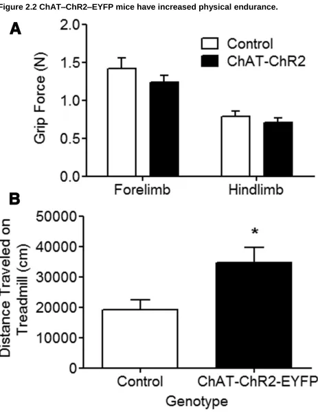
2.4.2 ChAT–ChR2–EYFP mice have improved motor endurance ... 118
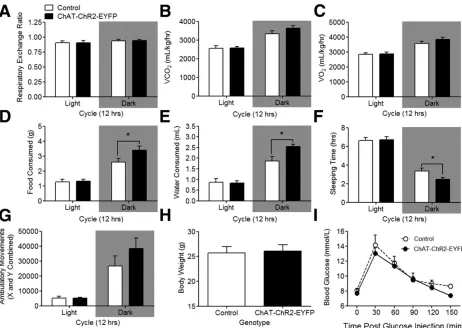
2.4.3 ChAT–ChR2–EYFP mice do not present gross alterations in metabolism ... 121
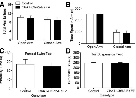
2.4.4 ChAT–ChR2–EYFP mice do not present anxiety or depression-like behavior ... 124
2.4.5 ChAT–ChR2–EYFP mice show normal locomotion but have impaired motor learning ... 127
2.4.6 ChAT–ChR2–EYFP mice have impaired spatial memory ... 130
2.4.7 ChAT–ChR2–EYFP mice have deficiencies in cue-directed memory ... 135
2.4.8 ChAT–ChR2–EYFP have impaired working memory ... 138
2.4.9 ChAT–ChR2–EYFP mice have impaired attentional processing ... 142
2.5 Discussion ... 146
2.6 Acknowledgements ... 150
2.7 References ... 150
Chapter 3 ... 158
Forebrain deletion of the vesicular acetylcholine transporter results in deficits in executive function, metabolic, and RNA splicing abnormalities in the prefrontal cortex ... 158
3.1 Chapter Summary ... 158
3.2 Introduction ... 159
3.3 Material and Methods ... 160
3.3.1 Animals ... 160
3.3.2 Western Blotting ... 161
3.3.3 Acetylcholine Release ... 161
3.3.4 qPCR ... 161
3.3.5 Magnetic resonance imaging ... 163
3.3.6 Metabolite analysis ... 163
3.3.7 Touchscreen behavioral assessment ... 163
3.3.7.1 Apparatus and task ... 163
3.3.7.2 Pretraining... 164
3.3.7.3 Pairwise visual discrimination and reversal ... 165
3.3.7.4 Training in 5-CSRT ... 165
ix
3.3.7.6 Distraction task... 166
3.3.7.7 5-CSRT task measures ... 167
3.3.7.8 Drug treatments ... 167
3.3.8 Statistical analysis ... 167
3.4 Results... 168
3.4.1 VAChTSix3-Cre-flox/flox mice have reduced VAChT and ACh release in the PFC... 168
3.4.2 Decreased forebrain cholinergic tone specifically disturbs reversal learning in the pairwise visual discrimination task ... 171
3.4.3 VAChTSix3-Cre-flox/flox mice have impaired acquisition of the 5-CSRT task 177 3.4.4 Deletion of forebrain VAChT results in inattentive but not impulsive or compulsive behavior ... 180
3.4.5 Deletion of forebrain VAChT impairs sustained attention ... 183
3.4.6 Deletion of forebrain VAChT increases susceptibility to distractions ... 186
3.4.7 Galantamine improves attention in wild-type mice on a demanding task ... 189
3.4.8 Galantamine does not improve attention deficits in VAChTSix3-Cre-flox/flox mice ... 189
3.4.9 Deletion of forebrain VAChT results in metabolic abnormalities in the PFC 193 3.4.10 Deletion of forebrain VAChT results in altered RNA metabolism in the PFC ... 196
3.5 Discussion ... 200
3.5.1 Cognitive flexibility in VAChTSix3-Cre-flox/flox mice ... 200
3.5.2 Attention deficits in VAChT-deficient mice ... 201
3.5.3 Alterations in PFC function in VAChT-deficient mice ... 203
3.6 Conclusion ... 204
3.7 Acknowledgements... 204
3.8 References ... 205
Chapter 4 ... 216
α7 nicotinic ACh receptor‐deficient mice exhibit sustained attention impairments that are reversed by β2 nicotinic ACh receptor activation ... 216
x
4.2 Chapter Summary ... 222
4.3 Introduction ... 222
4.4 Material and Methods ... 224
4.4.1 Animals ... 224
4.4.2 Five-choice serial reaction time task training ... 224
4.4.3 Probe trial ... 225
4.4.4 Drug injections ... 225
4.4.5 Analysis of 5-CSRT task ... 225
4.4.6 Food intake in food-deprived mice ... 226
4.4.7 qPCR ... 226
4.4.8 Western Blotting ... 226
4.4.9 Statistical analyses ... 227
4.5 Results... 227
4.5.1 α7nAChR-null mice present normal acquisition on the 5-CSRT task ... 227
4.5.2 α7nAChR-null mice have impaired sustained attention ... 228
4.5.3 Effect of α7nAChR agonists on attention ... 242
4.5.4 Positive and negative effects of α7nAChR agonists are abolished in CHRΝΑ7−/− mice ... 248
4.5.5 The β2nAChR agonist ABT-418 improves attention ... 254
4.6 Discussion ... 266
4.7 Acknowledgements ... 269
4.8 References ... 269
Chapter 5 ... 278
Cholinergic surveillance over hippocampal RNA metabolism and Alzheimer’s-like pathology ... 278
5.1 Chapter Summary ... 278
5.2 Introduction ... 279
5.3 Materials and Methods ... 280
5.3.1 Mouse lines ... 280
5.3.2 RNA Sequencing ... 280
5.3.3 Immunofluorescence ... 281
xi
5.3.5 Gene Ontology Analysis... 282
5.3.6 RNA Binding Protein Analysis ... 282
5.3.7 qPCR ... 283
5.3.8 Primary Neuronal Cultures ... 283
5.3.9 APP Processing... 283
5.3.10 ELISA ... 283
5.3.11 Congo Red Staining ... 284
5.3.12 Silver staining ... 284
5.3.13 Estimation of Hippocampal Volume ... 284
5.3.14 GSK3 Inhibition ... 284
5.3.15 Morris Water Maze ... 285
5.3.16 Protein Isolation from Human post-mortem brain tissue ... 286
5.3.17 Statistical analysis ... 286
5.4 Results... 286
5.4.1 Forebrain Cholinergic dysfunction modifies expression levels of hippocampal transcripts and alternative splicing ... 286
5.4.2 Cholinergic deficit triggers abnormal BACE1 alternative splicing and APP Processing ... 297
5.4.3 Cholinergic deficit leads to age-dependent hippocampal tauopathy ... 303
5.4.4 Cholinergic deficiency exacerbates age-dependent neuronal vulnerability and impaired learning ... 306
5.4.5 Cholinergic mediated age dependent pathology is partially mediated by GSK3 activation... 312
5.4.6 Cholinergic dysfunction in human AD brains ... 318
5.5 Discussion ... 325
5.6 Acknowledgements ... 332
5.7 References ... 332
Chapter 6 ... 348
Cholinergic Regulation of hnRNPA2/B1 Translation by M1 Muscarinic Receptors ... 348
6.1 Chapter Summary ... 348
6.2 Introduction ... 349
xii
6.3.1 Mouse lines...350
6.3.2 Immunofluorescence...350
6.3.3 Western Blotting...351
6.3.4 Subcellular Fractionation...351
6.3.5 Sarkosyl Insolubility...351
6.3.6 Ubiquitination Assay...351
6.3.7 Stereotaxic injections of adeno-associated virus...352
6.3.8 RNA Sequencing...352
6.3.9qPCR and RT-PCR...352
6.3.10 Isolation of Polysomal RNA...352
6.3.11Primary Neuronal Cultures...353
6.3.12 Pharmacological manipulations in Primary Neuronal Cultures...354
6.3.13 Statistical Analysis...354
6.4 Results...354
6.4.1 Cholinergic Modulation of hnRNPA2/B1 Protein Levels...354
6.4.2 Mechanisms of cholinergic modulation of hnRNPA2/B1...361
6.4.3 Muscarinic Signalling Regulates hnRNPA2/B1 Translation by an NMD Mechanism...367
6.5 Discussion...371
6.6 Acknowledgements...373
6.7 References...374
7 Chapter 7...381
Summary and Conclusion...381
7.1 Summary of Major Findings...381
7.2 Limitations and Future Studies...384
7.3 Significance of Research and Conclusion...387
References...388
Appendices………390
xiii
List of Figures
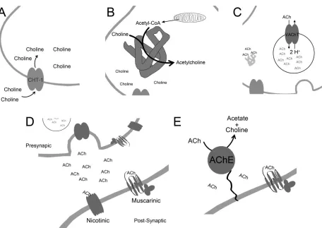
Figure 1.1 The Cholinergic Synapse
Figure 1.2 The Central Cholinergic System in the Murine Brain
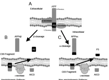
Figure 1.3 Processing of the Amyloid Precursor Protein
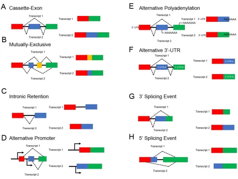
Figure 1.4 Common forms of Alternative Splicing
Figure 1.5 Workflow of an RNA-Seq Experiment
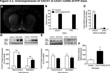
Figure 2.1- Overexpression of VAChT in ChAT–ChR2–EYFP mice.
Figure 2.2 ChAT–ChR2–EYFP mice have increased physical endurance.
Figure 2.3. Metabolic analysis in ChAT–ChR2–EYFP mice.
Figure 2.4. Assessment of anxiety and depressive-like behavior in ChAT–ChR2–EYFP
mice.
Figure 2.5. ChAT–ChR2–EYFP mice have normal locomotor activity but impaired motor
learning.
Figure 2.6. ChAT–ChR2–EYFP mice have spatial memory deficits in the Barnes maze.
Figure 2.7. ChAT–ChR2–EYFP mice have spatial memory deficits in the MWM.
Figure 2.8. ChAT–ChR2–EYFP mice have deficits in cue memory.
Figure 2.9. ChAT–ChR2–EYFP mice have deficits in working memory.
Figure 2.10. ChAT–ChR2–EYFP mice have deficits in attention.
Figure 3.1 Expression of VAChT in the PFC of VAChTSix3-Cre-flox/flox mice.
xiv
Figure 3.3. VAChTSix3-Cre-flox/flox mice have impaired reversal learning.
Figure 3.4. Training in the 5-CSRT task.
Figure 3.5. VAChTSix3-Cre-flox/flox mice have attentional deficits.
Figure 3.6. VAChTSix3-Cre-flox/flox mice have impaired sustained attention.
Figure 3.7 VAChTSix3-Cre-flox/flox mice are more susceptible to distraction.
Figure 3.8 Galantamine improves attention in wild-type, but not in VAChT
Six3-Cre-flox/flox mice.
Figure 3.9. VAChTSix3-Cre-flox/flox mice have metabolic abnormalities in the PFC.
Figure 3.10. VAChTSix3-Cre-flox/flox mice have abnormal RNA processing in the PFC.
Figure 4.1.1. Pre-training on the 5-CSRT task.
Figure 4.1.2. CHRNA7−/− mice have impaired sustained attention.
Figure 4.1.3. CHRNA7−/− mice have normal motivation and motor function during the
5-CSRT task.
Figure 4.1.4. Response patterns did not differ in α7nAChR null mice on the 5-CSRT
task probe trial.
Figure 4.1.5. Evaluation of the expression of cholinergic markers and relevant signalling pathways in the PFC of CHRNA7−/− mice.
Figure 4.2.1 α7nAChR agonists improve attention in wild-type mice.
Figure 4.2.2 α7nAChR agonists did not alter response patterns in wild-type mice.
Figure 4.3.1. α7nAChR agonists do not alter attention in mice lacking α7nAChR.
Figure 4.3.2. α7nAChR agonists did not alter response patterns in CHRNA7-null mice.
Figure 4.4.1. β2nAChR agonists improve attention in wild-type mice.
xv
Figure 4.4.3. Sustained attention deficits of CHRNA7 null mice are reversed by β2nAChR agonists.
Figure 4.4.4. ABT-418 did not alter response patterns in CHRNA7 null mice.
Figure 4.4.5. Performance of mice did not differ across all drug treatments.
Figure 5.1.1. Forebrain deletion of VAChT induces alterations in hippocampal
transcriptome.
Figure 5.1.2. qPCR validation of RNA-Seq data.
Figure 5.1.3. Global hippocampal expression of miRNA is not altered in VAChT deficient
mice.
Figure 5.2.1 Disrupted APP processing and alternative splicing of BACE1 in cholinergic
deficient mice.
Figure 5.2.2 Absence of altered APP processing in aged (11-14 month old)
C57/BJ6-Nkx2.1-Cre mice.
Figure 5.3. Hippocampal cholinergic failure triggers tauopathy in an age-dependent
manner.
Figure 5.4.1. Hippocampal cholinergic failure leads to increased neuronal vulnerability
and worsens cognitive functioning.
Figure 5.4.2 Estimation of neuronal volume in the CA3 and DG region of the
hippocampus of young (3-6) and aged (11-14) month old cholinergic deficient mice as
well as cognitive deficits in aged cholinergic deficient mice.
Figure 5.5.1 Cholinergic mediated tau hyperphosphorylation is regulated by GSK3
activation.
Figure 5.5.2 Cholinergic mediated age dependent pathology is partially mediated by
GSK3 hyper-phosphorylation.
Figure 5.6 Cholinergic Failure in the human AD Brain associates with loss of
xvi Figure 5.7. Summary of findings.
Figure 6.1. Analysis of hnRNPA2/B1 protein levels in genetically modified mice with
differential expression of VAChT.
Figure 6.2. Characterization of decreased hnRNPA2/B1 protein levels in the
hippocampus of VAChT-deficient mice.
Figure 6.3. Mechanisms of cholinergic regulation of hnRNPA2/B1 protein levels.
Figure 6.4. Forebrain cholinergic tone regulates translation of hNRNPA2/B1 in the
hippocampus.
Figure 6.5. Muscarinic Regulation of hnRNPA2/B1 Translation.
List of Tables
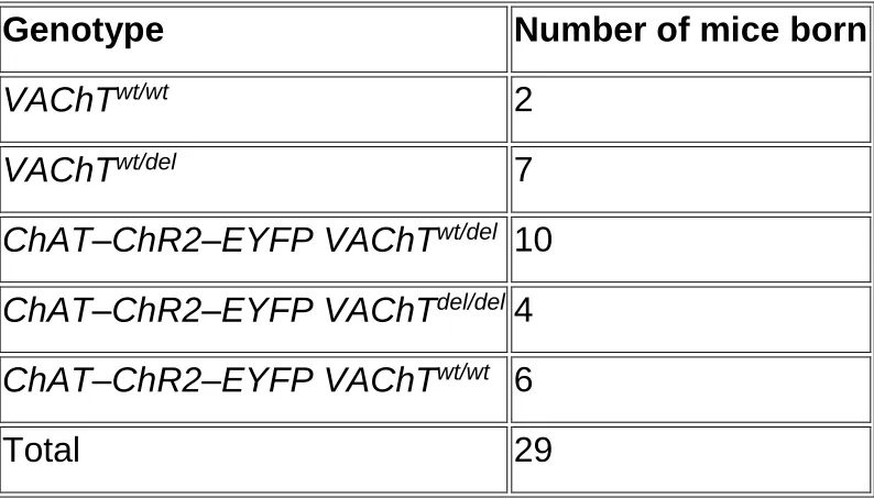
Table 2.1. Rescue of lethality in VAChTdel/del mice.
Table 3.1. Primers for alternative splicing assay
Table 5.1 AD Related Genes Identified in RNA-Seq Data
xvii
List of Abbreviations
ACh Acetylcholine
AChE Acetylcholinesterase
AD Alzheimer’s Disease
APP Amyloid Precursor Protein
ChAT Choline Acetyltransferase
CHT-1 High affinity choline transporter 1
CNS Central Nervous System
CTL1 Torpedo-like choline transporter
GSK-3 Glycogen-synthase kinase-3
hnRNP Heterogeneous nuclear ribonucleoproteins
LTD Long term depression
LTP Long term potentiation
mAChR Muscarinic acetylcholine receptors
MS Medial Septum
MSNs Medium Spiny Neurons
nAChR Nicotinic acetylcholine receptors
nBm Nucleus Basalis Magnocellularis
NFT Neurofibrillary tangles
OCT2 Organic Cation Transporter 2
xviii PNS Peripheral Nervous System
PPT Pedunculopontine Tegmentum
RNA-Seq RNA Sequencing
SR Serine/Arginine-Rich Protein
STP Short term potentiation
VAChT Vesicular Acetylcholine Transporter
VGLUT-3 Vesicular glutamate transporter-3
Chapter 1
1 Introduction
1.1 Central Cholinergic Tone
Acetylcholine (ACh) was the first neurotransmitter to be identified (Loewi 1921). Cells
which secrete, and thus signal via ACh, are deemed cholinergic cells. Cholinergic cells
are found throughout the mammalian body and they are both neuronal and
non-neuronal cell types. Neuronal ACh acts both in the peripheral nervous system (PNS)
and in the central nervous system (CNS). In the PNS ACh is the neurotransmitter which
activates skeletal muscles, in the neuromuscular junction. Furthermore, ACh in the PNS
is the neurotransmitter of the autonomic nervous system which regulates a host of
involuntary and unconscious bodily functions. CNS ACh is a neuromodulator affecting
synaptic plasticity and regulating many behaviours including learning and memory,
attention, and reward (Hasselmo 1999, Picciotto, Higley et al. 2012).
1.1.1 Regulation of ACh Release
Synthesis of ACh relies on the uptake of choline into the presynaptic cholinergic nerve
terminal. This process is mediated by the activity of the high affinity choline transporter
1 (CHT-1) (Yamamura and Snyder 1972) Kuhar and Murrin 1978). Choline in the
presynaptic cell is then combined with Acetyl-CoA and synthesized into ACh by the
choline acetyltransferase enzyme (ChAT) (Hersh 1982, Rylett and Schmidt 1993).
Newly synthesized molecules of ACh are then packaged into synaptic vesicles by the
vesicular acetylcholine transporter (VAChT) (Prado, Reis et al. 2002, Prado, Roy et al.
2013). VAChT represents the rate liming step in the release of ACh. Genetic elimination
of VAChT has demonstrated that VAChT is required for the storage and release of ACh
(Prado, Martins-Silva et al. 2006, de Castro, De Jaeger et al. 2009). Unlike monoamine
which have multiple and redundant transporters (Liu, Peter et al. 1992), VAChT is the
synaptic vesicles fuse with the presynaptic membrane and ACh is released into the
synapse (Katz and Miledi 1965, Varoqui and Erickson 1996). In the synapse, ACh can
bind and act upon pre and post synaptic ACh receptors. These receptors fall into two
main classes, ionotropic nicotinic receptors, and metabotropic muscarinic receptors.
Upon dissociation from the receptors, synaptic ACh is broken down into its constituents
by the activity of the acetylcholinesterase enzyme (AChE) (Marnay and Nachmansohn
Figure 1.1 The Cholinergic Synapse. (A) Choline is taken up into the pre-synaptic cholinergic neuron by the activity of the high affinity choline transporter, CHT-1. (B) Choline and acetyl-CoA are combined to form acetylcholine by the choline
acetyltransferase enzyme. (C) The Vesicular Acetylcholine Transporter packages acetylcholine into synaptic vesicles, exchanging one cytoplasmic molecule of acetylcholine for two vesicular H+ ions. (D) Following excitation of the pre-synaptic
cholinergic neurons, acetylcholine containing synaptic vesicle fuses with the plasma membrane and acetylcholine is released into the synapse. Acetylcholine can then bind to either ionotropic nicotinic receptors or metabotropic muscarinic receptors. These receptors can be expressed both pre- and post-synaptically. (E) Following dissociation of acetylcholine from its receptors, the molecule is broken down by the
1.1.2 The Vesicular Acetylcholine Transporter
To speed up synaptic transmission, neurotransmitters are packaged into synaptic
vesicles, and released as a quantum (Katz and Thesleff 1957, Stevens 1993). This
quantal release mechanism allows for the local concentration of neurotransmitters
released into the synapse to be orders of magnitude larger than if neurotransmitters
were simply released uniformly across the nerve terminal. Release events of ACh from
neurons into the synapse have been well characterized as quantal events (Katz and
Thesleff 1957, Van der Kloot and Molgo 1994). It has been estimated that roughly
10,000 molecules of ACh are packaged into each vesicle (Linder, Pennefather et al.
1984). As ACh is a cation, it will not simply diffuse passively through membranes. A
transport mechanism is required for the packaging of ACh into vesicles for synaptic
release. VAChT is the protein responsible for the packaging of ACh into synaptic
vesicles (Alfonso, Grundahl et al. 1993). Evidence of how VAChT carries out this
transport has been elucidated from models of the proteins structure.
Although the structure of the protein has not been resolved by crystallography or other
methods, models of the protein structure have been proposed to explain the
biochemical activity of the transporter. It has been suggested, based on the knowledge
of other transporters in the same protein family, that VAChT would assume a 12
transmembrane domain structure (Vardy, Arkin et al. 2004). Following the proposed
model, the protein would be separated into two distinct large structural halves, allowing
for a central transport path to be formed between them (Khare, Ojeda et al. 2010).
VAChT is able to pump ACh into synaptic vesicles, by exchanging two intra-vesicular
protons, for one cytoplasmic molecule of ACh (Nguyen, Cox et al. 1998). Experimental
evidence suggests that this exchange occurs within the described central transport path
of the protein (Ojeda, Kolmakova et al. 2004).
In vitro studies have indicated that the activity of VAChT is highly regulated. VAChT will
concentrate ACh into vesicles at a rate 30 folds lower than predicted, based on the free
energy formed by the exchange of two protons (Parsons 2000). The pharmacological
vesamicol have shown that the compound is able to block ACh storage in vesicles, and
also block release of ACh into the synapse (Anderson, King et al. 1983, Collier, Welner
et al. 1986, Whitton, Marshall et al. 1986, Prado, Gomez et al. 1992, Van der Kloot
2003). Therefore, synaptic release of ACh is dependent on VAChT packaging the
neurotransmitter into vesicles. These findings have been confirmed in vivo, where mice
with targeted deletions of VAChT no longer release ACh upon KCl stimulation (Prado,
Martins-Silva et al. 2006, de Castro, De Jaeger et al. 2009, Lima Rde, Prado et al.
2010, Guzman, De Jaeger et al. 2011). Taken together, these results place VAChT as
the rate limiting step in the release of ACh into the synapse.
The majority of total VAChT protein is found within the synaptic vesicles of cholinergic
neurons. The VAChT protein however is not locally translated at the synapse (Park,
Gondre-Lewis et al. 2011). The intracellular trafficking of VAChT is therefore a key
cellular process within cholinergic neurons (Prado and Prado 2002). VAChT trafficking
to synaptic vesicles has been shown to be a clathrin-mediated process (Santos,
Barbosa et al. 2001, Barbosa, Ferreira et al. 2002, Ferreira, Santos et al. 2005).
Experiments using a yeast two hybrid system to investigate protein-protein interaction,
have shown that the C-terminal domain of VAChT interacts with the AP-2 adaptor
complex (Barbosa, Ferreira et al. 2002). AP-2 is a multimeric protein that is a master
regulator of chlatrin-mediated endocytosis (Pearse, Smith et al. 2000). A di-leucine motif
has been characterized within the C-terminus of the VAChT protein, and plays a critical
role in the clathrin-mediated endocytosis of the transporter (Tan, Waites et al. 1998).
Mutations in this di-leucine motif abolish the interaction between VAChT and the AP-2
complex (Barbosa, Ferreira et al. 2002). Likewise, the C-terminal segment of vesicular
monoamine transporters (VMATs) regulates their trafficking (Tan, Waites et al. 1998).
This is not surprising as there is a large degree of homology between VAChT and
VMATs (Liu and Edwards 1997). Interestingly, the intracellular trafficking of these
proteins is vastly different. In the brain, VMATs are trafficked to many different types of
secretory vesicles (Nirenberg, Liu et al. 1995, Nirenberg, Chan et al. 1996). Conversely,
VAChT is localized almost exclusively to synaptic vesicles (Gilmor, Nash et al. 1996,
Weihe, Tao-Cheng et al. 1996). It appears that the phosphorylation of this C-terminus is
Ferreira, Santos et al. 2005). The kinase PKC has been shown to both modulate
VAChT phosphorylation as well as ACh release from hippocampal synaptosomes
(Tanaka, Fujiwara et al. 1986, Allgaier, Daschmann et al. 1988, Barbosa, Clarizia et al.
1997). VAChT phosphorylation can therefore be highly relevant to cholinergic signalling
in the brain (Van der Kloot and Molgo 1994).
1.1.3 Choline Acetyltransferase
Synthesis of ACh within neurons occurs within the cytoplasm of cholinergic nerve
terminals. ChAT is the enzyme responsible for the production of ACh within cells. As its
name suggests, choline acetyltransferase transfers an acetyl group from an acetyl-CoA
to a choline molecule to produce the neurotransmitter ACh (Nachmansohn and Berman
1946). Expression of the ChAT enzyme is extremely sparse, estimated to comprise less
than a ten thousandth of a percent of total brain proteins (Eckenstein and Thoenen
1982, Bruce, Wainer et al. 1985). Despite this low abundance, ChAT protein is found
within every cholinergic cell in the brain (Docherty, Bradford et al. 1985), its expression
pattern therefore follows the anatomical distribution of cholinergic neurons in the brain.
Interestingly, the genes for ChAT and VAChT are present in the same gene locus,
termed the cholinergic gene locus (Eiden 1998, Mallet, Houhou et al. 1998). Cloning of
the ChAT gene revealed a rather unique structure for the gene (Berrard, Brice et al.
1987, Strauss, Kemper et al. 1991). Upstream of the coding region for ChAT, within the
intron between the first and second exons, the VAChT 1590bp open reading frame was
discovered (Bejanin, Cervini et al. 1994, Erickson, Varoqui et al. 1994, Roghani,
Feldman et al. 1994). The VAChT gene is found in the same transcriptional orientation
as ChAT (Berrard, Varoqui et al. 1995). This unique gene organization is evolutionarily
conserved and can be found in C. elegans (Alfonso, Grundahl et al. 1994, Roghani,
Feldman et al. 1994), drosophila (Kitamoto, Wang et al. 1998), mice (Misawa, Ishii et al.
1992, Barbosa, Massensini et al. 1999), rats (Bejanin, Habert et al. 1992) and men
ChAT are transcribed as separate transcripts (Alfonso, Grundahl et al. 1994, Erickson,
Varoqui et al. 1994).
ChAT is localized predominantly in the cytoplasm (Bruce, Wainer et al. 1985). However,
membrane-bound forms of the protein have been reported (Bruce and Hersh 1987). In
addition, a variant of the human form of the enzyme can localize to the nucleus, wherein
it plays a role in the regulation of gene expression and chromatin modeling (Gill,
Bhattacharya et al. 2003, Matsuo, Bellier et al. 2011). The ChAT enzyme is organized
as a globular, single strand, protein (Govindasamy, Pedersen et al. 2004). Site-directed
mutagenesis studies have shown that a number of critical amino acid residues regulate
the catalytic function of the protein (Carbini and Hersh 1993, Dobransky, Davis et al.
2000). This work showed that a histidine residue is the acid/base catalytic residue of the
enzyme, while a nearby arginine residue interacts with, and binds the CoA molecule.
Further structural biology experiments have confirmed these findings (Kim, Rylett et al.
2006).
Interestingly, there appears to be no correlation between the mRNA level of ChAT
within a cellular population, and the enzymatic activity of the cells (Berrard, Brice et al.
1987). This finding suggests that regulation of ChAT protein levels and activity occur at
the post-transcriptional level. Translation of the ChAT protein occurs within the soma of
neurons (Berrard, Brice et al. 1987) and the enzyme is then transported by an
undetermined mechanism to the synapse. The transport of the enzyme represents a
means by which cells can regulate their production of ACh. A critical post-translational
modification to the ChAT enzyme is its phosphorylation (Bruce and Hersh 1989,
Dobransky and Rylett 2003). A number of phosphorylation sites have been
demonstrated on the enzyme and it appears that these modifications play a role in
regulating the catalytic activity of the enzyme (Dobransky and Rylett 2003). A key
phosphatase involved in the regulation of ChAT enzymatic activity is PKC (Dobransky,
Doherty-Kirby et al. 2004). Importantly ChAT phosphorylation in synaptosomes is
sensitive to Ca2+ levels (Dobransky and Rylett 2005). The enzymatic activity of ChAT,
and consequently ACh synthesis, can therefore be regulated in response to neuronal
lethal (Misgeld, Burgess et al. 2002). Studies of the embryos from these animals have
shown in the absence of ChAT there is dramatic restructuring of the PNS in these
animals (Brandon, Lin et al. 2003), indicating that ChAT, and therefore ACh, is essential
for proper developments of the nervous system.
1.1.4 The High Affinity Choline Transporter
Synthesis of ACh is dependent on the intracellular concentrations of choline. Neurons
are unable to synthesise enough of their own choline for cholinergic neurotransmission
(Yamamura and Snyder 1972, Collier and Katz 1974).Therefore, ACh releasing neurons
are dependent on the activity of a transporter to obtain their choline (Birks, Macintosh et
al. 1956, Birks and Macintosh 1957). The transporter responsible for the uptake of
choline into neurons is the CHT1 transporter. This transporter regulates choline levels
within a cell, by transporting choline into the cell in a Na+ dependant manner (Simon
and Kuhar 1975, Birks, Worsley et al. 1985). This protein is encoded by the SLC5A7
gene, which was first identified in C. elegans, and subsequently identified in the rat,
mouse and human (Apparsundaram, Ferguson et al. 2000, Okuda, Haga et al. 2000).
There is a very high degree of homology between the rat and human gene (Okuda and
Haga 2000) as well as between the rat and mouse gene (Apparsundaram, Ferguson et
al. 2001). Importantly, CHT1 is preferentially expressed in cholinergic neurons, and can
therefore be used as a specific marker for these cells (Misawa, Nakata et al. 2001).
CHT1 is not the sole choline transporter expressed in the mammalian brain. The
Torpedo-like choline transporter, CTL1 is highly expressed in oligodendrocytes, where it
provides choline for the synthesis of phospholipids (Traiffort, Ruat et al. 2005). CLT1
also differs from CHT1 in that its function is dependent on K+ ions rather than Na+ ions
(Fujita, Shimada et al. 2006). The other choline transporter expressed in the
mammalian brain is the organic cation transporter 2 (OCT2). Unlike CTL1, OCT2 is also
expressed in cholinergic neurons, being found on synaptic vesicles within these
neurons (Nakanishi, Haruta et al. 2011, Nakata, Matsui et al. 2013). OCT2 however is
not selective for choline, but rather can transport all organic cations. Given that OCT2
that these two transporters may collaborate to transport choline into cholinergic neurons
(Nakata, Matsui et al. 2013).
The CHT1 protein is 580 amino acids long and organized on plasma membranes in a
13 transmembrane domain structure (Apparsundaram, Ferguson et al. 2000, Okuda,
Haga et al. 2000, Torres, Gainetdinov et al. 2003). CHT1 has been shown to have
consensus PKA and PKC phosphorylation motifs, implicating these signalling pathways
in the regulation of this protein (Gates, Ferguson et al. 2004, Brock, Nickel et al. 2007).
Experimental inhibition of these signalling pathways has led to decreases in cell surface
levels of the transporter (Gates, Ferguson et al. 2004). Although CHT1 carries out its
physiological function at the plasma membrane, the vast majority of the protein is found
within intracellular vesicles, with only a small portion of the total protein pool found on
the plasma membrane (Ferguson, Savchenko et al. 2003, Ribeiro, Alves-Silva et al.
2003, Ribeiro, Black et al. 2005). Inclusion of CHT1 in the plasma membrane of
synapses is dependent on the activity of the neuron. Depolarization by action potential
of the neuron increases the levels of CHT1 at the plasma membrane in a calcium
dependant manner (Collier and Katz 1974, Simon and Kuhar 1975). The trafficking of
CHT1 to the plasma membrane is mediated by the clathrin endocytic pathway (Ribeiro,
Alves-Silva et al. 2003). Plasma membrane CHT1 then can be internalized in a clathrin
dependant manner to either be tagged for proteasome degradation, or to be trafficked
back to the plasma membrane (Ribeiro, Black et al. 2005).
Genetic elimination of CHT1 from the mouse genome results in post-natal lethality,
within an hour of birth, due to an inability of the animals to breathe (Ferguson,
Bazalakova et al. 2004). Mice with a heterozygous null mutation are however viable and
are able to maintain the same level of choline uptake as wild-type control animals
through posttranslational compensation of CHT1 function, by increased inclusion at the
plasma membrane (Ferguson, Bazalakova et al. 2004). Furthermore, in mice with a
heterozygous null deletion of the ChAT gene (and therefore a reduced capacity to
synthesize ACh), levels of the CHT1 protein were increased in compensation (Brandon,
Mellott et al. 2004). Analysis of the prefrontal cortex of rats performing attention based
(Apparsundaram, Martinez et al. 2005). These findings demonstrate that the trafficking
of CHT1 to the plasma membrane is a highly dynamic process which plays a role in
normal physiological functioning. Importantly polymorphisms in the CHT-1 gene have
been identified in patients with Attention Deficit Disorders, related to cholinergic deficits
in these patients (English, Hahn et al. 2009). The trafficking of CHT1 to the plasma
membrane is thus a critical determinant to the levels of choline within cholinergic cells
and a critical process for cognitive functioning.
1.1.5 Nicotinic Receptors
Nicotinic acetylcholine receptors (nAChRs) are ionotropic receptors which are selective
for ACh. nAChRs are expressed in both the peripheral and in the CNS. This class of
receptors are involved in neuronal excitability and regulation of neurotransmitter
release. There are nine different nicotinic receptor subunits that are expressed in the
CNS, they are either subunits (2, 3, 4, 5, 6, 7) or subunits (2, 3, 4) all encoded by
distinct genes (Le Novere and Changeux 1995, Dani and Bertrand 2007). These
subunits combine as either homomeric or heteromeric pentameric receptors. The most
prominent homomeric nicotinic receptor in the mammalian CNS is the 7 nicotinic
receptor (7nAChR). However the most prominently expressed nicotinic receptor in the
brain is the 42nAChR (Wada, Wada et al. 1989).
Regardless of their make-up, nicotinic receptors share the same general structure. They
are organised as a transmembrane receptor with a central ion channel, an extracellular
ligand binding domain with a ligand binding pocket (Karlin and Akabas 1995). Although
they share similar structures, 7nAChRs and 42nAChRs have drastically different
functional properties (Giniatullin, Nistri et al. 2005). 7nAChRs are quick to be activated
by ACh and are quickly desensitized (Pidoplichko, DeBiasi et al. 1997). These receptors
are permeable to Na+, and K+, but are highly permeable to Ca2+ (Seguela, Wadiche et
al. 1993). 42nAChRs on the other hand are slow to be activated by ACh and are also
slow to desensitize (Alkondon and Albuquerque 2005). These receptors are only
In situ hybridization studies have demonstrated that the 4 and 2 nAChR subunits are
expressed ubiquitously across the mammalian brain (Wevers, Jeske et al. 1994).
Expression of these receptors is highest in the thalamus and cortex and relatively low in
the hippocampus (Alkondon, Reinhardt et al. 1994). 2 nAChRs is essential for nicotine
evoked release of GABA and dopamine from synaptosomes collected from a number of
brain regions (Turner 2004, McClure-Begley, King et al. 2009). Furthermore nicotine
evoked striatal release of dopamine is abolished in mice lacking the 2 subunit (King,
Caldarone et al. 2004). This regulation of dopamine release by 2nAChRs is
functionally relevant as mice lacking this receptor do not display place preference
conditioning to cocaine (Zachariou, Caldarone et al. 2001). Young mice lacking the 2
nAChR subunit do not display memory impairments, however as these mice age they
display cortical atrophy and cell loss in the hippocampus (Zoli, Picciotto et al. 1999).
The age dependant neurodegeneration in these animals is associated with learning and
memory impairments.
The 7nAChR like the 4 and 2 is expressed throughout the brain. However the
distribution pattern of the 7nAChR is antithetical to the pattern of the 4 and 2
subunits. 7nAChRs are most expressed in the hippocampus and cortex, but are
almost absent in the thalamus (Seguela, Wadiche et al. 1993). Although 7nAChRs are
highly involved in synaptic plasticity in the hippocampus, 7-subunit knockout mice did
not differ from wild-type controls in a host of memory assays including contextual and
cued fear-conditioning and on the Morris water maze (Paylor, Nguyen et al. 1998,
Fernandes, Hoyle et al. 2006). 7nAChRs are also expressed on non-neuronal cells in
both the brain and in the periphery, including astrocytes and microglia. 7nAChRs on
these non-neuronal cells play a critical role in inflammation (de Jonge and Ulloa 2007)
and in neuroprotection (Ren, Puig et al. 2005). On immune cells activation of 7
receptors regulates the production of inflammatory cytokines (Wang, Yu et al. 2003).
1.1.6 Muscarinic Acetylcholine Receptors
ACh can act upon a class of metabotropic receptors, the muscarinic acetylcholine
identified, the M1, M2, M3, M4 and M5 receptors. All of these receptors function as
classical G-protein coupled receptors (Wess 1996). mAChRs can be subdivided into
two categories, the Gq coupled receptors which includes the M1, M3 and M5 receptors
(Berstein, Blank et al. 1992, Offermanns, Wieland et al. 1994, Qin, Dong et al. 2011),
and the Gi/o coupled M2 and M4 receptors (Winitz, Russell et al. 1993, Migeon and
Nathanson 1994). The Gq coupled receptors activate phospholipase C and increase
intracellular Ca2+ levels; these receptors are therefore deemed excitatory. The Gi/o
coupled receptors on the other hand act by decreasing cyclic nucleotide levels,
decreasing intracellular Ca2+ levels and promoting K+ efflux, thus inhibiting the neuron.
G proteins can also directly regulate K+ channels, which is a common mechanism for
M2 muscarinic receptors (Kunkel and Peralta 1995).
All five of the mAChR are expressed in the mammalian nervous system. These
receptors are found to be expressed on both neuronal and glial cell types in the nervous
system. The expression of the M1, M4 and M5 subtypes is enriched in the CNS,
whereas the M2 and M3 receptors are equally found in both the CNS and the PNS
(Levey 1993). mAChRs in the brain have been shown to play regulatory roles in many
cognitive processes. Each of the five muscarinic receptors are encoded by a unique
gene. This has allowed for the genetic manipulation of the individual receptor types and
from this their distinct physiological roles in the central nervous system have been
elucidated (Wess 2004).
M1 mAChRs are expressed throughout the mammalian forebrain, including cerebral
cortex, hippocampus, and in the striatum (Wolfe and Yasuda 1995). It is by its broad
level of expression that the receptor is thought to play a role in many cognitive
processes. Mice lacking the M1 mAChR have been shown to be hyperactive (Gerber,
Sotnikova et al. 2001) and have impaired working memory (Anagnostaras, Murphy et al.
2003) and consolidation (Gould, Dencker et al. 2015). These animals also present a
number of biochemical (Berkeley, Gomeza et al. 2001) and electrophysiological
M3 mAChRs are expressed throughout the brain but are most prominently expressed in
the hypothalamus (Wall, Yasuda et al. 1991). Hypothalamic M3 mAChRs regulate food
intake and appetite. These receptors are found on melanin-concentrating hormone
producing neurons of the hypothalamus, and their activations stimulates production of
the appetite regulating hormones (Yamada, Miyakawa et al. 2001). Mice lacking this
receptor are lean and have a pronounced reduction in bodyweight and food intake
compared to control animals (Yamada, Miyakawa et al. 2001). In the periphery these
receptors have been shown to regulate smooth muscle function (Matsui, Motomura et
al. 2002) and the salivary response (Bacman, Sterin-Borda et al. 1996).
M5 mAChRs have low levels of expression throughout the brain and are enriched in
limited cellular populations (Wei, Walton et al. 1994). M5 mAChRs are expressed on the
midbrain dopaminergic neurons, originating from the substantia nigra (Vilaro, Palacios
et al. 1990), and are therefore highly critical to the regulation of dopamine release in the
striatum. M5 mAChRs have been shown to directly regulate striatal dopamine release,
although other muscarinic subtypes can also modulate striatal dopamine by indirect
mechanisms (Zhang, Yamada et al. 2002). The ventral tegmental area of mice deficient
in the M5 mAChR do not respond to cholinergic stimulation demonstrating that these
receptors also regulate ventral tegmental area dopamine release (Yeomans, Forster et
al. 2001, Forster, Yeomans et al. 2002). Given the role of these receptors in regulating
dopamine signalling, mice lacking M5 mAChRs have altered responses to opioids and
cocaine (Basile, Fedorova et al. 2002, Thomsen, Woldbye et al. 2005).
The inhibitory M2 and M4 mAChRs function mainly as auto-receptors for ACh in the
brain (Starke, Gothert et al. 1989). These receptors are also both present on the
GABAergic medium spiny neurons in the striatum (Yan, Flores-Hernandez et al. 2001).
For the most part, these receptors are responsible for inhibiting release of ACh (Zhou,
cholinergic neurons (Levey, Edmunds et al. 1995). These receptors therefore play a
critical role in the regulation of the timing and release of ACh, processes which are
highly relevant to cognitive function (Sarter, Parikh et al. 2009). Mice lacking the M2
receptor show a broad range of hippocampal behavioural and electrophysiological
impairments (Seeger, Fedorova et al. 2004). Mice lacking the M4 receptor also present
with learning impairments and hyperactivity (Koshimizu, Leiter et al. 2012). Given that
these receptors regulate release of ACh; it is difficult to differentiate the specific roles of
these receptors to cognition, from general roles of cholinergic tone in the brain.
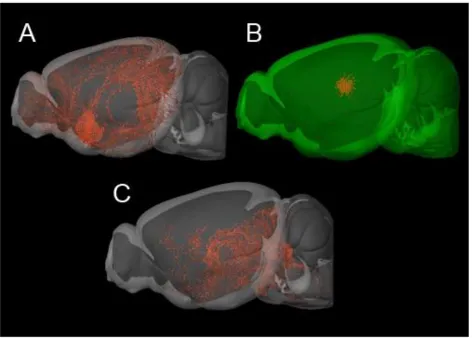
1.1.7 Basal Forebrain Cholinergic System
The basal forebrain is a collection of nuclei including the medial septum (MS), the
ventral pallidum (VP), the diagonal band nuclei, magnocellular preoptic nucleus, and the
nucleus basalis magnocellularis (nBm) (Zaborszky, Pang et al. 1999). The neurons
found within this brain region have been implicated in many cognitive functions including
learning and memory and executive function (Miyamoto, Shintani et al. 1985). The basal
forebrain is composed of a highly variable population of neurons. ACh releasing
neurons represent roughly 20% of the neurons in the basal forebrain (Semba 2004).
Other neurons in the basal forebrain release glutamate, GABA and neuropeptides
(Henny and Jones 2008).
The output of basal forebrain cholinergic neurons is dependent on the nuclei from which
these neurons are found (Figure 1.2A). Neurons originating in the MS and the diagonal
band nuclei provide the major cholinergic innervation to the hippocampus (Mesulam,
Mufson et al. 1983). Cholinergic neurons from the diagonal band and from the
magnocellular preoptic nucleus project to the olfactory bulb and to the enthorinal cortex
(Gaykema, Luiten et al. 1990), while the nBm cholinergic neurons project to both the
basolateral amygdala and the entire cortex (Boegman, Cockhill et al. 1992, Power, Thal
et al. 2002). Just as the basal forebrain is not a homogenous cell population, the
described projections are not purely cholinergic with both GABAergic and glutamatergic
projection neurons sharing the same projection tracks as the cholinergic cells (Huh,
Basal forebrain cholinergic neurons receive afferent input from midbrain and brainstem
as well as from the hypothalamus. Unlike the non-cholinergic neurons in the basal
forebrain, the cholinergic neurons in the basal forebrain do not appear to receive top
down input from the cortex (Zaborszky 1989, Zaborszky and Duque 2000).
Hypocretin/orexin positive neurons from the hypothalamus have been identified as the
main source of innervation to the basal forebrain (Henny and Jones 2006). These
neurons have been shown to synapse onto both cholinergic and non-cholinergic neuron
in the basal forebrain. These neurons are histaminergic and are important regulators of
arousal. Midbrain afferent connections to the basal forebrain originate from the
dopaminergic cells of the substantia nigra and from the ventral tegmental area (Fallon
and Moore 1978, Zahm and Trimble 2008). Brainstem structures which project to the
basal forebrain cholinergic cells are adrenergic in nature from the medulla and the locus
coeruleus and cholinergic from the pedunculopontine nucleus.
Basal forebrain cholinergic neurons role in regulating executive function and memory
has largely been investigated by immunotoxin lesioning of these cells (Baxter, Bucci et
al. 2013). The 192 IgG-saporin is a specific neurotoxin which, when delivered to the
basal forebrain, will selectively kill cholinergic neurons and spare the other cell types in
the region (Book, Wiley et al. 1992). An important caveat to the attribution of these
behaviours to cholinergic signalling is that these neurons may not be simply cholinergic.
In culture, these cholinergic neurons from the basal forebrain have been shown to be
able to release glutamate (Allen, Abogadie et al. 2006). Recent optogenetic
experiments have demonstrated that basal forebrain cholinergic which project to the
cortex co-release GABA from distinct pools of vesicles (Saunders, Granger et al. 2015).
The behavioural consequences of this co-transmission have yet to be investigated, but
suggest that GABA and/or glutamate co-released with ACh may influence behaviours
thought to be regulated by ACh signalling alone (Granger, Mulder et al. 2016).
1.1.8 Striatal Cholinergic System
Contrary to projection cholinergic neurons found in the basal forebrain and the
interneurons (Figure 1.2B). As such, they are characterized by shorter axons and form
circuits with nearby neuron within the same brain region (Zhou, Wilson et al. 2002). The
cholinergic interneurons of the striatum are the primary source of ACh in the striatum,
but only comprise less than 1% percent of all cells in the striatum (Bolam, Wainer et al.
1984). Cholinergic neurons in the striatum, relative to other cholinergic neurons, have
higher levels of the key cholinergic markers choline acetyltransferase (Hebb and Silver
1961) and acetylcholinesterase (Woolf and Butcher 1981). This enrichment in
cholinergic markers demonstrates the importance that these cells play in regulating
striatal function. 33 1.2 B.
A key role of the cholinergic interneurons in the striatum is to integrate information from
afferent inputs to the striatum. This is achieved by assimilating inputs of a large number
of neurons which release various different neurotransmitters, by expressing a large
number of different receptor classes for these neurotransmitters. These
neurotransmitters can either act in an excitatory manner, such as glutamate (Calabresi,
Centonze et al. 1998), serotonin (Bonsi, Cuomo et al. 2007), histamine (Bell,
Richardson et al. 2000) and substance P (Aosaki and Kawaguchi 1996).
Neurotransmitters can also inhibit cholinergic interneurons, such as GABA (DeBoer and
Westerink 1994), adenosine (Brown, James et al. 1990), and endogenous opioids
(Rada, Mark et al. 1991). Other neurotransmitters can be both excitatory and inhibitory,
depending on which class of receptors they activate such as with dopamine, excitatory
D1-like signalling (Aosaki, Kiuchi et al. 1998) and inhibitory D2-like (Chuhma, Mingote et
al. 2014). It is therefore the role of these cholinergic interneurons to integrate all of
these various signals.
Despite their relatively small cell number, striatal interneurons project throughout the
striatum and can therefore have broad physiological effects. GABA-ergic medium spiny
neurons (MSNs) are the primary output of the striatum (Chuhma, Tanaka et al. 2011).
These neurons are not thought to express nicotinic acetylcholine receptors (Luo,
Janssen et al. 2013), though nicotine can indirectly modulate their activity (Liu, Otsu et
activation of muscarinic receptors. Activation of M1 muscarinic receptors on these cells
is thought to reduce KCNQ potassium inhibition (Shen, Hamilton et al. 2005) and Ca2+
entry (Perez-Garci, Bargas et al. 2003), increasing the excitation of MSNs. Nicotinic
receptors in the striatum are highly concentrated on both glutamatergic and
dopaminergic nerve terminals. Glutamatergic nerve terminals in the striatum are
enriched in 7 nicotinic receptors, and activation of these receptors leads to release of
glutamate (Campos, Alfonso et al. 2010). Release of dopamine in the striatum is also
highly dependent on cholinergic signalling. Unlike glutamate, the release of dopamine in
the striatum has been shown to be dependent on the synchrony of cholinergic
interneuron firing, and the activation of 2 nicotinic receptors on dopaminergic terminals
(Cachope, Mateo et al. 2012).
An important caveat for many of the studies of striatal cholinergic neurons, is that these
cells co-transmit glutamate. This co-transmission is achieved through synergism
between VAChT and the vesicular glutamate transporter-3 (VGLUT-3) (El Mestikawy,
Wallen-Mackenzie et al. 2011). VAChT and VGLUT-3 are found on the same vesicles in
these neurons and it has been shown that VGLUT-3 enables these vesicles to package
additional ACh (Gras, Amilhon et al. 2008). Studies using genetically modified mice
have shown that the glutamate and the ACh released from these neurons can have
separate and potentially opposite physiological roles in regulating striatal function and
behaviour (El Mestikawy, Wallen-Mackenzie et al. 2011, Guzman, De Jaeger et al.
2011, Sakae, Marti et al. 2015). Behavioural studies of mice with lesioned striatal
cholinergic interneurons were found to be hyperactive, have a heightened response to
cocaine and do not show haloperidol induced catalepsy (Hikida, Kaneko et al. 2001,
Kitabatake, Hikida et al. 2003). These behaviours were initially attributed to the
cholinergic properties of these cells. However mice lacking release of ACh from the
striatum were not hyperactive and did not display heightened responses to cocaine
(Guzman, De Jaeger et al. 2011). In fact, it was mice lacking the VGLUT-3 transporter
which recapitulated the phenotype of the lesioned animals (Gras, Amilhon et al. 2008).
The ACh released from these cells seems to be playing a critical role in regulating
functional increase in dopamine receptor expression and activity, assessed by
pharmacological MRI (Guzman, De Jaeger et al. 2011). VGLUT-3 and VAChT have
opposing roles in the regulation of dopamine release. Elimination of VAChT from striatal
interneurons decreases KCl stimulated dopamine release, whereas elimination of
VGLUT-3 from these neurons potentiates the dopamine response (Sakae, Marti et al.
2015). These results suggest that other physiological properties of striatal ACh may
potentially be attributable to the glutamate released from these cholinergic interneurons
or may be dependent on the combined action of both neurotransmitters.
1.1.9 The Pedunculopontine Tegmentum Cholinergic System
The pedunculopontine tegmentum (PPT) is a collection of nuclei found within the
brainstem behind the substantia nigra. The nuclei found within the PPT are a
heterogeneous population of neurons that differ by the neurotransmitters they release.
The three major neurons within the PPT are cholinergic, GABAergic and glutamatergic
(Martinez-Gonzalez, Wang et al. 2012). These three types of neurons are largely
segregated into separate regions of the PPT. The majority of cholinergic neurons within
the PPT are located in the PPT pars compacta (Pahapill and Lozano 2000).
Much of the work to characterize the projection of the cholinergic neurons from the PPT
has been done in non-human primates. These experiments have shown that these
cholinergic neurons project predominantly to structures within the basal ganglia (Figure
1.2 C). Both the substantia nigra (Futami, Takakusaki et al. 1995) and the ventral
tegmental area receive significant input from the cholinergic neurons of the PPT
(Charara, Smith et al. 1996). This anatomical data place these cholinergic cells as key
regulators of dopamine signalling in the brain. The primary output of the cholinergic
neurons from the PPT is the thalamus (Parent, Pare et al. 1988). Additionally the
striatum also receives direct input from the cholinergic neurons of the PPT (Dautan,
Huerta-Ocampo et al. 2014). The PPT is therefore an alternate source of cholinergic
tone in the striatum, besides the cholinergic interneurons. Finally an important target of
Given the anatomical connectivity data, the cholinergic output from the PPT is placed to
be a key regulator of dopamine signalling in the brain. Experimental evidence for this
was first shown by studying the addictive properties of nicotine; by increasing dopamine
release acting specifically through 2 nicotinic acetylcholine receptors in the VTA
(Picciotto, Zoli et al. 1998). Further work has also demonstrated that M5-type
muscarinic receptors can also stimulate dopamine release from the VTA (Corrigall,
Coen et al. 2002). Since the cholinergic output from the PPT can robustly modulate
dopamine signaling, these neurons are important intermediaries of addiction and
reward. Not only do these neurons release more ACh into the VTA during cocaine
administration and cocaine seeking behaviours (You, Wang et al. 2008), but these
cholinergic neurons will also increase their firing rate in response to environmental cues
associated with reward (Goldberg and Reynolds 2011). Given these findings,
cholinergic PPT neurons have been proposed to be master regulators of dopamine
signalling in the brain (Maskos 2008).
The PPT cholinergic neurons also play a key role in regulating sleep and arousal. PPT
cholinergic neurons are completely inhibited during seizures, this inhibition is
accompanied by changes in EEG recordings (Motelow, Li et al. 2015). In line with these
findings, PPT cholinergic neurons play a critical role in increasing arousal states during
sleep. Recent optogenetic studies have found that activation of cholinergic neurons in
the PPT is an important modulator of REM sleep and plays a critical role in the initiation
Figure 1.2 The Central Cholinergic System in the Murine Brain. Sagittal 3D
reconstructions of the mouse brain. Image data detailing axonal projections labeled by rAAV tracers injections into ChAT-IRES-Cre mice, and visualized using serial
two-photon tomography. (A) Cholinergic projections from the basal forebrain. (B) Cholinergic interneurons in the striatum (C) Cholinergic projections from the pedunculopontine tegmental nucleus. Images generated from the Allen Brain Institute Mouse Connectivity Atlas. © 2016 Allen Institute for Brain Science. Allen Mouse Brain Connectivity Atlas [Internet]. Available from: http://connectivity.brain-map.org. Oh, S.W. et al. (2014) A mesoscale connectome of the mouse brain, Nature 508: 207-214.
1.2 Cholinergic Regulation of Brain Functions
1.2.1 Cholinergic Regulation of Hippocampal Function
The hippocampus has been extensively studied for its role in regulating learning and
memory (Squire 1992). This brain region plays an important role in the formation and maintenance of new memories. In Alzheimer’s disease (AD) the hippocampus is one of
the first brain regions to be affected by pathology (De Leon, George et al. 1997,
Padurariu, Ciobica et al. 2012). Hippocampal degeneration mediates some of the
learning and memory deficits in AD patients (Graham and Hodges 1997).
The finding in humans that drugs which block cholinergic receptors in brain impair
performance on task of learning and memory (Ghoneim and Mewaldt 1975, Atri,
Sherman et al. 2004), have placed this neurotransmitter as a central regulator of
memory. Functional MRI studies have shown that cholinergic modulation of the
hippocampus occurs during learning and memory tasks (Goekoop, Scheltens et al.
2006, Wink, Bernard et al. 2006). Much of the work delineating the mechanism of
cholinergic control of hippocampal function has been performed in rodents. In both
rodents and man, the cholinergic innervation to the hippocampus arises from the medial
septum nucleus within the basal forebrain cholinergic system (Lewis and Shute 1967).
The involvement of the basal forebrain cholinergic system in memory is supported by
several works employing various methods ranging from unspecific and specific lesion
methods (Wrenn, Lappi et al. 1999, Chudasama, Dalley et al. 2004), pharmacological
manipulations (Granon, Poucet et al. 1995) and more recently the use of genetic
manipulations (Fernandes, Hoyle et al. 2006, Martyn, De Jaeger et al. 2012, Al-Onaizi,
Parfitt et al. 2016).The observation of a transient increase of cholinergic activity during
and after a memory tasks can be observed in hippocampus (Durkin and Toumane 1992,
Durkin 1994). It is suggested that this increase in neurotransmitter release may support
memory (Fadda, Melis et al. 1996, Hironaka, Tanaka et al. 2001).
Synaptic plasticity is a process whereby the nervous system changes the weight of
functional connectivity; it can help form, eliminate, potentiate and weaken connections
of neuronal circuits (Abbott and Nelson 2000). The mechanism of cholinergic regulation
of hippocampal plasticity has been well studied (Hasselmo and Bower 1993, McEwen
1999, Yakel and Shao 2004, Hasselmo 2006, Drever, Riedel et al. 2011, Yakel 2012).
The addition of low concentrations of carbachol, a cholinergic mimetic, onto cultured
slices induces LTP, an effect dependent of muscarinic receptors (Auerbach and Segal
1996). In vivo experiments have consistently demonstrated the role of cholinergic tone
in the modulation of LTP. Free walking mice showed reduced LTP in the hippocampus,
after cholinergic denervation in the medial septum or following administration of
muscarinic receptors antagonists (Leung, Shen et al. 2003, Doralp and Leung 2008).
Furthermore, deletion of VAChT from basal forebrain neurons also disrupts
hippocampal LTP ex vivo (Martyn, De Jaeger et al. 2012) and in vivo (Al-Onaizi, Parfitt
et al. 2016).
The mechanisms by which cholinergic neurons regulate synaptic plasticity are complex
and are likely to involve short-term changes in membrane conductance and also on
second messengers. Carbachol can induce another type of synaptic plasticity in the
hippocampus and the cortex, long-term depression (LTD), once again requiring
muscarinic activation (Jo, Son et al. 2010, Caruana, Warburton et al. 2011). LTD is
correlated with learning and information storage, furthermore, it has been related to
processes which require cognitive flexibility such as extinction and behavior flexibility
(Collingridge, Peineau et al. 2010).
Reports have proposed the notion that cholinergic modulation of synaptic plasticity is
highly dependent on time, and that nicotinic receptors serve as a switch between LTP
and LTD. Ge and Dani (2005), using a protocol to induce a short-term, transient,
potentiation (STP) observed that the STP became an LTP response when ACh was