C A S E R E P O R T
Open Access
Angiolymphoid hyperplasia with
eosinophilia: a case report
Alexey Youssef
1*, Ali Ramez Hasan
1, Youssef Youssef
2, Lina Al-soufi
3, Yahya Elshimali
4and Zuheir Alshehabi
5Abstract
Background:Angiolymphoid hyperplasia with eosinophilia is a benign neoplasm that includes blood vessel
proliferation and a dense eosinophilic inflammatory infiltrate. Mostly, it affects middle-aged adults manifesting as flesh/plum-colored pruritic nodules and papules, most commonly affecting the ear and the periauricular area.
Case presentation: In this case, we report a 13-year-old Caucasian girl with bilateral, huge, protruding, and yellowish nostril masses which were peculiar in location and of gross appearance. At first, the disease proved to be a diagnostic dilemma. After making a diagnosis of angiolymphoid hyperplasia with eosinophilia, the disease also proved to be a therapeutic dilemma. It did not respond to oral prednisolone or to oral indomethacin, and it proved to be resistant to topical steroids. Although surgery is the standard therapeutic approach, it recurred despite multiple surgical attempts. However, the only regimen that seemed to partially control the lesion was intralesional steroids combined with topical tacrolimus ointment.
Conclusions: Angiolymphoid hyperplasia with eosinophilia proves a therapeutic dilemma, because there is a
large variety of proposed treatments, yet there is not enough data on most of them. Although the disease is not deadly by itself, it usually presents with disfiguring lesions that grimly affect the patient’s quality of life. This warrants further research and efforts to find an effective cure and a unified therapeutic approach.
Keywords: Angiolymphoid hyperplasia with eosinophilia, Therapeutic approach, Pediatric disorders, Peculiar presentation
Background
Angiolymphoid hyperplasia with eosinophilia (ALHE) is a benign neoplasm of debated etiology. Most com-monly, it affects the face and the periauricular area especially, where it usually manifests as papules or nodules. We describe here a distinctive case with regard to the location and gross appearance of the tumor.
Case presentation
Our 13-year-old patient is a Caucasian girl with an unremarkable medical history, yet her family history is significant for allergies and asthma. She initially presented, to another institute, with multiple pruritic facial skin lesions and a pruritic left intranasal lump. Apart from having two erythematous pruritic plaques
in the left suborbital region and a yellowish pruritic lump occupying the left nasal vestibule, her physical examination proved to be insignificant without any lymphadenopathies or salivary gland enlargements. Consequently, a laboratory workup was conducted in addition to an excisional biopsy of one dermatologic lesion and a needle biopsy of the nasal lesion. Both biopsies exhibited nonspecific inflammation with granulation, necrosis, and no signs of malignancy. No specific diagnosis was made. The patient was started on hydrogen peroxide treatment for the skin lesions, which resolved completely with no recurrence. Simultaneously, another lump started growing in the right nasal vestibule. Suspecting an inflammatory eti-ology, she was started on oral prednisolone 1 mg/kg/ day by mouth twice a day. Despite therapy, these nasal lumps continued growing. Hence, prednisolone was discontinued after 10 days of therapy to start an indomethacin trial of 1.5 mg/kg/day by mouth twice a * Correspondence:[email protected]
1Faculty of Medicine, Tishreen University, Lattakia, Syria
Full list of author information is available at the end of the article
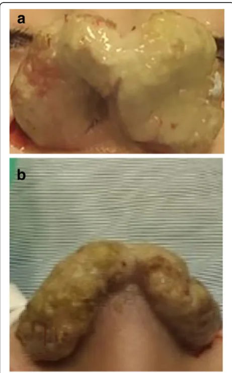
day for 14 days, which was also discontinued due to its inefficacy. Then a decision to perform surgery was taken, and the presurgical computed tomography (CT) scan revealed bilateral soft tissue masses arising from the right and left nasal vestibules. Although she had undergone many surgical attempts to remove the lumps, none of them succeeded and both lumps flared in size. After a couple of months, our patient presented to our institution with bilateral nasovestibu-lar lumps; they were massive in size, occluding nasal entrance and protruding outside the nose (Fig. 1). We did an extensive laboratory workup to exclude any comorbidities (Table 1). We did an fine-needle aspir-ation (FNA) biopsy of the lesion, which was diagnos-tic of ALHE (Fig. 2). Our following surgical attempt included complete mass resection. Despite surgery and postsurgical treatment with topical steroid creams, the lesion recurred. Consequently, we started the patient on intralesional prednisolone twice a
month and topical 0.1% tacrolimus ointment twice daily. This latter regimen seemed to slightly control the lesion’s growth, causing a limited regression in size after 4 months of treatment (Fig. 3). A timeline of the patient's case can be seen in (Fig. 4).
[image:2.595.301.539.99.623.2]Fig. 1Presurgical photograph. The mass appears as a congested and yellowish tumor, protruding outside the nostrils.acaudal view of the lesion.b. superior view of the lesion
Table 1Laboratory values throughout the disease course
Lab results at the other institution Lab results at time of diagnosis 4 months after surgery Units Reference range
Leukocytes 11,700 12,000 8000 C/uL 3500– 10,000
Neutrophils 72 68 60 % 34–71
Eosinophils 15.2 16 5 % 0.7–6
Lymphocytes 8 10 25 % 19–51
Erythrocytes 4.80 4.70 5 ×10³/
mm3 M 4.5F 4–5.4–6.2
Hemoglobin 14 14 15 g/dL M 13–18
F 12–16
CRP 3.2 4 1.5 mg/L 0.5–5
ESR 35 45 14 mm/hL Up to 15
Tuberculin test
Negative Negative
ANCA c 2.5
Negative U Positive more than 18 Negative less than 18
ANCA p 2.2
Negative U Positive more than 18 Less than 18 HHV8: IgG 8.1 Positive U Positive more than 1.1 IgM 0.2 Negative U Negative less than 0.9
IgE 1997 1200 250 IU/mL U to 120
Creatinine 0.6 0.7 0.6 mg/dL 0.5–1.2
Urea 25 23 20 mg/dL 5–45
ALT 6 U/L 5–40
AST 18 U/L 0–40
Urine protein Negative Negative Negative
Urine casts 0 0 Number/
field 0
Blood film Within
normal
CRPC-reactive protein,ESRerythrocytes sedimentation rate,ANCA pantineutrophil
cytoplasmic antibodies perinuclear,ANCA cantineutrophil cytoplasmic antibodies
cytoplasmic,HHV8human herpes virus 8,IgGimmunoglobulin G,IgMimmunoglobulin
M,IgEimmunoglobulin E,ALTalanine transferase,ASTaspartate transferase. We typed
[image:2.595.57.288.323.694.2]Our patient’s parents reported her full adherence to treatment. The patient herself reported decreased quality of life and impaired social interactions due to the disfig-uring lesions. She also reported marked fear and distress because of the ineffectiveness of multiple therapeutic regimens and surgeries. The patient’s family reported se-vere financial burden due to the high costs of the treatments.
Discussion
The term angiolymphoid hyperplasia with eosinophilia (ALHE) was first created (in 1969) by Wells and Whimster [1], who used it to describe a distinguished neoplasm. The tumor is characterized by a florid pro-liferation of blood vessels lined by plump endothelial cells and admixed with a dense inflammatory infiltrate of lymphocytes, eosinophils, and mast cells. They re-ported nine cases in which the neoplasm remained benign although they reported recurrence after exci-sion in some cases. Later, Weiss and Enzinger argued the nature of that entity, for they wanted to evidently differentiate the lesion from the malignant vascular tumor, epithelioid hemangioendothelioma. For this, they introduced the term epithelioid hemangioma (EH) in 1982 [1]. Since then, many hypotheses have been proposed to explain the etiology of the tumor, including a reactive process, a neoplastic process, and infectious mechanisms with possible association with HIV [1]. However, the argument that ALHE/EH may
Fig. 2Histopathologic study and description.aSkin biopsy, nose - pseudoepitheliomatous hyperplasia with ulceration and necrosis. Diffuse lymphohistiocytic infiltrate with numerous eosinophils in a fibrous stroma (hematoxylin and eosin, × 40).bSkin biopsy, nose - vascular proliferation with prominent (plump) endothelial cells and occasional cytoplasmic vacuoles (hematoxylin and eosin, × 60).c Skin biopsy, nose - vascular proliferation with intense eosinophilic infiltrate (hematoxylin and eosin, × 100)
[image:3.595.56.291.86.698.2] [image:3.595.306.539.567.702.2]represent a monoclonal T-cell process is supported in some cases [2]. Interestingly, peripheral T-cell lymph-oma has been reported to develop in a patient with ALHE/EH. Additionally, a certain association between ALHE/EH and follicular mucinosis has been reported. There are also cases of ALHE/EH in which T-cell re-ceptor gene (TCR) rearrangement and monoclonality have been detected [1].
In attempt to define the histogenesis of this dis-order, a team analyzed both the phenotype and the
genotype of the inflammatory infiltrate using immu-nohistochemistry and TCR gene rearrangement by polymerase chain reaction (PCR) and other methods. The results showed five out of seven patients with ALHE displaying a clonal T-cell population and pro-liferative T-cell activity in ALHE tissue; most of these cases were recurrent following a prolonged and therapy-resistant course. Unfortunately, these tests were not available for our case because of war restrictions [2].
[image:4.595.58.536.78.591.2]In 2015, a statistical analysis yielded no sex predomin-ance among 908 patients [3]. Over half of the patients presented with a single lesion, and most common loca-tions were the ear, and periauricular area; face; and scalp. Furthermore, the analysis revealed a consequential asso-ciation between the existence of multiple lesions and pruritus along with bleeding. Considering age, statistics has shown a wide prevalence range (0.7 months to 91 years) and the mean age of presentation was 37.6 years. According to the literature [3], cases of ALHE with earl-ier age of onset, longer duration, and multiple lesions were linked to higher recurrence rates after excision, which exactly fits the course of our case. Although the ear is the most common location, in our case the lesion was located in the nostrils. To the best of our know-ledge, only one similar case of ALHE in the nostrils has been reported in the medical literature [4]. While ALHE usually present as flesh- to plum-colored papules or nodules, our patient had bilateral soft tissue masses of congestive nature and a distinctive yellowish color. Kharkwal et al. have reported a similar unusual gross appearance [5].
ALHE is commonly associated with neither an ele-vated level of immunoglobulin E (IgE) nor an eosino-philia; in contrast, these two signs are common findings in Kiruma’s disease [1]. Eosinophilia is evi-dent in only 20% of ALHE cases and elevated IgE levels is quite a rare finding [2]. Unexpectedly, our patient had both signs (remarkably high IgE level and eosinophilia). While Kimura’s disease surmounts the differential diagnosis list, it also includes angiomatous lymphoid hamartoma, hemangioma, pyogenic granu-loma, Kaposi sarcoma, lymphoma, and epithelioid hemangioendothelioma [1].
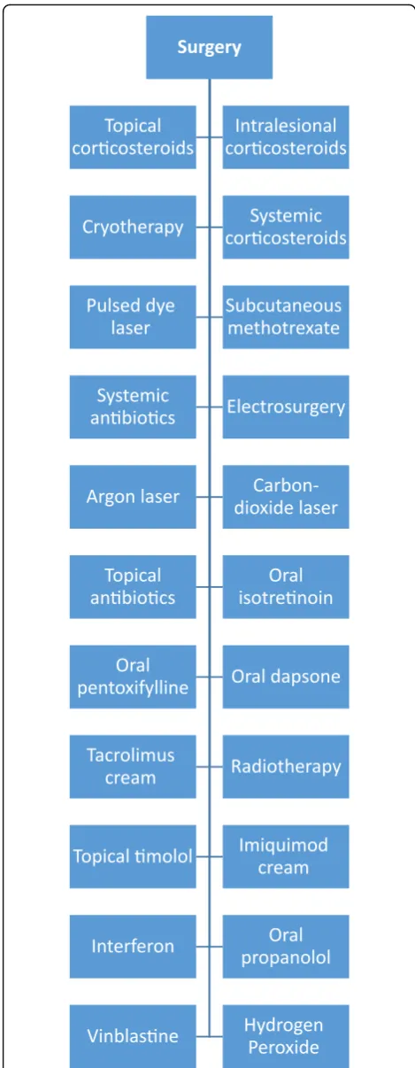
Considering the highly recurring nature of ALHE, there is no single best therapeutic approach. Surgery is the most commonly used one (with a 40% recurrence rate), followed by intralesional and topical corticoste-roids. In our case, our final approach was intralesional corticosteroids and topical tacrolimus cream, which eventually proved a slight effectiveness. Here, we provide a figure describing the available treatment options in addition to hydrogen peroxide, which was effective for the suborbital lesions in our case (Fig.5) [3,4,6–8].
Conclusions
What is peculiar about our case is the gross appearance and unconventional location of the intranasal lesions and the high levels of IgE and eosinophilia, which is ra-ther uncommon in ALHE. Taking into account the highly debilitating and disfiguring nature of the disease, more efforts should be aimed towards creating a stan-dardized and effective therapeutic approach that could help physicians treat such a recurring disease.
[image:5.595.55.290.86.690.2]Abbreviations
ALHE:Angiolymphoid hyperplasia with eosinophilia; ALT: Alanine transferase; ANA: Antinuclear antibodies; ANCA c: Antineutrophil cytoplasmic antibodies cytoplasmic; ANCA p: antineutrophil cytoplasmic antibodies perinuclear; AST: Aspartate transferase; CRP: C-reactive protein; EH: Epithelioid hemangioma; ESR: Erythrocytes sedimentation rate; FNA: Fine-needle aspiration; HHV8: Human herpes virus 8; IgE: Immunoglobulin E; IgG: Immunoglobulin G; IgM: Immunoglobulin M; PCR: Polymerase chain reaction; TCR: T-cell receptor gene
Acknowledgements Not applicable.
Funding Not applicable.
Availability of data and materials Not applicable.
Authors’contributions
AY: Drafted the article and collected the patient data. ARH: Participated in drafting the article and critically revising it. YY: Participated in drafting the article and critically revising it. LA: participated in data collection and drafting the article. YE: Critically revised the work for intellectual content and assisted with the diagnosis of the case. ZA: Performed the histologic examination of the biopsy and critically revised the article. All authors read and approved the final manuscript.
Authors’information
AY is a medical student. ARH is a medical student. YY is a professor of otolaryngology. LS is a dermatologist. ZA is a professor of pathology. YE is a professor of pathology/oncology.
Ethics approval and consent to participate Not applicable.
Consent for publication
Written informed consent was obtained from the patient’s legal guardian for publication of this case report and any accompanying images. A copy of the written consent is available for review by the Editor-in-Chief of this journal.
Competing interests
The authors declare that they have no competing interests.
Publisher’s Note
Springer Nature remains neutral with regard to jurisdictional claims in published maps and institutional affiliations.
Author details
1Faculty of Medicine, Tishreen University, Lattakia, Syria.2Department of
Otolaryngology and Head and Neck Surgery, Tishreen University Hospital, Tishreen University, Lattakia, Syria.3Ministry of Health, National Hospital of Lattakia, Lattakia, Syria.4Department of Pathology, University of California Los Angeles, Charles Drew University, Los Angeles, California, USA.5Department of Pathology, Tishreen University Hospital, Tishreen University, Lattakia, Syria.
Received: 11 May 2017 Accepted: 27 January 2018
References
1. Guo R, Gavino AC. Angiolymphoid hyperplasia with eosinophilia. Arch Pathol Lab Med. 2015;139(5):683–6.
2. Kempf W, Haeffner AC, Zepter K, Sander CA, Flaig MJ, Mueller B, Panizzon RG, Hardmeier T, Adams V, Burg G. Angiolymphoid hyperplasia with eosinophilia: evidence for a T-cell lymphoproliferative origin. Hum Pathol. 2002;33(10):1023–9.
3. Adler BL, Krausz AE, Minuti A, Silverberg JI, Lev-Tov H. Epidemiology and treatment of angiolymphoid hyperplasia with eosinophilia (ALHE): a systematic review. J Am Acad Dermatol. 2016;74(3):506–12. e511
4. Chiu SC. An unusual case of angiolymphoid hyperplasia with eosinophilia of the nose. Ear, Nose Throat J. 2013;92(9):E10–1.
5. Kharkwal N, Agarwal Y, Mathur NN, Rajni. Giant epithelioid hemangioma of the external auditory canal: Role of imaging in clinical management. Apollo Med. 2016;14(1):82–86.
6. Horst C, Kapur N. Propranolol: a novel treatment for angiolymphoid hyperplasia with eosinophilia. Clin Exp Dermatol. 2014;39(7):810–2. 7. Chacon A, Mercer J. Successful management of angiolymphoid hyperplasia
with eosinophilia in a split-face trial of topical tacrolimus and timolol solution. G Ital Dermatol Venereol. 2016;151(4):436–40.
8. Gencoglan G, Karaca S, Ertekin B. Angiolymphoid hyperplasia with eosinophilia successfully treated with imiquimod. A case report. Dermatology. 2007;215(3):233–5.
• We accept pre-submission inquiries
• Our selector tool helps you to find the most relevant journal • We provide round the clock customer support
• Convenient online submission • Thorough peer review
• Inclusion in PubMed and all major indexing services • Maximum visibility for your research
Submit your manuscript at www.biomedcentral.com/submit