ScholarWorks @ Georgia State University
ScholarWorks @ Georgia State University
Biology Theses Department of Biology
Spring 5-7-2011
Systematic Studies of Kir and TRP Channel mRNAs in the
Systematic Studies of Kir and TRP Channel mRNAs in the
Norepinephrenergic Neurons of the Locus Coeruleus
Norepinephrenergic Neurons of the Locus Coeruleus
Sakuntala Jyothirmayee Tadepalli
Georgia State University
Follow this and additional works at: https://scholarworks.gsu.edu/biology_theses
Part of the Biology Commons
Recommended Citation Recommended Citation
Tadepalli, Sakuntala Jyothirmayee, "Systematic Studies of Kir and TRP Channel mRNAs in the Norepinephrenergic Neurons of the Locus Coeruleus." Thesis, Georgia State University, 2011. https://scholarworks.gsu.edu/biology_theses/32
This Thesis is brought to you for free and open access by the Department of Biology at ScholarWorks @ Georgia State University. It has been accepted for inclusion in Biology Theses by an authorized administrator of
SYSTEMATIC STUDIES OF KIR AND TRP CHANNEL MRNAS IN THE
NOREPINEPHRENERGIC NEURONS OF THE LOCUS COERULEUS
by
SAKUNTALA JYOTHIRMAYEE TADEPALLI
Under the Direction of Chun Jiang
ABSTRACT
Neurons in the Locus coeruleus (LC) play an important role in the central CO2
chemosensitivity. However, the molecular mechanisms for neuronal CO2 chemosensitivity
remain unclear. To demonstrate the expression of pH/CO2 sensitive ion channels, we
screened the inward rectifier K+ channels (Kir) and transient receptor protein (TRP)
channels, as parallel studies in this lab suggested that certain Kir and TRP channels are
involved in neuronal responses to high levels of CO2. Our results showed that several
members of the Kir and TRP channel families were robustly expressed in the LC neurons
at the mRNA level. Of particular interest are TRPC5, Kir4.1 and Kir5.1 channels that are
all pH-sensitive. The rich expression of various pH-sensitive Kir and TRP channels
suggests that these ion channels are likely to play a role in the chemosensitivity of LC
neurons.
SYSTEMATIC STUDIES OF KIR AND TRP CHANNEL MRNAS IN THE
NOREPINEPHRENERGIC NEURONS OF THE LOCUS COERULEUS
by
SAKUNTALA JYOTHIRMAYEE TADEPALLI
A Thesis Submitted in Partial Fulfillment of the Requirements for the Degree of
Master of Science
In the College of Arts and Sciences
Georgia State University
Copyright by
Sakuntala Jyothirmayee Tadepalli
SYSTEMATIC STUDIES OF KIR AND TRP CHANNEL MRNAS IN THE
NOREPINEPHRENERGIC NEURONS OF THE LOCUS COERULEUS
by
SAKUNTALA JYOTHIRMAYEE TADEPALLI
Committee Chair: Chun Jiang
Committee: Andrew Clancy
William Walthall
Electronic Version Approved:
Office of Graduate Studies
College of Arts and Sciences
Georgia State University
ACKNOWLEDGEMENTS
I thank my advisor Dr. Chun Jiang, for his guidance that showed me a stairway to the
heaven of research, and his encouragement that motivates me to explore an unknown
scientific kingdom. The author also appreciates my committee members, Dr. Andrew
Clancy and Dr. Walter William Walthall, for their valuable discussions and generous
assistance in experimental techniques. The author would like to acknowledge the
members from Dr. Chun Jiang’s lab for technical assistance. Special thanks to Dr.
Ningren Cui for his help in experimental techniques. I appreciate Dr. Xiaoli Zhang, Dr.
Xin Jin and Dr. Yang Yang, for their assistance.
I also give special acknowledgement to Mrs. Debby Walthall for her instruction in real
time PCR. I would like to thank all members of the Department of Biology who provided
their assistance and shared their research experiences with me during my graduate
education.
The author would like to thank her family, especially her parents and Mr. Madhu
Kesireddy for their continuous encouragement and support. Finally, the author would
like to express her appreciation to her friends especially Ms. Anuhya Konduru for her
TABLE OF CONTENTS
ACKNOWLEDGEMENTS iv
LIST OF FIGURES vii
LIST OF TABLES viii
ABBREVIATIONS ix
1. INTRODUCTION 1
1.1. Rhythmicity 1
1.2. Plasticity 2
1.3. Chemosensitivity 3
1.4. LC as a chemosensitive site in the brainstem 5
1.5. Inhibition of Inward rectifier K+ channels 6
1.6. Activation of Transient Receptor Protein channels (TRP) 10
1.7. Significance 15
2. MATERIALS AND METHODS 16
2.1. Acute dissociation of LC neurons 16
2.2. Reverse transcription PCR and quantitative qPCR 16
2.3. Polymerase Chain Reaction 19
2.4. Single cell PCR 19
3. SPECIFIC AIM 21
4. RESULTS 22
4.1. Expression levels of various TRP channel mRNAs in LC using PCR and qPCR 22
RT-PCR 23
4.3. Expression levels of various Kir channel mRNAs in LC 24
4.4. Dissociation of LC neurons 26
4.5. Expression levels of various TRP and Kir channels in LC neurons
using scPCR 27
4.6. Negative controls for the PCR experiments 28
5. DISCUSSION 30
LIST OF FIGURES
Figure 1. The expression levels of Kir4.1 and Kir5.1 channel currents ... 8
Figure 2. The inhibition of Kir channel currents at low pH levels ... 9
Figure 3. The phylogenetic tree of TRP superfamily. ... 11
Figure 4. The expression levels of various TRP channels using random primers. ... 22
Figure 5. The expression levels of various TRP channels using specific primers. ... 24
Figure 6. The expression levels of various Kir channels in LC neurons ... 25
Figure 7. The dissociated LC neurons ... 26
Figure 8. The expression of various Kir and TRP channels in single LC neurons ... 27
Figure 9. Expression levels of various TRPC channel mRNAs in cardiac ventricular muscle tissue ... 28
LIST OF TABLES
Table 1. List of RT-PCR primers... 18
ABBREVIATIONS
cDNA complementary DNA
CO2 carbon dioxide
DMSO dimethyl sulfoxide
GAPDH glyceraldehyde 3-phosphate dehydrogenase
PCR polymerase chain reaction
qPCR quantitative real-time PCR
RQ relative quantitation
RT-PCR reverse transcriptase polymerase chain reaction
scPCR single cell PCR
TM transmembrane segment
TMD transmembrane domain
TRP transient receptor potential channels
WT wild type
Kir Inward rectifier potassium channels
LC locus coeruleus
PBC preBotzinger complex
NK1R neurokinin 1 receptor
PPE preproenkephalin
FN facial nucleus
NTS Nucleus of solitary tract
rVRG rostral ventral respiratory group
NA nucleus ambiguus
LRN lateral reticular nucleus
RTN retrotrapezoid nucleus
NE norepinephrine
1. INTRODUCTION
Breathing is a vital function controlled autonomically and voluntarily by several
brain areas. Central breathing activity is a continuous process starting from the stage of
utero. The control and regulation of breathing relies on the neural networks especially
those in the brainstem. The connections in the neural networks are changed according
to the age and maturity. Such a complex behavior is responsible for the maintenance of
stable levels of CO2, O2 and pH as well as the regulation of speech, singing, body
postures, etc.
There are three critical aspects of breathing, i.e., rhythmicity, plasticity and
chemosensitivity. Rhythmicity is the regular pattern of breathing. Plasticity is the
adaptive behavior of breathing according to the changes in the environment like altitude,
pregnancy and disease. Chemosensitivity is the regulation of the breathing in the neural
networks according to the changes in the levels of CO2, O2 and pH (Feldman et al.,
2003).
1.1. Rhythmicity
The breathing rhythm generation relies on neural networks and the pacemaker
properties of individual neurons. The brain stem neurons are crucial for the rhythm
generation. The neural kernel for the rhythmicity is located in the rostral ventrolateral
medulla in an area called preBotzinger complex (PBC)(Smith et al., 1991). The
lesioning of the PBC region in vitro resulted in abolishing the rhythmicity. The disruption
of the local synaptic transmission of PBC also disrupts the activity of rhythm generation
distinct patterns of discharge in the PBC neurons are important for the rhythm
generation for breathing. The absence of clear making for the PBC hindered the clear
understanding of its involvement in the rhythmicity. The expression of neurokinin 1
receptor (NK1R) in the ventral respiratory column in the rodent proposed the
identification of PBC neurons (Gray et al., 1999). These neurons in the region are
further divided depending upon preproenkephalin (PPE) expression. The neurons with
PPE are bulbospinal cells which are larger than the neurons without PPE which are
propriobulbar and more rostral (Guyenet et al., 2002). The bulbospinal neurons are not
involved in the rhythm generation, indicating that the PBC region contains mostly
propriobulbar neurons with neurokinin 1 receptors (Makeham et al., 2001). The
pacemaker neurons in the PBC region do not affect the normal rhythm generation (Del
Negro et al., 2002). The overlapping of different patterns of neural networks in the PBC
region is responsible for different patterns of respiration like eupnea, sighing, gasping,
etc. (Lieske et al., 2000; Ramirez et al., 2002). These different respiratory patterns are
produced because of articulation of synaptic and intrinsic properties in PBC region
(Rekling et al., 2000).
1.2. Plasticity
Plasticity is the property involved during the conditions like changes in the levels
of O2, hypoxia, hypercapnia, conditioning, neural injury (Cheng et al., 2002; Gozal and
Gozal, 2001; Mitchell and Johnson, 2003; Powell et al., 1998). The changes in the
neuroplasticity plays an important role. Neuroplasticity requires the activation of different
receptor complexes like serotonin, dopamine and norepinephrine (Baker et al., 2001;
Huey et al., 2000a; Huey et al., 2000b; Kinkead et al., 2001; Mitchell et al., 2001).
Serotonin is involved in neuroplasticity in different conditions like hypercapnic exercise,
intermittent hypoxia, chemoafferent denervation (Forster, 2003; Forster et al., 2000;
Golder et al., 2001; Ling et al., 2001; Mitchell and Johnson, 2003; Prabhakar, 2001).
Serotonin receptor complex is involved in long term facilitation (LFT) of respiration
(Bach and Mitchell, 1996). This involvement depends upon different conditions like age,
previous experience and gender. The prior experience shows the property of
metaplasticity in breathing (Kinkead et al., 1998). The response of LFT also depends
upon the arterial CO2 levels (Janssen and Fregosi, 2000).
1.3. Chemosensitivity
The chemosensitivity of breathing is regulated by chemoreceptors which sense
the changes in O2, CO2, and pH (Ballantyne and Scheid, 2001). The O2
chemosensitivity is regulated in carotid bodies that are located at the fork of the carotid
arteries. Although the CO2/pH chemoreceptors are found in the carotid bodies, most
are located in the brain stem known as central chemoreceptors. The central
chemoreceptors are extremely sensitive to the minor changes in the levels of CO2/pH.
The changes are indicated by the changes in the acid-base levels in the blood and the
brain (Nattie and Li, 2008). There are various locations in the brain which regulate the
chemoreception. Some of sites include fastigial nucleus (FN), locus coeruleus (LC),
ambiguus (NA), lateral reticular nucleus (LRN), and retrotrapezoid nucleus (RTN).
The changes in intracellular and extracellular pH are the key aspects of central
chemoreception. Some motor neurons that are modulated by respiration are sensitive
to the extracellular pH (Bayliss et al., 2001) whereas the neurons located in LC and
medullary raphe nuclei are involved in sensing intracellular levels of pH (Filosa et al.,
2002; Wang et al., 2001b). Amino acids like histidine present in the proteins are
involved in sensing the pH (Jiang et al., 2001). There are many locations that sense the
levels of pH like low resistance gap junctions with TASK channels and inward rectifying
K+ channels and pH sensitive ion transporter proteins (Bayliss et al., 2001; Dean et al.,
2001; Jiang et al., 2001; Solomon, 2003; Spengler et al., 2001). These various
locations sense the changes in the pH simultaneously. The lower brain is the major
location for the central chemoreception (Ballantyne and Scheid, 2001; Li et al., 1999;
Nattie and Li, 2002; Solomon, 2003). The response of the neurons to the increased
levels of CO2 indicates the role of chemosensitive neurons. The chemoreceptor sites
work together to produce increased ventilation with the increase in the levels of CO2.
The increase in the ventilation by 120% with the increase in the levels of CO2 at low
intensity show that most of the chemoreceptor sites work simultaneously which
produced overall high sensitivity to CO2 levels (Li and Nattie, 2002). Some of the
neurons in the PBC site are also involved in chemoreception (Gray et al., 2001). The
chemoreception is coupled to the breathing rhythmicity. The serotonergic and
noradrenergic medullary neurons are involved in different functions like neuroplasticity
and chemoreception. The serotonergic neurons play a role in the development of motor
1.4. LC neurons as chemosensitive site in the brainstem
The noradrenergic neurons present in the LC expressing c-fos, increases
breathing by focal acidosis and the neurons are excited by CO2 (Haxhiu et al., 2001).
The LC is the major source of noradrenergic neurons located in the dorsal pontine
region of brain stem adjacent to the fourth ventricle. The LC neurons are involved in
regulating the cardiovascular function during hypercapnic conditions which causes the
increase in the arterial blood pressure. The noradrenergic neurons present in the LC
form the highest group of neurons that are excited by high levels of CO2/pH. Among the
80% of the noradrenergic neurons present in the LC region 64% of them responded to
levels of CO2. This indicates the involvement of LC neurons in breathing. The change in
the levels of intracellular pH increases the firing rate of the LC neurons. The change in
the extracellular pH along with the intracellular pH modulates the firing activity in the LC
neurons. There are many signals that cause the chemosensitivity of LC neurons along
with the change in the pH. The chemosensitivity of LC neurons in turn affect multiple
targets. This will cause the increase in the firing activity in the LC neurons. The
norepinephrenergic neurons present in the LC region extend their projections to other
parts of the brain like frontal cortex, cerebellum, hippocampus, spinal cord and
brainstem (Berridge and Waterhouse, 2003; Dunn et al., 2004). The disruption of the
intrinsic properties of the LC neurons during diseased conditions is caused due to
changes in NE levels (Zhang et al., 2010). The firing activity of LC neurons during
hypercapnic conditions is because of the presence of different ions channels and their
1.5. Inhibition of Inward rectifier K+ channels
The inhibition of K+ channels produce depolarization. There are various
categories of K+ channel members that are present in the brain. The different
categories of K+ channel members include calcium activated K+ channels, inwardly
rectifying K+ channels (Walder et al.), voltage gated K+ channels and tandem pore
domain K+ channels. A group of K+ channels that are mostly present in the neurons are
the inwardly rectifying K+ channels (Walder et al.). The Kir channels preferably move K+
into the cell rather than out of the cell in the absence of extracellular Na+. The name
inwardly rectification indicates that the channel passes positive charge more easily in
the inward direction. These channels have a role in establishing the resting membrane
potential of the cell. Kir channels exist as tetramers and align to form a pore region to
allow the passage of ions. Each subunit of the tetramer consists of two transmembrane
domains (TM1, TM2). The subunits can form either homodimers or heterodimers. The
constitutively active K+ channels (Kir2.x), the G-protein coupled receptor Kir channels
(Kir3.x), kir channels (Kir6.x) that are involved in cellular metabolism and sensitive to
ATP and K+ transport channels (Kir1.x, Kir4.x, Kir5.x, and Kir7.x). The members of each
subfamily have identical amino acid sequence. Kir channels are commonly found to be
present in the endothelial cells, neurons, cardiac myocytes and kidneys. Their roles
vary according to the cell type in which they are present. Disruption of the channel
function due to mutations or blockers can lead to severe pathologies. Kir5.1 is often
co-expressed with Kir4.1 as Kir4.1/Kir5.1 heterodimers. These heterodimers are present in
properties, each member of the Kir channel has a different physiological role. There are
different members of Kir channels that are expressed in the LC neurons. These Kir
channels are expressed in different locations of brainstem including LC.
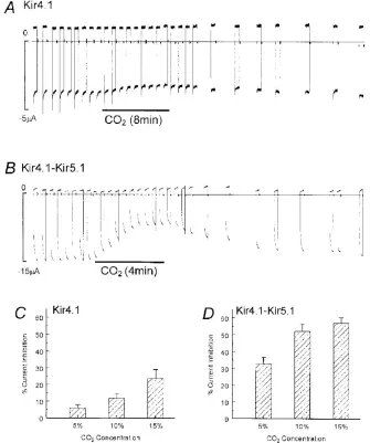
The previous studies show the inhibition of Kir4.1/Kir5.1 at high levels of CO2.
The Kir4.1 and Kir5.1 were microinjected into the Xenopus oocytes and were
incubated for 2-3days for the channel expression. The different levels of CO2 show
H. Xu, N. 728 Cui, Z. Yang, Z. Qu and C. Jiang; J. Physiol. 524.3
Figure 1: The expression levels of Kir4.1 and Kir5.1 channel currents.
The A and B show the inhibition of Kir 4.1 and Kir4.1-Kir5.1 channel currents at high levels of CO2 respectively. The C, D shows the % current inhibition to that of CO2
H. Xu, N. 728 Cui, Z. Yang, Z. Qu and C. Jiang; J. Physiol. 524.3
The decrease in the intracellular pH produced by hypercapnic conditions
cause the inhibition of various Kir channels like homomeric Kir 4.1 channels and
heteromeric Kir4.1-Kir5.1 channels (figure 2) (Xu et al., 2000).
1.6. Activation of Transient Receptor Potential channels (TRP)
The activation of cationic channels also produces depolarization. One of the
most interesting diversified superfamily of cationic channels are Transient Receptor
Potential (TRP) channels. The TRP channels are divided into seven families depending
upon the homology of their sequences (Clapham, 2003; Corey, 2003; Montell et al.,
2002) (Corey, 2003) (Clapham, 2003).The TRP super family is divided into two groups,
group 1 and group 2 (Palmer et al., 2001) (Denis and Cyert, 2002). The group 1 of TRP
superfamily consists of TRPA, TRPC, TRPM, TRPN, TRPV channels whereas the
group 2 consists of TRPML and TRPP channels. The cation selective ion channels are
formed by the homo or hetero-tetramer assembly of different channels. The selectivity
and permeability of different cations by the ion channels depends on the assembly of
the channels. The TRP channels are recognized to be important because of their role in
responses to external stimuli like light, chemicals, temperature and sounds. These TRP
channels are expressed in almost all multicellular organisms including yeast. The TRP
like protein expressed in yeast vacuole is recognized as TRPY which is also called yvc1
(Palmer et al., 2001). The families of TRP channels share some common features like
the six putative transmembrane domains and a pore region in between fifth and sixth
transient response to light was observed which led to the discovery of trp gene. The
study of the trp gene in different multicellular organisms led to the formation of TRP
superfamily.
S.F. Pedersen et al. / Cell Calcium 38 (2005) 233–252
TRPA family has only one member -TRPA1. The locations of TRPA channel are
hair cells (Corey et al., 2004), dorsal root ganglion and trigeminal ganglion. The TRPA1
protein is characterized by the presence of 14 N-terminal ankyrin repeats because of
which it is called ANKTM1 protein (Story et al., 2003). These proteins are mostly located
in the stereocilia of hair cells and are found to be involved in mechanosensing. These
channels are Ca2+ dependent and are voltage dependent to some extent. The TRPA1
gene has some similarity with TRPM8 gene. The TRPA1 channels are also involved
in sensation of cold along with TRPM8 channels (McKemy, 2005).
The TRP channel family members that are closely related to Drosophila TRP
channels are TRPC channel members. There are seven TRPC channels which are
grouped into 4 categories. TRPC1, TRPC2, TRPC3/6/7 and TRPC4/5 are the
categories of TRPC channels. They share a common feature of TRP box EWKFAR
sequence near the C-terminal and 3-4 ankyrin repeats near the N-terminal (Zimmer et
al., 2000). The TRP channels are non-selective to cations and are highly permeable to
Ca2+ compared to Na+. The permeability depends upon the complexity of the ion
channels where they form heterotetramers or homotetramers (Hofmann et al., 2002)
(Strubing et al., 2001). The TRPC heterotetramers are mostly formed by TRPC1 and
TRPC4/ TRPC5, TRPC4 and TRPC5 subfamilies. Some channels of TRPC are
activated by phospholipase C (Hofmann et al., 1999) and some are found to be
activated by diacylglycerol (DAG) (Venkatachalam et al., 2002). The TRPC1 is
expressed ubiquitously in multicellular organisms. It is found to be activated by different
mechanisms and it is the channel which is mostly activated by store operated Ca2+
2000) and diacylglycerol (Yuan et al., 2003). TRPC3 channel is highly expressed in
brain and is also present in smooth and cardiac muscles (Clapham, 2003). It is one of
the channels which is stimulated by DAG and is also activated by T-cell receptor
(Wedel et al., 2003). The TRPC3 channel is present in Na+/ Ca2+ exchanger (Rosker et
al., 2004). TRPC4 and TRPC5 channels are expressed in brain and are activated by
phospholipase C (Schaefer et al., 2000). High expression of TRPC6 is found in brain
(Jia et al., 2007; Li et al., 2005; Zhou et al., 2008b) and lungs whereas the TRPC7
channel is expressed in kidney and pituitary gland (Berg et al., 2007). The TRPC6
and TRPC7 channels are closely related to each other.
The TRPV channels also called vanilloid channels are found to be mostly
involved in nociception. There are six TRPV channels TRPV1, TRPV2, TRPV3,
TRPV4, TRPV5, and TRPV6. The expression levels of TRPV1 and TRPV2 channels
are high expressed in spinal and peripheral nerve terminals and they are found to be
sensitive to temperature and non-selective to cations (Planells-Cases et al., 2005). The
vanilloid compounds like capsaicin and higher temperatures activate TRPV1 whereas
TRPV2 is only activated by harmful temperatures (Caterina et al., 1999). The TRPV1
and TRPV2 channels are present in the brain (Caterina et al., 1999; Kowase et al.,
2002; Steenland et al., 2006). The translocation of TRPV2 to the plasma membrane
occurs when the channel is activated (Iwata et al., 2003; Kanzaki et al., 1999;
Nagasawa et al., 2007). The TRPV3 channels are Ca2+ activated cationic channels
which are sensitive to warm temperatures (Peier et al., 2002; Smith et al., 2002; Xu et
al., 2002). The repeated activation of TRPV3 channels will cause sensitization. TRPV4
CaM and ATP. The TRPV4 channels present in the hypothalamus, skin and primary
sensory neurons are involved in sensing warm temperatures. The TRPV4 knockout
mice express damaged bladder function. The other functions regulated by TRPV4
include vascular tone, bone deposition and remodeling.
The abundance of TRPV6 is much higher than TRPV5. These two members of
TRPV family are inwardly rectifying cationic channels and are insensitive to heat and
the highly permeable to Ca2+. These channels are sensitive to various secondary
messengers like CaM, ATP, Mg2+ and protein kinases. The TRPV family members are
expressed in kidneys. TRPV5 channels are crucial in active reabsorption and
transcellular transport of Ca2+ in the distal convoluted loop and connecting tubules of
kidneys. The TRPV6 reabsorbs Ca2+ in various areas of kidneys like cortical and
medullary ducts of nephron and convoluted tubules.
TRPM family consists of eight channel members which are grouped according to
their amino acid sequence similarities. TRPM1 and TRPM3 belong to a group where
both of these channel members are outwardly rectifying channels. These two channel
members are expressed in brain whereas TRPM1 is also present in melanocytes and
retina and TRPM3 is also expressed in pituitary and kidney (Oancea et al., 2009).
TRPM2 channel is expressed in neurons and mutations in this channel causes
neurodegenerative disorders (Kaneko et al., 2006; Olah et al., 2009). The levels of
intracellular Ca2+ and oxidative stress activate TRPM2 channel. TRPM2 channel has a
considerable role in monocytes chemotaxis (Hara et al., 2002). The only channel
members of TRPM family that are selective to monovalent ions are TRPM4 and
current. The decrease in the intracellular pH inhibits TRPM5 channel whereas the
increase in the levels of ATP inhibits TRPM4 channels. The expression levels of
TRPM4 channels are considerable in brain and kidney whereas TRPM5 channels are
mostly involved in taste reception (Perez et al., 2002; Reading and Brayden, 2007).
TRPM6 and TRPM7 channels have protein kinase activity along with ion channel
activity. These are outwardly rectifying channels inhibited by Mg2+ (Schlingmann and
Gudermann, 2005). The extracellular acidic pH potentiates these channel members.
The TRPM6 channels are highly expressed in kidneys and intestine where they play a
considerable role in the reabsorption of Mg2+ in these areas. The role of TRPM6 in the
closure of neural tube during development is critical (Walder et al., 2009). TRPM7 is
mostly expressed in vascular smooth muscles and in sympathetic neurons where they
have a considerable role in the release of neurotransmitters (Li et al., 2007). TRPM8
channels are activated by cold temperatures and are expressed in many tissues
(Bautista et al., 2007; Colburn et al., 2007; Dhaka et al., 2007). The TRPML channels
are involved in compartment trafficking and are expressed intracellularly (Kim et al.,
2009). Most of the TRP channel currents are modulated at acidic pH conditions.
1.7. Significance
The current study was performed to test the expression of the pH/CO2-sensitive
Kir and TRP channels in the LC neurons. Our results showed that several members of
these two ion channel families were expressed in the LC neurons. The presence of
these pH sensitive ion channels in the cells suggests their contributions to CO2
2. MATERIALS AND METHODS
2.1. Acute Dissociation of LC Neurons
LC-containing brain slices were obtained from C57BL/6 mice at age of 2-4
weeks as described above (Zhang et al. 2010). The slices were then digested at 35oC
for 30min-45min with papain (0.25%, type XI, Sigma) in oxygenated dissociation buffer
containing (in mM) 140 NaCl, 2.5 KCl, 1 MgCl2, 1 CaCl2, 25 D-glucose, and 10 HEPES,
pH 7.40. The slices were transferred to oxygenated dissociation buffer containing 1
mg/ml papain inhibitor and washed twice with dissociation buffer. The LC area was
micropunched and gently triturated in dissociation buffer with fire-polished Pasteur
pipettes. The dissociation buffer containing triturated LC was dropped into 35-mm petri
dishes and kept at room temperature for 10 min before being observed with Hoffman
modulation optics. Individual neurons were harvested with patch pipettes and put into
eppendorf tubes with solution containing 10× RT buffer, RNase free water and RNase
OUT (4.5:4.5:1). The neurons were immediately frozen with liquid nitrogen and kept at -
80oC for further experiments.
2.2. Reverse transcription PCR and quantitative real-time PCR
The LC regions were obtained by micro puncture of the pontine sections from
WT mice. The tissue obtained from two mice was homogenized for 2min in solution
provided in the RNeasy mini Kit (Qiagen). The total mRNAs from the tissue was
extracted according to the manufacturer’s instructions (Qiagen). The concentration of
the mRNA obtained was determined using spectrophotometer (absorption at 260 nm
random hexamers as primers. The cDNAs obtained were used either for quantitative
PCR (qPCR) or regular PCR. The qPCR primers were designed for the target genes
using Primer3.0 software (Applied Biosystems, Warrington, UK). The qPCR was
performed using Platinum SYBR Green qPCR SuperMix-UDG (Invitrogen) on the ABI
PRISM 7500 (Applied Biosystems) in the fast mode for the amplification of cDNAs
according to the manufacturer’s instructions with 500nM prime. The endogenous
control used in the quantitation of target genes was a house-keeping gene, GAPDH.
The endogenous reference gene was run parallel with the targeted genes. Each gene
was performed in quadruplets obtained from WT animal. The ∆CT method (where CT is
threshold cycle) and GeneAmp 5700 SDS software were used for obtaining the data.
The expression levels of the targeted genes were normalized to that of GAPDH (∆CT).
The same set of primers was used for both qPCR and regular PCR with the difference
in concentrations (Zhang et al. 2010). The other set of qPCR experiments were
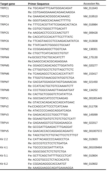
Table 1. List of RT-PCR primers.
Target gene Primer Sequence Accession No.
TRPC1 Fw: TGCAGATTTCAATGGGACAGAT NM_011643
Re: CGTGAAGGAAAACAGAACAAGGA
TRPC3 Fw: GAAAGACACGGGCACAAAGC NM_019510
Re: GGGTGAGCCACAAACTTTTTG
TRPC4 Fw: TTTCCACGTTATTCGAGACACTACA NM_016984
Re: CGTGCTGGGCTTTGACATT
TRPC5 Fw: AAGGACCTCCCCCAACTGTT NM_009428
Re: CACCATCCCACATTTCCTTTATC
TRPC6 Fw: TTCAGTAACCCTCCAAGACAATATCA NM_013838
Re: TTATCAATCTGGGCCTGCAAT
TRPM2 Fw: CCGGAGAGGCTTGGTCAA NM_138301
Re: CTGCTTCATCACGCCTGTGT
TRPM4 Fw:GCGCCTGCTGCACATCTT NM_175130
Re:CAAGCCACACGCAGAGGAA
TRPM5 Fw: GGAGCCAGACAGCTTGGATATG NM_020277
Re: CTTGGTGCCTCTGTTAGCTTCTC
TRPM6 Fw: TCAAGAGCCTCACCACCATTATT NM_153417
Re: TTGGTGTAAACGGTATGGTCTGA
TRPM7 Fw: GACGATGGAGGATAGTGAAAACAA NM_021450
Re: CCATCACTGCTGTCCAAAATCTT
TRPM8 Fw: CCCTGGCCAAAGTTAAGAATGAT NM_134252
Re: CACTGCTCGGGTCTCATATTCA
TRPV1 Fw: GGCGACCATCCCTCAAGAG NM_001001445
Re: GTACCACAGACACCAGCATGAAC
TRPV2 Fw:CCAGCCATTCCCTCATCAAA NM_011706
Re: ATACCCCCAAGCAGGATCAGA
TRPV3 Fw: GAGACACCCCTGGCTTTGG NM_145099
Re: GGAAGTGATGTCTGTCTGCTCATT
TRPV4 Fw: GAGAAAGGTCGTGGAGAAGCA NM_022017
Re: GCCGATTGAAGACTTTGAGGAT
TRPV5 Fw: GAACACCACCAGGAGCAGAATC NM_001007572
Re: TAGCTGCTCTTGTACTTCCTCTTTGT
Kir 2.2 Fw: CATTACAGCCCCAAGCCTCA NM_010603
Re: GCCCGTCCTCCTCTGATGA
Kir 4.1 Fw: TGCCCCGCGATTTATCA NM_001039484
Re: GGGCGGCTCTCTGTCTGA
Kir 5.1 Fw: GCTTCAGCTATTTTGCCCTCAT NM_010604
Re: ACCGTGCCCTCTACCACATG
Kir 6.2 Fw: CGGAGAGGGCACCAATGT NM_010602
2.3. Polymerase Chain Reaction
The polymerase chain reaction was performed to study the expression levels of
the different TRP channels. The PCR reaction mixture contained 1.25µl dNTP, 2.5µl of
DMSO (Dimethyl sulphoxide), 4µl MgCl2, 10µl of 5X green GoTaq Flexi byffer, 0.25µl
Taq Polymerase enzyme (5U/µl), 2µl primer mix (1.0µg/µl), 2µl of template, 28µl of
ddH2O for two tubes. The thermal cycling included initial activation at 95°C for 2min
followed by denature, annealing and extension at 95°C for 30sec, 60°C for 30 sec and
72°C for 2 sec respectively. The final extension was at 70°C for 10min and the
number of cycles was 36.
2.4. Single cell PCR
scPCR was performed for LC neurons obtained by acute dissociation as
described above. Two sets of primers were designed for the targeted genes using
Primer3.0 software. The first PCR was performed using the One Step RT-PCR kit
(Qiagen) for obtaining the cDNAs from the LC neuron cells. The second PCR was
performed using Hotstar Taq DNA polymerase (Qiagen). The One Step RT-PCR
reaction mix contained 10µl of 5X OneStep RT-PCR Buffer (pH 8.7), 10 µl of 5X
Q-solution, 2 µl of dNTP mix (containing 10mM of each dNTP), 1µl of primer mix
(1.0µg/µl), 2 µl of OneStep RT-PCR Enzyme Mix (pH 9.0), 15µl of RNase Free H2O and
finally 10µl of template was added to the mixture. The thermal cycling conditions
included 30min of reverse transcription at 50°C followed by initial PCR activation at
sec and 53°C for 45 sec respectively followed by extension at 72°C for 1 min and the
final extension at 72°C for 10 min. The number of cycles performed was 30. The
primers used were specific for the targeted genes. The Hotstar PCR reaction mixture
contained 5µl of 10× PCR buffer, 1 µl of dNTP mix (containing 10mM of each dNTP),
0.5µl of HotStar Taq DNA Polymerase, 10µl of 5X Q-solution, 1µl primer mix
(1.0µg/µl), 0.5µl of template (cDNA) and made to 50µl with ddH2O. The thermal
cycling included 40 cycles with initial PCR activation at 95°C for 15min followed by
denature, annealing and extension steps at 94°C for 45sec, 53°C for 45sec and 72°C
[image:32.612.80.456.377.701.2]for 1min respectively. The final extension was at 72°C for 10min.
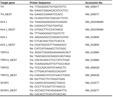
Table 2. List of single-cell PCR primers.
Target gene Primer Sequence Accession No.
TH Fw: TTGGAGGCTGTGGTATTC NM_009377
Re: GAAGTGAGACACATCCTCC
TH_NEST Fw: GAAGCCAAAATCCACC NM_009377
Re: CCAGGTGGTGACACTTATC
Kir4.1 Fw: TAAGAAGAGGGCCGAGAC NM_001039484 Re: CAGACGTTGCTGATGC
Kir4.1_NEST Fw: GTGGCTTCCCATAACG NM_001039484 Re: TTTAAGGGGCTGGTCTC
Kir5.1 Fw: AAGAGAGCCCAGACCATAC NM_010604 Re: CTCACAGCTGCTCACCA
Kir5.1_NEST Fw: CGATGGCGTTTAAAGACC NM_010604 Re: CATCATGAAACCTGTGGC
TRPC4 Fw: AGGCTGGAGGAGAAGACACT NM_016984 Re: TAGCAGCACGAGGCAGTAGA
TRPC4_NEST Fw: CACACAGCCTCCTATCTGAC NM_016984 Re: TCAAGGAGATTGTTGCCAGA
TRPC5 Fw: TCCCAGCAATGTAAGCTC NM_009428
Re: CTTGACATAGGCCACGAT
TRPC5_NEST Fw: CAGAAGTATCATGACCTGGC NM_009428 Re: GGTTGCTTCTGGGTGAG
GFAP2 Fw: CAAGCACGAAGCTAACG NM_010277
Re: CCCTTCCAATTCTAACCC
3. SPECIFIC AIM: To demonstrate the mRNA expression of various Kir and TRP
channels in the LC neurons.
A series of studies was performed to address this aim. First, the presence of
specific Kir and TRP mRNAs in the LC tissue was determined using regular PCR.
Second, the expression levels of the channels were quantified using qPCR. Third, sc
PCR analysis was carried out to show that the channel mRNAs identified above were
indeed located in catecholaminergic neurons in the LC, as specific antibodies are still
4. RESULTS
[image:34.612.95.508.152.400.2]4.1. Expression levels of various TRP channel mRNAs in LC using PCR and qPCR
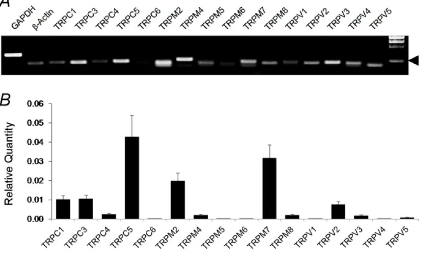
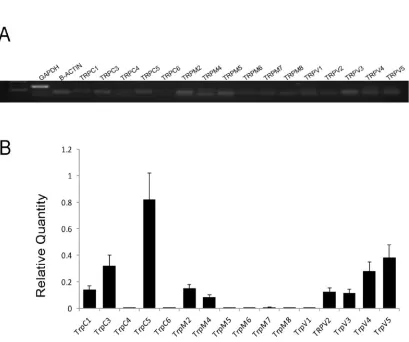
Figure 4. Expression levels of various TRP channels in LC neurons using random primers in RT-PCR.
A. All representative members in the TRPC, TRPM and TRPV families were included in the RT-PCR analysis. Several TRP channels were clearly detected in the mRNA level. Arrow head indicates 100bp. B. Analysis of TRP mRNA expression in the LC tissue with quantitative PCR (qPCR) indicated that the expression of TRPC5, TRPV2 and TRPV5 were several folds higher than other TRP channels. Data are presented as means ± s.e. (n = 16-20 samples from 5 experiments).
Previous studies have shown the presence of TRP channels in the central
nervous system (Kunert-Keil et al., 2006). However, the expression of specific
members of the TRP channels in the LC is still elusive. To investigate the expression
of specific TRP channels in the LC area, we systematically studied all representative
TRP channels in terms of the presence of mRNAs of these TRP channels in the LC
identified based on its pontine location with the IV ventricle as a landmark, the LC
area was isolated using micro-puncture. The LC tissue was then used for the mRNA
extraction. The mRNAs obtained were subjected to RT- PCR to obtain cDNAs using
random primers. The cDNAs produced were used as templates to detect the
expression of TRP channels. To prove that the tissue contained LC neurons, a
specific enzyme (tyrosine hydroxylase or TH) of these catecholaminergic neurons was
examined in each sample obtained. Only those that showed a clear expression of TH
were accepted for further studies. With such an approach, we found that several TRP
channel mRNAs were highly expressed in the LC (Figure 8).
Subsequently, the expression levels of various TRP channels were quantified
using qPCR on the basis of the ∆∆CT method (Figure 4) .Consistent with our regular
PCR tests, the TRPC5 mRNA was readily detected in the qPCR, whose expression
level was highest among all TRP channels. The TRPM7 and TRPM2 mRNAs were
found to be expressed at a moderate level in the LC tissue. The expression levels of
TRPC1, TRPC3 and TRPV2 mRNAs were also detectable at low levels, whereas the
expression levels of other TRP channel mRNAs were insignificant.
4.2. Confirmation of TRP channel expressions using specific primers in RT-PCR
To further analyze of the TRP expression in the LC neurons, the expression of
various TRP channel mRNAs was determined using primers specific for each TRP
channel mRNA (Table 1). These studies also showed the highest expression of
TRPC5 mRNA in the LC. The expression levels of several TRPV channels were
and TRPM4 mRNAs were low, while the expression levels of other TRP channel
mRNAs were undetectable. These results thus were consistent with our data in
[image:36.612.90.499.158.500.2]Section 4.1
Figure 5. Expression levels of various TRP channels using specific primers in RT-PCR A. All representative members in the TRPC, TRPM and TRPV families were included in the RT-PCR analysis. Several TRP channels were clearly detected in the mRNA level. Arrow head indicates 100bp. B. Analysis of TRP mRNA expression in the LC tissue with quantitative PCR (qPCR) indicated that the expression of TRPC5 were several folds higher than other TRP channels. Data are presented as means ± s.e. (n = 16 samples from 4
experiments).
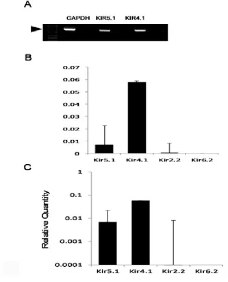
4.3. Expression levels of various Kir channel mRNAs in LC neurons
using regular PCR and q PCR. Figure 6 shows the expression of Kir mRNAs in the LC
tissue. The expression levels of Kir4.1 and Kir5.1 mRNAs in the LC neurons were high,
especially the Kir4.1, whereas the expression levels of other Kir channel mRNAs were
[image:37.612.85.418.194.605.2]very low (Figure 6).
Figure 6. Expression levels of various Kir channels in the LC neurons
A. The members of Kir channel family included for the study were Kir4.1, Kir5.1 due to their significant expression levels in the qPCR. Arrow head indicates 650bp. B. Analysis of various Kir channel mRNA expression in the LC tissue with quantitative PCR (qPCR) indicated that the expression of Kir4.1 were several folds higher than other Kir channels. Data are
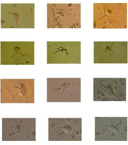
4.4. Dissociation of LC neurons
Since antibodies for the channels are still not available, the scPCR was
performed to prove the presence of the expression of the Kir channels and TRP
channels in the LC neurons. The LC neurons were dissociated from the LC tissue
from mice. The LC tissue was digested with papain for 30min-45min at 37˚C and the
inhibitor was then added to suppress the papain. The LC neurons were isolated in the
dissociation buffer. After morphological studies with the Hoffman modulation optics
(Figure 7), single LC neurons were harvested with patch pipettes and stored at -80˚C
[image:38.612.186.436.323.605.2]for further scPCR analysis.
Figure 7. The dissociated LC neurons
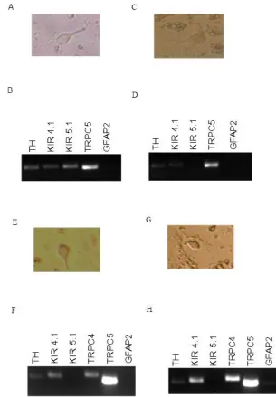
4.5. Expression levels of various TRP and Kir channels in LC neurons using
[image:39.612.145.450.125.564.2]scPCR
Each single neuron was subjected to scPCR analysis for the expression levels
of the Kir and TRP channel mRNAs with high levels of expression in the LC as shown
in Sections 4.1 and 4.2. We found the high expression of TRPC5 mRNAs in the LC
neurons. The expression levels of Kir4.1 and Kir5.1 mRNAs were rather heterogenous
in LC neurons, with some cells showing the expression of Kir4.1 mRNA alone, Kir5.1
mRNA alone, or both Kir4.1 and Kir5.1 mRNAs. The expression of TRPC4 mRNAs was
much lower than TRPC5 (Figure 8). The high expression of TRPC5 mRNA was
consistently seen in all LC neurons.
[image:40.612.114.422.367.451.2]4.6. Negative controls for the PCR experiments
Figure 9. Expression levels of various TRPC channel mRNAs in cardiac ventricular muscle tissue.
All representative members in the TRPC families were included in RT-PCR. Only TRPC3 and TRPC1 mRNAs were detected in the cardiac ventricular muscle tissue. Arrow head indicates 100bp. (n = 3 experiments).
The control experiments were performed to assure the conditions of the PCR.
The previous studies have shown that the expression levels of various TRP channels in
various tissues. The lack of expression of TRPC5 was found in the cardiac muscle cells
the conditions of the PCR for the negative expression of TRPC5 in the cardiac muscle
tissue. Consistent with previous reports, our results did not produce positive bands in
the cardiac ventricular muscles for TRPC5. The cardiac ventricular muscle tissue
[image:41.612.141.430.214.339.2]expressed TRPC3 mRNAs at a relatively high level followed by TRPC1 mRNAs.
Figure 10. Negative control PCR experiments for highly expressed TRP channels.
The negative control experiments were performed for three most abundant TRPs in LC. In the absence of template cDNAs, a weak and fuzzy band was found. In the presence of cDNA templates, a strong band was produced in each TRP, which was clearly larger than the primer band with expected size. Arrow head indicates 100bp. (n = 3 experiments).
The second set of control experiment was performed to test whether the
contaminations from primers affect the PCR reactions. The PCR experiments were
performed without cDNA template in parallel to that of with cDNA template. In the
absence of cDNA template, weak primer bands were observed. These fussy bands
were greater than primers, suggesting the primer multimerization. In the presence of
cDNA templates, strong bands were found. These bands with expected sizes for each
PCR product cannot be missed with the primer bands, because they were clearly larger
than the primers bands and none of the primer bands were seen in the presence of
5. DISCUSSION
The breathing is a complex behavior with critical aspects like plasticity,
rhythmicity and chemosensitivity. The chemosensitivity is important for the breathing
process as it controls the levels of O2, CO2 and pH. According to the previous studies
the LC neurons were involved in sensing the intracellular pH (Wang et al., 2001b). The
pH sensing property of LC neurons indicates the involvement of LC neurons in
controlling the chemosensitivity. The noradrenergic neurons present in the LC form the
highest group of neurons that are excited by high levels of CO2/pH. Among the 80% of
the noradrenergic neurons present in the LC region 64% of them responded to levels
of CO2. This indicates the involvement of LC neurons in breathing as one of the
chemosensitive sites. There are many signals that cause the chemosensitivity of LC
neurons along with the change in the pH and levels of CO2. The hypercapnic
conditions cause the increase in the firing activity of LC neurons. The sensing of the pH
and the levels of CO2 is mostly due to the presence of various ion channels like inward
rectifying K+ channels (Jiang et al., 2001) and TRP channels. The Kir channels, Kir 4.1
and Kir 5.1 are found to be highly expressed in the brain (Xu et al., 2000). The Kir 4.1
channel member and Kir4.1-Kir5.1 heterodimers are inhibited at the high levels of CO2
(Xu et al., 2000). So the presence of various Kir channels was studied in the LC
neurons. The expression of Kir channel members in the LC neurons indicates their role
in the chemosensitivity. The experiments performed included qPCR and scPCR which
shows the expression of mRNAs in the LC neurons. The results indicated the presence
chemosensitivity. The activation of cationic channels also causes the firing activity of
the LC neurons. The major cationic channels include the TRP channel members. Most
of the TRP channel members are expressed in the brain (Strubing et al., 2001; Zhou et
al., 2008a). Some of the TRP channels respond to the changes in the levels of pH
(Andersson et al., 2004; Kim et al., 2008; Semtner et al., 2007). So the experiments
were performed to study the expression of various TRP channel members. The data
obtained shows the expression of most of the representative members of TRP
channels in the LC neurons with the high expression levels of TRPC5 mRNA.
The control experiments in the cardiac ventricular muscle tissue with the
negative expression of TRPC5 mRNA confirmed the conditions of the PCR. The
expression of TRPC3 and TRPC1 mRNAs without the presence of TRPC5 mRNAs in
the cardiac ventricular muscle tissue according to the previous studies assure the high
expression of TRPC5 mRNAs in the LC tissue. The negative expression of TRPC5
mRNAs in the cardiac ventricular muscle tissue even with the second set of TRPC5
primers assures the accuracy of the primers used in the PCR. The absence of strong
bands without cDNA template in the PCR reactions assures the absence of
contamination from primers. The fuzzy bands with sizes greater than that of primers
show the multimerization of primers. The previous studies show the ability of Taq
polymerase for the reverse transcription and direct amplification of mRNA (Jones, 1993;
Tse and Forget, 1990). Therefore, the negative control experiments cannot be
produced without the reverse transcriptase in RT-PCR reactions.
The high expression levels of TRPC5 in the LC neurons were confirmed with
and scPCR which indicates the presence of TRPC5 in LC neurons. So the expression
of various TRP channel members and Kir channel members in the LC neurons
6. REFERENCES
Andersson, D.A., Chase, H.W., and Bevan, S. (2004). TRPM8 activation by menthol, icilin, and cold is differentially modulated by intracellular pH. J Neurosci 24, 5364-5369. Bach, K.B., and Mitchell, G.S. (1996). Hypoxia-induced long-term facilitation
of respiratory activity is serotonin dependent. Respir Physiol 104, 251-260.
Baker, T.L., Fuller, D.D., Zabka, A.G., and Mitchell, G.S. (2001). Respiratory plasticity: differential actions of continuous and episodic hypoxia and hypercapnia. Respir
Physiol 129, 25-35.
Ballantyne, D., and Scheid, P. (2001). Central chemosensitivity of respiration: a brief overview. Respir Physiol 129, 5-12.
Bautista, D.M., Siemens, J., Glazer, J.M., Tsuruda, P.R., Basbaum, A.I., Stucky, C.L., Jordt, S.E., and Julius, D. (2007). The menthol receptor TRPM8 is the principal detector of environmental cold. Nature 448, 204-208.
Bayliss, D.A., Talley, E.M., Sirois, J.E., and Lei, Q. (2001). TASK-1 is a highly modulated pH-sensitive 'leak' K(+) channel expressed in brainstem respiratory neurons. Respir Physiol 129, 159-174.
Berg, A.P., Sen, N., and Bayliss, D.A. (2007). TrpC3/C7 and SlO2.1 are
molecular targets for metabotropic glutamate receptor signaling in rat striatal cholinergic interneurons. J Neurosci 27, 8845-8856.
Berridge, C.W., and Waterhouse, B.D. (2003). The locus coeruleus-noradrenergic system: modulation of behavioral state and state-dependent cognitive processes. Brain Res Brain Res Rev 42, 33-84.
Bongianni, F., Mutolo, D., Carfi, M., and Pantaleo, T. (2002). Respiratory responses to ionotropic glutamate receptor antagonists in the ventral respiratory group of the rabbit. Pflugers Arch 444, 602-609.
Caterina, M.J., Rosen, T.A., Tominaga, M., Brake, A.J., and Julius, D. (1999). A
capsaicin-receptor homologue with a high threshold for noxious heat. Nature 398, 436-441.
Cheng, Z., Guo, S.Z., Lipton, A.J., and Gozal, D. (2002). Domoic acid lesions in nucleus of the solitary tract: time-dependent recovery of hypoxic ventilatory response and peripheral afferent axonal plasticity. J Neurosci 22, 3215-3226.
Clapham, D.E. (2003). TRP channels as cellular sensors. Nature 426, 517-524. Colburn, R.W., Lubin, M.L., Stone, D.J., Jr., Wang, Y., Lawrence, D., D'Andrea, M.R., Brandt, M.R., Liu, Y., Flores, C.M., and Qin, N. (2007). Attenuated cold sensitivity in TRPM8 null mice. Neuron 54, 379-386.
Corey, D.P. (2003). New TRP channels in hearing and mechanosensation. Neuron 39, 585-588.
candidate for the mechanosensitive transduction channel of vertebrate hair cells. Nature
432, 723-730.
Dean, J.B., Kinkade, E.A., and Putnam, R.W. (2001). Cell-cell coupling in CO(2)/H(+)-excited neurons in brainstem slices. Respir Physiol 129, 83-100. Del Negro, C.A., Koshiya, N., Butera, R.J., Jr., and Smith, J.C. (2002). Persistent sodium current, membrane properties and bursting behavior of pre-botzinger complex inspiratory neurons in vitro. J Neurophysiol 88, 2242-2250.
Denis, V., and Cyert, M.S. (2002). Internal Ca(2+) release in yeast is triggered by hypertonic shock and mediated by a TRP channel homologue. J Cell Biol 156, 29-34. Dhaka, A., Murray, A.N., Mathur, J., Earley, T.J., Petrus, M.J., and Patapoutian, A. (2007). TRPM8 is required for cold sensation in mice. Neuron 54, 371-378.
Dunn, A.J., Swiergiel, A.H., and Palamarchouk, V. (2004). Brain circuits involved in corticotropin-releasing factor-norepinephrine interactions during stress. Ann N Y Acad Sci 1018, 25-34.
Feldman, J.L., Mitchell, G.S., and Nattie, E.E. (2003). Breathing: rhythmicity, plasticity, chemosensitivity. Annu Rev Neurosci 26, 239-266.
Filosa, J.A., Dean, J.B., and Putnam, R.W. (2002). Role of intracellular and extracellular pH in the chemosensitive response of rat locus coeruleus neurones. J Physiol 541, 493-509.
Forster, H.V. (2003). Plasticity in the control of breathing following sensory denervation. J Appl Physiol 94, 784-794.
Forster, H.V., Pan, L.G., Lowry, T.F., Serra, A., Wenninger, J., and Martino, P. (2000). Important role of carotid chemoreceptor afferents in control of breathing of adult and neonatal mammals. Respir Physiol 119, 199-208.
Fung, M.L., Wang, W., and St John, W.M. (1994). Medullary loci critical for expression of gasping in adult rats. J Physiol 480 ( Pt 3), 597-611.
Golder, F.J., Reier, P.J., and Bolser, D.C. (2001). Altered respiratory motor drive after spinal cord injury: supraspinal and bilateral effects of a unilateral lesion. J Neurosci 21, 8680-8689.
Gozal, E., and Gozal, D. (2001). Respiratory plasticity following intermittent hypoxia: developmental interactions. J Appl Physiol 90, 1995-1999.
Gray, P.A., Janczewski, W.A., Mellen, N., McCrimmon, D.R., and Feldman, J.L. (2001). Normal breathing requires preBotzinger complex neurokinin-1 receptor-expressing neurons. Nat Neurosci 4, 927-930.
Gray, P.A., Rekling, J.C., Bocchiaro, C.M., and Feldman, J.L. (1999). Modulation of respiratory frequency by peptidergic input to rhythmogenic neurons in the
preBotzinger complex. Science 286, 1566-1568.
Hara, Y., Wakamori, M., Ishii, M., Maeno, E., Nishida, M., Yoshida, T., Yamada, H., Shimizu, S., Mori, E., Kudoh, J., et al. (2002). LTRPC2 Ca2+-permeable channel
activated by changes in redox status confers susceptibility to cell death. Mol Cell 9, 163-173.
Haxhiu, M.A., Tolentino-Silva, F., Pete, G., Kc, P., and Mack, S.O. (2001).
Monoaminergic neurons, chemosensation and arousal. Respir Physiol 129, 191-209. Hofmann, T., Obukhov, A.G., Schaefer, M., Harteneck, C., Gudermann, T., and Schultz, G. (1999). Direct activation of human TRPC6 and TRPC3 channels by diacylglycerol. Nature 397, 259-263.
Hofmann, T., Schaefer, M., Schultz, G., and Gudermann, T. (2002). Subunit
composition of mammalian transient receptor potential channels in living cells. Proc Natl Acad Sci U S A 99, 7461-7466.
Huey, K.A., Brown, I.P., Jordan, M.C., and Powell, F.L. (2000a). Changes in dopamine D(2)-receptor modulation of the hypoxic ventilatory response with chronic hypoxia. Respir Physiol 123, 177-187.
Huey, K.A., Low, M.J., Kelly, M.A., Juarez, R., Szewczak, J.M., and Powell, F.L. (2000b). Ventilatory responses to acute and chronic hypoxia in mice: effects of dopamine D(2) receptors. J Appl Physiol 89, 1142-1150.
Iwata, Y., Katanosaka, Y., Arai, Y., Komamura, K., Miyatake, K., and Shigekawa, M. (2003). A novel mechanism of myocyte degeneration involving the Ca2+-permeable growth factor-regulated channel. J Cell Biol 161, 957-967.
Janssen, P.L., and Fregosi, R.F. (2000). No evidence for long-term facilitation after episodic hypoxia in spontaneously breathing, anesthetized rats. J Appl Physiol 89, 1345-1351.
Jia, Y., Zhou, J., Tai, Y., and Wang, Y. (2007). TRPC channels promote cerebellar granule neuron survival. Nat Neurosci 10, 559-567.
Jiang, C., Xu, H., Cui, N., and Wu, J. (2001). An alternative approach to the
identification of respiratory central chemoreceptors in the brainstem. Respir Physiol 129, 141-157.
Jones, M.D. (1993). Reverse transcription of mRNA by Thermus aquaticus DNA
polymerase followed by polymerase chain reaction amplification. Methods Enzymol 218, 413-419.
Kaneko, S., Kawakami, S., Hara, Y., Wakamori, M., Itoh, E., Minami, T., Takada, Y., Kume, T., Katsuki, H., Mori, Y., et al. (2006). A critical role of TRPM2 in neuronal cell death by hydrogen peroxide. J Pharmacol Sci 101, 66-76.
Kanzaki, M., Zhang, Y.Q., Mashima, H., Li, L., Shibata, H., and Kojima, I. (1999). Translocation of a calcium-permeable cation channel induced by insulin-like growth factor-I. Nat Cell Biol 1, 165-170.
Kim, M.J., Jeon, J.P., Kim, H.J., Kim, B.J., Lee, Y.M., Choe, H., Jeon, J.H., Kim, S.J., and So, I. (2008). Molecular determinant of sensing extracellular pH in classical transient receptor potential channel 5. Biochem Biophys Res Commun 365, 239-245.
Kinkead, R., Bach, K.B., Johnson, S.M., Hodgeman, B.A., and Mitchell, G.S. (2001). Plasticity in respiratory motor control: intermittent hypoxia and hypercapnia activate opposing serotonergic and noradrenergic modulatory systems. Comp Biochem Physiol A Mol Integr Physiol 130, 207-218.
Kinkead, R., Zhan, W.Z., Prakash, Y.S., Bach, K.B., Sieck, G.C., and Mitchell, G.S. (1998). Cervical dorsal rhizotomy enhances serotonergic innervation of phrenic
motoneurons and serotonin-dependent long-term facilitation of respiratory motor output in rats. J Neurosci 18, 8436-8443.
Kowase, T., Nakazato, Y., Yoko, O.H., Morikawa, A., and Kojima, I. (2002).
Immunohistochemical localization of growth factor-regulated channel (GRC) in human tissues. Endocr J 49, 349-355.
Kunert-Keil, C., Bisping, F., Kruger, J., and Brinkmeier, H. (2006). Tissue-specific expression of TRP channel genes in the mouse and its variation in three different mouse strains. BMC Genomics 7, 159.
Launay, P., Fleig, A., Perraud, A.L., Scharenberg, A.M., Penner, R., and Kinet, J.P. (2002). TRPM4 is a Ca2+-activated nonselective cation channel mediating cell membrane depolarization. Cell 109, 397-407.
Li, A., and Nattie, E. (2002). CO2 dialysis in one chemoreceptor site, the RTN: stimulus
intensity and sensitivity in the awake rat. Respir Physiol Neurobiol 133, 11-22. Li, A., Randall, M., and Nattie, E.E. (1999). CO(2) microdialysis in retrotrapezoid nucleus of the rat increases breathing in wakefulness but not in sleep. J Appl Physiol
87, 910-919.
Li, M., Du, J., Jiang, J., Ratzan, W., Su, L.T., Runnels, L.W., and Yue, L. (2007). Molecular determinants of Mg2+ and Ca2+ permeability and pH sensitivity in TRPM6 and TRPM7. J Biol Chem 282, 25817-25830.
Li, Y., Jia, Y.C., Cui, K., Li, N., Zheng, Z.Y., Wang, Y.Z., and Yuan, X.B. (2005). Essential role of TRPC channels in the guidance of nerve growth cones by brain-derived neurotrophic factor. Nature 434, 894-898.
Lieske, S.P., Thoby-Brisson, M., Telgkamp, P., and Ramirez, J.M. (2000). Reconfiguration of the neural network controlling multiple breathing patterns: eupnea, sighs and gasps [see comment]. Nat Neurosci 3, 600-607.
Ling, L., Fuller, D.D., Bach, K.B., Kinkead, R., Olson, E.B., Jr., and Mitchell, G.S. (2001). Chronic intermittent hypoxia elicits serotonin-dependent plasticity in the central neural control of breathing. J Neurosci 21, 5381-5388.
27799-27805.
Makeham, J.M., Goodchild, A.K., and Pilowsky, P.M. (2001). NK1 receptor and the ventral medulla of the rat: bulbospinal and catecholaminergic neurons. Neuroreport 12, 3663-3667.
McKemy, D.D. (2005). How cold is it? TRPM8 and TRPA1 in the molecular logic of cold sensation. Mol Pain 1, 16.
Mitchell, G.S., Baker, T.L., Nanda, S.A., Fuller, D.D., Zabka, A.G., Hodgeman, B.A., Bavis, R.W., Mack, K.J., and Olson, E.B., Jr. (2001). Invited review: Intermittent hypoxia and respiratory plasticity. J Appl Physiol 90, 2466-2475.
Mitchell, G.S., and Johnson, S.M. (2003). Neuroplasticity in respiratory motor control. J Appl Physiol 94, 358-374.
Montell, C., Birnbaumer, L., and Flockerzi, V. (2002). The TRP channels, a remarkably functional family. Cell 108, 595-598.
Nagasawa, M., Nakagawa, Y., Tanaka, S., and Kojima, I. (2007). Chemotactic peptide fMetLeuPhe induces translocation of the TRPV2 channel in macrophages. J Cell Physiol 210, 692-702.
Nattie, E.E., and Li, A. (2002). CO2 dialysis in nucleus tractus solitarius region of rat
increases ventilation in sleep and wakefulness. J Appl Physiol 92, 2119-2130. Nattie, G., and Li, A. (2008). Multiple central chemoreceptor sites: cell types and function in vivo. Adv Exp Med Biol 605, 343-347.
Oancea, E., Vriens, J., Brauchi, S., Jun, J., Splawski, I., and Clapham, D.E. (2009). TRPM1 forms ion channels associated with melanin content in melanocytes. Sci Signal
2, ra21.
Olah, M.E., Jackson, M.F., Li, H., Perez, Y., Sun, H.S., Kiyonaka, S., Mori, Y., Tymianski, M., and MacDonald, J.F. (2009). Ca2+-dependent induction of TRPM2 currents in hippocampal neurons. J Physiol 587, 965-979.
Palmer, C.P., Zhou, X.L., Lin, J., Loukin, S.H., Kung, C., and Saimi, Y. (2001). A TRP homolog in Saccharomyces cerevisiae forms an intracellular Ca(2+)-permeable channel in the yeast vacuolar membrane. Proc Natl Acad Sci U S A 98, 7801-7805.
Peier, A.M., Reeve, A.J., Andersson, D.A., Moqrich, A., Earley, T.J., Hergarden, A.C., Story, G.M., Colley, S., Hogenesch, J.B., McIntyre, P., et al. (2002). A heat-sensitive TRP channel expressed in keratinocytes. Science 296, 2046-2049.
Perez, C.A., Huang, L., Rong, M., Kozak, J.A., Preuss, A.K., Zhang, H., Max, M., and Margolskee, R.F. (2002). A transient receptor potential channel expressed in taste receptor cells. Nat Neurosci 5, 1169-1176.
Planells-Cases, R., Garcia-Sanz, N., Morenilla-Palao, C., and Ferrer-Montiel, A. (2005). Functional aspects and mechanisms of TRPV1 involvement in neurogenic inflammation that leads to thermal hyperalgesia. Pflugers Arch 451, 151-159.
Prabhakar, N.R. (2001). Oxygen sensing during intermittent hypoxia: cellular and molecular mechanisms. J Appl Physiol 90, 1986-1994.
Ramirez, J.M., Zuperku, E.J., Alheid, G.F., Lieske, S.P., Ptak, K., and McCrimmon, D.R. (2002). Respiratory rhythm generation: converging concepts from in vitro and in vivo approaches? Respir Physiol Neurobiol 131, 43-56.
Reading, S.A., and Brayden, J.E. (2007). Central role of TRPM4 channels in cerebral blood flow regulation. Stroke 38, 2322-2328.
Rekling, J.C., Shao, X.M., and Feldman, J.L. (2000). Electrical coupling and excitatory synaptic transmission between rhythmogenic respiratory neurons in the preBotzinger complex. J Neurosci 20, RC113.
Rosker, C., Graziani, A., Lukas, M., Eder, P., Zhu, M.X., Romanin, C., and Groschner, K. (2004). Ca(2+) signaling by TRPC3 involves Na(+) entry and local coupling to the Na(+)/Ca(2+) exchanger. J Biol Chem 279, 13696-13704.
Schaefer, M., Plant, T.D., Obukhov, A.G., Hofmann, T., Gudermann, T., and Schultz, G. (2000). Receptor-mediated regulation of the nonselective cation channels TRPC4 and TRPC5. J Biol Chem 275, 17517-17526.
Schlingmann, K.P., and Gudermann, T. (2005). A critical role of TRPM channel-kinase for human magnesium transport. J Physiol 566, 301-308.
Semtner, M., Schaefer, M., Pinkenburg, O., and Plant, T.D. (2007). Potentiation of TRPC5 by protons. J Biol Chem 282, 33868-33878.
Smith, G.D., Gunthorpe, M.J., Kelsell, R.E., Hayes, P.D., Reilly, P., Facer, P., Wright, J.E., Jerman, J.C., Walhin, J.P., Ooi, L., et al. (2002). TRPV3 is a temperature-sensitive vanilloid receptor-like protein. Nature 418, 186-190.
Smith, J.C., Ellenberger, H.H., Ballanyi, K., Richter, D.W., and Feldman, J.L. (1991). Pre-Botzinger complex: a brainstem region that may generate respiratory rhythm in mammals. Science 254, 726-729.
Solomon, I.C. (2003). Connexin36 distribution in putative CO2-chemosensitive
brainstem regions in rat. Respir Physiol Neurobiol 139, 1-20.
Solomon, I.C., Edelman, N.H., and Neubauer, J.A. (1999). Patterns of phrenic motor output evoked by chemical stimulation of neurons located in the pre-Botzinger complex in vivo. J Neurophysiol 81, 1150-1161.
Spengler, C.M., Gozal, D., and Shea, S.A. (2001). Chemoreceptive mechanisms elucidated by studies of congenital central hypoventilation syndrome. Respir Physiol 129, 247-255.
Steenland, H.W., Ko, S.W., Wu, L.J., and Zhuo, M. (2006). Hot receptors in the brain. Mol Pain 2, 34.
Story, G.M., Peier, A.M., Reeve, A.J., Eid, S.R., Mosbacher, J., Hricik, T.R., Earley, T.J., Hergarden, A.C., Andersson, D.A., Hwang, S.W., et al. (2003). ANKTM1, a TRP-like channel expressed in nociceptive neurons, is activated by cold temperatures. Cell
Strubing, C., Krapivinsky, G., Krapivinsky, L., and Clapham, D.E. (2001). TRPC1 and TRPC5 form a novel cation channel in mammalian brain. Neuron 29, 645-655.
Tse, W.T., and Forget, B.G. (1990). Reverse transcription and direct amplification of cellular RNA transcripts by Taq polymerase. Gene 88, 293-296.
Venkatachalam, K., van Rossum, D.B., Patterson, R.L., Ma, H.T., and Gill, D.L. (2002). The cellular and molecular basis of store-operated calcium entry. Nat Cell Biol 4, E263-272.
Walder, R.Y., Yang, B., Stokes, J.B., Kirby, P.A., Cao, X., Shi, P., Searby, C.C., Husted, R.F., and Sheffield, V.C. (2009). Mice defective in Trpm6 show embryonic mortality and neural tube defects. Hum Mol Genet 18, 4367-4375.
Wang, H., Stornetta, R.L., Rosin, D.L., and Guyenet, P.G. (2001a). Neurokinin-1 receptor-immunoreactive neurons of the ventral respiratory group in the rat. J Comp Neurol 434, 128-146.
Wang, W., Tiwari, J.K., Bradley, S.R., Zaykin, R.V., and Richerson, G.B. (2001b). Acidosis-stimulated neurons of the medullary raphe are serotonergic. J Neurophysiol
85, 2224-2235.
Wedel, B.J., Vazquez, G., McKay, R.R., St, J.B.G., and Putney, J.W., Jr. (2003). A calmodulin/inositol 1,4,5-trisphosphate (IP3) receptor-binding region targets TRPC3 to the plasma membrane in a calmodulin/IP3 receptor-independent process. J Biol Chem 278, 25758-25765.
Xu, H., Cui, N., Yang, Z., Qu, Z., and Jiang, C. (2000). Modulation of kir4.1 and kir5.1 by hypercapnia and intracellular acidosis. J Physiol 524 Pt 3, 725-735.
Xu, H., Ramsey, I.S., Kotecha, S.A., Moran, M.M., Chong, J.A., Lawson, D., Ge, P., Lilly, J., Silos-Santiago, I., Xie, Y., et al. (2002). TRPV3 is a calcium-permeable temperature-sensitive cation channel. Nature 418, 181-186.
Yuan, J.P., Kiselyov, K., Shin, D.M., Chen, J., Shcheynikov, N., Kang, S.H., Dehoff, M.H., Schwarz, M.K., Seeburg, P.H., Muallem, S., et al. (2003). Homer binds TRPC family channels and is required for gating of TRPC1 by IP3 receptors. Cell 114, 777-789.
Zhang, X., Cui, N., Wu, Z., Su, J., Tadepalli, J.S., Sekizar, S., and Jiang, C. (2010). Intrinsic membrane properties of locus coeruleus neurons in Mecp2-null mice. Am J Physiol Cell Physiol 298, C635-646.
Zhou, F.W., Matta, S.G., and Zhou, F.M. (2008a). Constitutively active TRPC3 channels regulate basal ganglia output neurons. J Neurosci 28, 473-482.
Zhou, J., Du, W., Zhou, K., Tai, Y., Yao, H., Jia, Y., Ding, Y., and Wang, Y. (2008b). Critical role of TRPC6 channels in the formation of excitatory synapses. Nat Neurosci
11, 741-743.