Targeted and restricted complement activation
on acrosome-reacted spermatozoa
Rebecca C. Riley-Vargas, … , Susan Lanzendorf, John P.
Atkinson
J Clin Invest. 2005;
115(5)
:1241-1249.
https://doi.org/10.1172/JCI23213
.
A specific hypoglycosylated isoform of the complement regulator membrane cofactor protein
(MCP; CD46) is expressed on the inner acrosomal membrane (IAM) of spermatozoa. This
membrane is exposed after the acrosome reaction, an exocytosis event that occurs upon
contact with the zona pellucida. We initiated this investigation to assess MCP’s regulatory
function in situ on spermatozoa. Upon exposure of human spermatozoa to autologous
serum or follicular fluid, we unexpectedly observed that acrosome-reacted spermatozoa
activated the complement cascade efficiently through C3 but not beyond. Using FACS to
simultaneously evaluate viability, acrosomal status, and complement deposition, we found
that complement activation was initiated by C-reactive protein (CRP) and was C1q, C2, and
factor B dependent. This pattern is consistent with engagement of the classical pathway
followed by amplification through the alternative pathway. C3b deposition was targeted to
the IAM, where it was cleaved to C3bi. Factor H, and not MCP, was the cofactor responsible
for C3b cleavage. We propose that this localized deposition of complement fragments aids
in the fusion process between the spermatozoa and egg, in a role akin to that of
complement in immune adherence. In addition, we speculate that this “targeted and
restricted” form of complement activation on host cells is a common strategy to handle
modified self.
Article
Immunology
Find the latest version:
Targeted and restricted complement activation
on acrosome-reacted spermatozoa
Rebecca C. Riley-Vargas,1 Susan Lanzendorf,2 and John P. Atkinson1
1Department of Medicine, Rheumatology Division, and 2Department of Obstetrics and Gynecology,
Washington University School of Medicine, St. Louis, Missouri, USA.
A specific hypoglycosylated isoform of the complement regulator membrane cofactor protein (MCP; CD46)
is expressed on the inner acrosomal membrane (IAM) of spermatozoa. This membrane is exposed after the
acrosome reaction, an exocytosis event that occurs upon contact with the zona pellucida. We initiated this
investigation to assess MCP’s regulatory function in situ on spermatozoa. Upon exposure of human
matozoa to autologous serum or follicular fluid, we unexpectedly observed that acrosome-reacted
sper-matozoa activated the complement cascade efficiently through C3 but not beyond. Using FACS to
simul-taneously evaluate viability, acrosomal status, and complement deposition, we found that complement
activation was initiated by C-reactive protein (CRP) and was C1q, C2, and factor B dependent. This pattern
is consistent with engagement of the classical pathway followed by amplification through the alternative
pathway. C3b deposition was targeted to the IAM, where it was cleaved to C3bi. Factor H, and not MCP,
was the cofactor responsible for C3b cleavage. We propose that this localized deposition of complement
fragments aids in the fusion process between the spermatozoa and egg, in a role akin to that of complement
in immune adherence. In addition, we speculate that this “targeted and restricted” form of complement
activation on host cells is a common strategy to handle modified self.
Introduction
Membrane cofactor protein (MCP; CD46) is a widely expressed complement regulator with cofactor activity for the factor I– mediated cleavage of C3b and C4b (1–4). In addition, MCP is a receptor for a growing list of human pathogens, including 4 viruses (5–10) and 3 bacteria (11, 12). Cross-linking MCP produces intracellular signals in diverse cell types including macrophages (reviewed in ref. 13), CD4+ T cells (14), and
epithe-lial cells (reviewed in ref. 13). Recently, a deficiency of MCP has been linked to the hemolytic uremic syndrome (15, 16).
MCP plays a role in reproduction (reviewed in ref. 13). The MCP isoform expressed on spermatozoa is independent of the inherited size polymorphism observed in somatic cells (17) and is hypoglyco-sylated secondary to trimming of the mature N-linked sugars (18). Its binding affinity and cofactor activity for C3b and C4b, how-ever, are comparable to those of MCP expressed by somatic cells (2). Expression of MCP by human spermatozoa is restricted to the inner acrosomal membrane (IAM) (19, 20), which is exposed follow-ing bindfollow-ing of spermatozoa to the zona pellucida (Figure 1). Varia-tions in MCP expression on spermatozoa have been associated with infertility (21, 22), and MCP is a target for anti-sperm Abs (23). In mice (24), rats (25), and probably guinea pigs (26), MCP is expressed exclusively on the IAM of spermatozoa. In these species, another related protein, Crry, is widely expressed and performs MCP’s com-plement-regulatory role (27, 28). Also, Abs against MCP inhibit binding to and penetration of zona-free hamster eggs (19, 29, 30)
and human zona (31) by human spermatozoa. In particular, Abs against complement control protein repeat 1 (CCP1), the amino-terminal CCP, block binding more efficiently than function-block-ing Abs that bind to CCPs 3 and 4 (30). This observation led us to evaluate MCP expression on spermatozoa of New World monkeys (32), since they express a splice alternative of MCP lacking CCP1 on somatic cells (33). This variant may protect against infection by measles virus, whose hemagglutinin requires CCP1 to bind MCP (33). However, the loss of this repeat does not affect MCP’s comple-ment-regulatory ability (33–35). Surprisingly, New World monkeys retain expression of CCP1 on spermatozoa (32). This conservation of structure suggests that MCP participates in fertilization, in addi-tion to being a complement regulator.
There are several explanations for MCP’s expression on the IAM. First, as noted, the retention of CCP1 on spermatozoa of New World monkeys and the Ab inhibition data both suggest that CCP1 might be playing a role in fertilization independent of complement activation (32). Second, in 1993 Anderson et al. described a model whereby MCP might be one half of a C3b dimer bridge between the spermatozoa and the egg (29). The expres-sion of complement receptors 1 and 3 on the plasma membrane of human eggs adds support to this possibility (29). Third, the signaling capabilities of MCP, especially the ability to induce a calcium flux (reviewed in ref. 13), may be important on the IAM. Fourth, MCP’s complement-regulatory activity could protect acrosome-reacted (AR) spermatozoa during the final steps of fertilization. Relatedly, the concentration of complement com-ponents in follicular fluid is equivalent to that in serum (36–39), and acrosin, a protease released during the acrosome reaction, can cleave C3 (29). However, with anti-sperm Abs and other potential complement-activating agents in follicular fluid, it is curious that MCP is not also expressed on the plasma membrane of spermato-zoa as are 2 other complement-regulatory proteins, decay-acceler-ating factor (DAF) (40) and CD59 (41).
Nonstandard abbreviations used: AR, acrosome-reacted; CCP1, complement con-trol protein repeat 1; CRP, C-reactive protein; DAF, decay-accelerating factor; GVB++, gelatin veronal buffer supplemented with Mg2+ and Ca2+; IAM, inner acrosomal membrane; MCP, membrane cofactor protein; mHTF, modified human tubal fluid; TRACS, targeted and restricted activation of the complement system.
Conflict of interest: The authors have declared that no conflict of interest exists.
research article
To better define the role of MCP on spermatozoa, we set out to evaluate MCP in situ as a complement regulator on spermatozoa. Early in the course of these experiments, we recognized that AR human spermatozoa spontaneously activate complement, and this report represents our initial studies of this phenomenon.
Results
The IAM of spermatozoa is exposed following the acrosome tion. Since MCP’s expression is limited to this structure, the reac-tion of anti-MCP specific mAb TRA-2-10 with spermatozoa pro-vides a means to define acrosomal status (i.e., AR or nonreacted). Spermatozoa that do not stain for MCP have an intact plasma membrane and therefore are not AR. MCP-positive spermatozoa, as determined by FACS, have an exposed IAM because they either are AR or have a compromised plasma membrane. The latter pos-sibility was assessed using membrane-impermeable dyes to mark nonviable (i.e., membrane-compromised) spermatozoa. Consistent with the literature, we found that approximately 25% of the sper-matozoa in fresh ejaculates were in this category.
To induce the acrosome reaction, human spermatozoa were incubated with the calcium ionophore A23187. This reagent con-verts between 30% and 70% of viable, nonreacted spermatozoa to AR spermatozoa. A representative histogram of MCP expres-sion by ionophore-treated spermatozoa is shown in Figure 2A. Using primary labeled Abs, there is approximately a 1-log shift in MCP expression following incubation of spermatozoa with the ionophore. This shift represents exposure of the IAM and hence MCP. In this sample, approximately 60% of the spermatozoa were induced to acrosome-react. A small percentage of cells (approxi-mately 20% for this donor) in the untreated sample were also MCP positive. This is expected and represents the population of spermatozoa that have spontaneously undergone the acrosome reaction. The proportion of spontaneously AR spermatozoa var-ied among donors and ranged from 20% to 45% in the 4 donors regularly used for these studies.
To examine the complement-regulatory activity of MCP on spermatozoa in situ, experiments were designed in which sper-matozoa were to be sensitized with anti-sperm Abs. As a control, unsensitized ionophore-treated and untreated spermatozoa were exposed to 10% autologous serum and analyzed by FACS. An
[image:3.585.44.376.82.282.2]mAb against C3d was used as a marker of complement activation. Unexpectedly, this mAb reacted with unsensitized, ionophore-treated spermatozoa. This suggests that, following the acrosome reaction, spermatozoa activate complement (Figure 2B). To fur-ther analyze this finding, we stained for MCP and C3d on the same population. There was a direct relationship between MCP staining and presence of C3d antigen (Figure 3A). No staining for C3d was observed on viable nonreacted spermatozoa (Figure 3A, box in top left panel). This result further establishes that C3-derived fragments deposit on AR spermatozoa. Although there was vari-ability in the percentage of AR spermatozoa among individu-als, the parallel relationship between MCP and C3d staining was observed for each donor (Figure 3A), and the pattern was reproducible for the same donor. The deposition on MCP-posi-tive (AR) spermatozoa was greater (mean ± SD fluorescence for 4 donors was 313 ± 65) than on spermatozoa incubated with heat-inactivated serum (mean ± SD fluorescence for 4 donors
Figure 1
[image:3.585.305.542.507.639.2]Schematic representation of the acro-some reaction of human spermatozoa. (A) The acrosome reaction of capacitated spermatozoa occurs after binding to the zona pellucida. (B) Upon this event, the outer acrosomal membrane fuses with the plasma membrane, releasing the acro-some contents. (C) MCP is expressed on the IAM and thus is exposed following the acrosome reaction.
Figure 2
was 34 ± 17, which is equivalent to that for the no-Ab control samples) (Figure 3B). Complement deposition was rapid, being maximal by 15 minutes, and there was no decrease in spermato-zoa viability at 15 or 30 minutes. In addition, after 15 minutes only approximately 1% of the viable C3-positive spermatozoa were positive for C5b-9 neoepitope (data not shown), and there was no decrease in spermatozoa motility. Also, we were not able to detect C5b or C9 deposition on ionophore-treated sperma-tozoa by Western blot, even if the cofactor activity of MCP was blocked with an mAb (data not shown). These data establish that the complement deposition on AR spermatozoa is restricted to the early complement components (through C3) and does not decrease spermatozoa viability.
Following binding to the zona pellucida in the fallopian tubes, which induces the acrosome reaction, the IAM of spermatozoa is exposed to follicular fluid. We obtained neat follicular fluid from 1 donor and follicular fluid diluted approximately 1:10 from several donors to determine whether the complement deposition occurs in this physiologically relevant source of com-plement. We incubated both untreated and ionophore-treated spermatozoa with follicular fluid and observed deposition on AR spermatozoa (Figure 4). Results were similar whether neat or diluted follicular fluid was used. The mean (± SD)
fluores-cence for C3d was 263 ± 19 and 219 (see legend to Figure 4) for spermatozoa from 2 different donors incubated with follicular fluid, compared with 387 ± 17 and 229 ± 39, respectively, for spermatozoa incubated with 10% serum.
[image:4.585.56.532.81.282.2]To evaluate the pathway responsible for complement acti-vation, deficient, depleted, and treated human sera were used (Table 1). Deposition of C3 fragments was prevented if the autologous serum was heat-inactivated or treated with EDTA. These conditions inactivate the proteases in the complement cascade but leave C3 intact. In these conditions there was no complement activation; this indicates that the initial activation
Figure 3
FACS analysis of C3d deposition on viable spermatozoa exposed to 10% autologous serum. (A) Each FACS plot represents a single donor and is gated on viable spermatozoa. The correlation between the acrosome reaction (as measured by MCP staining) and complement deposition (as measured by C3d staining) is observed in all 4 donors. The inset box in each FACS plot represents the MCP- and C3d-negative population. (B) Mean C3d fluorescence of viable AR and non-AR (NAR) spermatozoa exposed to 10% normal human serum (NHS) or heat-inactivated (HI) human serum. Mean ± SD for a minimum of 3 experiments per donor is shown.
Figure 4
[image:4.585.303.531.512.744.2]research article
of C3 is unlikely to be secondary to cleavage by an acrosomal protease such as acrosin (29). Deposition was not observed with C1q-depleted serum; this rules out the lectin pathway as a con-tributing cascade and establishes a role for the classical pathway. This conclusion was substantiated by the finding that neither C2-deficient sera nor Mg2+-EGTA–treated serum permitted
complement activation. Factor B–depleted sera also prevented C3 fragment deposition, demonstrating a requirement for the alternative pathway. As anticipated, a deficiency of a late com-plement component, in this case C7, did not affect C3 fragment deposition. Taken together, these data support initial comple-ment activation via the classical pathway and then amplifica-tion of the activaamplifica-tion process through the feedback loop of the alternative pathway.
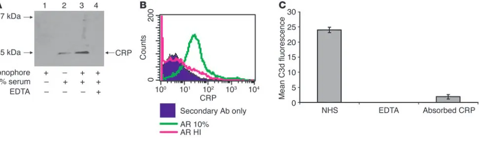
There are several means to initiate complement activation through C1q. First, we examined spermatozoa, following expo-sure to serum, for the presence of natural Ab. FACS, ELISA, and confocal microscopy using Abs against human IgM or IgG did not detect Ab bound to AR spermatozoa (data not shown). Second, because C-reactive protein (CRP) initiates the classical pathway via C1q (42) and is present in follicular fluid at levels similar to those in serum (43, 44), we performed Western blots and FACS analysis on ionophore-treated spermatozoa exposed to 10% autologous serum. A CRP-specific band at 25 kDa was observed in AR spermatozoa exposed to serum (Figure 5A). The fainter band in the untreated sample is likely derived from spon-taneously AR spermatozoa (typically 10–20% of the zoa). In addition, FACS showed CRP binding to AR spermato-zoa that had been exposed to 10% serum (Figure 5B). Because of these results, CRP was absorbed from serum with beads linked to phosphocholine. The resulting serum had a reduced ability to activate complement on AR spermatozoa (Figure 5C). These data demonstrate that CRP is responsible for initiating the clas-sical complement pathway on AR spermatozoa.
Next, we used confocal microscopy to define the location of the complement fragments. An mAb against MCP, TRA-2-10, was used to identify the IAM (Figure 6A). After spermatozoa were exposed to 10% serum, an mAb against C3d exclusively stained the IAM (Fig-ure 6, B and C). This demonstrates that complement activation is restricted to the IAM and supports the FACS profile of Figure 3 in which AR spermatozoa are shown to activate complement.
[image:5.585.52.275.122.226.2]The Ab used in the FACS and confocal experiments recog-nizes C3d, an epitope present in all membrane-bound cleavage fragments of C3 (Figure 7A). To determine the complement fragments containing C3d on the IAM, we performed Western blots with a polyclonal Ab against C3. In the lane derived from ionophore-treated spermatozoa, the α′ chain is cleaved and α2 is present, indicating that the C3b has been cleaved to C3bi (Fig-ure 7B). As expected, the α1 band is barely visible, as it would be attached via an ester linkage to proteins of varying molecular weights expressed on the IAM. Since MCP is a cofactor for the
Table 1
Complement activation by AR spermatozoa under different serum conditions
Serum C3d Fluorescence (mean ± SD)
10% NHS Yes 313 ± 65 Heat-inactivated No 34 ± 17 EDTA No 31 ± 18 MgEGTA No 32 ± 17 C1q-depleted No 29 ± 14 C2-deficient No 33 ± 12 Factor B–depleted No 32 ± 18 C7-deficient Yes 289 ± 54
Calcium ionophore–treated human spermatozoa were exposed for 15 minutes to the sera noted above and then incubated with an mAb to C3d and analyzed by FACS. Heat inactivation and EDTA treatment block all complement activation pathways. Mg++EGTA-treated serum,
C1q-deplet-ed serum, and C2-deficient serum prevent classical-pathway activation. Factor B–depleted serum prevents alternative-pathway activation. The data in the first, second, and last rows represent 10 separate experi-ments, while the results in the other rows are from 2 experiments. NHS, normal human serum.
Figure 5
[image:5.585.53.539.508.652.2]cleavage of C3b to C3bi, we blocked MCP’s cofactor activity with the mAb GB24 (45). TRA-2-10, used as a control, binds to CCP1 and does not block function (45). Neither Ab inhibited the cleav-age of C3b to C3bi (Figure 7B). Factor H, a soluble complement regulator that is present in follicular fluid at levels comparable to those in serum (39), also cleaves C3b to C3bi. When the mAb MH-10, which blocks the function of factor H, was used (46), the cleavage of C3b was inhibited (Figure 7B). Thus, factor H was responsible for the majority of the C3b cleavage.
Discussion
The acrosome reaction is a large exocytosis event that is induced in vivo when spermatozoa bind to the zona pellucida of the egg (Figure 1). This reaction releases enzymes that degrade the protein matrix of the zona and thereby allows the spermatozoa to trans-verse the zona and enter the perivitelline space. Our data show that when this event occurs spontaneously or is induced in vitro, com-plement activation is triggered, resulting in deposition of comple-ment fragcomple-ments specifically on the IAM. Deposition was depen-dent on C1q, C2, and factor B, consistent with initial activation of the classical pathway followed by engagement of the alternative pathway amplification loop. The observation that complement deposition on viable AR spermatozoa was C1q dependent, but Ab independent, led us to investigate CRP as the activating
pro-tein, because this lectin initiates the complement cascade through C1q (42). Also, CRP concentrations in follicular fluid are comparable to those in serum and fluctuate with the female cycle, peaking during ovulation (43, 44). Fur-ther, since CRP activates complement on apoptotic cells (42, 47) and nonviable spermatozoa activate complement (48), we used 3 different nucleic dyes and examined motili-ty to establish that viable, motile, AR spermatozoa activate complement. CRP bound specifically to the IAM of viable, AR spermatozoa. Consistent with its biochemical proper-ties, heat inactivation or EDTA treatment of serum abro-gated the binding of CRP to AR spermatozoa. In addition, serum incubated with phosphocholine beads to absorb CRP prevented C3d deposition. Taken together, these data indicate that CRP mediates the complement activation on the IAM of AR spermatozoa.
At first glance, complement activation on viable, AR spermatozoa is perplexing. If the membrane attack com-plex were to be formed on the IAM, there is the risk of membrane perturbation and possibly lysis of sperma-tozoa in the final steps of fertilization. Second, spermasperma-tozoa opsonized with C3b and C3bi are subject to immune adherence and phagocytosis. Third, the anaphylatoxins produced during complement activation could produce an inflammatory cell influx. Each of these points will now be addressed.
[image:6.585.46.315.81.209.2]Surprisingly, complement activation on AR spermatozoa did not lead to loss of motility or cell death. These data suggest that the complement deposition on AR spermatozoa is largely restricted to early complement components (C1, C4, C2, and C3) and does not efficiently proceed to the membrane attack com-plex. The presence of 2 complement regulators on the IAM, MCP (19, 20, 49) and DAF (40), makes it difficult to envision spon-taneous complement deposition on this membrane. However, activation by the classical pathway can overwhelm these regula-tory proteins (46). Their presence, though, is in concert with the hypothesis of “restricted activation” (see below) and may account
Figure 6
[image:6.585.335.501.497.742.2]Confocal microscopy of AR spermatozoa exposed to 10% serum. Spermatozoa were incubated with calcium ionophore to induce the acrosome reaction and then exposed to 10% autologous serum. (A) The mAb TRA-2-10 labeled with Alexa Fluor 488 recognizes MCP on the IAM. (B) An mAb to C3d labeled with Alexa Fluor 647 binds to the IAM. (C) Overlay of A and B. C3d deposition is limited to the IAM. Magnification, ×63.
Figure 7
research article
for why the late complement components are inefficiently depos-ited on viable, AR spermatozoa. These results are also consistent with studies showing that complement activation by CRP does not deplete the late complement components (42, 47).
Nonviable spermatozoa are known to activate the complement cascade, producing the anaphylatoxins C3a and C5a (50, 51). These peptides are potent chemotaxins and cellular activators that could serve to attract neutrophils to clear the female repro-ductive tract of nonviable spermatozoa (50, 51). It is also likely that these neutrophils assist in eliminating pathogens introduced to the female genital tract. In concert with this idea, the concen-tration of C3 in follicular fluid increases during ovulation (52). Under this scenario, C3b-opsonized viable spermatozoa might be at risk for phagocytosis. However, complement activation on AR spermatozoa occurs following binding to the zona pellucida of the egg. There is limited, if any, opportunity for these sperma-tozoa to interact with a phagocyte after entering the zona pel-lucida. As outlined above, complement activation on nonviable spermatozoa is likely a means of affecting their clearance. Viable AR spermatozoa, in contrast, induce restricted targeted comple-ment activation for a different purpose, presumably to facilitate sperm-egg interactions.
The purpose of complement activation on AR spermatozoa may be to coat the IAM with opsonins. Complement fragments on spermatozoa would then facilitate egg binding, similar to complement’s role in mediating immune adherence. Comple-ment receptors 1 and 3 are expressed on the plasma membrane of the egg (29).Complement is visualized as part of a highly redundant process to facilitate sperm-egg binding. This line of reasoning could explain why the mice deficient in C3 (53, 54), C4 (55), MCP (24), or integrins (56) are fertile. We hypothesize that dismantling parts of the complement system in these mice elimi-nates a supportive pathway for fertilization but does not obliter-ate it. Hence, by the broad measurement of litter size, there is no defect observed. This hypothesis provides the impetus to further assess the sperm-egg binding in these deficient mouse strains.
The possibility of complement activation fragments interact-ing with complement receptors to facilitate sperm-egg bindinteract-ing was first proposed in 1993 by Anderson and colleagues (29). In that pivotal report, several mechanisms were identified whereby spermatozoa could be coated with C3b, including alternative path-way activation, direct cleavage of C3 by the protease acrosin, and binding of dimeric C3b to complement regulators/receptors. We have extended and refined this study, particularly in relation to the complement activation on AR spermatozoa. In an experimen-tal system that quantitatively assesses viability, acrosomal status, and complement deposition using follicular fluid, we demonstrate that CRP binds to the IAM of viable AR spermatozoa and initiates the classical pathway. These results are consistent with a “seeding” of C3b by the classical pathway that in turn triggers the feedback loop of the alternative pathway. The bound C3b is rapidly con-verted to C3bi by factors H and I. We therefore hypothesize that C3bi, a ligand for integrin-related complement receptors, facili-tates sperm-egg interactions.
Complement activation on viable AR spermatozoa, whether spontaneous or induced by the calcium ionophore, has several features that distinguish it from complement activation on for-eign antigens. First, on spermatozoa, the fragment deposition is targeted to a specific cellular location. Second, complement activation is restricted beyond the C3 step, thereby limiting the
formation of membrane-damaging pores and release of the C5a chemotactic factor and anaphylaxation. Third, the overall mag-nitude of activation up to the C3 step appears to be more tightly controlled. It is as if this system on spermatozoa was specifically designed to allow a measured amount of C3b to be deposited at a specific site for a critical purpose. This concept of complement activation on self tissue (CAST), which we propose to also call TRACS for highly targeted and restricted activation of the com-plement system, is not well described as such in the literature; however, we believe it is a common and underappreciated func-tion of the complement system’s interacfunc-tion with self tissue. In conditions like apoptosis, ischemia/reperfusion injury, and prob-ably other types of alterations in host cell structure, TRACS is a preferable means to activate the complement system. In contrast to the desirable explosion of the cascade on microbes, comple-ment activation on an apoptotic cell is less robust, yet sufficient to facilitate the safe and relatively noninflammatory removal of altered self (42, 47). In the setting of ischemia/reperfusion inju-ry, one can envision a gradient of complement activation that correlates with the degree of cellular damage. In this example, the goal is to rapidly and safely remove dead and dying tissue in order to enhance wound healing and promote tissue sterility. As previously outlined, activation on spermatozoa may represent a situation whereby complement activation plays a role in repro-duction. In these settings, regulatory proteins are present on the cells, and it is likely the ratio of the strength and type of activa-tion to the quantity and efficiency of the regulators that deter-mines the outcome. Complement activation on the membrane of
Mycobacterium tuberculosis may represent an abuse of this type of a system (57). This pathogen salvages the catalytic complement fragment C2a of the classical pathway C3 convertase and uses it to then deposit a limited amount of C3b on its membrane. This deposition of C3b aids the pathogen by facilitating internaliza-tion through complement receptors.
evidenced by a role for MCP in the acceleration of the rate of the acrosome reaction in MCP-deficient mice (24).
In summary, viable AR spermatozoa activate complement. The complement deposition is triggered by CRP activation of the classical pathway, followed by amplification of C3 deposi-tion through the alternative pathway feedback loop. Comple-ment deposition is localized to the IAM and is curtailed beyond the C3 activation step. The purpose of this reaction remains problematic. However, we propose that this type of targeted and restricted complement activation is a common phenomenon on modified self tissue.
Methods
Spermatozoa samples. Spermatozoa samples were obtained with informed consent, and all studies were approved by the Washington University Human Studies Committee (protocol number 94-0420). Human sper-matozoa samples were repeatedly obtained from 4 healthy donors ranging in age from 18 to 38. Fresh ejaculates were allowed to liquefy at room temperature for 45 minutes, and then washed twice with modi-fied human tubal fluid (mHTF) supplemented with 5% serum substitute supplement (Irvine Scientific) and 5% HSA (100 mg/ml) (Irvine Scien-tific). Unless stated otherwise, centrifugation steps were performed at 200 g for 10 minutes at 4°C.
Serum preparation. Blood samples were obtained from donors that pro-vided informed consent, and all studies were approved by the Washing-ton University Human Studies Committee (protocol number 94-0420). Human blood was collected in glass tubes and allowed to clot at room temperature for 30 minutes. Samples were then centrifuged at 200 g. The serum was placed in sterile 1.5-ml microcentrifuge tubes and maintained at –20°C for short-term or –70°C for long-term storage.
Follicular fluid collection. Follicular fluid samples were obtained from patients that provided informed consent, and all studies were approved by the Washington University Human Studies Committee (protocol number 02-1004). Follicular fluids were collected from patients under-going in vitro fertilization. Ovarian stimulation was performed using gonadotropins in combination with a gonadotropin-releasing hormone (GnRH) antagonist or GnRH agonist according to established proto-cols. Serum estradiol was measured on cycle days 3, 5, and 7 and daily thereafter until retrieval. Vaginal ultrasound was performed daily from cycle day 7 onward. Human chorionic gonadotropin (5,000–10,000 IU, Profasi; Serono Inc.) was administered intramuscularly when 3 or more follicles were at least 16 mm in largest diameter. Transvaginal follicular aspiration was performed 35–36 hours later. Follicles were aspirated using a Wallace catheter (Irvine Scientific), and contents of each follicle were collected into a 15-ml tube (Falcon 2057; Cardinal Health) contain-ing approximately 1.0 ml of mHTF (Irvine Scientific) supplemented with 0.3 ml of sodium heparin. After the removal of oocytes, the follicular fluids were stored at 4°C until processed. To collect pure follicular fluid, the dominant follicle of a patient(s) was aspirated directly into a tube that did not contain mHTF.
Acrosome reaction. The acrosome reaction was induced by incubation of spermatozoa in supplemented mHTF media with 20 mM calcium ionophore A23187 (Sigma-Aldrich) at 37°C for 45 minutes.
Antibodies. The mouse anti-MCP mAb TRA-2-10 was prepared at Wash-ington University. The mouse mAb against factor H (MH-10) was donat-ed by J. Lambris (University of Pennsylvania, Philadelphia, Pennsylvania, USA). The mouse mAbs against human C3d and C5b-9 neoantigen were purchased from Quidel Corp. The polyclonal goat anti–human C9 Ab was purchased from Quidel Corp. The polyclonal goat anti–human C5 Ab was purchased from Advanced Research Technologies. Polyclonal
goat anti–human CRP was purchased from Affinity BioReagents. HRP-conjugated rabbit anti-goat IgG (Sigma-Aldrich) was used as the second-ary Ab for Western blotting. Zenon labeling kits (Invitrogen Corp.) were used to link fluorochromes (Alexa Fluor 488 or Alexa Fluor 647) to pri-mary Abs according to the manufacturer’s instructions (1 μg of primary Ab was used per labeling reaction). One microgram of TRA-2-10 was used per 1 × 10⁶ spermatozoa, and 1 μg of either the C3d or the C5b-9
mAb was used per 2 × 10⁶ spermatozoa for both FACS (BD Biosciences)
and confocal microscopy.
Complement deposition. AR or untreated spermatozoa were resuspended in gelatin veronal buffer supplemented with Mg2+ and Ca2+ (GVB++)
(Sigma-Aldrich). After incubation in 10% autologous serum or follicular fluid for 15 minutes at 37°C, the samples were washed with supplemented mHTF media. Gelatin veronal buffer supplemented with 10 mM EDTA (Sigma-Aldrich) and gelatin veronal buffer supplemented with 10 mM EGTA and 7 mM magnesium chloride were also used. Sera were heat-inactivated by 30 minutes of incubation at 56°C. Sources of the C2- and C7-deficient sera were as described in ref. 46. C1q- and factor B–depleted sera were obtained from Advanced Research Technologies.
Viability dyes. Propidium iodide (Invitrogen Corp.), SYTOX Green (Invitrogen Corp.), and SYBR 14 (Invitrogen Corp.) were used to assess viability according to the manufacturer’s instructions. Results were com-parable with all 3 dyes, but the different fluorescent ranges of the 3 dyes allowed for flexibility in fluorochrome usage.
FACS analysis. Spermatozoa, after exposure to a complement source, were incubated with Zenon-labeled primary Abs (described above) for 30 minutes at 37°C in supplemented mHTF and then washed twice. Flow cytometry was performed with a FACSCalibur (BD Biosciences) using forward and side scatter thresholds optimized for spermatozoa. A minimum of 10,000 events were collected per sample. Samples were gated on viable spermatozoa.
Lysates. Spermatozoa were resuspended in lysis buffer (PBS containing 1% NP-40, 0.05% SDS, and Complete protease inhibitor cocktail tablets [Roche Diagnostics Corp.]) and incubated for 30 minutes at 4°C. Samples were centrifuged at 13,500 g for 15 minutes at 4°C. Supernatants were transferred to 1.5-ml microcentrifuge tubes and stored at –20°C.
Immunoabsorption. Polyclonal goat anti–human C3 was coupled to CNBr-activated Sepharose (Amersham Biosciences) according to the manufac-turer’s suggestion. Spermatozoa lysate (50 μl), coupled Sepharose (50 μl), protease inhibitor tablets (Roche Diagnostics Corp.), and cell lysis buffer were combined and incubated at 4°C overnight with “end over end” mixing. The Sepharose beads were next washed 3 times with 0.3% NP-40. Eluates, obtained by boiling of the Sepharose beads in SDS loading buffer, were used for Western blotting as described below.
Western blotting. Cell lysates were separated in a 10–20% SDS-polyacrylamide gel under reducing conditions and then transferred onto nitrocellulose mem-branes using the NOVEX MiniCell II wet blotting system (Invitrogen Corp.). The membranes were blocked with 5% nonfat dry milk powder in 1× Tris-buff-ered saline (TBS) and 0.05% Tween-20 (TBS-T) (Sigma-Aldrich) overnight at 4°C. After 3 washes in TBS-T, the membranes were incubated in blocking buf-fer for 1 hour at 37°C with either purified polyclonal goat anti-CRP (1:1,000) or polyclonal goat anti-C3 Ab (1:7,500). Blots were washed 3 times and then incubated in blocking buffer for 1 hour at room temperature with HRP-con-jugated rabbit anti-goat IgG secondary Ab (Sigma-Aldrich) (1:10,000). After washing, the signals were developed using the enhanced chemiluminescence kit from Pierce Biotechnology Inc. The bands were visualized by exposure of the membranes to BioMax MR film (Eastman Kodak Co.).
research article
Confocal microscopy. Spermatozoa were incubated in the fluid phase with Zenon-labeled primary Abs (described above) for 30 minutes at 37°C in supplemented mHTF. After the samples were washed twice with supple-mented mHTF media, a 1:1 dilution of sample and ProLong Antifade Reagent (Invitrogen Corp.) was placed on a slide and analyzed with an LSM 510 META confocal microscope (Carl Zeiss Inc.).
CRP absorption. Immobilized p-aminophenyl phosphorylcholine beads (150 μl; Pierce Biotechnology Inc.) were added to human serum diluted 1:10 with GVB++ buffer. The samples were incubated at room temperature
for 30 minutes with gentle mixing. After centrifugation at 200 g for 5 minutes, the supernatant was removed and used as a complement source for FACS experiments.
Acknowledgments
We thank Madonna Bogacki for excellent secretarial support and Wandy Beaty from the Washington University Imaging Core for guid-ance in confocal microscopy. This work was supported by NIH grant AI37618-10.
Received for publication August 30, 2004, and accepted in revised form March 1, 2005.
Address correspondence to: John P. Atkinson, Washington Uni-versity School of Medicine, 660 South Euclid Avenue, Campus Box 8045, St. Louis, Missouri 63110, USA. Phone: (314) 362-8391; Fax: (314) 362-1366; E-mail: [email protected].
1. Seya, T., Turner, J., and Atkinson, J.P. 1986. Puri-fication and characterization of a membrane pro-tein (gp45-70) that is a cofactor for cleavage of C3b and C4b. J. Exp. Med.163:837–855. 2. Ballard, L.L., Bora, N.S., Yu, G.H., and Atkinson,
J.P. 1988. Biochemical characterization of mem-brane cofactor protein of the complement sys-tem. J. Immunol.141:3923–3929.
3. Seya, T., and Atkinson, J.P. 1989. Functional prop-erties of membrane cofactor protein of comple-ment. Biochem. J.264:581–588.
4. Oglesby, T.J., Allen, C.J., Liszewski, M.K., White, D.J., and Atkinson, J.P. 1992. Membrane cofactor protein (CD46) protects cells from complement-mediated attack by an intrinsic mechanism.
J. Exp. Med.175:1547–1551.
5. Dorig, R.E., Marcil, A., Chopra, A., and Richard-son, C.D. 1993. The human CD46 molecule is a receptor for measles virus (Edmonston strain).
Cell.75:295–305.
6. Naniche, D., et al. 1993. Human membrane cofac-tor protein (CD46) acts as a cellular recepcofac-tor for measles virus. J. Virol.67:6025–6032.
7. Manchester, M., Liszewski, M.K., Atkinson, J.P., and Oldstone, M.B.A. 1994. Multiple isoforms of CD46 (membrane cofactor protein) serve as receptors for measles virus. Proc. Natl. Acad. Sci. U. S. A.91:2161–2165.
8. Santoro, F., et al. 1999. CD46 is a cellular receptor for human herpesvirus 6. Cell.99:817–827. 9. Segerman, A., et al. 2003. Adenovirus type
11 uses CD46 as a cellular receptor. J. Virol.
77:9183–9191.
10. Gaggar, A., Shayakhmetov, D.M., and Lieber, A. 2003. CD46 is a cellular receptor for group B adenoviruses. Nat. Med.9:1408–1412.
11. Okada, N., Liszewski, M.K., Atkinson, J.P., and Caparon, M. 1995. Membrane cofactor protein (CD46) is a keratinocyte receptor for the M pro-tein of the group A streptococcus. Proc. Natl. Acad. Sci. U. S. A.92:2489–2493.
12. Kallstrom, H., Liszewski, M.K., Atkinson, J.P., and Jonsson, A.B. 1997. Membrane cofactor protein (MCP or CD46) is a cellular pilus receptor for pathogenic Neisseria. Mol. Microbiol.25:639–647. 13. Riley-Vargas, R.C., and Atkinson, J.P. 2003. Expres-sion of membrane cofactor protein (MCP; CD46) on spermatozoa: just a complement inhibitor?
Modern Aspects of Immunobiology.3:75–78. 14. Kemper, C., et al. 2003. Activation of human CD4+
cells with CD3 and CD46 induces a T-regulatory cell 1 phenotype. Nature.421:388–392. 15. Richards, A., et al. 2003. Mutations in human
complement regulator, membrane cofactor pro-tein (CD46), predispose to development of famil-ial hemolytic uremic syndrome. Proc. Natl. Acad. Sci. U. S. A.100:12966–12971.
16. Noris, M., et al. 2003. Familial haemolytic urae-mic syndrome and an MCP mutation. Lancet.
362:1542–1547.
17. Cervoni, F., et al. 1992. Identification and
charac-terization of membrane cofactor protein (MCP) on human spermatozoa. J. Immunol.148:1431–1437. 18. Riley, R.C., Kemper, C., Leung, M., and Atkinson,
J.P. 2002. Characterization of human membrane cofactor protein (MCP; CD46) on spermatozoa.
Mol. Reprod. Dev.62:534–546.
19. Anderson, D.J., Michaelson, J.S., and Johnson, P.M. 1989. Trophoblast/leukocyte-common anti-gen is expressed by human testicular germ cells and appears on the surface of acrosome-reacted sperm. Biol. Reprod.41:285–293.
20. Fenichel, P., et al. 1990. Localization and charac-terization of the acrosomal antigen recognized by GB24 on human spermatozoa. Mol. Reprod. Dev.
27:173–178.
21. Kitamura, M., et al. 1997. Possible association of infertility with sperm-specific abnormality of CD46. J. Reprod. Immunol.33:83–88.
22. Nomura, M., et al. 2001. Genomic analysis of idiopathic infertile patients with sperm-spe-cific depletion of CD46. Exp. Clin. Immunogenet.
18:42–50.
23. Jiang, H., and Pillai, S. 1998. Complement regulato-ry proteins on the sperm surface: relevance to sperm motility. Am. J. Reprod. Immunol.39:243–248. 24. Inoue, N., et al. 2003. Disruption of mouse CD46
causes an accelerated spontaneous acrosome reaction in sperm. Mol. Cell. Biol.23:2614–2622. 25. Mizuno, M., Harris, C.L., and Morgan, B.P. 2004.
Immunohistochemical analysis of membrane complement regulatory proteins in rat testis: unique roles for DAF and MCP in spermatozoal function? Abstracts from the XXth International Complement Workshop, 172. Mol. Immunol.
41:280.
26. Hosokawa, M., Nonaka, M., Okada, N., and Okada, H. 1996. Molecular cloning of guinea pig membrane cofactor protein: preferential expres-sion in testis. J. Immunol.157:4946–4952. 27. Foley, S., Li, B., Dehoff, M., Molina, M., and
Hol-ers, V.M. 1993. Mouse Crry/p65 is a regulator of the alternative pathway of complement activa-tion. Eur. J. Immunol.23:1381–1384.
28. Xu, C., et al. 2000. A critical role for the murine complement regulator Crry in fetomaternal tol-erance. Science.287:498–501.
29. Anderson, D.J., Abbott, A.F., and Jack, R.M. 1993. The role of complement component C3b and its receptors in sperm-oocyte interaction. Proc. Natl. Acad. Sci. U. S. A.90:10051–10055.
30. Taylor, C.T., Biljan, M.M., Kingsland, C.R., and Johnson, P.M. 1994. Inhibition of human sper-matozoon-oocyte interaction in vitro by mono-clonal antibodies to CD46 (membrane cofactor protein). Hum. Reprod.9:907–911.
31. D’Cruz, O.J., Lambert, H., and Haas, G.G., Jr. 1997. Expression of CD15 (Lewisx) antigen on human sperm and its role in sperm-egg interaction. Am. J. Reprod. Immunol.37:172–183.
32. Riley, R.C., Tannenbaum, P.L., Abbott, D.H., and Atkinson, J.P. 2002. Cutting edge. Inhibiting
mea-sles virus infection but promoting reproduction: an explanation for splicing and tissue-specific expression of CD46. J. Immunol.169:5405–5409. 33. Hsu, E.C., et al. 1997. Artificial mutations and
natural variations in the CD46 molecules from human and monkey cells define regions important for measles virus binding. J. Virol.71:6144–6154. 34. Adams, E., Brown, M.C., Nunge, M., Krych,
M., and Atkinson, J.P. 1991. Contribution of the repeating domains of membrane cofactor protein (CD46) of the complement system to ligand binding and cofactor activity. J. Immunol.
147:3005–3011.
35. Iwata, K., et al. 1995. Diversity of sites for measles virus binding and for inactivation of comple-ment C3b and C4b on membrane cofactor pro-tein CD46. J. Biol. Chem.270:15148–15152. 36. Vanderpuye, O.A., Labarrere, C.A., and McIntyre,
J.A. 1992. The complement system in human reproduction. Am. J. Reprod. Immunol.27:145–155. 37. Clarke, G.N., Hsieh, C., Koh, S.H., and Cauchi,
M.N. 1984. Sperm antibodies, immunoglobulins, and complement in human follicular fluid. Am. J. Reprod. Immunol.5:179–181.
38. Perricone, R., et al. 1992. Functionally active com-plement is present in human ovarian follicular fluid and can be activated by seminal plasma.
Clin. Exp. Immunol.89:154–157.
39. Perricone, R., et al. 1990. Complement, comple-ment activation and anaphylatoxins in human ovarian follicular fluid. Clin. Exp. Immunol.
82:359–362.
40. Cervoni, F., et al. 1993. Expression of decay acceler-ating factor (CD55) of the complement system on human spermatozoa. J. Immunol.151:939–948. 41. Fenichel, P., et al. 1994. Expression of the
comple-ment regulatory protein CD59 on human sper-matozoa: characterization and role in gametic interaction. Mol. Reprod. Dev.38:338–346. 42. Mold, C., Gewurz, H., and Du Clos, T.W. 1999.
Regulation of complement activation by C-reac-tive protein. Immunopharmacology.42:23–30. 43. Jilma, B., et al. 1997. Menstrual cycle-associated
changes in blood levels of interleukin-6, alpha1 acid glycoprotein, and C-reactive protein. J. Lab. Clin. Med.130:69–75.
44. Orvieto, R., et al. 2004. C-reactive protein levels in patients undergoing controlled ovarian hyperstim-ulation for IVF cycle. Hum. Reprod.19:357–359. 45. Liszweski, M.K., et al. 2000. Dissecting sites
important for complement regulatory activity in membrane cofactor protein (MCP; CD46). J. Biol. Chem. 275:37692–37701.
46. Barilla-LaBarca, M.L., Liszewski, M.K., Lambris, J.D., Hourcade, D., and Atkinson, J.P. 2002. Role of membrane cofactor protein (CD46) in regula-tion of C4b and C3b deposited on cells. J. Immunol.
168:6298–6304.
terminal complement components, and sustains an antiinflammatory innate immune response: implications for systemic autoimmunity [erratum 2001, 193:1439]. J. Exp. Med.192:1353–1364. 48. Vogelpoel, F.R., et al. 1987. Antibody and
comple-ment-binding activity of viable and nonviable human spermatozoa. Arch. Androl.18:189–197. 49. Cervoni, F., et al. 1992. Identification and
char-acterization of membrane cofactor protein on human spermatozoa. J. Immunol.148:1431–1437. 50. Clark, R.A., and Klebanoff, S.J. 1976. Generation
of a neutrophil chemotactic agent by spermato-zoa: role of complement and regulation by semi-nal plasma factors. J. Immunol.117:1378–1386. 51. Vogelpoel, F.R., and Verhoef, J. 1985. Activation of
polymorphonuclear leukocytes by spermatozoa.
Arch. Androl.14:123–131.
52. Lee, Y.-L., et al. 2004. The embryotrophic activity of oviductal cell-derived complement C3b and iC3b, a novel function of complement protein in
reproduction. J. Biol. Chem.279:12763–12768. 53. Wessels, M.R., et al. 1995. Studies of group B
streptococcal infection in mice deficient in com-plement component C3 or C4 demonstrate an essential role for complement in both innate and acquired immunity. Proc. Natl. Acad. Sci. U. S. A.
92:11490–11494.
54. Circolo, A., et al. 1999. Genetic disruption of the murine complement C3 promoter region gener-ates deficient mice with extrahepatic expression of C3 mRNA. Immunopharmacology.42:135–149. 55. Fischer, M., et al. 1996. Regulation of the B cell
response to T-dependent antigens by classical pathway complement. J. Immunol.157:549–556. 56. He, Z.Y., et al. 2003. None of the integrins known
to be present on the mouse egg or to be ADAM receptors are essential for sperm-egg binding and fusion. Dev. Biol.254:226–237.
57. Schorey, J.S., Carroll, M.C., and Brown, E.J. 1997. A macrophage invasion mechanism of pathogenic
mycobacteria. Science.277:1091–1093.
58. Seya, T., et al. 1991. Preferential inactivation of the C5 convertase of the alternative complement pathway by factor I and membrane cofactor pro-tein (MCP). Mol. Immunol.28:1137–1147. 59. Kurita-Taniguchi, M., et al. 2002. Molecular
assembly of CD46 with CD9, alpha 3-beta 1 integrin and protein tyrosine phosphatase SHP-1 in human macrophages through differentiation by GM-CSF. Mol. Immunol.38:689–700. 60. Lozahic, S., et al. 2000. CD46 (membrane cofactor
protein) associates with multiple beta 1 integrins and tetraspans. Eur. J. Immunol.30:900–907. 61. LeNaour, F., Rubinstein, E., Jasmin, C., Prenant,
M., and Boucheix, C. 2000. Severely reduced female fertility in CD9-deficient mice. Science.
287:319–321.
62. Miyado, K., et al. 2000. Requirement of CD9 on the egg plasma membrane for fertilization. Science.