Endotoxin and cytokines decrease serum levels
and extra hepatic protein and mRNA levels of
cholesteryl ester transfer protein in syrian
hamsters.
I Hardardóttir, … , K Feingold, C Grünfeld
J Clin Invest.
1996;
97(11)
:2585-2592.
https://doi.org/10.1172/JCI118707
.
Endotoxin alters the metabolism of lipoproteins, including that of high density lipoprotein
(HDL). Cholesteryl ester transfer protein (CETP) facilitates exchange of HDL cholesterol for
very low density lipoprotein (VLDL) triglyceride, leading to catabolism of HDL. We
investigated the effects of endotoxin and cytokines on CETP in Syrian hamsters. Endotoxin
induced a rapid and progressive decrease in serum CETP levels, by 48 h CETP had
decreased to < 20% of control levels. Endotoxin also decreased CETP mRNA and protein
levels in adipose tissue, heart, and muscle, the tissues with highest levels of CETP mRNA,
providing a plausible mechanism for the endotoxin-induced decrease in circulating CETP.
Dexamethasone did not mimic the effects of endotoxin on CETP, but the combination of
tumor necrosis factor and interleukin-1 did, indicating that these cytokines may in part
mediate the effects of endotoxin on CETP. The endotoxin-induced decrease in CETP may
help maintain HDL cholesterol levels during infection and inflammation when increased
triglyceride levels could drive the exchange of HDL cholesteryl ester for VLDL triglyceride.
Maintaining circulating HDL may be important because HDL protects against the toxic
effects of endotoxin and provides cholesterol for peripheral cells involved in the immune
response and tissue repair.
Research Article
Find the latest version:
The Journal of Clinical Investigation Volume 97, Number 11, June 1996, 2585–2592
Endotoxin and Cytokines Decrease Serum Levels and Extra Hepatic Protein and
mRNA Levels of Cholesteryl Ester Transfer Protein in Syrian Hamsters
Ingibjörg Hardardóttir,* Arthur H. Moser,* John Fuller,* Christopher Fielding,‡ Kenneth Feingold,* and Carl Grünfeld*
*Department of Medicine, University of California San Francisco and Metabolism Section, Medical Service, Department of Veterans Affairs Medical Center, San Francisco, California 94121; and ‡Cardiovascular Research Institute, University of California Medical Center,
San Francisco, California 94143
Abstract
Endotoxin alters the metabolism of lipoproteins, including that of high density lipoprotein (HDL). Cholesteryl ester transfer protein (CETP) facilitates exchange of HDL cho-lesterol for very low density lipoprotein (VLDL) triglycer-ide, leading to catabolism of HDL. We investigated the ef-fects of endotoxin and cytokines on CETP in Syrian hamsters. Endotoxin induced a rapid and progressive de-crease in serum CETP levels; by 48 h CETP had dede-creased to , 20% of control levels. Endotoxin also decreased CETP mRNA and protein levels in adipose tissue, heart, and mus-cle, the tissues with highest levels of CETP mRNA, provid-ing a plausible mechanism for the endotoxin-induced de-crease in circulating CETP. Dexamethasone did not mimic the effects of endotoxin on CETP, but the combination of tumor necrosis factor and interleukin-1 did, indicating that these cytokines may in part mediate the effects of endotoxin on CETP. The endotoxin-induced decrease in CETP may help maintain HDL cholesterol levels during infection and inflammation when increased triglyceride levels could drive the exchange of HDL cholesteryl ester for VLDL triglycer-ide. Maintaining circulating HDL may be important be-cause HDL protects against the toxic effects of endotoxin and provides cholesterol for peripheral cells involved in the immune response and tissue repair. (J. Clin. Invest. 1996.
97:2585–2592.) Key words: lipopolysaccharide •
immuno-logic factors • acute phase reaction • cholesteryl ester trans-fer protein • corticosteroid
Introduction
Infection and inflammation induce dramatic changes in plasma levels of the acute phase proteins as well as alterations in lipid metabolism that are thought to have beneficial effects in host defense (1, 2). Endotoxin administration, which mimics infec-tion, stimulates the production of cytokines that mediate many of the metabolic responses to infection and inflammation. For example, endotoxin and cytokines increase serum triglycer-ides, predominantly in very low density lipoproteins (VLDL), by increasing hepatic lipogenesis and VLDL production and/
or by decreasing clearance of triglyceride-rich lipoproteins through inhibition of lipoprotein lipase (reviewed in reference 2). Endotoxin and cytokines also increase serum cholesterol levels in rodents, an effect that is due in part to an increase in the activity of hydroxymethylglutaryl coenzyme A reductase, the rate-limiting enzyme in cholesterol synthesis (3, 4) and a decrease in cholesterol 7a hydroxylase, the rate-limiting en-zyme in bile acid synthesis (Feingold, K., manuscript submit-ted for publication).
In contrast to serum triglyceride and cholesterol levels, high density lipoprotein (HDL) cholesterol levels show small decreases after administration of endotoxin and cytokines to rodents. The decrease in HDL cholesterol levels is often ac-companied by a decrease in apolipoprotein (apo) A-I (re-viewed in reference 2). Because apo A-I plays a major role in HDL-mediated cholesterol efflux from cells, decreased apo A-I during infection may lead to decreased reverse cholesterol transport. In contrast, two other apolipoproteins associated with HDL are increased after administration of endotoxin or cytokines: apo serum amyloid A (apoSAA)1 and apo J. Both
of these apolipoproteins may be important for regulating HDL cholesterol levels and HDL function. ApoSAA-rich HDL are rapidly cleared from plasma (5), thus increased apoSAA could contribute to endotoxin-induced decrease in HDL cholesterol levels. SAA-rich HDL are also more readily taken up by mac-rophages and less readily taken up by hepatocytes (6). HDL cholesterol may thus preferentially be directed to macrophages during the acute phase response.
Metabolism of HDL is also dependent on several key en-zymes the activity of which are modified by endotoxin. The ac-tivity of hepatic lipase is decreased by endotoxin and inflam-mation (7–10). Hepatic lipase is responsible for metabolizing large triglyceride-rich HDL into smaller HDL2, which is more
rapidly cleared from the circulation, and to preb1HDL, which
is believed to play a key role in mediating the transfer of free cholesterol from cell membranes (11–13). In addition endo-toxin decreases the activity of lecithin:cholesterol acyl trans-ferase (LCAT), which is responsible for esterifying free cho-lesterol in HDL (14–16). The decrease in LCAT activity leads to an increase in free cholesterol and a decrease in esterified cholesterol in HDL.
Cholesteryl ester transfer protein (CETP) mediates the ex-change of HDL cholesteryl ester for VLDL triglycerides (17, 18). Subsequently, HDL becomes depleted of cholesterol ester and enriched in triglycerides. Hepatic lipase acts on the trigly-ceride rich HDL, generating remnant HDL2, which is taken up
by the liver. The effect of endotoxin on CETP has been exam-ined in transgenic mice overexpressing the human CETP gene.
Address correspondence to Carl Grünfeld, M.D., Ph.D., Department of Veterans Affairs Medical Center 111F, 4150 Clement Street, San Francisco, CA 94121. Phone: 415-750-2005; FAX: 415-750-6927; E-mail: [email protected]
Received for publication 3 October 1995 and accepted in revised form 30 January 1996.
In these mice, endotoxin decreases mRNA levels for CETP in the liver and CETP protein in plasma (19). However, endo-toxin increased the mRNA levels for CETP in peripheral tis-sues in the transgenic mice. The effects of cytokines on CETP has not been studied.
We have examined the effects of both endotoxin and cy-tokines on expression of the native gene for CETP in Syrian hamsters. Hamsters have very low levels of mRNA for CETP in liver but higher levels in adipose tissue, muscle, and heart, which may contribute to serum levels of CETP in these ani-mals (20). We have used the Syrian hamsters to investigate the effects of endotoxin and cytokines on lipid and lipoprotein me-tabolism because in contrast to other rodents, cholesterol and lipoprotein metabolism in Syrian hamsters resembles that in humans. Moreover, hamsters, unlike mice and rats, have cho-lesteryl ester transfer activity in plasma (21).
Methods
Materials.{32P}dCTP (3,000 Ci/mmol, 10 mCi/ml) was purchased
from New England Nuclear (Boston, MA). Endotoxin (Escherichia coli 55:B5) was purchased from Difco Laboratories (Detroit, MI) and was freshly diluted to desired concentrations in pyrogen-free 0.9% sa-line (Kendall McGraw Laboratories, Inc., Irvine, CA). Human tumor necrosis factor (TNF)-a with a sp act of 5 3 107 U/mg was kindly
pro-vided by Genentech, Inc. (South San Francisco, CA). Recombinant human interleukin (IL)-1b with a sp act of 1 3 109 U/mg was
gener-ously provided by Immunex (Seattle, WA). Dexamethasone sodium phosphate was from Lyphomed (Deerfield, IL). Multiprime DNA la-beling system was purchased from Amersham International (Amer-sham, United Kingdom). Mini spin columns were purchased from Worthington Biochemical Corp. (Freehold, NJ). Oligo (dt)-cellulose, type 77F was purchased from Pharmacia LKB Biotechnology AB (Uppsala, Sweden). Western light chemiluminescent detection sys-tem was purchased from Tropix, Inc. (Bedford, MA). A mAb against human CETP that reacts with hamster CETP was obtained from In-ternational Immunodiagnostics (Foster City, CA), and the IgG puri-fied on protein A-agarose was from Bio-Rad Laboratories (Affigel-Protein A, MAPSII kit; Hercules, CA). Nitrocellulose was purchased from Schleicher and Schuell (Keene, NH). Kodak XAR5 film was used for autoradiography. The DNA for CETP was prepared as de-scribed (22).
Animals. Male Syrian hamsters (z 100–150 g) were purchased
from Simonsen Laboratories (Gilroy, CA). The animals were main-tained in a room with lights on from 6:00 a.m. to 6:00 p.m. and were provided with rodent chow and water ad lib. Animals were injected intraperitoneally (i.p.) with endotoxin, dexamethasone, TNF 1 IL-1, TNF, or IL-1 at the indicated doses in 0.5 ml 0.9% saline or with sa-line alone. After administration of endotoxin, dexamethasone, or cy-tokines, food was withdrawn from both control and treated animals, because endotoxin and cytokines can induce anorexia. Animals were studied between 90 min and 48 h after endotoxin administration as in-dicated in the text.
Isolation of RNA and Northern blotting. Total RNA was iso-lated by a variation of the guanidinium thiocyanate method (23). To-tal RNA from adipose tissue was used for Northern blotting, but poly A RNA from all other tissues was isolated using oligo dT cellulose. Poly A RNA or RNA was quantified by measuring adsorption at 260 nm. Equal amounts of RNA (10 mg) were loaded on 1% agarose-formal-dehyde gels and electrophoresed. The uniformity of sample applica-tions was checked by ultraviolet visualization of the ethidium bro-mide–stained gel before transfer to nitrocellulose membranes. We and others have found that endotoxin increases mRNA levels of actin in liver (4, 24) and in adipose tissue (unpublished observations by this laboratory). Endotoxin also increases hepatic mRNA levels for cyclo-philin by 2.7-fold (unpublished observations by this laboratory). The
mRNA levels of actin and cyclophilin, which are widely used for nor-malizing data, can therefore not be used during studies of endotoxin induction of acute phase proteins. However, the magnitude of the change in mRNA levels after endotoxin and cytokine administration and the relatively small standard error of the mean make it unlikely that the changes observed are due to unequal loading of RNA. Fur-thermore, the regulation of mRNA by LPS and cytokines is specific, as the mRNAs for key proteins show different degrees of induction or repression, differential induction by cytokines, different dose re-sponse curves, and tissue-specific rere-sponses (4, 16, 28–30, Hardardóttir, I., et al., manuscript submitted for publication). RNA probe hybrid-ization was performed in 0.75 M sodium chloride, 0.075 M sodium ci-trate, 2% SDS, 10% dextran sulfate, 23 Denhardt’s solution and 100
mg/ml sheared salmon sperm DNA at 658C overnight. Blots were washed at 428C in 0.3 M sodium chloride, 0.03 M sodium citrate, and 0.1% SDS. All blots were exposed to x-ray film, and bands were quantified by densitometry. Duration of film exposure was varied to allow measurements on the linear portion of the curve.
Western blotting. For the determination of CETP protein, SDS-PAGE was performed as described by Clarke et al. (25) with modifi-cations described previously (4). Serum was diluted with loading buffer before analysis on a SDS-PAGE. Tissues (1 g) were homoge-nized on ice with a Wheaton Dounce tissue grinder in 2 ml of buffer containing 20 mM sodium phosphate, pH 7.5, 0.2 mM NaCl, 2% Tri-ton X-100, vol/vol, 1% sodium deoxycholate, 0.2% SDS, 2 mM di-ethyl p-nitrophenyl phosphate, 0.2 mM leupeptin, and 600 U/ml apro-tinin (26). After 15-min incubation on ice, the homogenate was centrifuged, and the clear infranatant was collected. Serum samples and infranatants from tissues were analyzed in the reduced state us-ing 7% polyacrylamide gel with a 4% stackus-ing gel. Protein was trans-ferred to nitrocellulose (0.2-mm pore size). CETP protein was de-tected with mAb generated against human CETP, using the Tropix Western Light chemiluminescent detection system. After x-ray film development, band density was measured using a Biorad GS-670 Im-aging Densitometer (Bio-Rad Laboratories).
Statistics. The results are expressed as means6SEM. Statistical significance was determined using a two-tailed Student’s t test.
Results
To investigate the effects of endotoxin on serum levels of CETP, Syrian hamsters were injected intraperitoneally with endotoxin (100 mg/100 g body wt [bw]). CETP was detected by Western blotting using an mAb against human CETP that cross-reacts with hamster CETP (see Methods). As shown in Fig. 1, endotoxin administration markedly decreased serum levels of CETP. The CETP levels were significantly reduced 4 h after endotoxin administration (by 18%) and continued to de-crease, such that by 48 h after endotoxin administration, serum CETP levels were , 20% of control levels.
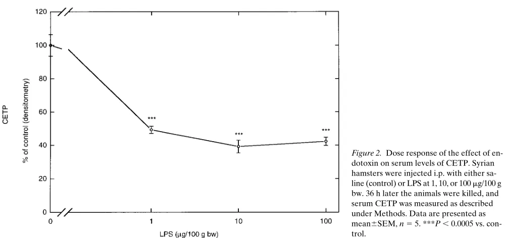
We next determined the dose response for endotoxin-induced decreases in serum CETP levels at 36 h after treat-ment. As shown in Fig. 2, 10 and 100 times lower doses of endo-toxin (1 and 10 mg/100 g bw) decreased serum CETP protein levels 36 h after administration. A dose of 0.1 mg/100 g bw was ineffective when studied 24 and 48 h after administration, indi-cating that the half-maximal dose of the effect of endotoxin on serum CETP levels is between 0.1 and 1 mg/100 g bw.
tis-sues may contribute to serum levels of CETP (20). CETP pro-tein levels in adipose tissue, heart, and muscle were measured by Western blotting after homogenization in a detergent buffer as detailed in Methods. As shown in Fig. 3, endotoxin administration significantly decreased CETP levels in adipose tissue, heart, and muscle by 50–60%, suggesting that the decrease in CETP levels in the circulation may be due to decreased CETP production in these tissues.
To further determine the mechanism by which CETP pro-tein levels are decreased, we next measured the mRNA levels for CETP in the key tissues of the hamster after administration
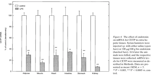
of endotoxin. In accordance with previous reports, mRNA for CETP was not detected in the liver or spleen of the hamsters (26). RNA for CETP was, however, detected in adipose tissue, muscle, heart, intestine, stomach, and kidney. As shown in Fig. 4, 16 h after administration of endotoxin, mRNA levels for CETP were markedly decreased in all of these tissues.
[image:4.612.62.557.58.322.2]Endotoxin induces glucocorticoids, and glucocorticoids have been shown to mediate some of the effects of endotoxin (27). In mice overexpressing the human CETP gene, cortisone administration mimics the effects of endotoxin on plasma CETP levels. Additionally, treatment with cortisone decreased Figure 1. Time course of the effect of endotoxin on serum levels of CETP. Syrian hamsters were in-jected i.p. with either saline (s) or 100 mg/100 g bw endotoxin (d). At the times indicated, the animals were killed and serum CETP was measured as described under Meth-ods. Data are presented as mean6SEM, n 5 5. *P , 0.05, **P , 0.005, ***P , 0.0005 vs. control.
[image:4.612.56.556.503.739.2]hepatic mRNA levels for CETP (19). It was therefore of inter-est to tinter-est whether corticosteroids would mimic the effects of endotoxin on CETP levels in hamsters, where regulation of the native gene can be studied and where serum levels of CETP are likely to be derived from extra hepatic sources. Hamsters were injected intraperitoneally with dexamethasone (5 mg/kg) or with saline (control), and plasma CETP and mRNA for CETP in adipose tissue were measured 16 h later. Dexametha-sone did not affect serum levels of CETP in the Syrian ham-sters (control (C): 10068.4%, dexamethasone: 123610.2%) and increased mRNA levels for CETP in adipose tissue (C: 10064.5%; dexamethasone: 20867.6%), indicating that
gluco-corticoids do not mediate the effects of endotoxin on CETP in the hamster.
[image:5.612.54.559.64.319.2]As TNF and IL-1 mediate many of the effects of endotoxin, we next investigated whether administration of these cytokines individually or in combination affected serum levels of CETP. The doses of TNF and IL-1 used (17 mg and 1 mg/100 g bw, re-spectively) have been shown previously to alter lipid metabo-lism in Syrian hamsters (28) and to regulate apo J (29). In addi-tion, TNF at this dose decreases hepatic LCAT mRNA levels and LCAT activity in Syrian hamsters (16). However, adminis-tration of the cytokines individually did not significantly affect serum CETP levels (Fig. 5). In contrast, the combination of
Figure 3. Effect of endotoxin on CETP protein levels in tissues. Syr-ian hamsters were injected i.p. with either saline (control, open bars) or 100 mg/100 g bw endotoxin (hatched bars). 16 h later the animals were killed, and the respective organs were collected. A portion (1 g) of the tissues were homogenized on ice with a Wheaton Dounce tissue grinder in 2 ml of buffer containing 20 mM sodium phosphate, pH 7.5, 0.2 mM NaCl, 2% Triton X-100, vol/vol, 1% sodium deoxycholate, 0.2% SDS, 2 mM diethyl p -nitro-phenyl phosphate, 0.2 mM leupep-tin, and 600 U/ml aprotinin (26). After 15-min incubation, the homo-genate was centrifuged, and the clear infranatant used for determi-nation of CETP by Western blot-ting. CETP was measured as de-scribed under Methods. Data are presented as mean6SEM, n 5 5. **P , 0.005, ***P , 0.0005 vs. con-trol.
[image:5.612.59.556.493.744.2]TNF 1 IL-1 significantly decreased serum levels of CETP to 69% of that of controls 16 h after their administration (Fig. 5). We have previously reported that the combination of TNF 1 IL-1 is more effective in mimicking the effects of endotoxin on cholesterol metabolism in Syrian hamsters (28, 29).
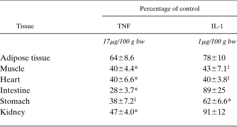
When TNF and IL-1 were administered alone at the doses described earlier, there was some effect of these cytokines in reducing the levels of mRNA for CETP in a few of the tissues
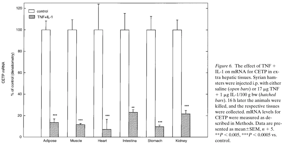
[image:6.612.58.557.60.347.2]studied (Table I). However, when TNF 1 IL-1 were adminis-tered together they were much more effective than the individ-ual cytokines; the combination reduced mRNA levels for CETP in all the tissues studied by . 75% (Fig. 6). Thus simul-taneous administration of TNF and IL-1 together is more ef-fective at decreasing both serum protein levels and tissue mRNA levels of CETP than either cytokine individually. The combination mimics LPS treatment.
Figure 5. The effect of TNF and IL-1 on serum levels of CETP. Syrian hamsters were injected i.p. with sa-line, TNF (17 mg/100 g bw), IL-1 (1
mg/100 g bw), or TNF 1 IL-1 (17 mg
1 1 mg/100 g bw). 16 h later the ani-mals were killed, and serum CETP was measured as described under Methods. Data are presented as mean6SEM, n 5 5. ***P , 0.0005 vs. control.
Figure 6. The effect of TNF 1
IL-1 on mRNA for CETP in ex-tra hepatic tissues. Syrian ham-sters were injected i.p. with either saline (open bars) or 17 mg TNF
[image:6.612.55.544.491.740.2]Discussion
The data presented in this paper demonstrate that endotoxin and cytokines decrease CETP levels in the serum and CETP protein and mRNA levels in extra hepatic tissues in Syrian hamsters. Circulating CETP levels began to decrease rapidly after administration of endotoxin and progressively decreased with time for at least 48 h. This pattern of early, progressive, and sustained effect after single administration of endotoxin is common. The changes in serum triglyceride levels after endo-toxin administration follow a similar pattern in the opposite di-rection. 2 h after endotoxin administration, serum triglyceride levels are increased (30) and continue to increase with time (16), reaching levels fivefold higher than control levels at 48 h (C: 100619;% endotoxin: 509640%; unpublished observa-tions by this laboratory). The changes in serum triglyceride levels are thus a mirror image of the changes in CETP levels after endotoxin administration. The changes in HDL choles-terol levels follow a different pattern decreasing at 4 and 8 h (16) but returning to control levels by 48 h after endotoxin ad-ministration (C: 10065.6%; endotoxin: 9368.7%; unpublished observations by this laboratory).
The decrease in HDL cholesterol levels may be linked to the increase in serum triglycerides as hypertriglyceridemia is often associated with decreased HDL cholesterol levels (31– 33). CETP facilitates the exchange of cholesterol ester in HDL for VLDL triglyceride, thus the abundant VLDL triglyceride in hypertriglyceridemia can promote this exchange and con-tribute to the decrease in HDL cholesterol levels. In fact, hy-pertriglyceridemia in humans is often associated with in-creased CETP activity (34). In contrast, the endotoxin-induced hypertriglyceridemia in the hamsters, although associated with decreased HDL cholesterol levels, is associated with a marked decrease in CETP levels. Endotoxin thus appears to have an effect on CETP that is independent of the hypertriglyceri-demic effect and potentially serves to prevent or blunt the de-crease in HDL cholesterol levels. That decreasing CETP levels may increase HDL cholesterol levels is suggested by an in-verse relationship between plasma CETP levels and HDL cho-lesterol levels in CETP-deficient humans (35, 36). In addition, overexpression of CETP in transgenic mice causes decreased
HDL cholesterol levels (37, 38), and cholesterol feeding of hamsters which increases CETP activity is associated with de-creased HDL cholesterol levels (20).
The decrease in CETP is a sensitive response to endotoxin, with ID50 between 0.1 and 1 mg/100 g bw. These doses of
en-dotoxin are far below the doses required to cause death in ro-dents in our laboratory. We have previously demonstrated that the alterations in lipid metabolism are among the most sensi-tive host responses to endotoxin. For example, in Syrian ham-sters, endotoxin at 0.1 mg/100 g bw increases serum choles-terol, triglyceride, and apo J levels while decreasing serum LCAT activity (4, 16, 29). The increase in apo J and decrease in LCAT activity were preceded by increased and decreased mRNA levels for these proteins. However, higher doses of en-dotoxin (10 mg/100 g bw) are required for decreasing HDL cholesterol levels (16). A lower dose of endotoxin is able to de-crease CETP levels compared to what is needed to dede-crease HDL cholesterol levels.
Endotoxin-induced decreases in circulating CETP levels may be mediated at least in part by marked decreases in mRNA levels for CETP in extra hepatic tissues and decreases in CETP protein levels in adipose tissue, muscle and heart. Adipose tissue, muscle, and heart are abundant sources of mRNA for CETP and adipose tissue mRNA levels strongly correlate with plasma CETP concentrations (20, 26). Thus the decrease in CETP protein and mRNA levels in adipose and other extra hepatic tissues is likely to explain the decrease in plasma CETP seen here in response to endotoxin. However, it is possible that plasma CETP levels are also affected by changes in translation, secretion, or clearance of CETP.
The effects of endotoxin on serum levels of CETP in the present study are consistent with the reported results of Ma-succi-Magoulas et al. (19), who demonstrated that endotoxin reduces plasma CETP levels and hepatic mRNA levels for CETP in mice overexpressing the human CETP gene. How-ever, there are significant differences in the results from the two studies. First, the decrease in hepatic mRNA levels for CETP in the transgenic mice was transient, with mRNA levels returning to control levels by 48 h after the higher dose of en-dotoxin (200 mg) and actually being significantly increased af-ter lower doses of endotoxin (25 mg). Here we found the de-crease in CETP to be progressive over 48 h. Second, in contrast to our results in the hamster, in the transgenic mice endotoxin increased mRNA levels for CETP in extra hepatic tissues. Endotoxin thus decreased mRNA levels for CETP in the liver, the major contributor to plasma CETP in humans, but increased mRNA levels for CETP in other tissues. In the present study in hamsters, where extra hepatic tissues are a major source of plasma CETP, endotoxin decreased mRNA for CETP in these tissues.
Third, Masucci-Magoulas et al. (19) demonstrated that glu-cocorticoids mediated the effects of endotoxin on CETP levels in the transgenic mice. Although glucocorticoids may mimic some effects of endotoxin, dexamethasone administration (5 mg/kg) did not produce the same effect as endotoxin on CETP in Syrian hamsters, suggesting that in hamsters glucocorticoids do not mediate this effect of endotoxin. There are several pos-sible reasons for the disparate results with regard to glucocorti-coids. For example, many aspects of the acute phase response are species specific, and it is possible that there are species dif-ferences in the mechanisms by which endotoxin acts in ham-sters and humans/mice. In fact, we have previously
demon-Table I. Effects of TNF or IL-1 on mRNA Levels for CETP in Peripheral Tissues in Syrian Hamsters
Percentage of control
Tissue TNF IL-1
17mg/100 g bw 1mg/100 g bw
Adipose tissue 6468.6 78610 Muscle 4064.4* 4367.1‡
Heart 4066.6* 4063.8‡
Intestine 2863.7* 89625 Stomach 3867.2‡ 6266.6*
Kidney 4764.0* 91612
[image:7.612.57.297.83.212.2]strated that the effects of endotoxin on lipid metabolism in mice and rats may have different mediators, with TNF being an important mediator of endotoxin-induced hypertriglyceri-demia in mice but not in rats, where catecholamines seem to be of primary importance (3, 39). Furthermore, the difference in mediators of the endotoxin-induced decrease in CETP could be due to the fact that in the hamster serum levels of CETP are derived from extra hepatic tissues, whereas in hu-mans serum levels of CETP are of hepatic origin. Glucocorti-coids are known to regulate genes differently in the liver com-pared to extra hepatic tissues.
TNF and IL-1 may mediate the effects of endotoxin on CETP in the Syrian hamsters. Although ineffective when ad-ministered alone, when adad-ministered together these cytokines decreased CETP levels in the serum and mRNA levels for CETP in extra hepatic tissues. While TNF and IL-1 alone may mimic some of the effects of endotoxin the combination is more effective. For example TNF 1 IL-1 increase serum tri-glyceride levels, serum cholesterol levels, and decrease HDL cholesterol levels in Syrian hamsters more effectively when ad-ministered together than either cytokine alone (unpublished data from this laboratory) (28). In addition, the administration of TNF 1 IL-1 together acts synergistically to produce hemo-dynamic shock (40). Experiments with inhibitors of hamster cytokines will be necessary to directly demonstrate whether TNF and IL-1 mediate the effects of LPS on CETP in ham-sters. In mice, TNF mediates most of the effects of LPS on lipid metabolism (3).
Endotoxin-induced decreases in plasma CETP may help to preserve HDL levels after exposure to endotoxin. In addition to the decrease in CETP levels, a decrease in hepatic lipase ac-tivity (7–10) may also serve to maintain HDL cholesterol levels by decreasing formation of remnant HDL2 and thus decreasing
clearance of HDL. Preserving HDL cholesterol levels during in-fection and inflammation may be important because of the ability of HDL to prevent the toxic effects of endotoxin (41–43) and reduce oxidative stress (44, 45). Maintaining HDL choles-terol levels may also be important for delivery of cholescholes-terol to peripheral tissues that may have increased needs for cholesterol during infection and inflammation. Increased apoSAA on HDL during infection and inflammation may lead to increased uptake of cholesterol by peripheral cells involved in the immune re-sponse as apoSAA-rich HDL have increased affinity for macro-phages and decreased affinity for hepatocytes (6). These changes may be analogous to the increased glucose utilization in mac-rophage-rich tissues after administration of endotoxin (46–48). In summary, endotoxin and cytokines decrease serum lev-els and extra hepatic protein and mRNA levlev-els for CETP in Syrian hamsters. The decrease in CETP may serve to maintain HDL cholesterol levels under conditions where they would otherwise be significantly decreased through exchange for abundant VLDL triglyceride. Maintaining HDL cholesterol levels during infection and inflammation may be important be-cause HDL protects against the toxic effects of endotoxin and may serve as a source of cholesterol for cells involved in the immune response or tissue repair.
Acknowledgments
This work was supported by grants from the Research Service of the Department of Veterans Affairs and the National Institutes of Health (DK-40990).
References
1. Mackiewicz, A., I. Kushner, and H. Baumann, editors. 1993. Acute Phase Proteins. Molecular Biology, Biochemistry, and Clinical Applications. CRC Press. Boca Raton, FL.
2. Hardardóttir, I., C. Grünfeld, and K.R. Feingold. 1994. Effects of endo-toxin and cytokines on lipid metabolism. Curr. Opin. Lipidol. 5:207–215.
3. Memon, R.A., C. Grunfeld, A.H. Moser, and K.R Feingold. 1993. Tumor necrosis factor mediates the effects of endotoxin on cholesterol and triglyceride metabolism in mice. Endocrinology. 132:2246–2253.
4. Feingold, K.R., I. Hardardóttir, R. Memon, E.J.T. Krul, A.H. Moser, J.M. Taylor, and C. Grunfeld. 1993. Effect of endotoxin on cholesterol biosyn-thesis and distribution in serum lipoproteins in syrian hamsters. J. Lipid Res. 34: 2147–2158.
5. Hoffman, J.S., and E.P. Benditt. 1983. Plasma clearance kinetics of the amyloid-related high density lipoprotein apoprotein, serum amyloid protein (apoSAA), in the mouse. Evidence for rapid apoSAA clearance. J. Clin. Invest.
71:926–934.
6. Kisilevsky, R., and L. Subrahmanyan. 1992. Serum amyloid A changes high density lipoprotein’s cellular affinity. A clue to serum amyloid A’s princi-pal function. Lab. Invest. 66:778–785.
7. Kawakami, M., T. Murase, H. Itakura, N. Yamada, N. Ohsawa, and F. Takaku. 1986. Lipid metabolism in endotoxic rats: decrease in hepatic triglycer-ide lipase activity. Microbiol. Immunol. 30:849–854.
8. Magilavy, D.B., R. Zhan, and DD. Black. 1993. Modulation of murine hepatic lipase activity by exogenous and endogenous Kupffer-cell activation.
Biochem. J. 292:249–252.
9. Meraïhi, Z., O. Lutz, J.-M. Scheftel, A. Frey, J. Ferezou, and A.C. Bach. 1991. Decreased lypolytic activity in tissues during infectious and inflammatory stress. Nutrition. 7:93–97.
10. Sammalkorpi, K., V. Valtonen, Y. Kerttula, E. Nikkilä, and M.-R. Task-inen. 1988. Changes in serum lipoprotein pattern induced by acute infections.
Metabolism. 37:859–865.
11. Barrans, A., X. Collet, R. Barbaras, B. Jaspard, J. Manent, C. Vieu, H. Chap, and B. Perret. 1994. Hepatic lipase induces the formation of pre-b1 high density lipoprotein (HDL) from triacylglycerol-rich HDL2. J. Biol. Chem. 269: 11572–11577.
12. Castro, G.R., and C.J. Fielding. 1988. Early incorporation of cell-derived cholesterol into pre-beta-migrating high-density lipoprotein. Biochem-istry. 27:25–29.
13. Francone, O.L., A. Gurakar, and C. Fielding. 1989. Distribution and functions of lecithin:cholesterol acyltransferase and cholesterol ester transfer protein in plasma lipoproteins. Evidence for a functional unit containing these activities together with apolipoproteins A-I and D that catalyzes the esterifica-tion and transfer of cell-derived cholesterol. J. Biol. Chem. 264:7066–7072.
14. Ettinger, W.H., L.D. Miller, J.J. Albers, T.K. Smith, and J.S. Parks. 1990. Lipopolysaccharide and tumor necrosis factor cause a fall in plasma
con-centration of lecithin:cholesterol acyltransferase in cynomolgus monkeys. J.
Lipid Res. 31:1099–1107.
15. Auerbach, B.J., and J.S. Parks. 1989. Lipoprotein abnormalities associ-ated with lipopolysaccharide-induced lecithin:cholesterol acyltransferase and li-pase deficiency. J. Biol. Chem. 264:10264–10270.
16. Ly, H., O.L. Francone, C.J. Fielding, J.K. Shigenaga, A.H. Moser, C. Grunfeld, and K.R. Feingold. 1995. Endotoxin and TNF lead to reduced plasma LCAT activity and decreased hepatic LCAT mRNA levels in Syrian hamsters.
J. Lipid Res. 36:1254–1263.
17. Tall, A.R. 1993. Plasma cholesteryl ester transfer protein. J. Lipid Res.
34:1255–1274.
18. Fielding, C.J. 1993. Lipid transfer proteins: catalysts, transmembrane carriers and signalling intermediates for intracellular and extracellular lipid re-actions. Curr. Opin. Lipidol. 4:218–222.
19. Masucci-Magoulas, L., P. Moulin, X.C. Jiang, H. Richardson, A. Walsh, J.L. Breslow, and A. Tall. 1995. Decreased cholesteryl ester transfer protein (CETP) mRNA and protein and increased high density lipoprotein after
li-popolysaccharide administration in human CETP transgenic mice. J. Clin.
In-vest. 95:1587–1594.
20. Quinet, E.M., P. Huerta, D. Nancoo, A.R. Tall, Y.L. Marcel, and R. McPherson. 1993. Adipose tissue cholesteryl ester transfer protein mRNA in
response to probucol treatment: cholesterol and species dependence. J. Lipid
Res. 34:845–852.
21. Quig, D.W., C.M. Arbeeny, and D.B. Zilversmit. 1991. Effects of hyper-lipidemias in hamsters on lipid transfer protein activity and unidirectional cho-lesteryl ester transfer in plasma. Biochim. Biophys. Acta. 1083:257–264.
22. Au-Young, J., and C.J. Fielding. 1992. Synthesis and secretion of wild-type and mutant human plasma cholesteryl ester transfer protein in baculovi-rus-transfected insect cells: The carboxyl-terminal region is required for both li-poprotein binding and catalysis of transfer. Proc. Natl. Acad. Sci. USA. 89: 4094–4098.
23. Chomczynski, P., and N. Sacchi. 1987. Single-step method of RNA isola-tion by acid guanidinium thiocyanate-phenol-chloroform extracisola-tion. Anal. Bio-chem. 162:156–159.
In-duction of hepatic synthesis of serum amyloid A protein and actin. Proc. Natl. Acad. Sci. USA. 78:4718–4722.
25. Clarke, C.F., P.A. Edwards, S.-F. Lan, R.D. Tanaka, and A.M. Fogel-man. 1983. Regulation of 3-hydroxy-3-methylglutaryl-coenzyme A reductase mRNA levels in rat liver. Proc. Natl. Acad. Sci. USA. 80:3305–3308.
26. Jiang, X.C., P. Moulin, E. Quinet, I.J. Goldberg, L.K. Yacoub, L.B. Agellon, D. Compton, R. Schnitzer-Polokoff, and A. Tall. 1991. Mammalian adipose tissue and muscle are major sources of lipid transfer protein in mRNA.
J. Biol. Chem. 266:4631–4639.
27. Baumann, H., C. Richards, and J. Gauldie. 1987. Interaction among hepatocyte-stimulating factors, interleukin-1 and glucocorticoids for regulation
of acute phase plasma proteins in human hepatoma (HepG2) cells. J. Immunol.
139:4122–4128.
28. Hardardóttir, I., A.H. Moser, R. Memon, C. Grünfeld, and F.R. Fein-gold. 1994. Effects of TNF, IL-1, and the combination of both cytokines on cho-lesterol metabolism in Syrian hamsters. Lymphokine Cytokine Res. 13:161–166. 29. Hardardóttir, I., S.T. Kunitake, A.H. Moser, W.T. Doerrler, J.H. Rapp, C. Grünfeld, and K.R. Feingold. 1994. Endotoxin and cytokines increase he-patic messenger RNA levels and serum concentrations of apolipoprotein J (Clusterin) in Syrian hamsters. J. Clin. Invest. 94:1304–1309.
30. Feingold, K.R., I. Staprans, R.A. Memon, A.H. Moser, J.K. Shigenaga, W. Doerrler, C.A. Dinarello, and C. Grunfeld. 1992. Endotoxin rapidly induces changes in lipid metabolism that produce hypertriglyceridemia: low doses stim-ulate hepatic triglyceride production while high doses inhibit clearance. J. Lipid Res. 33:1765–1776.
31. Goldberg, I.J., W.S. Blaner, T.M. Vanni, M. Moukides, and R. Ra-makrishnan. 1990. Role of lipoprotein lipase in the regulation of high density li-poprotein apolili-poprotein metabolism. Studies in normal and lili-poprotein lipase-inhibited monkeys. J. Clin. Invest. 86:463–473.
32. Albrink, M.J., R.M. Krauss, F.T. Lindgren, and V.D. Groeben. 1980. In-ter correlations among high density lipoprotein, obesity and triglycerides in a normal population. Lipids. 15:668–678.
33. Schaefer, E.J., R.I. Levy, D.W. Anderson, R.N. Danner, J.H.B. Brewer, and W.C. Blackwelder. 1978. Plasma-triglycerides in regulation of H.D.L.-cho-lesterol levels. Lancet. ii:391–392.
34. Iglesias, A., J.A. Contreras, M. Marínes-Pardo, A. Entrala, E. Herrera, and M.A. Lasunción. 1993. Cholesteryl ester transfer activity in lipoprotein li-pase deficiency and other primary hypertriglyceridemias. Clin. Chim. Acta. 221: 73–89.
35. Yamashita, S., Y. Matsuzawa, M. Okazaki, H. Kato, T. Yasugi, H. Ak-ioka, K. Hirano, and S. Tarui. 1988. Small polydisperse low density lipoproteins in familial hyperalphalipoproteinemia with complete deficiency of cholesteryl ester transfer activity. Atherosclerosis. 70:7–12.
36. Brown, M.L., A. Inazu, C.B. Hesler, L.B. Agellon, C. Mann, M.E. Whit-lock, Y.L. Marcel, R.W. Milne, J. Koizumi, H. Mabuchi, R. Takeda, and A.R. Tall. 1989. Molecular basis of lipid transfer protein deficiency in a family with increased high-density lipoproteins. Nature (Lond.). 342:448–451.
37. Agellon, L.B., A. Walsh, T. Hayek, P. Moulin, X.C. Jiang, S.A. Shelan-ski, J.L. Breslow, and A.R. Tall. 1991. Reduced high density lipoprotein choles-terol in human cholesteryl ester transfer protein transgenic mice. J. Biol. Chem.
266:10796–10801.
38. Hayek, T., T. Chajek-Shaul, A. Walsh, L.B. Agellon, P. Moulin, A.R.
Tall, and J.L. Breslow. 1992. An interaction between the human cholesteryl es-ter transfer protein (CETP) and apolipoprotein A-I genes in transgenic mice results in a profound CETP-mediated depression of high density lipoprotein cholesterol levels. J. Clin. Invest. 90:505–510.
39. Nonogaki, K., A.H. Moser, K.R. Feingold, and C. Grunfeld. 1994. a -adren-ergic receptors mediate the hypertriglyceridemia induced by endotoxin, but not tumor necrosis factor, in rats. Endocrinology. 135:2644–2650.
40. Okusawa, S., J.A. Gelfand, T. Ikejima, R.J. Connolly, and C.A. Dinarello. 1988. Interleukin 1 induces a shock-like state in rabbits. Synergism with tumor necrosis factor and the effect of cyclooxygenase inhibition. J. Clin. Invest. 81:1162–1172.
41. Levine, D.M., T.S. Parker, T.M. Donnelly, A. Walsh, and A.L. Rubin. 1993. In vivo protection against endotoxin by plasma high density lipoprotein.
Proc. Natl. Acad. Sci. USA. 90:12040–12044.
42. Parker, T.S., D.M. Levine, J.C. Chang, J. Laxer, C.C. Coffin, and A.L. Rubin. 1995. Reconstituted high-density lipoprotein neutralizes gram-negative
bacterial lipopolysaccharides in human whole blood. Infect. Immun. 63:253–
258.
43. Harris, H.W., C. Grunfeld, K.R. Feingold, and J.H. Rapp. 1990. Human very low density lipoproteins and chylomicrons can protect against endotoxin-induced death in mice. J. Clin. Invest. 86:696–702.
44. Steinbrecher, U.P., J.L. Witztum, S. Parthasarathy, and D. Steinberg. 1987. Decrease in reactive amino groups during oxidation or endothelial cell modification of LDL. Arteriosclerosis. 7:135–143.
45. Parthasarathy, S., J. Barnett, and L.G. Fong. 1990. High-density lipopro-tein inhibits the oxidative modification of low-density lipoprolipopro-tein. Biochim. Biophys. Acta. 1044:275–283.
46. Spolarics, Z., P.H. Pekala, G.J. Bagby, and J.J. Spitzer. 1993. Brief en-dotoxemia markedly increases expression of GLUT1 glucose transporter in
Kupffer, hepatic endothelial and parenchymal cells. Biochem. Biophys. Res.
Commun. 193:1211–1215.
47. Spolarics, Z., and J.J. Spitzer. 1995. Acute endotoxin tolerance is accom-panied by stimulated glucose use in macrophage rich tissues. Biochem. Biophys. Res. Commun. 211:340–346.
48. Orlinska, U., and R.C. Newton. 1993. Role of glucose in interleukin-1
beta production by lipopolysaccharide-activated human monocytes. J. Cell.
Physiol. 157:201–208.
49. Zamir, O., P.O. Hasselgren, D.V. Allmen, and J.E. Fishcer. 1993. The effect of interleukin-1a and the glucocorticoid receptor blocker RU 38486 on total and myofibrillar protein breakdown in skeletal muscle. J. Surg. Res. 50: 579–583.
50. Zamir, O., P.O. Hasselgren, D. Allmen, and J.E. Fischer. 1993. In vivo
administration of interleukin-1a induces muscle proteolysis in normal and
adrenalectomized rats. Metabolism. 42:204–208.
51. Zamir, O., P.O. Hasselgren, S.L. Kunkel., J. Frederick, T. Higashiguchi, and J.E. Fischer. 1992. Evidence that tumor necrosis factor participates in the regulation of muscle proteolysis during sepsis. Arch. Surg. 127:170–174.
52. Hall-Angeras, M., U. Angeras, O. Zamir, P.O. Hasselgren, and J.E. Fischer. 1990. Interaction between corticosterone and tumor necrosis factor stimulated protein breakdown in rat skeletal muscle, similar to sepsis. Surgery.