Emerging horizons for tick-borne pathogens: from the "one pathogen-one disease" vision to the pathobiome paradigm
Muriel Vayssier-Taussat1*, Maria Kazimirova2, Zdenek Hubalek3, Sándor Hornok4,
Robert Farkas4, Jean-François Cosson1, Sarah Bonnet1, Gwenaël Vourch5, Patrick
Gasqui5, Andrei Daniel Mihalca6, Olivier Plantard7, Cornelia Silaghi8, Sally Cutler9,
Annapaola Rizzoli10
1: INRA, UMR BIPAR,INRA,ANSES, ENVA Maisons-Alfort,France
2: Institute of Zoology, Slovak Academy of Sciences, Bratislava, Slovakia 3: Institute of Vertebrate Biology, Academy of Sciences of the Czech Republic ,v.v.i.,Brno, Czech Republic
4: Department of Parasitology and Zoology, Faculty of Veterinary Science, Szent István University, Budapest, Hungary
5: INRA, UR 346 Epidémiologie Animale, Saint Genès Champanelle, France 6: University of Agricultural Sciences and Veterinary Medicine Cluj-Napoca, department of Parasitology and Parasitic Diseases, Cluj-Napoca, Romania 7: INRA, UMR 1300 BioEpAR, Nantes, France
8: National Centre for Vector Entomology, Institute of Parasitology, Vetsuisse-Faculty, University of Zurich, Zürich, Switzerland
9: University of East London, School of Health, Sport and Bioscience, London, UK 10 :Fondazione Edmund Mach, Research and Innovation Centre, San Michele all’Adige, Trento, Italy
Abstract
Ticks as vectors of several notorious zoonotic pathogens, represent an important and increasing threat for human, animal health in Europe. Recent application of new technology revealed the complexity of the tick microbiome that might impact upon its vectorial capacity. Appreciation of these complex systems is expanding our vision of tick-borne pathogens leading us to evolve a more integrated view that embraces the “pathobiome” representing the pathogenic agent integrated within its abiotic and biotic environments.
In this review, we will explore how this new vision will revolutionize our understanding of tick-borne diseases. We will discuss the implications in terms of research
Recent application of next generation sequencing technology revealed the
complexity of the tick microbiome that might impact upon its vectorial capacity and consequently affecting the vector-reservoir host interactions. Appreciation of these complex systems is expanding our vision of tick-borne pathogens leading us to evolve a more integrated view that embraces the “pathobiome” representing the pathogenic agent integrated within its abiotic and biotic environments including other pathogens, commensals, or mutualists. In this review, we will explore how this
emerging vision of borne pathogens will revolutionize our understanding of tick-borne diseases which are a growing concern given their exponential increase since the discovery of the Lyme disease agent. We will discuss the implications in terms of research approach for the future in order to efficiently prevent and control the threat posed by ticks.
CURRENT STATE OF THE ART KNOWLEDGE OF TICK-BORNE PATHOGENS USING “CONVENTIONAL VISION”
Expanding horizons of Tick borne pathogens. In Europe, the most prevalent tick-borne disease in humans is Lyme borreliosis (LB), caused by a group of bacteria belonging to the Borrelia burgdorferi sensu Iato group with at least 5 different species infecting humans in Europe [1]. Recently, B. miyamotoi, belonging to the more
distantly related relapsing fever group, has been detected in patients in USA, Japan, Russia and The Netherlands [2-5] and is transmitted by the tick species involved in LB. Ticks can also be infected with other pathogens that might be transmitted to humans [6] (see Table 1). Amongst them, Anaplasma phagocytophilum is
emerging human tick borne pathogen in USA, has also been identified in ticks in Europe, with one single human case to date [14]. Tick species also transmit arboviruses, the tick-borne encephalitis virus being the most notorious in terms of public health in Europe [15, 16]. Beside TBEV, many tick-borne viruses are known to be transmitted by other ticks. Among them, Crimean-Congo haemorrhagic fever virus (CCHFV) is considered to be one of the major emerging disease threats spreading within the European Union following an expanding distribution of its main tick vector, the genus Hyalomma [17]. More anecdotally, Omsk virus, an endemic virus from rural regions in Siberia and transmitted by Dermancentor species, is expanding its range. This virus caused capillary damage responsible for the haemorrhagic
manifestations [15]. Other European tick-borne viruses are less well established as causes of disease but case reports are emerging. Among them, Powassan virus, a member of the genus Flavivirus, has been recovered from the brains of patients following fatal infection [15]. Louping ill virus, also member of the genus Flavivirus causes encephalitis in sheep, while exposed humans developed asymptomatic infection [15].
An increasing number of new species, strains or genetic variants of other
microorganisms are being detected in ticks, resulting in an ever-increasing list of (potential) pathogens capable of infecting livestock, companion animals and humans. However, it needs to be taken into account that a significant portion of these "new" species/genotypes are not truly emerging, but only newly detected. This increasing recognition of pathogen biodiversity is not generating answers, but instead raising rather complex questions regarding ecological cycles of pathogens, their
polymicrobial cross-talk, and their influence upon infection mechanisms, clinical differential diagnosis and intervention opportunities.
Identification of microorganisms in ticks has been largely dominated by the use of conventional molecular approaches mostly using specific primers combined with (real-time) PCR, and less frequently by culture-dependent methods. However,
biology, maintenance of tick colonies and their infection with micro-organisms is not easy. However, several methods have been successfully developed and used to infect hard ticks with pathogens, e.g. feeding ticks on infected animals, injecting pathogens through the cuticule, by using thin capillary tubes, and feeding ticks on infected blood through artificial or animal-derived membranes [18]. These methods have been successfully employed to validate vector competence for a number of tick-borne pathogens, including Lyme spirochaetes [19], A. phagocytophilum [20],
Babesia sp. EU1 (or B. venatorum) [21] Bartonella sp. [22, 23] and Tick-borne
encephalitis virus [24, 25]. However, for some established tick-borne pathogens such as Ca. N. mikurensis or R. helvetica (both of which currently lack any cultivable strain), the tick vector competence remains to be proven. These are consequently considered “de facto” tick-borne pathogens under more or less strong
“epidemiological evidence”.
Diagnostic challenges posed by Tick borne Pathogens/Diseases. Given a clinical history of tick bites, Lyme borreliosis is the primary consideration, but in some this diagnosis remains elusive being unconfirmed by conventional serological tests [26].
People bitten by ticks can also be infected by tick-borne encephalitis virus (TBEV) causing severe encephalitis, which is readily diagnosed by serological tests [15]. TBE can be successfully prevented by active immunization, but no specific treatment is available [27]. As already mentioned, ticks are capable of transmitting the largest variety of pathogens amongst arthropod vectors, and pathogens other than the Lyme or tick-borne encephalitis agents might be involved in Tick Borne Diseases (TBD). Interestingly, the majority of those pathogens have been discovered during the last 20 years. The symptoms induced by those pathogens are often mild and non-specific (high fever, fatigue, body aches, chills…) and can be confused with symptoms
most recently in the Netherlands [3-5, 28]. Circulation of B. miyamotoi between I. ricinus and wild animals has been confirmed in other European countries such as France, Estonia, Poland and Switzerland [29], which has confirmed that the French genotype is identical to an isolate from a Dutch patient [30]. Despite this apparent absence of human cases of B. miyamotoi infections among these countries, this is likely to reflect the absence of serological or molecular tests for B. miyamotoi
combined with the lack of knowledge of these bacteria among medical practitioners. Thus, it is likely that the absence of human infections is rather due to missed
diagnoses than to an actual absence of infection.
Those patients bitten by ticks are additionally at risk for co-infection by several
pathogens. For instance, Horowitz et al [31] described co-infection rates ranging from
2 to 5% for Borrelia species and A. phagocytophilum among patients with erythema
migrans, the diagnostic hallmark for Lyme borreliosis. Co-infections between B.
afzelii and R. monacensis were also identified in skin biopsy of erythema migrans patients in The Netherlands [32]. However, co-infections are rarely diagnosed in routine practice, alerting us to the problem that co-infection in humans a relevant, albeit understudied issue, with important implications for public health.
In consequence, people infected by pathogens other than Lyme borreliosis
THE TECHNOLOGY-DRIVEN REVOLUTION OF TICK BORNE PATHOGEN’S VISION:
From pathogen to pathobiome. Until now, most studies detecting pathogens in ticks have used assays able to assess only limited number of agents simultaneously [33, 34]. This is partly due to technological limitations making complete screens of microorganisms in their natural vector/reservoir populations out of reach using standard laboratory procedures. Within the last few years, the rapid development of NGS methods has revolutionized the research field of epidemiology and diagnosis of infectious diseases facilitating complete screening of pathogens within their hosts, discovery of new pathogens, or the detection of unexpected ones. NGS has recently been successfully used to identify the bacterial communities associated with I. ricinus [35-38] based on the amplification and sequencing of hyper-variable regions of the 16S rRNA encoding genes (metagenomic profile), revealing a highly diverse
microbial community (108 genera representing all bacterial phyla). As expected, those approaches have allowed detection without a priori established tick-borne
pathogens such as the Borrelia, Anaplasma, Coxiella, Francisella or Rickettsia genus. Among those genera, mostly known as pathogenic for vertebrates, whilst other
species are considered as endosymbionts (e.g. the Rickettsia-endosymbiont of I. scapularis) [39] underscoring the challenge of differentiating between pathogens and endosymbionts. Adding further complexity, some authors consider Rickettsia species as endosymbionts that are transmitted vertically in arthropods, and only secondarily serve as pathogens of vertebrates [40]. For the Coxiella genus, the species C.
burnetii is mostly considered as a vertebrate pathogen while numerous other Coxiella species have been found associated to ticks [41]. Phylogenetic analyses combined with experimental approaches suggested that these might also be considered as endosymbionts of ticks [11, 42]. Thus the pathogenic nature of C. burnetii could be rather an exception within the genus [43]. Beside the well-known vertebrate
(Ixodiphagus hookeri) [48]. The role of Arsenophonus as tick endosymbionts has still to be demonstrated. Finally, the endosymbiont Midichloria mitochondrii was initially observed within tick cells (especially in ovarian cells of I. ricinus; [49]. Use of
molecular probes specific for this alphaproteobacteria have demonstrated their presence in almost 100% of I. ricinus females derived from natural populations [50], but also in other tick species [51]. Furthermore, M. mitochondri has recently been implicated as potential vertebrate pathogen [52].
Use of NGS technology will undoubtedly shed new lights on the intriguing bacterial communities associated with ticks [37]. The clear-cut boundries between the so-called “vertebrate-pathogens”, “arthropod-pathogens” or “arthropod-symbionts” may thus fade into a more dynamic and complex vision of bacterial-vector-vertebrate communities. Better knowledge of the role of these bacteria could even constitute useful resources for developing anti-vectorial control measures.
Besides the known micro-organisms (either belonging to pathogens, endosymbionts or both), NGS also revealed that the majority of RNA/DNA sequences carried by ticks belonged to unknown micro-organisms. For instance, 80% of the viral nucleic
sequences detected from tick extracts represented as-yet unidentified
microorganisms (Vayssier-Taussat et al., unpublished data). Among these new viral sequences, we identified genera transmissible to humans and/or animals via
arthropods, including Bunyaviridae (Nairovirus and Phlebovirus), Rhabdoviridae (Vesiculovirus) and Reoviridae (Coltivirus) (Vayssier-Taussat et al., unpublished data). A similar study undertaken by Lipkin et al. in the USA characterized the virome of different tick species. Powassan virus, a well-known human pathogenic tick-borne virus, and eight novel viruses belonging to nairovirus, phlebovirus and
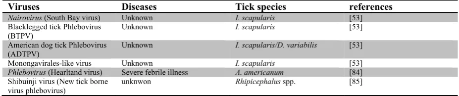
mononegavirus genera were identified among the three ticks assessed [53]. New viruses recently identified in ticks by NGS are listed in Table 2.
By having sight of the entire tick microbial community, we can identify that pathogens are intimately associated to the vast community of micro-organisms (including other pathogens) and that by elucidating their influence tick biology, pathogen persistence, transmission, and virulence justifies the need to shift from the study of isolated
[54] (See figure 1). Taking into account the multifactorial pathobiome requires comprehensive knowledge of the microbial community comprising the pathobiome, the network of interactions between microbes and the biological relevance of these interactions.
Deciphering microbial interactions within the tick ecosystem
pathways [63, 64], in computer science: peer to peer networks [65] or in social science: scientific collaboration [66]). They also offer an attractive representation of assessing dynamics of multiple pathogen relationships. They provide indices of association such as connectance [67], nestedness [68] or betweeness [69]. However, up to date, statistical tests regarding the networks parameters have rarely been used, but developments in this field are promising.
PATHOBIOME APPROACH FOR SURVEILLANCE, DIAGNOSIS AND PREVENTION OF TICK BORNE DISEASES
Surveillance and Diagnosis. Considering the vast number of potential tick borne pathogens that can result in disease, either alone or in association, there is an urgent need to develop methods that are capable to accommodate this diversity, but also provide insights into the biology of borne pathogens. For instance, many tick-borne pathogens colonize blood (residing within either intra- or extracellular niches) of vertebrate hosts. Thus it makes sense to detect the presence of their DNA in the blood of infected human patients of animals. However, blood infection does not occur for all tick-borne pathogens. A notable exception is the Lyme spirochaete that does not stably infect blood of human hosts, therefore detection of DNA in the blood of patient bitten by ticks is unhelpful necessitating use of more specific samples (such as skin biopsies) or use of serological tests even though their specificity and
Conclusion
The tick pathobiome vision, thanks to powerful molecular and technological
Future perspective
EXECUTIVE SUMMARY
Current Knowledge on Tick-borne pathogens:
An increasing number of “new” species, strains or genetic variants of
microorganisms are being detected in ticks, resulting in an ever-increasing list of potential pathogens.
This increasing recognition of pathogen diversity is raising complex questions regarding ecological cycles of pathogen, polymicrobial cross-talk, diagnosis and intervention opportunities.
The new vision:
Next generation technology shed new lights on bacterial communities associated with ticks.
The majority of DNA/RNA sequences carried by ticks belong to unknown microorganisms.
Pathogens are intimately associated with the tick microbial community. This justifies the need to shift from the study of isolated pathogens to a more
integrated pathobiome approach.
Future research directions in term of surveillance, diagnosis and prevention of tick borne diseases:
New tools enabling high-throughput monitoring of tick-borne pathogens are an urgent priority
Given the vast number of pathogens that could be transmitted by the same tick species, deployment of tick vaccines would be a smart and
Acknowledgment:
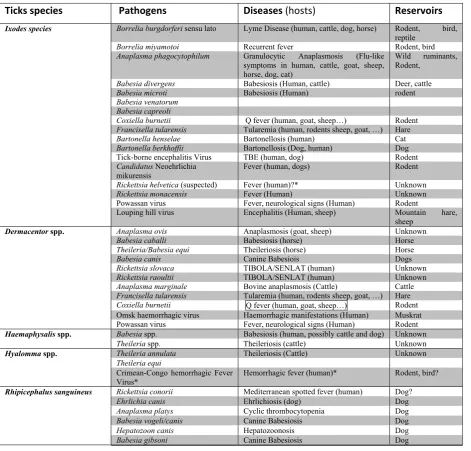
Table 1: The predominant tick species present in North hemisphere, the pathogens they transmit, associated diseases, animal hosts as well as animal reservoirs of the corresponding pathogens.
Ticks species Pathogens Diseases (hosts) Reservoirs
Ixodes species Borrelia burgdorferi sensu lato Lyme Disease (human, cattle, dog, horse) Rodent, bird,
reptile
Borrelia miyamotoi Recurrent fever Rodent, bird
Anaplasma phagocytophilum Granulocytic Anaplasmosis (Flu-like symptoms in human, cattle, goat, sheep, horse, dog, cat)
Wild ruminants, Rodent,
Babesia divergens Babesiosis (Human, cattle) Deer, cattle
Babesia microti Babesiosis (Human) rodent
Babesia venatorum Babesia capreoli
Coxiella burnetii Q fever (human, goat, sheep…) Rodent
Francisella tularensis Tularemia (human, rodents sheep, goat, …) Hare
Bartonella henselae Bartonellosis (human) Cat
Bartonella berkhoffii Bartonellosis (Dog, human) Dog
Tick-borne encephalitis Virus TBE (human, dog) Rodent Candidatus Neoehrlichia
mikurensis Fever (human, dogs) Rodent
Rickettsia helvetica (suspected) Fever (human)?* Unknown
Rickettsia monacensis Fever (Human) Unknown
Powassan virus Fever, neurological signs (Human) Rodent
Louping hill virus Encephalitis (Human, sheep) Mountain hare, sheep
Dermacentor spp. Anaplasma ovis Anaplasmosis (goat, sheep) Unknown
Babesia caballi Babesiosis (horse) Horse
Theileria/Babesia equi Theileriosis (horse) Horse
Babesia canis Canine Babesiois Dogs
Rickettsia slovaca TIBOLA/SENLAT (human) Unknown
Rickettsia raoultii TIBOLA/SENLAT (human) Unknown
Anaplasma marginale Bovine anaplasmosis (Cattle) Cattle
Francisella tularensis Tularemia (human, rodents sheep, goat, …) Hare
Coxiella burnetii Q fever (human, goat, sheep…) Rodent
Omsk haemorrhagic virus Haemorrhagic manifestations (Human) Muskrat Powassan virus Fever, neurological signs (Human) Rodent
Haemaphysalis spp. Babesia spp. Babesiosis (human, possibly cattle and dog) Unknown
Theileria spp. Theileriosis (cattle) Unknown
Hyalomma spp. Theileria annulata Theileriosis (Cattle) Unknown
Theileria equi
Crimean-Congo hemorrhagic Fever
Virus* Hemorrhagic fever (human)* Rodent, bird?
Rhipicephalus sanguineus Rickettsia conorii Mediterranean spotted fever (human) Dog?
Ehrlichia canis Ehrlichiosis (dog) Dog
Anaplasma platys Cyclic thrombocytopenia Dog
Babesia vogeli/canis Canine Babesiosis Dog
Hepatozoon canis Hepatozoonosis Dog
Table 2: New viruses recently identified in ticks by NGS.
Viruses Diseases Tick species references
Nairovirus (South Bay virus) Unknown I. scapularis [53]
Blacklegged tick Phlebovirus
(BTPV) Unknown I. scapularis [53]
American dog tick Phlebovirus
(ADTPV) Unknown I. scapularis/D. variabilis [53]
Monongavirales-like virus Unknown I. scapularis [53]
Phlebovirus (Hearltand virus) Severe febrile illness A. americanum [84]
Shibuinji virus (New tick borne
References
Papers of special note have been highlighted as: Of interest
1. Rizzoli A, Hauffe H, Carpi G, Vourc HG, Neteler M, Rosa R: Lyme borreliosis in Europe.EuroSurveill 16(27), (2011).
2. Platonov AE, Karan LS, Kolyasnikova NM etal.: Humans infected with relapsing fever spirochete Borrelia miyamotoi, Russia.EmergInfectDis 17(10), 1816‐1823 (2011).
3. Chowdri HR, Gugliotta JL, Berardi VP etal.: Borrelia miyamotoi infection presenting as human granulocytic anaplasmosis: a case report.AnnInternMed
159(1), 21‐27 (2013).
4. Krause PJ, Narasimhan S, Wormser GP etal.: Human Borrelia miyamotoi infection in the United States.TheNewEnglandjournalofmedicine 368(3), 291‐293
(2013).
5. Hovius JW, De Wever B, Sohne M etal.: A case of meningoencephalitis by the relapsing fever spirochaete Borrelia miyamotoi in Europe.Lancet 382(9892), 658 (2013).
firstcaseof B.miyamotoiinEurope
6. Heyman P, Cochez C, Hofhuis A etal.: A clear and present danger: tick‐borne diseases in Europe.ExpertRevAntiInfectTher 8(1), 33‐50 (2010).
7. Welinder‐Olsson C, Kjellin E, Vaht K, Jacobsson S, Wenneras C: First case of human "Candidatus Neoehrlichia mikurensis" infection in a febrile patient with chronic lymphocytic leukemia.JClinMicrobiol 48(5), 1956‐1959 (2010).
8. Von Loewenich FD, Geissdorfer W, Disque C etal.: Detection of "Candidatus Neoehrlichia mikurensis" in two patients with severe febrile illnesses: evidence for a European sequence variant.JClinMicrobiol 48(7), 2630‐2635 (2010). 9. Parola P, Raoult D: Tick‐borne bacterial diseases emerging in Europe.Clin
MicrobiolInfect 7(2), 80‐83 (2001).
10. Parola P, Raoult D: Ticks and tickborne bacterial diseases in humans: an emerging infectious threat.ClinInfectDis 32(6), 897‐928 (2001).
11. Duron O, Jourdain E, Mccoy KD: Diversity and global distribution of the Coxiella intracellular bacterium in seabird ticks.TicksTickBorneDis 5(5), 557‐563 (2014). 12. Angelakis E, Raoult D: Q Fever.VetMicrobiol 140(3‐4), 297‐309 (2010).
13. Hildebrandt A, Hunfeld KP: [Human babesiosis ‐ a rare but potentially dangerous zoonosis].DtschMedWochenschr 139(18), 957‐962 (2014).
14. Hildebrandt A, Hunfeld KP, Baier M etal.: First confirmed autochthonous case of human Babesia microti infection in Europe.EurJClinMicrobiolInfectDis 26(8), 595‐601 (2007).
15. Charrel RN, Attoui H, Butenko AM etal.: Tick‐borne virus diseases of human interest in Europe.ClinMicrobiolInfect 10(12), 1040‐1055 (2004).
16. Hubalek Z, Rudolf I: Tick‐borne viruses in Europe.Parasitologyresearch 111(1), 9‐36 (2012).
18. Liu XY, Bonnet SI: Hard tick factors implicated in pathogen transmission.PLoS
NeglTropDis 8(1), e2566 (2014).
19. Burgdorfer W: Discovery of the Lyme disease spirochete and its relation to tick vectors.YaleJBiolMed 57(4), 515‐520 (1984).
20. Massung RF, Mather TN, Priestley RA, Levin ML: Transmission efficiency of the AP‐variant 1 strain of Anaplasma phagocytophila.AnnNYAcadSci 990, 75‐79 (2003).
21. Bonnet S, Brisseau N, Hermouet A, Jouglin M, Chauvin A: Experimental in vitro transmission of Babesia sp. (EU1) by Ixodes ricinus.VetRes 40(3), 21 (2009). 22. Cotte V, Bonnet S, Le Rhun D etal.: Transmission of Bartonella henselae by Ixodes
ricinus.EmergInfectDis 14(7), 1074‐1080 (2008).
23. Reis C, Cote M, Le Rhun D etal.: Vector competence of the tick Ixodes ricinus for transmission of Bartonella birtlesii.PLoSNeglTropDis 5(5), e1186 (2011). 24. Alekseev AN, Chunikhin SP: [The experimental transmission of the tick‐borne
encephalitis virus by ixodid ticks (the mechanisms, time periods, species and sex differences)].Parazitologiia 24(3), 177‐185 (1990).
25. Alekseev AN, Chunikhin SP: [The exchange of the tick‐borne encephalitis virus between ixodid ticks feeding jointly on animals with a subthreshold level of viremia].Meditsinskaiaparazitologiiaiparazitarnyebolezni (2), 48‐50 (1990). 26. Perronne C: Lyme and associated tick‐borne diseases: global challenges in the
context of a public health threat.Frontiersincellularandinfectionmicrobiology 4, 74 (2014).
27. Kaiser R: Tick‐borne encephalitis: Clinical findings and prognosis in adults.Wien MedWochenschr 162(11‐12), 239‐243 (2012).
28. Gugliotta JL, Goethert HK, Berardi VP, Telford SR, 3rd: Meningoencephalitis from Borrelia miyamotoi in an immunocompromised patient.NEnglJMed 368(3), 240‐245 (2013).
29. Vayssier‐Taussat M, Moutailler S, Michelet L etal.: Next generation sequencing uncovers unexpected bacterial pathogens in ticks in western Europe.PloSone
8(11), e81439 (2013).
30. Cosson Jf ML, Chotte J, Le Naour E, Cote M, Devillers E, Poulle Ml, Huet D, Galan M, Geller J, Moutailler S and Vayssier‐Taussat M.: Genetic characterization of the Human Relapsing Fever Spirochete
Borrelia miyamotoi in vectors and animal reservoirs of Lyme disease spirochetes in France. Vectors&Parasites, (In press).
31. Horowitz HW, Aguero‐Rosenfeld ME, Holmgren D etal.: Lyme disease and human granulocytic anaplasmosis coinfection: impact of case definition on coinfection rates and illness severity.Clinicalinfectiousdiseases:anofficialpublicationofthe InfectiousDiseasesSocietyofAmerica 56(1), 93‐99 (2013).
illustratesthepossibleco‐infectionofhumanbytick‐bornepathogensand
theirimpactonillnessseverity
32. Tijsse‐Klasen E, Sprong H, Pandak N: Co‐infection of Borrelia burgdorferi sensu lato and Rickettsia species in ticks and in an erythema migrans patient.Parasit Vectors 6, 347 (2013).
34. Reis C, Cote M, Paul RE, Bonnet S: Questing ticks in suburban forest are infected by at least six tick‐borne pathogens.VectorBorneZoonoticDis 11(7), 907‐916 (2011).
35. Carpi G, Cagnacci F, Wittekindt NE etal.: Metagenomic profile of the bacterial communities associated with Ixodes ricinus ticks.PloSone 6(10), e25604 (2011). 36. Nakao R, Abe T, Nijhof AM etal.: A novel approach, based on BLSOMs (Batch
Learning Self‐Organizing Maps), to the microbiome analysis of ticks.ISMEJ 7(5), 1003‐1015 (2013).
37. Hawlena H, Rynkiewicz E, Toh E etal.: The arthropod, but not the vertebrate host or its environment, dictates bacterial community composition of fleas and ticks.
ISMEJ 7(1), 221‐223 (2013).
38. Williams‐Newkirk AJ, Rowe LA, Mixson‐Hayden TR, Dasch GA: Characterization of the bacterial communities of life stages of free living lone star ticks
(Amblyomma americanum).PLoSOne 9(7), e102130 (2014).
39. Gillespie JJ, Joardar V, Williams KP etal.: A Rickettsia genome overrun by mobile genetic elements provides insight into the acquisition of genes characteristic of an obligate intracellular lifestyle.JBacteriol 194(2), 376‐394 (2012).
40. Perlman SJ, Hunter MS, Zchori‐Fein E: The emerging diversity of Rickettsia.Proc
BiolSci 273(1598), 2097‐2106 (2006).
41. Duron O, Noel V, Mccoy KD etal.: The Recent Evolution of a Maternally‐Inherited Endosymbiont of Ticks Led to the Emergence of the Q Fever Pathogen, Coxiella burnetii.PLoSPathog 11(5), e1004892 (2015).
42. Almeida AP, Marcili A, Leite RC etal.: Coxiella symbiont in the tick Ornithodoros rostratus (Acari: Argasidae).TicksTickBorneDis 3(4), 203‐206 (2012).
43. Machado‐Ferreira E, Dietrich G, Hojgaard A etal.: Coxiella symbionts in the Cayenne tick Amblyomma cajennense.MicrobEcol 62(1), 134‐142 (2011). 44. Kreizinger Z, Hornok S, Dan A etal.: Prevalence of Francisella tularensis and
Francisella‐like endosymbionts in the tick population of Hungary and the genetic variability of Francisella‐like agents.VectorBorneZoonoticDis 13(3), 160‐163 (2013).
45. Rudolf I, Mendel J, Sikutova S etal.: 16S rRNA gene‐based identification of cultured bacterial flora from host‐seeking Ixodes ricinus, Dermacentor
reticulatus and Haemaphysalis concinna ticks, vectors of vertebrate pathogens. FoliaMicrobiol(Praha) 54(5), 419‐428 (2009).
firstidentificationoftickmicroflorausinga16Sapproach
46. Werren JH, Baldo L, Clark ME: Wolbachia: master manipulators of invertebrate biology.NatRevMicrobiol 6(10), 741‐751 (2008).
47. Ahantarig A, Trinachartvanit W, Baimai V, Grubhoffer L: Hard ticks and their bacterial endosymbionts (or would be pathogens).FoliaMicrobiol(Praha) 58(5), 419‐428 (2013).
48. Plantard O, Bouju‐Albert A, Malard MA, Hermouet A, Capron G, Verheyden H: Detection of Wolbachia in the tick Ixodes ricinus is due to the presence of the hymenoptera endoparasitoid Ixodiphagus hookeri.PLoSOne 7(1), e30692 (2012). 49. Lewis D: The detection of rickettsia‐like microorganisms within the ovaries of
female Ixodes ricinus ticks.ZParasitenkd 59(3), 295‐298 (1979).
51. Epis S, Sassera D, Beninati T etal.: Midichloria mitochondrii is widespread in hard ticks (Ixodidae) and resides in the mitochondria of phylogenetically diverse species.Parasitology 135(4), 485‐494 (2008).
52. Bazzocchi C, Mariconti M, Sassera D etal.: Molecular and serological evidence for the circulation of the tick symbiont Midichloria (Rickettsiales: Midichloriaceae) in different mammalian species.ParasitVectors 6, 350 (2013).
53. Tokarz R, Williams SH, Sameroff S, Sanchez Leon M, Jain K, Lipkin WI: Virome analysis of Amblyomma americanum, Dermacentor variabilis, and Ixodes scapularis ticks reveals novel highly divergent vertebrate and invertebrate viruses.Journalofvirology 88(19), 11480‐11492 (2014).
54. Vayssier‐Taussat M, Albina E, Citti C etal.: Shifting the paradigm from pathogens to pathobiome: new concepts in the light of meta‐omics.Frontiersincellularand infectionmicrobiology 4, 29 (2014).
55. Rigaud T, Perrot‐Minnot MJ, Brown MJ: Parasite and host assemblages:
embracing the reality will improve our knowledge of parasite transmission and virulence.JournaloftheRoyalStatisticalSociety:SeriesB 277(1701), 3693‐3702 (2010).
56. Bordes F, Morand S: The impact of multiple infections on wild animal hosts: a review.Infectionecology&epidemiology 1, (2011).
57. Telfer S, Lambin X, Birtles R etal.: Species interactions in a parasite community drive infection risk in a wildlife population.Science 330(6001), 243‐246 (2010). 58. Gauch HG: Multivariate analysis in community ecology. Cambridge University
Press, (1982).
59. Tollenaere C, Bryja J, Galan M etal.: Multiple parasites mediate balancing selection at two MHC class II genes in the fossorial water vole: insights from multivariate analyses and population genetics.Journalofevolutionarybiology
21(5), 1307‐1320 (2008).
60. Salvador AR, Guivier E, Xuereb A etal.: Concomitant influence of helminth
infection and landscape on the distribution of Puumala hantavirus in its reservoir, Myodes glareolus.BMCmicrobiology 11(1), 30 (2011).
61. Vaumourin E, Vourc'h G, Telfer S etal.: To be or not to be associated: power study of four statistical modeling approaches to identify parasite associations in cross‐ sectional studies.Frontiersincellularandinfectionmicrobiology 4, 62 (2014). 62. Bascompte J: Networks in ecology.BasicandAppliedEcology 8(6), 485‐490
(2007).
63. Ravasz E, Somera AL, Mongru DA, Oltvai ZN, Barabasi AL: Hierarchical
organization of modularity in metabolic networks.Science 297(5586), 1551‐1555 (2002).
64. Qin J, Li Y, Cai Z etal.: A metagenome‐wide association study of gut microbiota in type 2 diabetes.Nature 490(7418), 55‐60 (2012).
65. Fox G: Peer to Peer Networks.ComputinginScience&Engineering 3(3), 75‐77 (2001).
66. Newman ME: Coauthorship networks and patterns of scientific collaboration. ProceedingsoftheNationalAcademyofSciences 101 Suppl 1, 5200‐5205 (2004). 67. Yodzis P: The connectance of real ecosystems.Nature 284(5756), 544‐545
(1980).
68. Bascompte J, Jordano P, Melian CJ, Olesen JM: The nested assembly of plant‐ animal mutualistic networks.ProceedingsoftheNationalAcademyofSciences
69. Freeman LC: A Set of Measures of Centrality Based on Betweenness.Sociometry
40(1), 35‐41 (1977).
70. Childs JE, Paddock CD: Passive surveillance as an instrument to identify risk factors for fatal Rocky Mountain spotted fever: is there more to learn? The Americanjournaloftropicalmedicineandhygiene 66(5), 450‐457 (2002).
71. Klyachko O, Stein BD, Grindle N, Clay K, Fuqua C: Localization and visualization of a coxiella‐type symbiont within the lone star tick, Amblyomma americanum. Appliedandenvironmentalmicrobiology 73(20), 6584‐6594 (2007).
72. Narasimhan S, Rajeevan N, Liu L etal.: Gut microbiota of the tick vector Ixodes scapularis modulate colonization of the Lyme disease spirochete.Cellhost& microbe 15(1), 58‐71 (2014).
73. Cirimotich CM, Dong Y, Clayton AM etal.: Natural microbe‐mediated
refractoriness to Plasmodium infection in Anopheles gambiae.Science 332(6031), 855‐858 (2011).
74. Michelet L, Delannoy S, Devillers E etal.: High‐throughput screening of tick‐borne pathogens in Europe.Frontiersincellularandinfectionmicrobiology 4, 103
(2014).
75. Liu XY, Bonnet SI: Hard tick factors implicated in pathogen transmission.PLoS
NeglTropDis 8(1), e2566 (2014).
76. De La Fuente J, Merino O: Vaccinomics, the new road to tick vaccines.Vaccine
31(50), 5923‐5929 (2013).
77. De La Fuente J, Almazan C, Canales M, Perez De La Lastra JM, Kocan KM,
Willadsen P: A ten‐year review of commercial vaccine performance for control of tick infestations on cattle.Animalhealthresearchreviews/ConferenceofResearch WorkersinAnimalDiseases 8(1), 23‐28 (2007).
78. Decrem Y, Mariller M, Lahaye K etal.: The impact of gene knock‐down and vaccination against salivary metalloproteases on blood feeding and egg laying by Ixodes ricinus.Internationaljournalforparasitology 38(5), 549‐560 (2008). 79. Liu XY, De La Fuente J, Cote M etal.: IrSPI, a tick serine protease inhibitor
involved in tick feeding and Bartonella henselae infection.PLoSNeglTropDis
8(7), e2993 (2014).
80. Dai J, Wang P, Adusumilli S etal.: Antibodies against a tick protein, Salp15, protect mice from the Lyme disease agent.CellHostMicrobe 6(5), 482‐492 (2009).
81. Merino O, Antunes S, Mosqueda J etal.: Vaccination with proteins involved in tick‐pathogen interactions reduces vector infestations and pathogen infection. Vaccine 31(49), 5889‐5896 (2013).
82. Labuda M, Trimnell AR, Lickova M etal.: An antivector vaccine protects against a lethal vector‐borne pathogen.PLoSPathog 2(4), e27 (2006).
83. De La Fuente J, Almazan C, Blouin EF, Naranjo V, Kocan KM: Reduction of tick infections with Anaplasma marginale and A. phagocytophilum by targeting the tick protective antigen subolesin.ParasitolRes 100(1), 85‐91 (2006).
84. Mcmullan LK, Folk SM, Kelly AJ etal.: A new phlebovirus associated with severe febrile illness in Missouri.TheNewEnglandjournalofmedicine 367(9), 834‐841 (2012).
85. Matsuno K, Weisend C, Kajihara M etal.: Comprehensive molecular detection of tick‐borne phleboviruses leads to the retrospective identification of