
pyogenes
Infections in North and South India
Axana Haggar,aAndreas Nerlich,bRajesh Kumar,cVinod J. Abraham,dKootallur N. Brahmadathan,ePallab Ray,cVanita Dhanda,c John Melbin Jose Joshua,eNarinder Mehra,fRene Bergmann,bG. Singh Chhatwal,band Anna Norrby-Teglunda
Center for Infectious Medicine, Karolinska Institutet, Stockholm, Swedena
; Helmholtz Centre for Infection Research, Braunschweig, Germanyb
; School of Public Health, Postgraduate Institute of Medical Education and Research, Chandigarh, Indiac
; Department of Community Health, Christian Medical College, Vellore, Indiad
; Department of Microbiology Christian Medical College, Vellore, Indiae
; and Department of Transplant Immunology and Immunogenetics, All Indian Institute of Medical Sciences, New Delhi, Indiaf
The lack of epidemiologic data on invasiveStreptococcus pyogenes infections in many developing countries is concerning, asS. pyogenesinfections are commonly endemic in these areas. Here we present the results of the first prospective surveillance study of invasiveStreptococcus pyogenesinfections in India. Fifty-four patients with invasiveS. pyogenesinfections were prospectively enrolled at two study sites, one in the north and one in the south of India. Sterile-site isolates were collected, and clinical infor-mation was documented using a standardized questionnaire. Available acute-phase sera were tested for their ability to inhibit superantigens produced by the patient’s own isolate using a cell-based neutralizing assay. The most common clinical presenta-tions were bacteremia without focus (30%), pneumonia (28%), and cellulitis (17%). Only two cases of streptococcal toxic shock syndrome and no cases of necrotizing fasciitis were identified. Characterization of the isolates revealed great heterogeneity, with 32 differentemmsubtypes and 29 different superantigen gene profiles being represented among the 49 sterile-site isolates. Anal-yses of acute-phase sera showed that only 20% of the cases in the north cohort had superantigen-neutralizing activity in their sera, whereas 50% of the cases from the south site had neutralizing activity. The results demonstrate that there are important differences in both clinical presentation and strain characteristics between invasiveS. pyogenesinfections in India and invasive S. pyogenesinfections in Western countries. The findings underscore the importance of epidemiologic studies on streptococcal infections in India and have direct implications for current vaccine developments.
S
treptococcus pyogenesis a significant human pathogen capable of causing a wide spectrum of diseases ranging from uncom-plicated infections of the throat and skin to severe life-threatening diseases and poststreptococcal sequelae. Streptococcal pharyngitis is one of the most common childhood diseases worldwide, ac-counting for several millions of cases each year. Inadequatetreat-ment ofS. pyogenesinfections, predominantly throat infections,
can result in the serious postinfectious sequela acute rheumatic fever, which may lead to rheumatic heart disease. The burden of poststreptococcal sequelae is great in developing countries but relatively rare in developed countries, although isolated outbreaks
have been reported (5, 12). Furthermore,S. pyogenescauses
inva-sive infections, including the two most severe invainva-sive manifesta-tions, streptococcal toxic shock syndrome (STSS) and necrotizing fasciitis, associated with high morbidity and mortality.
S. pyogenesinvasive disease in North America and Europe has been an area of intense research since its reemergence in the late 1980s (1, 9). In contrast, these infections have not received much attention in developing countries. In an attempt to estimate the
global burden ofS. pyogenesinfections, Carapetis et al. (5)
re-viewed available databases and estimated that more than 18
mil-lion people currently suffer from a seriousS. pyogenesdisease, with
about 2 million new cases occurring each year and with an annual mortality of more than 100,000. Added to this are 111 million
cases of streptococcal pyoderma and 616 million new cases ofS.
pyogenespharyngitis each year. It was noteworthy that the vast majority of cases were in resource-limited countries. This report especially highlighted the fact that epidemiologic data from less developed countries are scarce, thus emphasizing the need for studies in these regions (5).
In India, the disease burden of streptococcal infections is con-siderable (26) and the incidence of acute rheumatic fever and rheumatic heart disease ranges from 0.3 to 5.4 per 1,000 children
(24).Streptococcus pyogenespharyngitis has a high prevalence in
north India (14), whereas pyoderma is more frequent in south India (4). In light of invasive infection, this is a completely ne-glected field in India, and the only data available in the literature are from one retrospective study of invasive beta-hemolytic
strep-tococcal infections (19). To advance our understanding ofS.
pyo-genesinfections in India and to obtain epidemiologic data that could direct the design of an effective vaccine, a European Com-mission-funded project, ASSIST, was launched in 2007. The proj-ect included surveillance of acute rheumatic fever/rheumatic
heart disease as well as invasiveS. pyogenesinfections at study sites
in north and south India. Here we present the results of the first
prospective surveillance study of invasiveS. pyogenesinfections
conducted in India, with the results showing important differ-ences in relation to surveillance data from Western countries.
Received14 December 2011Returned for modification20 January 2012 Accepted16 February 2012
Published ahead of print22 February 2012
Address correspondence to Anna Norrby-Teglund, [email protected].
Supplemental material for this article may be found athttp://jcm.asm.org/.
Copyright © 2012, American Society for Microbiology. All Rights Reserved.
doi:10.1128/JCM.06697-11
on May 16, 2020 by guest
http://jcm.asm.org/
MATERIALS AND METHODS
Setting and study population.Surveillance of invasiveS. pyogenes infec-tions was established at the two ASSIST study sites, the Postgraduate In-stitute of Medical Education and Research (PGIMER), Chandigarh, in the north of India, and the Christian Medical College (CMC), Vellore, in the south of India. All consenting patients with invasiveS. pyogenesinfection (n⫽56) identified at the two sites during the study period (January 2007 to February 2010) were enrolled in the study. Invasive cases were defined as those with isolation ofS. pyogenesfrom normally sterile sites, and cases of STSS were defined according to published definition criteria (30). Iden-tification of cases was achieved by daily contact with the microbiology laboratory serving the hospital. Isolates were collected (n⫽49), and clin-ical information obtained from the patients’ medclin-ical records was docu-mented in a standardized questionnaire designed to harmonize with data collected in the recently published StrepEuro study of invasiveS. pyogenes infections in Europe (15). Acute-phase sera were obtained from 10 and 22 invasive cases from the north and south sites, respectively.
The ethics of the study were approved by the human research ethics committees at the respective hospitals in India, as well as in Stockholm, Sweden. Written informed consent was obtained from all enrolled pa-tients.
Strain characterization:emmsequencing and superantigen gene profile.Clinical isolates were confirmed to beS. pyogenesby use of con-ventional bacteriologic techniques. Genomic DNA was isolated from strains grown overnight in Todd-Hewitt medium plus yeast extract (THY) using zirconia beads in combination with a DNeasy kit (Qiagen, Hilden, Germany). Direct sequencing of the 5=end of theemmgene was done as detailed athttp://www.cdc.gov/ncidod/biotech/strep/M-Protein Gene_typing.htm, using the oligonucleotidesemm_fwd (5=-TATTCGCT TAGAAAATTAA-3=) and emm_rev (5=-GCAAGTTCTTCAGCTTGTT T-3=). Theemmtype was obtained as described athttp://www.cdc.gov /ncidod/biotech/strep/strepblast.htm by BLAST comparison to emm sequences in the database.
Superantigen genes were detected by PCR using the primers listed in
Table S1 in the supplemental material. As positive controls for the respec-tive genes, the sequenced strains MGAS315, MGAS6708, and MGAS8232 were used.
Proliferation and neutralization assays.Supernatants were prepared from overnight bacterial cultures in Todd-Hewitt broth supplemented with yeast extract (THY) and tested for mitogenic activity in proliferation assays using peripheral blood mononuclear cells (PBMCs) from healthy donors as detailed elsewhere (22).
Patient sera were tested for their ability to inhibit superantigens pro-duced by the patients’ own isolates by use of a cell-based neutralizing assay previously described in detail (10, 22, 29). In brief, PBMCs from healthy donors were stimulated with superantigen-containing bacterial culture supernatants (SUPs; 1:50 dilution) in the presence of 2.5% heat-inacti-vated patient serum (PS) supplemented with 2.5% fetal calf serum (FCS) or 5% FCS. After 72 h, [3H]thymidine uptake was determined. Phytohe-magglutinin-L (1g/ml; Sigma) was included as a control for nonspecific toxicity and PBMC quality. All samples were assayed in triplicate, and the data are presented as mean counts per minute of [3H]thymidine uptake⫾ standard deviation (SD). Serum neutralizing activity was calculated by the equation [1⫺(cpmPS⫹SUP⫺cpmPS⫹medium)/(cpmFCS ⫹SUP⫺ cpmFCS⫹medium)]⫻100. Significant neutralizing activity was defined as 50% inhibition of proliferative responses, as previously established (22). The analyses also included sera from 58 healthy controls (median age, 18 years; provided by PGIMER), which were tested for neutralizing activity against SUPs from two selected isolates (isolates 01 and 01-1400-08), both from the north India study site, as that was the region of the controls. Intravenous polyspecific immunoglobulin (IVIG; 2.5 mg/ml; Baxter) was tested for neutralizing activity against bacterial culture super-natants using medium supplemented with 5% FCS.
Statistical analyses.Data were analyzed using Prism software (version 5.01; Graphpad). The D’Agostino and Pearson omnibus normality test was used to assess the Gaussian distribution. Student’sttest or the Mann-Whitney U test was used to compare the two patient cohorts, and the2 test or Fisher’s exact test was used to compare the distribution of categor-ical data.
RESULTS
A total of 56 invasive cases were enrolled at the two Indian study sites, including 26 and 30 patients from the north and south sites,
respectively. Sterile-site isolates were obtained from blood (n⫽
41, 73%), pleural fluid (n ⫽4), synovial fluid (n ⫽3), tissue
aspirates (n ⫽ 3), peritoneal fluid (n ⫽2), bile (n ⫽1), and
vitreous fluid (n⫽2). Upon further analyses, two isolates were
reclassified as group G streptococcus, and consequently, the two cases were excluded from the study. The median age of all invasive
cases was 30 years (range, 1 day to 80 years), with 24% beingⱕ4
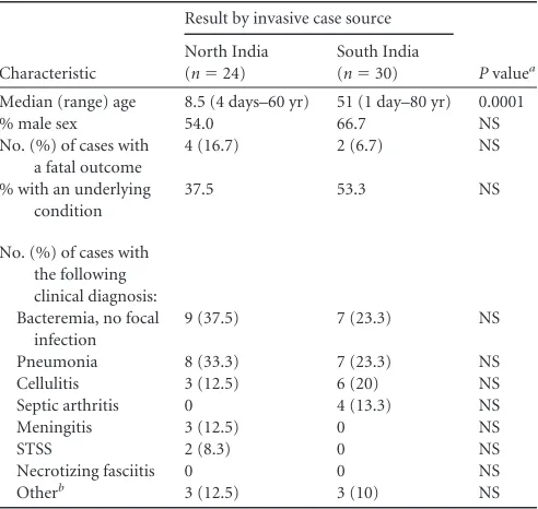
years old (Table 1 and Fig. 1). The two cohorts differed
signifi-TABLE 1Demographics and characteristics of invasive cases at the two Indian study sites
Characteristic
Result by invasive case source
Pvaluea
North India (n⫽24)
South India (n⫽30)
Median (range) age 8.5 (4 days–60 yr) 51 (1 day–80 yr) 0.0001
% male sex 54.0 66.7 NS
No. (%) of cases with a fatal outcome
4 (16.7) 2 (6.7) NS
% with an underlying condition
37.5 53.3 NS
No. (%) of cases with the following clinical diagnosis: Bacteremia, no focal
infection
9 (37.5) 7 (23.3) NS
Pneumonia 8 (33.3) 7 (23.3) NS
Cellulitis 3 (12.5) 6 (20) NS
Septic arthritis 0 4 (13.3) NS
Meningitis 3 (12.5) 0 NS
STSS 2 (8.3) 0 NS
Necrotizing fasciitis 0 0 NS
Otherb 3 (12.5) 3 (10) NS
a
NS, not significant; significant,P⬍0.05 using the Mann-Whitney U test or Fisher’s exact test.
b
Including empyema, endophthalmitis, puerperal sepsis, nephrotic syndrome, peritonitis, and follicular cholecystitis.
FIG 1Age distribution among invasiveS. pyogenescases enrolled at the north and south study sites in India.
on May 16, 2020 by guest
http://jcm.asm.org/
[image:2.585.41.287.86.319.2] [image:2.585.327.513.570.702.2]cantly with respect to age, as children and adolescents were more common among patients enrolled at the north site than the south site (Table 1 and Fig. 1).
Clinical characteristics of invasive cases.Forty-six percent of the invasive cases reported underlying conditions, including can-cer (13%), chronic liver disease (7.4%), malnourishment (7.4%), surgery (5.6%), and burns (5.6%). The most common clinical diagnoses were bacteremia with no identified focus (30%) and pneumonia (28%), followed by cellulitis (17%). Septic arthritis cases were found only in the south site patient cohort, whereas meningitis cases were exclusively represented in the north cohort (Table 1). No necrotizing fasciitis cases and only two cases of STSS were identified. The two STSS cases were both children who pre-sented with cellulitis and with meningitis and pneumonia. Nine patients were treated at the intensive care unit (16.7%). A fatal outcome was recorded in 6 of the 54 invasive cases, including 1 of the STSS cases, for a fatality rate of 11.1%. However, it should be noted that the study did not include a day 28 follow-up, and many patients declined hospitalization due to the associated cost; hence, the recorded fatalities are likely underestimated.
Microbiologic findings. From the 54 invasive S. pyogenes
cases, 49 isolates were retrieved and further characterized with
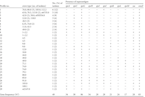
respect toemmtype and toxin gene profiling (Table 2). Theemm
sequencing revealed a great heterogeneity among the isolates, with
32 differentemmtypes being represented in the cohort. The most
common subtypes wereemm49.0 (n ⫽ 4),emm74.0 (n ⫽4),
emm80.0 (n⫽4),emm12.0 (n⫽3), andemm28.5 (n⫽3). The
fatal cases were caused by various subtypes, including the two
emm1-2.2 isolates and one isolate each ofemm28.5,emm74.0, and
emm80.0. The type distribution differed between isolates from the
north and south sites, with all isolates of theemm12.0 (n⫽3),
emm28.5 (n⫽3), andemm49.0 (n⫽4) subtypes being identified
in the south cohort, whereasemm74.0 (n⫽3),emm80.0 (n⫽3),
andemm1-2.2 (n⫽2) were predominantly in the north isolate
cohort.
Exotoxin gene profiling of the 49 invasive isolates identified 29 distinct profiles with unique combinations of superantigen genes
evenly distributed among the 32 differentemmsubtypes (Table 2).
The 29 profiles contained between 1 and 9 superantigen genes per profile, with the greatest variation occurring in genes encoding prophage. There were five profiles that harbored only one or two
exotoxin genes. Profile 5 harbored onlysmeZand was exclusively
represented byemm28.0 strains.speGandsmeZwere the most
common superantigen genes, being present in 86% and 83% of the profiles, respectively.
Superantigen-containing culture supernatants from all isolates were potent triggers of proliferative responses, and there was no statistical difference between the cohorts (Fig. 2). There was no
[image:3.585.41.550.79.421.2]relation between degree of proliferative response and theemm
TABLE 2Superantigen gene profiles related toemmsubtypes of invasiveS. pyogenesisolates
Profile no. emmtype (no. of isolates)
No. (%) of isolates
Presence of superantigen
speA speC speG speH speI speJ speK speL speM ssa smeZ
1 76.0, 80.0 (3), 105.0, 112.2 6 (12) ⫺ ⫺ ⫹ ⫺ ⫺ ⫹ ⫺ ⫺ ⫺ ⫺ ⫹
2 63.0, 78.3, 113.0 (2), st6735.0 5 (10) ⫺ ⫺ ⫹ ⫺ ⫺ ⫺ ⫺ ⫺ ⫺ ⫺ ⫹
3 42.0 (2), 58.8, stNS554.0 4 (8) ⫺ ⫹ ⫹ ⫺ ⫺ ⫺ ⫺ ⫺ ⫺ ⫺ ⫹
4 12.0 (2), 118.0 3 (6) ⫺ ⫹ ⫹ ⫺ ⫺ ⫺ ⫺ ⫺ ⫺ ⫹ ⫹
5 28.5 (3) 3 (6) ⫺ ⫺ ⫺ ⫺ ⫺ ⫺ ⫺ ⫺ ⫺ ⫺ ⫹
6 6.35, 74.0 (2) 3 (6) ⫹ ⫹ ⫹ ⫺ ⫺ ⫺ ⫺ ⫺ ⫺ ⫺ ⫹
7 11.0, 43.3 2 (4) ⫺ ⫹ ⫹ ⫺ ⫺ ⫺ ⫺ ⫹ ⫺ ⫺ ⫹
8 49.0 (2) 2 (4) ⫹ ⫺ ⫹ ⫹ ⫹ ⫺ ⫺ ⫺ ⫺ ⫺ ⫺
9 1–2.2 1 (2) ⫹ ⫺ ⫹ ⫹ ⫹ ⫹ ⫺ ⫹ ⫹ ⫹ ⫹
10 1–2.2 1 (2) ⫹ ⫺ ⫹ ⫺ ⫺ ⫹ ⫺ ⫺ ⫺ ⫺ ⫹
11 4.5 1 (2) ⫺ ⫺ ⫺ ⫺ ⫺ ⫺ ⫹ ⫺ ⫺ ⫹ ⫹
12 4.5 1 (2) ⫺ ⫺ ⫺ ⫺ ⫺ ⫺ ⫺ ⫹ ⫹ ⫹ ⫹
13 9.0 1 (2) ⫺ ⫺ ⫹ ⫺ ⫺ ⫺ ⫹ ⫺ ⫺ ⫺ ⫹
14 9.0 1 (2) ⫺ ⫹ ⫹ ⫺ ⫺ ⫺ ⫹ ⫺ ⫺ ⫺ ⫹
15 12.0 1 (2) ⫺ ⫹ ⫹ ⫹ ⫹ ⫺ ⫺ ⫺ ⫺ ⫹ ⫹
16 18.8 1 (2) ⫹ ⫺ ⫹ ⫺ ⫺ ⫺ ⫺ ⫺ ⫺ ⫺ ⫹
17 44.0 1 (2) ⫺ ⫹ ⫹ ⫹ ⫺ ⫹ ⫺ ⫺ ⫺ ⫺ ⫺
18 49.0 1 (2) ⫺ ⫺ ⫹ ⫺ ⫺ ⫺ ⫺ ⫹ ⫺ ⫺ ⫺
19 49.0 1 (2) ⫺ ⫺ ⫹ ⫹ ⫹ ⫺ ⫺ ⫹ ⫹ ⫺ ⫺
20 71.1 1 (2) ⫹ ⫺ ⫹ ⫺ ⫺ ⫹ ⫺ ⫺ ⫹ ⫺ ⫹
21 74.0 1 (2) ⫹ ⫹ ⫹ ⫹ ⫹ ⫺ ⫺ ⫹ ⫹ ⫹ ⫹
22 74.0 1 (2) ⫹ ⫹ ⫹ ⫹ ⫹ ⫺ ⫺ ⫹ ⫺ ⫺ ⫹
23 79.1 1 (2) ⫺ ⫹ ⫹ ⫺ ⫺ ⫺ ⫺ ⫺ ⫺ ⫺ ⫺
24 80.0 1 (2) ⫺ ⫺ ⫹ ⫺ ⫺ ⫹ ⫹ ⫺ ⫺ ⫺ ⫹
25 85.0 1 (2) ⫺ ⫺ ⫹ ⫹ ⫹ ⫺ ⫺ ⫺ ⫺ ⫺ ⫹
26 91.0 1 (2) ⫺ ⫹ ⫹ ⫹ ⫹ ⫹ ⫺ ⫺ ⫺ ⫺ ⫹
27 103.0 1 (2) ⫺ ⫺ ⫹ ⫹ ⫺ ⫹ ⫹ ⫺ ⫺ ⫹ ⫹
28 109.1 1 (2) ⫹ ⫺ ⫺ ⫺ ⫺ ⫺ ⫺ ⫺ ⫺ ⫺ ⫹
29 st2147.0 1 (2) ⫹ ⫺ ⫹ ⫺ ⫺ ⫺ ⫹ ⫺ ⫺ ⫹ ⫹
Gene frequency (%a) 49 34 38 86 34 28 28 21 24 17 28 83
a
Percentage related to number of profiles.
on May 16, 2020 by guest
http://jcm.asm.org/
subtype, number of superantigen genes harbored, or superantigen profile.
Superantigen-neutralizing activity in acute-phase sera.As a lack of protective humoral immunity against superantigens has
been shown to be a risk factor for invasiveS. pyogenesinfections in
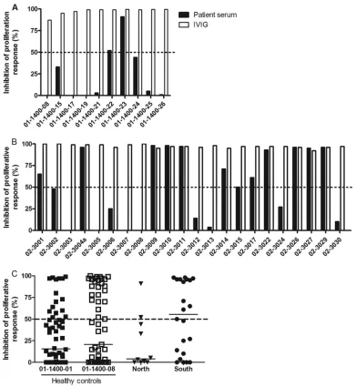
Europe and in Canada (2, 18, 22), it was of interest to assess pro-tective antibody levels among Indian patients. Available acute-phase sera were tested for their ability to inhibit superantigens produced by the patient’s own isolate using a cell-based neutral-izing assay previously employed in epidemiologic studies (2, 18, 22, 29). Only 20% of the cases in the north cohort had superanti-gen-neutralizing activity in their sera (Fig. 3A), whereas 50% of the cases from the south cohort showed neutralizing activity (Fig. 3B). Equally low neutralizing activities were seen in the healthy controls, with 26% and 40% showing neutralizing activity against isolates 01-1400-01 and 01-1400-08, respectively (Fig. 3C). IVIG was included as a source of neutralizing superantigen antibodies
that completely inhibits the superantigenic activity of invasiveS.
pyogenesisolates in Europe and in Canada (7, 13, 21). Similarly, superantigens produced by Indian invasive isolates were com-pletely neutralized by IVIG (Fig. 3).
DISCUSSION
Here we present the results of the first prospective study of inva-siveS. pyogenesinfections in India, including infections at two study sites, one in the north and one in the south. At both sites, the most common clinical presentations were bacteremia without
fo-cus, pneumonia, and cellulitis. Three meningitis cases, all from the north site, were reported, consistent with children being more prevalent in this patient cohort than that at the south site. In contrast, four septic arthritis cases were found in the south patient cohort, whereas none was found in the north. It was noteworthy that only two STSS cases and no cases of necrotizing fasciitis were identified, numbers which are considerably less than those that have been reported from Western countries (8, 15). Equally low numbers were reported in a retrospective study of beta-hemolytic streptococcal infections in India, in which 3 cases of STSS were found among 225 invasive cases and there was no mention of necrotizing fasciitis cases (19). In contrast, results from the 2003-2004 European surveillance covering 11 countries reported 13% and 8% of cases with STSS and necrotizing fasciitis, respectively (15), and similar figures have been observed in Canada (8, 11) and the United States (23). Two recent prospective studies from de-veloping countries, including one from New Caledonia, which reported 43% of cases with necrotizing fasciitis but only low fre-quencies of STSS (3%) (16), and one from Fiji, where 5% and 7% of cases with STSS and necrotizing fasciitis, respectively, were noted (27).
The lack of necrotizing fasciitis cases was particularly unex-pected, considering that the south of India, like many other trop-ical settings, is known to have a high prevalence of streptococcal
skin infections. It is recognized that differentS. pyogenes emm
types are associated with specific tissue tropism (3) and particular
disease manifestations, such as the association betweenemm1 and
FIG 2Mitogenic activity induced by overnight bacterial culture supernatants. Peripheral blood mononuclear cells from healthy donors were stimulated with bacterial supernatants prepared from invasive isolates collected at the north site (A) and south site (B). Proliferative responses were determined after 72 h of culture by measurement of [3H]thymidine uptake. The data are shown as mean counts per minute⫾SD of triplicate experiments for unstimulated cells (unst.)
and cells stimulated with supernatants of all invasive isolates (indicated by patient identifier) or phytohemagglutinin (PHA) as a positive control.
on May 16, 2020 by guest
http://jcm.asm.org/
[image:4.585.102.484.67.364.2]emm3 with STSS and necrotizing fasciitis cases (reviewed in
refer-ence 9).emmsubtyping of the Indian invasive isolates revealed
great heterogeneity, with a total of 32 different subtypes being represented among the 49 isolates. Importantly, there were no
emm1 oremm3 types among the invasive isolates, nor were there
anyemm15 types that were predominant in the study from New
Caledonia and responsible for 19% of the necrotizing fasciitis
cases (16). Thus, theemmdistribution indicates a lack of types
previously found to be associated with STSS and necrotizing fas-ciitis, which might explain the low number of cases with these severe manifestations in this material. However, the small size of the patient material does not allow robust conclusions, and future studies are warranted to confirm these data.
The heterogeneity inS. pyogenes emmtypes is in agreement
with other studies from developing countries and indigenous
populations, all of which report highly diverseemmtypes among
throat, skin, and invasive isolates (16, 25, 27). Not only are the
isolates more heterogenic, but also theemmtypes are strikingly
different from those in the Western population (17, 28). This has implications for the coverage of the 26-valent vaccine that has completed clinical phase I and II trials (20), inasmuch as only 22% of the Indian isolates in this study are accounted for in the vaccine, compared to 69% and 76% for isolates in Europe and the United States, respectively (17, 23). However, it should be noted that a recent report of a 30-valent M-protein-based vaccine showed that vaccine-elicited bactericidal antibodies cross-reacted to some
ex-FIG 3Superantigen-neutralizing activity in acute-phase patient sera and in healthy controls. Peripheral blood mononuclear cells from healthy donors were stimulated with bacterial culture supernatants in the presence or absence of patient sera, and proliferative responses were assessed. The data are presented as percent inhibition of bacterial supernatant in the presence of patient serum (2.5%) or IVIG (2.5 mg/ml) in relation to the response obtained in the presence of fetal calf serum. Serum from each patient was tested for neutralizing activity against the patient’s own isolate, whereas healthy control sera were analyzed using two selected isolates, as indicated. The dashed lines indicate 50% inhibition of mitogenic activity, which is considered significant neutralizing activity. Results for samples from the north site (A) and from the south site (B) are shown. (C) Compiled data for controls and patients. The horizontal lines denote median values in each group. Phytohemagglutinin was included as a control for nonspecific toxicity and in the presence of patient sera induced responses as high as those obtained with FCS.
on May 16, 2020 by guest
http://jcm.asm.org/
[image:5.585.97.488.64.489.2]tent with nonvaccine serotypes, indicating that the efficacy may extend beyond the included serotypes (6).
Similar heterogeneity in the superantigen gene profile was seen, with 29 distinct profiles identified. Despite different profiles, all isolates were prominent triggers of T cell activation, and the degree of response was not associated with either the number of
superantigen genes, the presence ofsmeZ, or particular profiles or
emmtypes. Lack of superantigen-neutralizing antibodies has been
identified to be a significant risk factor for invasiveS. pyogenes
infections in Western countries (2, 18, 22, 29). We hypothesized that the antibody levels should be higher in India, considering the endemic nature and thus likely greater exposure to streptococcal infections in this area. However, analyses of acute-phase sera and sera from healthy controls revealed surprisingly low levels of su-perantigen-neutralizing activity in both patients and controls. Thus, despite an expected high exposure to streptococcal infec-tions, the Indian cases were as susceptible to streptococcal supe-rantigens as the Western population. Here we assessed neutraliz-ing activity only in a functional cell-based assay, but in future studies it will be of interest to investigate enzyme-linked immu-nosorbent assay (ELISA) titers of antibodies against superantigens as well as other streptococcal factors to address how neutralization relates to prior exposure. It has previously been shown that ELISA titers of antibodies to streptococcal superantigens frequently do not correlate with neutralizing activity (22), but the underlying rationale for this as of yet remains unknown.
ACKNOWLEDGMENTS
This work was supported by grants from the European Union’s 6th Framework Program (grant number 032390), Torsten and Ragnar Söder-berg’s Foundation, the Swedish Research Council (grant number 12610), and Karolinska University Hospital.
We thank Jonathan Sampath Franklyne for important contributions to this work.
REFERENCES
1.Aziz RK, Kotb M.2008. Rise and persistence of global M1T1 clone of Streptococcus pyogenes.Emerg. Infect. Dis.14:1511–1517.
2.Basma H, et al.1999. Risk factors in the pathogenesis of invasive group A streptococcal infections: role of protective humoral immunity. Infect. Im-mun.67:1871–1877.
3.Bessen DE, Sotir CM, Readdy TL, Hollingshead SK. 1996. Genetic correlates of throat and skin isolates of group A streptococci. J. Infect. Dis. 173:896 –900.
4.Brahmadathan KN, Koshi G.1988. Epidemiology of streptococcal pyo-derma in an orphanage community of a tropical country. J. Trop. Med. Hyg.91:306 –314.
5.Carapetis J, Steer AC, Mulholland EK, Weber M. 2005. The global burden of group A streptococcal diseases. Lancet Infect. Dis.5:685– 694. 6.Dale JB, Penfound TA, Chiang EY, Walton WJ.2011. New 30-valent M
protein-based vaccine evokes cross-opsonic antibodies against non-vaccine serotypes of group A streptococci. Vaccine29:8175– 8178. 7.Darenberg J, et al.2003. Intravenous immunoglobulin G therapy in
streptococcal toxic shock syndrome—an European randomized double-blind placebo-controlled trial. Clin. Infect. Dis.37:333–340.
8.Davies DH, et al. 1996. Invasive group A streptococcal infections in Ontario, Canada. N. Engl. J. Med.135:547–554.
9.Efstratiou A.2000. Group A streptococci in the 1990s. J. Antimicrob. Chemother.45:3–12.
10. Ekelund K, Skinhoj P, Madsen J, Konradsen HB.2005. Invasive group A, B, C and G streptococcal infections in Denmark 1999-2002: epidemio-logical and clinical aspects. Clin. Microbiol. Infect.11:569 –576. 11. Hollm-Delgado M-G, Allard R, Pilon PA.2005. Invasive group A
strep-tococcal infections, clinical manifestations and their predictors, Montreal 1995-2001. Emerg. Infect. Dis.11:77– 82.
12. Jackson SJ, Steer AC, Campbell H.2011. Systematic review: estimation of global burden of non-suppurative sequelae of upper respiratory tract in-fection: rheumatic fever and post-streptococcal glomerulonephritis. Trop. Med. Int. Health16:2–11.
13. Kaul R, et al.1999. Intravenous immunoglobulin therapy for streptococ-cal toxic shock syndrome—a comparative observational study. Clin. In-fect. Dis.28:800 – 807.
14. Kumar R, et al.2009. Epidemiology of group A streptococcal pharyngitis & impetigo: a cross-sectional & follow up study in a rural community of northern India. Indian J. Med. Res.130:765–771.
15. Lamagni TL, et al.2008. Epidemiology of severeStreptococcus pyogenes disease in Europe. J. Clin. Microbiol.46:2359 –2367.
16. Le Hello S, et al.2010. Clinical and microbial characteristics of invasive Streptococcus pyogenesdisease in New Caledonia, a region in Oceania with a high incidence of acute rheumatic fever. J. Clin. Microbiol.48:526 –530. 17. Luca-Harari B, et al.2009. Clinical and microbiological characteristics of severeStreptococcus pyogenesdisease in Europe. J. Clin. Microbiol.47: 1155–1165.
18. Mascini EM, et al.2000. Invasive group A streptococcal disease in the Netherlands: evidence for a protective role of anti-exotoxin A antibodies. J. Infect. Dis.181:631– 638.
19. Mathur P, et al.2002. Invasive beta-haemolytic streptococcal infections in a tertiary care hospital in northern India. J. Med. Microbiol.51:791– 792.
20. McNeil SA, et al.2005. Safety and immunogenicity of 26-valent group A streptococcus vaccine in healthy adult volunteers. Clin. Infect. Dis.41: 1114 –1122.
21. Norrby-Teglund A, et al.1998. Varying titres of neutralizing antibodies to streptococcal superantigens in different preparations of normal poly-specific immunoglobulin G (IVIG): implications for therapeutic efficacy. Clin. Infect. Dis.26:631– 638.
22. Norrby-Teglund A, Pauksens K, Holm SE, Norgren M.1994. Relation between low capacity of human sera to inhibit streptococcal mitogens and serious manifestation of disease. J. Infect. Dis.170:585–591.
23. O’Loughlin RE, et al.2007. The epidemiology of invasive group A strep-tococcal infection and potential vaccine implications: United States, 2000-2004. Clin. Infect. Dis.45:853– 862.
24. Padmavati S.2001. Rheumatic fever and rheumatic heart disease in India at the turn of the century. Indian Heart J.53:35–37.
25. Sagar V, Kumar R, Ganguly NK, Chakraborti A.2008. Comparative analysis ofemmtype pattern of group A Streptococcus throat and skin isolates from India and their association with closely related SIC, a strep-tococcal virulence factor. BMC Microbiol.8:150.
26. Shet A, Kaplan E.2004. Addressing the burden of group A streptococcal disease in India. Indian J. Pediatr.71:41– 48.
27. Steer AC, et al.2009. Prospective surveillance of invasive group A strep-tococcal disease, Fiji, 2005-2007. Emerg. Infect. Dis.15:216 –222. 28. Steer AC, et al.2009. Globalemmtype distribution of group A
strepto-cocci: systematic review and implications for vaccine development. Lancet Infect. Dis.9:611– 616.
29. Vikerfors A, et al.2009. Severe group A streptococcal infections in Upp-sala County, Sweden: clinical and molecular characterization of a case cluster from 2006-2007. Scand. J. Infect. Dis.41:822– 830.
30. Working Group on Severe Streptococcal Infections.1993. Defining the group A streptococcal toxic shock syndrome. Rationale and consensus definition. JAMA269:390 –391.

![Hydrogen bonding and π–π stacking in di μ isophthalato bis[bis(isonicotinamide)copper(II)] trihydrate](data:image/gif;base64,R0lGODlhAQABAIAAAP///wAAACH5BAEAAAAALAAAAAABAAEAAAICRAEAOw==)