Cryptococcus
from HIV-Infected Patients in South Africa
Marelize Van Wyk,aNelesh P. Govender,a,bThomas G. Mitchell,cAnastasia P. Litvintseva,c*GERMS-SA
National Institute for Communicable Diseases, Centre for Opportunistic, Tropical and Hospital Infections,aand Faculty of Health Sciences, University of the Witwatersrand,bJohannesburg, South Africa; Department of Molecular Genetics and Microbiology, Duke University Medical Center, Durham, North Carolina, USAc
Patients with cryptococcal meningitis in sub-Saharan Africa frequently relapse following treatment. The natural history and etiology of these recurrent episodes warrant investigation. Here, we used multilocus sequence typing (MLST) to compare the molecular genotypes of strains ofCryptococcus neoformansandCryptococcus gattiiisolated from serial episodes of cryptococcal meningitis that were separated by at least 110 days. The most common MLST genotypes among the isolates were the dominant global clinical genotypes (M5 and M4) of molecular type VNI, as well as the VNI genotypes apparently restricted to southern Af-rica. In addition, there was considerable genetic diversity among these South African isolates, as 15% of the patients had unique genotypes. Eleven percent of the patients were reinfected with a genetically different strain following their initial diagnosis and treatment. However, the majority of serial episodes (89%) were caused by strains with the same genotype as the original strain. These results indicate that serial episodes of cryptococcosis in South Africa are frequently associated with persistence or relapse of the original infection. Using a reference broth microdilution method, we found that the serial isolates of 11% of the patients infected with strains ofC. neoformansvar.grubiiwith identical genotypes exhibited>4-fold increases in the MICs to flucona-zole. Therefore, these recurrent episodes may have been precipitated by inadequate induction or consolidation of antifungal treatment and occasionally may have been due to increased resistance to fluconazole, which may have developed during the chronic infection.
C
ryptococcus neoformansis an opportunistic pathogen that ex-hibits profound neurotropism and causes life-threatening dis-ease among persons with HIV/AIDS (1–3). This encapsulated ba-sidiomycetous yeast is widespread in the environment, where it is associated most commonly with avian excreta and tree hollows (4). Cryptococcosis is not contagious but is acquired by inhalation of organisms from the environment, and the most serious mani-festation of disease is cryptococcal meningoencephalitis (CM). The U.S. Centers for Disease Control and Prevention (CDC) has estimated that approximately one million new infections (range, 371,700 to 1,544,000) occur each year, and the vast majority of these cases occur in sub-Saharan Africa, which has the largest prevalence of HIV-infected individuals (5). In sub-Saharan Af-rica, CM accounts for 13% to 40% of deaths among patients with HIV/AIDS, and according to CDC estimates, mortality from CM in the region may be similar to the death rates from tuberculosis and other tropical diseases (5). Most cases of CM are caused byCryptococcus neoformans. The sibling pathogenic species, Crypto-coccus gattii, exhibits a similar natural history but is considerably less prevalent thanC. neoformansboth clinically and environmen-tally (4).
Without treatment, CM is fatal. However, even when treat-ment is available, 20% to 60% of patients die from the disease, and many patients develop recurrent disease (6–8). The immune re-constitution inflammatory syndrome (IRIS), which occurs after initiation of antiretroviral therapy (ART) and is associated with an exuberant immune reaction to the residual pathogen or its anti-gens in the host and the sudden reappearance of symptoms, may account for up to 30% of recurrent episodes of disease (7,9). The other common reason for recurrent cryptococcosis is inadequate treatment with the primary antifungal drug(s) and/or nonadher-ence to secondary fluconazole prophylaxis (7).
Although South Africa recognizes internationally accepted
guidelines for treating cryptococcosis, there is significant variation in the care provided by individual facilities that treat patients with CM. The standard antifungal regimen for a first episode of CM includes a 2-week induction phase with amphotericin B (admin-istered intravenously at 0.7 to 1.0 mg/kg/day) and flucytosine fol-lowed by a consolidation phase with 400 mg/day of oral flucona-zole (3,10). However, flucytosine is not available in South Africa, and during the period of this study, some public hospitals with limited resources treated patients with oral fluconazole from the outset (11,12). Even in the areas with high standards of in-hospi-tal medical care, adherence to prophylaxis therapy is exceptionally low, which results in recurrent disease and significant mortality (8). The actual rates of mortality from cryptococcosis are un-known due to the difficulties of following patients in the commu-nity; however, survival at 90 days has been estimated to be as low as 41% (13).
Surveillance data suggest that approximately 10% to 23% of patients in South Africa present with recurrent cryptococcosis (8,
13). Here we report the largest comparison to date of the
treat-Received12 November 2013Returned for modification29 December 2013 Accepted14 March 2014
Published ahead of print19 March 2014
Editor:G. A. Land
Address correspondence to Thomas G. Mitchell, [email protected].
* Present address: Anastasia P. Litvintseva, Mycotic Diseases Branch, Centers for Disease Control and Prevention, Atlanta, Georgia, USA.
Supplemental material for this article may be found athttp://dx.doi.org/10.1128 /JCM.03177-13.
Copyright © 2014, American Society for Microbiology. All Rights Reserved.
doi:10.1128/JCM.03177-13
on May 16, 2020 by guest
http://jcm.asm.org/
ment and molecular genotypes of serial isolates from patients with recurrent CM. Recurrent cryptococcosis can be caused by reinfec-tion, persistence of the initial infecting strain, or relapse of an original strain or strains following clearance. Persistence and re-lapse are associated with suboptimal antifungal treatment, resis-tance to the antifungal treatment, or IRIS. Alternatively, a novel infecting strain can be acquired from the environment and cause a new episode of disease. Environmental sampling suggests thatC. neoformansandC. gattiiare widespread in the environment in South Africa, which results in constant exposure of immunosup-pressed patients to these fungi (12,14). Multilocus sequence typ-ing (MLST) has been established as the gold standard for identi-fying the major molecular types or populations ofC. neoformans
andC. gattii(namely, VNI-VNIV, VNB, and VGI-VGIV) as well as the genotypes of individual strains (15,16). A consensus set of seven primer pairs are utilized worldwide to determine the geno-types of isolates of the pathogenicC. neoformans/C. gattiispecies complex (15).
In this study, we determined the genotypes of initial and serial isolates ofC. neoformansandC. gattiifrom patients with CM and estimated the proportion of relapses due to persistent or novel infections. We also measured the susceptibility of isolates ofC. neoformansto fluconazole to investigate the development of anti-fungal drug resistance in serial isolates.
(Preliminary results of this study were presented at the 8th International Conference on Cryptococcusand Cryptococcosis, Charleston, SC, 1 to 5 May 2011.)
MATERIALS AND METHODS
Isolates.Clinical isolates were obtained from the Mycology Reference Laboratory at the National Institute for Communicable Diseases (NICD) in South Africa through an active, population-based, national surveillance program (the Group for Enteric, Respiratory, and Meningeal Disease Sur-veillance in South Africa [GERMS-SA]). At enhanced surSur-veillance hospitals, nurse surveillance officers collected detailed case information, including HIV infection status, hospital antifungal treatment, and in-hospital outcome (survival or death). Serial isolate pairs or triads were identified from patients who were diagnosed with cryptococcal disease from 2005 through 2009 at an enhanced surveillance site and who had a second or third strain isolated⬎30 days after isolation of the initial strain. However, due to the large number of cases that met these criteria (841 isolates representing 402 cases), we selected only cases in which a second or third strain was isolated more than 110 days after the first strain. Be-cause clearance of the initial infection from the cerebrospinal fluid was not documented, persistence and relapse could not be clinically differentiated. This national surveillance program was approved by the Human Research Ethics Committee (Medical) of the University of the Witwatersrand, Jo-hannesburg, and by other appropriate university and provincial ethics committees.
Banked and stored clinical isolates ofCryptococcusspecies were sub-cultured at least twice on Sabouraud agar (Diagnostic Media Products [DMP], National Health Laboratory Service, Johannesburg, South Af-rica). Isolates were confirmed asC. neoformansusing standard phenotypic tests, including development of brown-pigmented colonies on Staib’s ni-ger-seed medium (DMP) and a positive test for urease on urea-containing agar (DMP).C. gattiiwas distinguished fromC. neoformansby growth and appearance on canavanine-glycine-bromothymol blue plates (17). Phenotype-based identifications were subsequently confirmed by analy-ses of DNA sequences as described below. In addition, DNA from a num-ber of reference strains ofC. neoformanswere included in the phylogenetic analyses (see Table S1 in the supplemental material) (18,19).
DNA isolation and MLST.Isolates were streaked onto plates of yeast peptone dextrose (YPD) agar and incubated at 30°C until single colonies
were observed. A single colony from each isolate was transferred to a new YPD plate and incubated overnight prior to DNA extraction. DNA was isolated with the MasterPure yeast DNA purification kit (Epicentre, Bio-technologies, Madison, WI) according to the manufacturer’s instructions. MLST was performed using seven consensus loci plus theTEF1locus, which is particularly helpful in differentiating closely related strains that are indistinguishable by the consensus loci (15,18). The amplified prod-ucts were cleaned with ExoSAP (USB Prodprod-ucts, Affymetrix, Inc., Cleve-land, OH).
Sequence analyses.Sequences were manually edited with Chromas Lite 2.01 (Techelysium, Brisbane, Australia) and verified automatically with Geneious 5.4.5 software (Biomatters, Ltd., Auckland, New Zealand). The forward and reverse amplicons were aligned and regions with low-quality sequencing data were removed. Sequences of the eight MLST loci were concatenated, aligned, and analyzed by the neighbor-joining (NJ) method, as previously described (19). Strains were considered to have the same genotype if 100% identity was observed among all eight loci. To establish their genotypic identity, the clinical isolates were compared with reference strains with previously characterized MLST genotypes (18–21). When the incident and serial isolate(s) set from the same patient pos-sessed different genotypes, the patient may have been subsequently rein-fected with a new strain or initially inrein-fected with more than one strain. To investigate the latter possibility, we repeated the MLST genotyping on the initial sample without single-colony purification to detect evidence of mixed infection. For these experiments, DNA was extracted and PCR amplified with primers for thePLB1locus, which evinces more strain variation than the other MLST loci (18). We then compared these se-quences with those of the single-colony purified analysis of the patient’s initial culture. The presence of more than one haplotype would indicate that the incident isolate consisted of more than one strain.
Ploidy and mating type.The ploidy of each isolate was determined by measurement of DNA content using a fluorescence-activated cell sorter (FACS) as described previously (22). Mating types were determine by PCR and confirmed by mating type assays on V8 medium as described previously (21).
Antifungal susceptibility testing.Strains ofC. neoformansvar.grubii
with identical MLST genotypes that were isolated from the same patient were tested for antifungal susceptibility. MICs to fluconazole (FLZ) were determined using the Clinical and Laboratory Standards Institute M27–A3 microdilution broth method; microtiter plates were prepared at the NICD (23). As clinical breakpoints are not available for FLZ, we com-pared the MICs of incident and serial isolates and defined resistance as a 4-fold increase in the MIC. Based on the distribution of MICs to FLZ among wild-type and clinical isolates, epidemiologic cutoff values (ECV) have been determined for nontyped isolates ofC. neoformans(16g/ml FLZ) and isolates of the most prevalent molecular type VNI and for mo-lecular type VNIII (8g/ml and 16g/ml, respectively) (24). MICs to amphotericin B (AMB) were not assessed because no resistance was ob-served in our recent analysis of nearly 500 isolates (23).
RESULTS
Patients.From 2005 through 2009, a national cryptococcal sur-veillance program in South Africa identified 26,850 cases of cryp-tococcosis. A total of 402 incident cases of cryptococcosis with a second or third strain isolated⬎30 days after isolation of the ini-tial strain were identified at enhanced surveillance sites. For this study, we selected patients who had serial isolates that were col-lected⬎110 days after the initial isolates were obtained. Using these criteria, we selected 89 cases, and they were represented by 185 isolates. Most cases had two isolates, but eight cases were represented by three serially collected isolates. Among these cases, the time between incident and serial isolates ranged from 111 to 290 days. The study included patients who were diagnosed in each of the 5 years and sought hospital care in all nine South African
on May 16, 2020 by guest
http://jcm.asm.org/
provinces. However, as expected, most patients sought care in the two most populous provinces, Gauteng and KwaZulu-Natal.
Seventy-three of the 85 patients (86%) had been tested for HIV infection, and all were seropositive. The HIV status of the other 12 patients was unknown. The prevalence of HIV/AIDS in South Africa is higher among women than men, and this patient cohort included more women (n⫽49; 58%). The age distribution was also consistent with that of South Africans with HIV/AIDS (me-dian, 35 years; range, 10 to 63 years). Reflecting the inadequate availability of ART during this period (12), only 9 of the 85 pa-tients were known to be receiving ART, 62 papa-tients were not re-ceiving ART, and the other 14 patients had unknown treatment status. At the time of diagnosis, counts of circulating CD4⫹T lymphocytes were obtained for 50 of the 85 cases. Half of these patients (26 of 50) had CD4⫹cell counts of⬍50 cells/l, and 10 of the tested patients had CD4⫹counts between 51 and 100 cells/l. The counts of only three patients exceeded 200 CD4⫹cells/l. In addition, 28 (33%) patients were being treated for tuberculosis, 39 were not undergoing treatment for tuberculosis, and 21 patients lacked a record of tuberculosis.
Isolates ofCryptococcus.As summarized inTable 1, 81 pa-tients were infected with isolates ofC. neoformansvar.grubii(i.e., molecular type VNI, VNII, or VNB), 4 patients were infected with diploid hybrids ofC. neoformansvar.grubiiandC. neoformansvar.
neoformans(i.e., AD hybrids or molecular type VNIII), and 4 were infected withC. gattii(molecular type VGI or VGIV). Because the AD hybrids were few in number and problematic to analyze, the seven isolates and those patients infected with them were removed from the study. However, AD hybrids have been previously re-ported from South Africa (12). The remaining 85 patients with CM had a total of 178 incident and serial isolates. There were 170 isolates ofC. neoformansvar.grubiiand 8 isolates ofC. gattii. The isolates ofC. neoformansvar.grubiiconsisted of 139 strains of the globally predominant VNI molecular type, 22 isolates of the VNII molecular type, and 9 strains of the VNB molecular type. Consis-tent with previous surveys of the molecular types in South Africa, there were no isolates of VNIV (C. neoformansvar.neoformans), VGII, or VGIII (12,25).
Antifungal treatment of patients.Table 2summarizes some of the demographic, clinical, molecular, and laboratory data associ-ated with each patient and isolate. The treatments administered to these patients for CM varied considerably. Although most of the patients were treated with AMB and/or FLZ, the dosages and
du-rations of their administration were not uniform. This inconsis-tency, which was largely attributable to drug availability and vari-able prescribing practices, was common during this period in South Africa (12). Of the 81 patients infected withC. neoformans, only 20 (24%) were treated with AMB followed by FLZ in hospital, and 14 (17%) of these patients received AMB for at least 7 days before receiving FLZ. Twenty-two patients (27%) received only AMB, and all but five of these patients were treated for 7 or more days. Twenty-nine patients (36%) were treated only with FLZ in hospital. Of the total of 49 patients who received FLZ, most (36 patients) were treated with 400 mg per day, 8 patients were treated with 200 mg per day, and 5 received 800 mg per day. Treatment with FLZ was initiated during hospitalization, as recorded inTable 2, and many of the patients probably continued to take FLZ after being discharged. However, patients were not followed after dis-charge, and information about the duration of FLZ treatment was not available. Four patients (cases 19, 34, 63, and 95) received neither AMB nor FLZ, and six patients (cases 13, 30, 41, 45, 47, and 75) lacked a history of treatment.
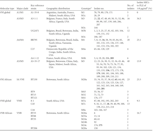
Multilocus sequence typing of incident and serially collected isolates.The MLST alleles at each locus of each isolate ofC. neo-formansvar.grubiiare depicted in Fig. S1A in the supplemental material. Variations in DNA sequences at these loci permitted the identification of multiple, distinct sequence types. We concate-nated the sequences of each isolate at all eight loci and analyzed the phylogenetic relationships of these 170 isolates to one another as well as their relatedness to more than 50 previously genotyped strains from around the world (see Table S1 in the supplemental material) (18–21). The resulting neighbor-joining phylogram is presented in Fig. S1B in the supplemental material. Most geno-types were identical or differed only slightly (e.g., in one or more single nucleotide polymorphisms) from reference strain(s) with established genotypes (Table S1). As shown inTable 3, this com-parative analysis enabled us to identify the genotypes of these iso-lates.
[image:3.585.43.547.87.205.2]The overall diversity of these isolates is consistent with previ-ous analyses of southern African isolates. The 139 isolates of mo-lecular type VNI included 14 distinct multilocus sequence types or genotypes. The 22 isolates of molecular type VNII were repre-sented by at least three genotypes, and there were six unique ge-notypes among the VNB isolates (Table 3). However, two of the three most prevalent VNI genotypes in this sample were identical to the globally dominant VNI genotypes, M5 and M4 (19,26). The
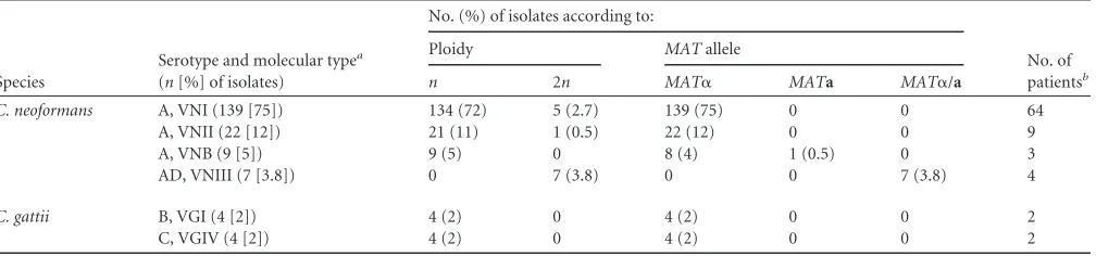
TABLE 1Serotype, molecular type, ploidy, and mating type of the 185 incident and serial isolates ofCryptococcusobtained from cerebrospinal fluid specimens of 89 patients with cryptococcal meningitis
Species
Serotype and molecular typea (n[%] of isolates)
No. (%) of isolates according to:
No. of patientsb
Ploidy MATallele
n 2n MAT␣ MATa MAT␣/a
C. neoformans A, VNI (139 [75]) 134 (72) 5 (2.7) 139 (75) 0 0 64
A, VNII (22 [12]) 21 (11) 1 (0.5) 22 (12) 0 0 9
A, VNB (9 [5]) 9 (5) 0 8 (4) 1 (0.5) 0 3
AD, VNIII (7 [3.8]) 0 7 (3.8) 0 0 7 (3.8) 4
C. gattii B, VGI (4 [2]) 4 (2) 0 4 (2) 0 0 2
C, VGIV (4 [2]) 4 (2) 0 4 (2) 0 0 2
a
No isolates of molecular types VNIV (serotype D), VGII, or VGIII were obtained.
bIn addition, two patients had serial isolates of VNI and VNII, and three patients had serial isolates of VNI and VNB.
on May 16, 2020 by guest
http://jcm.asm.org/
TABLE 2Serial isolates from cerebral spinal fluid specimens of 81 patients from South Africa with cryptococcal meningitis due toC. neoformans
var.grubii(170 isolates) and four patients withC. gattii(eight isolates)
Case
no.a Sexb Age (yr) Provincec
No. of days on treatment
(dosage [mg/day]) Isolate no. (days after initial isolate)e
Molecular type
Geographic
distribution Genotypef
FLZ MIC (g/ml)
AMBd FLZ
1 F 24 MP 8 (45) 1 (400) 1 VNI Global M3b 4
2 (238) VNI Global M3b 4
2 F 31 GA None 12 (400) 3 VNI Global M4b 0.5
4 (121) VNI Global M4b 1
3 M 63 GA None 7 (200) 5 VNI Global M3b NT
6 (157) VNB African M26a NT
4 F 37 GA Unknown 32 (400) 7 VNI Global M4 2
8 (180) VNI Global M4 4
5 M 11 KZ 16 (14) None 9 VNII Global M7c 0.5
10 (153) VNII Global M7c 0.5
6 F 33 GA Unknown 3 (400) 11 VNII Global M7c NT
12 (170) VNI Global M5 NT
7 M 38 GA None 8 (400) 13 VNB African M35a 1
14 (137) VNB African M35a 1
8 M 45 KZ None 2 (400) 15 VNI Global M4 4
16(144) VNI Global M4 16
9 F 10 KZ None 7 (200) 17 VNII Global M7c 4
18(138) VNII Global M7c ⱖ64
10 M 49 EC 2 (50) 17 (400) 19 VNI Global M4b 2
20 (131) VNI Global M4b 2
11 F 29 GA None 17 (400) 21 VNI Global M3 4
22(155) VNI Global M3 16
12 F 31 GA None 7 (400) 23 VNI Global M5 4
24 (121) VNI Global M5 2
13 M 35 KZ Unknown Unknown 25 VNI Global M3b 8
27 (286) VNI Global M3b 8
14 M 27 NC None 8 (400) 28 VNI Global M1c 2
29 (145) VNI Global M1c 4
15 F 24 GA None 2 (800) 30 VNI Global M5 2
31 (166) VNI Global M5 4
32 (223) VNI Global M5 1
16 F 26 GA None 1 (400) 33 VNI African M28a 4
34 (169) VNI African M28a 4
17 F 23 GA 2 (35) 3 (800) 35 VNI African M28a NT
36 (184) VNI Global M5 NT
18 M 34 GA None 5 (400) 37 VNI African M28a 1
38 (171) VNI African M28a 4
19 M 42 GA None None 39 VNII Global M7c 0.5
40 (154) VNII Global M7c 1
21 F 29 GA 12 (25) None 45 VNI Global M5 4
46 (266) VNI Global M5 4
22 M 34 WC 13 (60) 7 (800) 47 VNI Global M3 4
48 (151) VNI Global M3 4
23 F 32 GA None 2 (400) 49 VNI Global M3 4
50 (221) VNI Global M3 8
24 M 44 GA None 3 (800) 51 VNI Global M3 8
52 (191) VNI Global M3 8
25 M 31 GA None 3 (200) 53 VNI Global M5 2
54 (149) VNI Global M5 2
26 F 41 EC 7 (50) 7 (400) 55 VNI African M43 4
56 (135) VNI African M43 4
57 (226) VNI African M43 4
27 M 36 WC 12/14 (36) 13 (200) 58 VNI Global M4 4
59 (228) VNI Global M4 8
28 M 31 NC 10/11 (37.5) None 50 VNB African M24a 1
61 (129) VNB African M24a 1
(Continued on following page)
on May 16, 2020 by guest
http://jcm.asm.org/
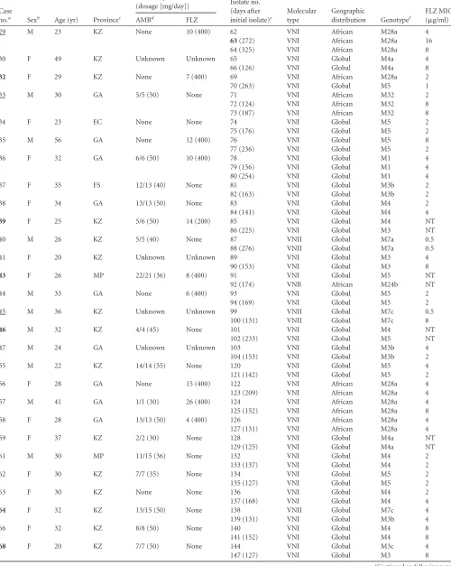
TABLE 2(Continued)
Case
no.a Sexb Age (yr) Provincec
No. of days on treatment
(dosage [mg/day]) Isolate no. (days after initial isolate)e
Molecular type
Geographic
distribution Genotypef
FLZ MIC (g/ml)
AMBd FLZ
29 M 23 KZ None 10 (400) 62 VNI African M28a 4
63(272) VNI African M28a 16
64 (325) VNI African M28a 8
30 F 49 KZ Unknown Unknown 65 VNI Global M4a 4
66 (126) VNI Global M4a 8
32 F 29 KZ None 7 (400) 69 VNI African M28a 2
70 (263) VNI Global M5 1
33 M 30 GA 5/5 (50) None 71 VNI African M32 2
72 (124) VNI African M32 8
73 (187) VNI African M32 8
34 F 23 EC None None 74 VNI Global M5 2
75 (176) VNI Global M5 2
35 M 56 GA None 12 (400) 76 VNI Global M5 8
77 (236) VNI Global M5 2
36 F 32 GA 6/6 (50) 10 (400) 78 VNI Global M1 4
79 (156) VNI Global M1 4
80 (254) VNI Global M1 4
37 F 35 FS 12/13 (40) None 81 VNI Global M3b 2
82 (163) VNI Global M3b 2
38 F 34 GA 13/13 (50) None 83 VNI Global M4 2
84 (141) VNI Global M4 4
39 F 25 KZ 5/6 (50) 14 (200) 85 VNI Global M4 NT
86 (225) VNI Global M3 NT
40 M 26 KZ 5/5 (40) None 87 VNII Global M7a 0.5
88 (276) VNII Global M7a 0.5
41 F 20 KZ Unknown Unknown 89 VNI Global M3 4
90 (153) VNI Global M3 8
43 F 26 MP 22/21 (36) 8 (400) 91 VNI Global M5 NT
92 (174) VNB African M24b NT
44 M 33 GA None 6 (400) 93 VNI Global M5 2
94 (169) VNI Global M5 2
45 M 36 KZ Unknown Unknown 99 VNII Global M7c 0.5
100 (131) VNII Global M7c 8
46 M 32 KZ 4/4 (45) None 101 VNI Global M4 NT
102 (233) VNI Global M5 NT
47 M 24 GA Unknown Unknown 103 VNI Global M3b 4
104 (153) VNI Global M3b 2
55 M 22 KZ 14/14 (55) None 120 VNI Global M5 4
121 (142) VNI Global M5 2
56 F 28 GA None 15 (400) 122 VNI African M28a 4
123 (209) VNI African M28a 4
57 M 41 GA 1/1 (30) 26 (400) 124 VNI African M28a 4
125 (152) VNI African M28a 8
58 F 28 GA 13/13 (50) 4 (400) 126 VNI African M28a 4
127 (131) VNI African M28a 4
59 F 37 KZ 2/2 (30) None 128 VNI Global M4a NT
129 (125) VNI Global M4a NT
61 M 30 MP 11/15 (36) None 132 VNI Global M4 2
133 (137) VNI Global M4 2
62 F 30 KZ 7/7 (35) None 134 VNI Global M5 2
135 (127) VNI Global M5 2
63 F 30 KZ None None 136 VNI Global M4 2
137 (168) VNI Global M4 4
64 F 32 KZ 13/15 (50) None 138 VNII Global M7c 4
139 (131) VNI Global M3b 4
66 F 32 KZ 8/8 (50) None 140 VNI Global M4 8
141 (152) VNI Global M4 8
68 F 20 KZ 7/7 (50) None 144 VNI Global M3c 4
147 (127) VNI Global M3 8
(Continued on following page)
on May 16, 2020 by guest
http://jcm.asm.org/
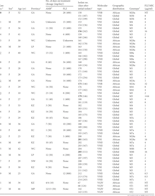
TABLE 2(Continued)
Case
no.a Sexb Age (yr) Provincec
No. of days on treatment
(dosage [mg/day]) Isolate no. (days after initial isolate)e
Molecular type
Geographic
distribution Genotypef
FLZ MIC (g/ml)
AMBd FLZ
70 F 52 GA None 28 (400) 150 VNB African M20a NT
151 (187) VNI Global M3b 8
152 (199) VNI Global M3b 8
71 M 39 GA Unknown 15 (400) 153 VNI Global M4 4
154 (136) VNI Global M4 2
72 F 34 GA 11 (50) 13 (400) 155 VNI Global M5 8
156(242) VNI Global M5 16
74 F 41 GA None 4 (400) 159 VNI Global M3 2
160 (246) VNI Global M3 4
75 F 30 WC Unknown Unknown 161 VNI African M28a 4
162 (178) VNI African M28a 2
76 M 39 LP None 21 (400) 163 VNI African M28a 2
164 (150) VNI African M28a 4
77 F 40 WC 13 (32) 1 (400) 165 VNII Global M8a 1
166 (182) VNII Global M8a 4
167 (290) VNII Global M8a 1
78 F 28 GA 8 (40) 36 (400) 168 VNI African M28a 4
169 (134) VNI African M28a 2
79 F 28 GA None 21 (400) 170 VNI Global M1 2
171 (144) VNI Global M1 4
80 F 28 GA None 10 (400) 172 VNI Global M5 2
173 (254) VNI Global M5 4
81 M 49 GA None 14 (400) 174 VNB African M24c 1
175(146) VNB African M24c 32
82 F 29 WC 14 (50) None 176 VNI African M41 8
177 (182) VNI African M41 4
83 F 10 WC 23 (18) 8 (200) 178 VNI Global M5 2
179(146) VNI Global M5 ⱖ64
84 F 27 GA 11 (40) 1 (400) 180 VNI Global M5 4
181 (119) VNI Global M5 8
85 F 53 KZ 3 (50) None 182 VNI Global M4 1
183 (131) VNI Global M4 1
86 F 26 KZ 14 (50) None 184 VNI Global M5 1
185 (175) VNI Global M5 2
87 F 29 KZ 10 (45) None 186 VNI Global M3c 2
187 (136) VNI Global M3c 2
88 M 36 GA 7 (50) 10 (200) 188 VNI Global M5 1
189 (184) VNI Global M5 2
90 F 40 EC 1 (50) 18 (400) 192 VNII Global M7a 1
193 (147) VNII Global M7a 1
93 F 23 KZ 7 (30) 3 (400) 200 VNI African M28a 4
201(165) VNI African M28a 16
94 M 49 KZ 10 (45) None 202 VNII Global M7a 4
203 (167) VNII Global M7a 2
95 M 42 WC None None 204 VNI Global M4b 8
205(111) VNI Global M4b 16
96 M 36 LP 12 (50) 6 (200) 206 VNI Global M3 2
207 (197) VNI Global M3 2
97 F 29 NW 14 (50) None 208 VNI Global M5 4
209 (159) VNI Global M5 4
98 F 30 KZ 9 (50) None 210 VNI Global M5 2
211 (142) VNI Global M5 2
99 M 25 GA None 12 (800) 212 VNII Global M7c 0.5
213 (174) VNII Global M7c 0.5
214 (243) VNII Global M7c 1
31 M 34 KZ 4/4 (10) None 67 VGIV African ST1 NT
68 (124) VGIV African ST1 NT
67 M 46 MP 12/13 (50) None 142 VGIV African ST2 NT
143 (135) VGIV African ST2 NT
(Continued on following page)
on May 16, 2020 by guest
http://jcm.asm.org/
third most common genotype, M28a, is closely related to refer-ence strain BT150 (see Table S1 and Figure S1B in the supplemen-tal material), which belongs to the subpopulation that is geneti-cally and ecologigeneti-cally related to the global strains of VNI but is restricted to southern Africa (19). At the other extreme, several unique genotypes were quite rare and isolated only from individ-ual patients. Analyses of the clinical data inTable 2revealed no association of cryptococcal genotypes with patients’ gender, age, or provincial location.
A comparison of the molecular genotypes of strains from serial episodes due toC. neoformansvar.grubiiindicated that in 72 of the 81 cases, the genotypes of the initial and serially collected strains were identical, which is consistent with persistence or relapse of the original infection. Because 70% of the isolates were repre-sented by the five most common genotypes (viz., M5, M28a, M4, M3, and M7c), it is possible that some patients with identical serial isolates may have been reinfected with another isolate of the same genotype. However, nine patients (Table 2) (cases 7, 14, 26, 28, 33, 77, 81, 82, and 89) were infected with identical and unique serial isolates that were found in no other patient, and each of these cases was undoubtedly a relapse of the original infection. Conversely, in nine (11%) other cases, the initial and serial strains had different genotypes, which is consistent with reinfection. (The possibility of an original infection with different strains was excluded by rean-alyzing the original cultures without single-colony purification.)
We considered the hypothesis that the length of time between collection of the incident and serial isolates was associated with the probability of reinfection with a new genotype. However, this pos-sibility could not be addressed because the serial isolates were obtained only when CSF cultures were clinically warranted; they were not routinely obtained at regular intervals. Consequently, we observed no significant differences in times between the collec-tions of serial isolates from patients who had identical (range, 111 to 286 days; median, 153 days; mean⫾standard deviation [SD], 163⫾41.8 days) or different genotypes (range, 127 to 263 days; median, 184 days; mean⫾SD, 173⫾44.5 days).
The isolates ofC. gattiiwere obtained from four HIV-positive patients and included four strains of the VGI molecular type and four strains of the VGIV molecular type (Tables 1and2). Each
patient withC. gattiihad identical initial and serial isolates with unique genotypes (see Fig. S2 in the supplemental material).
Mating types.Mating type- and serotype-specific primers were used to determine the mating type of each isolate (Table 1). All eight isolates ofC. gattii, and all but one isolate ofC. neoformans, had the dominantMAT␣mating type allele. The exception was an isolate of the African molecular type VNB (genotype M20a) with the rareMATaallele (Table 2) (isolate number 150), which was confirmed by mating assays in the laboratory. This isolate was responsible for the incident infection in patient 70; she was treated only with FLZ and subsequently relapsed with a global strain of VNI. (In addition, each of the seven AD hybrid strains possessed bothMAT␣andMATaalleles.)
Ploidy. The ploidy of C. neoformans has been reported to change during the course of infection (22,27). The six diploid isolates of molecular type VNI or VNII (allMAT␣/␣) were ob-tained from five patients: the incident and serial isolates of patient 9 had the same relatively common genotype of VNII, but the in-cident isolate (Table 2) (isolate number 17) was haploid, and the serial isolate (isolate number 18), which was collected 138 days later, was diploid, and its MIC to FLZ had increased from 4 toⱖ64
g/ml. This was the only case in which a change in ploidy was accompanied by increased resistance to FLZ. The incident isolates of patients 16 and 24 were haploid VNI strains of the African genotype M28a and the global genotype M3, respectively. Their serial isolates were diploid, but there was no change in their geno-types or MICs to FLZ. The incident and serial isolates of patient 55 were identical diploid strains of the most prevalent global VNI genotype, M5. However, patient 17 was undoubtedly reinfected with a different strain of VNI. The genotype of her incident isolate
(Table 2) (isolate number 35) was a diploid strain of the African
VNI clade (genotype M28a), and the serial isolate (isolate number 36) was a haploid strain of the global VNI clade with the common M5 genotype.
Susceptibility of isolates ofC. neoformansvar.grubiito FLZ. The MIC to FLZ was assayed in isolates ofC. neoformansvar.
grubiifrom patients whose incident and serial isolates had identi-cal genotypes. Several isolates had relatively high MICs to FLZ. The persistence of these infections may have been attributable to
TABLE 2(Continued)
Case
no.a Sexb Age (yr) Provincec
No. of days on treatment
(dosage [mg/day]) Isolate no. (days after initial isolate)e
Molecular type
Geographic
distribution Genotypef
FLZ MIC (g/ml)
AMBd FLZ
69 F 39 MP Unknown 3 (400) 148 VGI Global ST3 NT
149 (134) VGI Global ST3 NT
91 M 36 KZ 10 (50) None 194 VGI Global ST4 NT
195 (224) VGI Global ST4 NT
aIncident and serial isolates of nine patients differed in their genotypes (the case numbers of those patients [3, 17, 32, 39, 43, 46, 64, 68, and 70] are in bold type). Among the 72
patients whose incident and serial isolates ofC. neoformansvar.grubiihad the same genotype, in 11 cases, the MICs to FLZ of the serial isolates wereⱖ4-fold higher than the MIC of the incident isolate (the case numbers of those patients [8, 9, 11, 18, 29, 33, 45, 77, 81, 83, 93] are underlined).
b
F, female; M, male.
cAbbreviations of provinces: EC, Eastern Cape; FS, Free State; GA, Gauteng; KZ, KwaZulu-Natal; LP, Limpopo; MP, Mpumalanga; NW, North West; WC, Western Cape. Other
abbreviations: AMB, amphotericin B; FLZ, fluconazole; MIC, minimum inhibitory concentration; NT, not tested.
dWhen 2 days are listed and separated by a slash, the first number indicates the number of doses administered, and the second number indicates the number of days for which
treatment was prescribed.
eThe nine isolates ofC. neoformans(numbers 16, 18, 22, 63, 156, 175, 179, 201, and 205) with MICs to FLZ ofⱖ16g/ml are in bold type. All but six isolates are haploid; the six diploid (2n) isolates are numbers 18, 34, 35, 52, 120, and 121. With one exception, all the isolates possess the dominantMAT␣mating type allele; African VNB isolate no. 150 has the rareMATamating type.
f
For explanations of the designations of MLST genotypes, refer to the text, Fig. S1B, and Table S1 in the supplemental material.
on May 16, 2020 by guest
http://jcm.asm.org/
insufficient treatment and/or the acquisition of resistance within the patient. Of 155 isolates that were tested, 31 (20%) had MICs to FLZ ofⱖ8g/ml, and nine (5.8%) had MICs to FLZ ofⱖ16
g/ml. The latter were all relapse isolates, and they were distrib-uted among seven different genotypes (see the bolded isolate numbers inTables 2and3). Of the 128 tested isolates of molecular type VNI, 28 (21.9%) had MICs ofⱖ8g/ml. The last column of
Table 3shows the percentages of tested isolates in each major clade
with MICs ofⱖ8g/ml For example, 10 of the 29 (34.5%) tested isolates within the global VNI clade of A3/M3 genotypes had MICs ofⱖ8g/ml, but only 5 of 36 tested isolates within the A5/M5 clade had MICs ofⱖ8g/ml.
[image:8.585.42.547.77.507.2]A common metric of acquired resistance to FLZ is aⱖ4-fold increase in the MIC. The serial isolates of 11 patients (15%) were significantly more resistant (i.e., with MICs atⱖ4-fold higher) than their genetically identical incident isolates (seeTable 1, un-derlined patient numbers 8, 9, 11, 18, 29, 33, 45, 77, 81, 83, and 93). Because eight of these patients (cases 8, 9, 11, 18, 29, 45, 83, and 93) were infected with strains that possessed more prevalent genotypes (M3, M4, M5, M7c, and M28a), it is also possible that during the time between obtaining the incident and serial isolates, some of these patients could have become reinfected with a more resistant strain of the same genotype. However, three patients (cases 33, 77, and 81) were infected with strains of relatively rare
TABLE 3MLST genotypes of 170 incident and serial clinical isolates ofC. neoformansvar.grubii
Molecular type Major clade
Key reference
strain Geographic distribution Genotypea Isolate no.
No. of isolates
Isolate MICs to FLZ of
ⱖ8g/mlb(%) VNI global A1/M1 A1-35-8 Argentina, Australia, France,
Malawi, South Africa, USA
M1 78, 79, 80, 170, 171 5 0
M1c 28, 29 2
A3/M3 A3-1-1 Belgium, France, Italy, South Africa, Uganda, USA
M3 21,22, 47, 48, 49, 50, 51, 52, 86, 89, 90, 147, 159, 160, 206, 207
16 34.5
M3c 144 1
UG2471 Belgium, Brazil, Botswana, India, South Africa, Uganda
M3b 1, 2, 5, 25, 27, 81, 82, 103, 104, 139, 151, 152
12
M3c 186, 187 2
A4/M4 BR795 Belgium, Botswana, Brazil, India, South Africa, Tanzania, Uganda
M4 7, 8, 15,16, 58, 59, 83, 84, 85, 101, 132, 133, 136, 137, 140, 141, 153, 154, 182, 183
20 23.1
C27 Democratic Republic of the Congo, India, South Africa, USA
M4a 65, 66, 128, 129 4
A4-1-12 France, South Africa, USA M4b 3, 4, 19, 20, 204,205 6
A5/M5 A5-35-17 Belgium, Botswana, China, Italy, Japan, Malawi, South Africa, USA
M5 12, 23, 24, 30, 31, 32, 36, 45, 46, 53, 54, 70, 74, 75, 76, 77, 91, 93, 94. 102, 120, 121, 134, 135, 155,156, 172, 173, 178, 179, 180, 181, 184, 185, 188, 189, 208, 209, 210, 211
40 13.9
VNI African SA VNI BT150 Botswana, South Africa M28a 33, 34, 35, 37, 38, 62,63, 64, 69, 122, 123, 124, 125, 126, 127, 161, 162, 163, 164, 168, 169, 200,201
23 23.3
BT9 M43 55, 56, 57 3
BT15 M32 71, 72, 73 3
BT121 M41 176, 177 2
VNII global VNII 8-1 South Africa, USA M7a 87, 88, 192, 193, 202, 203 6 9.5
C45 M7c 9, 10, 11, 17,18, 39, 40, 99, 100,
138, 212, 213, 214
13
C44 M8a 165, 166, 167 3
VNB African VNB BT33 Botswana, South Africa M26a 6 1 16.7
BT88 M35a 13, 14 2
BT35 M24a 60, 61 2
M24b 92 1
M24c 174,175 2
BT206 M20a 150 1
aThe MLST allelic profile of each isolate can be observed in Figure S1A in the supplemental material. As shown by the subsequent phylogenetic analysis in Figure S1B, most strains
were identical or closely related to reference strains (also see Table S1 in the supplemental material) that were previously genotyped by amplified fragment length polymorphisms (AFLP) and MLST methods and designated with an A and M genotype, respectively (12, 18, 19, and 26). Strains with minor allelic variations are denoted with a lowercase suffix (a, b, etc.). Genotypes within the SA VNI and VNB clades have been found only in southern Africa. Most of the VNII strains in this study are closely related to previously genotyped global isolates of VNII, but the database of genotyped VNII isolates is too small to be certain of their distribution.
b
Of the 170 isolates, 155 were tested for susceptibility to FLZ, and 31 (20%) had an MIC ofⱖ8g/ml. Nine isolates (5.2%) had an MIC to FLZ ofⱖ16g/ml, and they are in bold type (isolate numbers 16, 18, 22, 63, 156, 175, 179, 201, and 205). The last column indicates the percentage of tested isolates within each major clade that had an MIC to FLZ ofⱖ8
g/ml.
on May 16, 2020 by guest
http://jcm.asm.org/
genotypes (M32, M8a, and M24c, respectively). Considering their comparative rarity, these cases of infection with more resistant serial isolates were most likely not reinfections but relapses caused by the proliferation of resistant clones during the course of chronic infection.
As noted above, because of the stochastic timing of serial iso-lations, it was not possible to assess whether the number of days between incident and serial isolations was associated with the ac-quisition of resistance to FLZ. Indeed, there were no appreciable differences in the timing of their collection from the 11 patients with FLZ-resistant serial isolates (range, 124 to 272 days; median, 146 days; mean⫾SD, 151⫾39.7 days) and the other patients for whom MIC values were available (range, 111 to 286 days; median, 153 days; mean⫾SD, 165⫾42.2 days). Consequently, it is im-possible to conclude from these data that exposure to FLZ poses a risk of resistance.
DISCUSSION
Recurrent cryptococcosis is well documented among HIV-in-fected patients in South Africa (8,12,13,28). However, genetic relationships between the incident and recurrent strains have not been previously evaluated. We used the MLST method to geno-type the initial or incident isolates and serially collected recurrent isolates that were obtained⬎110 days after the incident isolate. Seventy-two of 81 patients withC. neoformansvar.grubiiand four patients withC. gattiihad identical MLST genotypes. There are three alternative explanations for the isolation of strains with identical genotypes from serial disease episodes. First, these cases may represent persistence of the disease, in which the cryptococcal strain causing the initial infection was not eradicated due to inad-equate treatment, treatment failure, IRIS, or resistance of the iso-late to antifungal drugs. IRIS is less likely to have developed in these patients because only very few received ART (29). Second, even among cases where eradication was achieved by antifungal treatment, relapse for the same reasons may also be possible. Third, it is also possible that patients were reinfected by a strain with an identical genotype. This possibility is difficult to refute because most patients were infected with relatively prevalent ge-notypes However, strains with rare gege-notypes (i.e., gege-notypes that occurred only once in the entire collection) were isolated from serial episodes of nine patients withC. neoformansvar.grubiiand all four patients withC. gattii. In these cases, reinfection with strains of the same rare genotypes would require regular exposure to these strains, which is not consistent with the reported distri-bution of genotypes in the environment (18,19,25).
Resistance of strains ofC. neoformansvar.grubiito FLZ may contribute to recurrent disease among South African patients. MIC testing by the CLSI broth microdilution method demon-strated that recurrent isolates from 11 patients became at least 4-fold more resistant to FLZ than did the original strains isolated from the same patients (see underlined cases inTable 2). Twenty-two percent of the tested isolates had MICs ofⱖ8g/ml, and 5.8% had MICs ofⱖ16g/ml. Our recent epidemiological survey of 487 incident isolates ofC. neoformans, obtained from patients with serial cases of cryptococcal meningitis separated by at least 30 days, indicated that only 0.6% of strains had MICs ofⱖ16g/ml (23), which was consistent with other reports that⬍1% of all strains in Africa had MICs ofⱖ16g/ml (30). In this study, the higher proportion and number of resistant strains were isolated from a biased selection of patients with serial episodes separated
by at least 110 days. These results indicate that changes in suscep-tibility levels may occur during chronic infection, and the emer-gence of resistant strains may occasionally contribute to relapse of the disease.
FLZ is a fungistatic drug, and exposure to FLZ could have se-lected resistant mutants that subsequently proliferated, leading to clinical relapse. Mechanisms of resistance include altered expres-sion of certain genes (e.g.,ERG1andAFR1), efflux pumps, and gene duplications. It is known thatin vitrogrowth ofC. neofor-mansin the presence of sublethal concentrations of FLZ induces the selection of resistant colonies with elevated MICs to FLZ (31,
32), and a similar increase was recently shown to occur in infected mice that were treated with FLZ (33). However, 40 of the 49 pa-tients who received FLZ in the hospital relapsed with the same isolate, but there was no significant increase in the MIC to FLZ. Unfortunately, no follow-up records were available to document which, if any, patients received treatment with FLZ after they were discharged from hospital.
The other factors that contribute to the recurrence of crypto-coccosis among South African patients are limited access to treat-ment and inadequate treattreat-ment (7,8,12). Although none of the patients received Infectious Diseases Society of America (IDSA)-and World Health Organization (WHO)-recommended induc-tion therapy with AMB and flucytosine, most received AMB fol-lowed by FLZ. Indeed, some patients received ample dosages of AMB and/or FLZ (e.g., patients 4, 22, 27, 58, and 78 inTable 2), and their relapse strains had the same genotypes and MICs to FLZ as the original isolates. Clearly, the original infecting strain may persist despite apparently adequate treatment and the absence of
in vitroantifungal resistance. These observations emphasize the need for patients to be monitored for any signs of relapse after completion of the initial treatment.
The incident and serial isolates of nine patients had differing genotypes. There are three nonexclusive explanations for the iso-lation of strains with different MLST genotypes from serial epi-sodes of cryptococcosis. It is possible that these cases represent subsequent new infections that were acquired from the environ-ment. Our data suggest thatC. neoformansvar.grubiiis common in the environment in southern Africa (12,26), and people who do not receive or adhere to ART may become infected again with different strains. It is also possible that patients were simultane-ously infected with more than one strain. Recent evidence indi-cates that approximately 30% of cryptococcal infections in France are caused by multiple strains (27). To test for any evidence of mixed infections in our samples, we obtained DNA from the en-tire population of cryptococcal cells isolated from each patient (not from single colonies) and performed MLST to detect any evidence of more than a one strain. If a multiple infection were present, we would have observed mixed MLST patterns similar to those obtained from the direct sequencing of the AD hybrid strains. However, identical MLST patterns were obtained from the individual colonies and mixed populations of cells, suggesting that mixed infections with multiple strains are not common in South Africa. Alternatively, it is possible that a single strain may have been selected from the original cerebrospinal fluid (CSF) culture for long-term storage.
Results of molecular epidemiological analysis of strains in this study are consistent with our previous observations and confirm the high genetic diversity among strains ofC. neoformansvar.
grubiiin South Africa (18,19,25,26). Although MLST genotyping
on May 16, 2020 by guest
http://jcm.asm.org/
has been somewhat standardized (15), many studies vary the pro-tocol or employ other methods, and there is no universal nomen-clature for designating VNI genotypes. However, where similar methods have been used or genotyping strains have been analyzed phylogenetically for similarity to reference strains with known ge-notypes, the global dominance of a small number of VNI geno-types has been demonstrated. For example, MLST analyses of sev-eral hundred VNI isolates from the United States and a dozen other countries yielded only a few distinct genotypes (designated M1 to M5), of which genotype M5 was the most prevalent (18,20). Subsequent investigations have shown that the M5 is the domi-nant genotype among clinical isolates from China, Japan, Thai-land, and South Korea (34–37). In contrast to the limited genetic diversity observed among global isolates of VNI, two subpopula-tions of VNI coexist in southern Africa: (i) strains with SA VNI genotypes that are closely related to the global genotypes of VNI (see Fig. S1B in the supplemental material) and associated with the same environmental niches (e.g., avian feces), but are more di-verse and apparently confined to southern Africa, and (ii) a genetically diverse, endemic population (VNB genotypes) that is associated with indigenous African trees (19). Predictably, the majority of the patients reported here were infected with strains of the globally dominant VNI genotypes of M5 and M4, as well as the southern African SA VNI genotypes (19).
To our knowledge, this study represents the first molecular epidemiological investigation of strains isolated from serial epi-sodes of cryptococcosis in Africa. The results indicate that the majority of infections were caused by strains with identical mo-lecular genotypes and suggest persistence or relapse of the original infecting strain rather than independent infections with multiple strains. Within this highly selected group, there were also rela-tively high percentages of strains with reduced susceptibilities to fluconazole. These results highlight the importance of properly treating cryptococcosis and monitoring patients after the comple-tion of treatment.
ACKNOWLEDGMENTS
We thank all laboratory and clinical personnel throughout South Africa for contributing to the surveillance. Members of GERMS-SA, 2005 to 2008, included S. Vasaikar (Eastern Cape); N. Janse van Rensberg, A. Möller, P. Smith, and A. M. Pretorius (Free State); K. Ahmed, A. Hoosen, R. Lekalakala, P. P. Sein, C. Feldman, A. S. Karstaedt, O. Perovic, J. Wadula, M. Dove, K. Lindeque, L. Meyer, and G. Weldhagen (Gauteng); S. Harvey and P. Jooste (Northern Cape); D. Cilliers and A. Rampe (North West Province); W. Sturm, T. Vanmali, P. Bhola, P. Moodley, S. Sithole, and H. Dawood (KwaZulu Natal); K. Hamese (Limpopo); K. Bauer, G. Hoyland, J. Lebudi, and C. Mutanda (Mpumalanga); R. Hoffmann, S. Martin, L. Liebowitz, and E. Wasserman (Western Cape); A. Whitelaw (Western Cape); A. Brink, I. Zietsman, M. Botha, X. Poswa, M. da Silva, and S. Budavari (Ampath laboratories); C. Heney and J. Smit (Lancet laboratories); M. Senekal (Pathcare laboratories); A. Schuchat and S. Schrag (CDC); K. P. Klugman (Emory); and C. Cohen, L. Dini, L. de Gouveia, J. Frean, S. Gould, K. Keddy, K. M. McCarthy, J. Patel, S. T. Meiring, E. G. Prentice, V. C. Quan, J. Ramalivhana, A. Sooka, A. von Gottberg, and N. P. Govender (National Institute for Communicable Dis-eases).
We thank Tom M. Chiller for critically reading the manuscript and providing helpful comments.
This investigation was initiated and supported by an International Fellowship from the American Society for Microbiology (Washington, DC) to M.V.W. The study also received crucial support from several other funding sources. In South Africa, the work was partially funded from 2005
through 2006 by the U.S. Agency for International Development’s Anti-microbial Resistance Initiative, transferred via Cooperative Agreement U60/CCU022088 from the U.S. Centers for Disease Control and Preven-tion (CDC), Atlanta, GA. From 2005 through 2008, the study was also partially supported by the CDC, National Center for HIV/AIDS, Viral Hepatitis, STD, and TB Prevention (NCHHSTP) and the Global AIDS Program (GAP) Cooperative Agreement U62/PSO022901. This study was also supported by U.S. Public Health Service grants from the National Institute of Allergy and Infectious Diseases (R01 AI 25783 [to T.G.M.] and R01 AI 93257 [to A.P.L.].
The contents are solely the responsibility of the authors and do not necessarily represent the official views of the CDC.
We have no commercial or any other associations that may pose a conflict of interest.
The use of product names in this article does not imply their endorse-ment by the U.S. Departendorse-ment of Health and Human Services. The findings and conclusions in this article are those of the authors and do not neces-sarily represent the views of the CDC.
REFERENCES
1.Lin X, Heitman J.2006. The biology of the Cryptococcus neoformans
species complex. Annu. Rev. Microbiol.60:69 –105.http://dx.doi.org/10 .1146/annurev.micro.60.080805.142102.
2.Perfect JR.2007. Management of cryptococcosis: how are we doing? PLoS Med.4:e47.http://dx.doi.org/10.1371/journal.pmed.0040047.
3.Perfect JR, Dismukes WE, Dromer F, Goldman DL, Graybill JR, Hamill RJ, Harrison TS, Larsen RA, Lortholary O, Nguyen MH, Pappas PG, Powderly WG, Singh N, Sobel JD, Sorrell TC.2010. Clinical practice guidelines for the management of cryptococcal disease: 2010 update by the Infectious Diseases Society of America. Clin. Infect. Dis.50:291–322.http: //dx.doi.org/10.1086/649858.
4.Mitchell TG, Castañeda E, Nielsen K, Wanke B, Lazéra MS. 2011. Ecological niches forCryptococcus neoformansandC. gattii, p 237–259.In
Heitman J, Kozel TR, Kwon-Chung KJ, Perfect JR, Casadevall A (ed),
Cryptococcus: from human pathogen to model yeast. ASM Press, Wash-ington, DC.
5.Park BJ, Wannemuehler KA, Marston BJ, Govender NP, Pappas PG,
Chiller TM.2009. Estimation of the current global burden of cryptococcal meningitis among persons living with HIV/AIDS. AIDS23:525–530.http: //dx.doi.org/10.1097/QAD.0b013e328322ffac.
6.Lessells RJ, Mutevedzi PC, Heller T, Newell M-L.2011. Poor long-term outcomes for cryptococcal meningitis in rural South Africa. S. Afr. Med. J.
101:251–252.
7.Jarvis JN, Dromer F, Harrison TS, Lortholary O. 2008. Managing cryptococcosis in the immunocompromised host. Curr. Opin. Infect. Dis.
21:596 – 603.http://dx.doi.org/10.1097/QCO.0b013e3283177f6c. 8.Jarvis JN, Meintjes GA, Williams Z, Rebe K, Harrison TS.2010.
Symp-tomatic relapse of HIV-associated cryptococcal meningitis in South Af-rica: the role of inadequate secondary prophylaxis. S. Afr. Med. J.100:
378 –382.
9.Shelburne SA, III, Visnegarwala F, Darcourt J, Graviss EA, Giordano TP, White AC, Jr, Hamill RJ.2005. Incidence and risk factors for im-mune reconstitution inflammatory syndrome during highly active anti-retroviral therapy. AIDS19:399 – 406.http://dx.doi.org/10.1097/01.aids .0000161769.06158.8a.
10. McCarthy KM, Meintjes GA, Arthington-Skaggs BA, Bicanic T, Chiller TM, Cotton MP, Govender NP, Harrison TS, Karstaedt AS, Maartens G, Varavia E, Venter F, Vismer HF.2007. Guidelines for the diagnosis, management and prevention of cryptococcal meningitis and disseminated cryptococcosis in HIV-infected patients. South. Afr. J. HIV Med.8:25–35. 11. Govender NP.2009. Trends in antifungal drug susceptibility of Crypto-coccusspecies in South Africa, 2002–2008, p 201. Abstr. EP-01–5. 17th Congress of the International Society for Human and Animal Mycology, Tokyo, Japan.
12. Govender NP, Mitchell TG, Litvintseva AP, Miglia KJ.2011. Crypto-coccosis in Africa, p 269 –285.InHeitman J, Kozel TR, Kwon-Chung KJ, Perfect JR, Casadevall A (ed),Cryptococcus: from human pathogen to model yeast. ASM Press, Washington, DC.
13. Park BJ, Shetty S, Ahlquist AM, Greenbaum A, Miller JL, Motsi A, McCarthy KM, Govender NP; Gauteng Cryptococcal Surveillance Ini-tiative Group.2011. Long-term follow-up and survival of
on May 16, 2020 by guest
http://jcm.asm.org/
naive patients with cryptococcal meningitis in the preantiretroviral ther-apy era, Gauteng Province, South Africa. Int. J. STD AIDS22:199 –203.
http://dx.doi.org/10.1258/ijsa.2010.010235.
14. Litvintseva AP, Xu J, Mitchell TG.2011. Population structure and ecol-ogy ofCryptococcus neoformansandC. gattii, p 97–111.InHeitman J, Kozel TR, Kwon-Chung KJ, Perfect JR, Casadevall A (ed),Cryptococcus: from human pathogen to model yeast. ASM Press, Washington, DC.
15. Meyer W, Aanensen DM, Boekhout T, Cogliati M, Diaz MR, Esposto
MC, Fisher MC, Gilgado F, Hagen F, Kaocharoen S, Litvintseva AP, Mitchell TG, Simwami SP, Trilles L, Viviani MA, Kwon-Chung KJ.
2009. Consensus multilocus sequence typing scheme forCryptococcus neo-formansandCryptococcus gattii. Med. Mycol.47:561–570.http://dx.doi .org/10.1080/13693780902953886.
16. Mitchell TG, Litvintseva AP.2010. Typing species ofCryptococcusand epidemiology of cryptococcosis, p 167–190.InAshbee HR, Bignell EM (ed), Pathogenic yeasts. Springer-Verlag, Berlin Heidelberg, Germany. 17. Kwon-Chung KJ, Polacheck I, Bennett JE.1982. Improved diagnostic
medium for separation ofCryptococcus neoformansvar.neoformans (sero-types A and D) andCryptococcus neoformansvar.gattii(serotypes B and C). J. Clin. Microbiol.15:535–537.
18. Litvintseva AP, Thakur R, Vilgalys RJ, Mitchell TG.2006. Multilocus sequence typing reveals three genetic subpopulations ofCryptococcus neo-formansvar.grubii(serotype A), including a unique population in Bo-tswana. Genetics172:2223–2238.http://dx.doi.org/10.1534/genetics.105 .046672.
19. Litvintseva AP, Carbone I, Rossouw J, Thakur R, Govender NP, Mitch-ell TG.2011. Evidence that the human-pathogenic fungusCryptococcus neoformansvar.grubiimay have evolved in Africa. PLoS One6:e19688.
http://dx.doi.org/10.1371/journal.pone.0019688.
20. Litvintseva AP, Kestenbaum L, Vilgalys RJ, Mitchell TG.2005. Com-parative analysis of environmental and clinical populations of Cryptococ-cus neoformans. J. Clin. Microbiol.43:556 –564.http://dx.doi.org/10.1128 /JCM.43.2.556-564.2005.
21. Litvintseva AP, Marra RE, Nielsen K, Heitman J, Vilgalys RJ, Mitchell TG.2003. Evidence of sexual recombination amongCryptococcus neofor-mansserotype A isolates in sub-Saharan Africa. Eukaryot. Cell2:1162– 1168.http://dx.doi.org/10.1128/EC.2.6.1162-1168.2003.
22. Lin X, Patel S, Litvintseva AP, Floyd A, Hicks R, Mitchell TG, Heitman J.2009. Diploids in theCryptococcus neoformansserotype A population homozygous for the␣mating type originate via unisexual mating. PLoS Pathog.5:e1000283.http://dx.doi.org/10.1371/journal.ppat.1000283. 23. Govender NP, Patel J, van Wyk M, Chiller TM, Lockhart SR, Group for
Enteric, Respiratory and Meningeal Disease Surveillance in South Af-rica (GERMS-SA).2011. Trends in antifungal drug susceptibility of Cryp-tococcus neoformansobtained through population-based surveillance, South Africa, 2002–2003 and 2007–2008. Antimicrob. Agents Chemother.
55:2606 –2611.http://dx.doi.org/10.1128/AAC.00048-11.
24. Espinel-Ingroff A, Aller AI, Cantón E, Castanon-Olivares LR, Chowd-hary A, Cordoba S, Cuenca-Estrella M, Fothergill AW, Fuller JA, Govender NP, Hagen F, Ilnait Zaragozi MT, Johnson E, Kidd SE, Lass-Flörl C, Lockhart SR, Martins MA, Meis JFGM, Melhem MSC, Ostrosky-Zeichner L, Peláez T, Pfaller MA, Schell WA, St Germain G, Trilles L, Turnidge J.2012.Cryptococcus neoformans-Cryptococcus gattii
species complex: an international study of wild-type susceptibility end-point distributions and epidemiological cutoff values for fluconazole, itra-conazole, posaconazole and voriconazole. Antimicrob. Agents Che-mother.56:5898 –5906.http://dx.doi.org/10.1128/AAC.01115-12.
25. Miglia KJ, Govender NP, Rossouw J, Meiring ST, Mitchell TG; Group for Enteric, Respiratory and Meningeal Disease Surveillance in South Africa.2011. Analysis of pediatric isolates ofCryptococcus neoformans
from South Africa. J. Clin. Microbiol.49:307–314.http://dx.doi.org/10 .1128/JCM.01277-10.
26. Litvintseva AP, Mitchell TG.2012. Population genetic analyses reveal the African origin and strain variation ofCryptococcus neoformansvar.
grubii. PLoS Pathog.8:e1002495.http://dx.doi.org/10.1371/journal.ppat .1002495.
27. Desnos-Ollivier M, Patel S, Spaulding AR, Charlier C, Garcia-Hermoso D, Nielsen K, Dromer F.2010. Mixed infections and in vivo evolution in the human fungal pathogenCryptococcus neoformans. mBio1:e00091–10.
http://dx.doi.org/10.1128/mBio.00091-10.
28. Govender NP.2007. HIV-associated opportunistic fungal infections: a guide to using the clinical microbiology laboratory. South. Afr. J. HIV Med.8:18 –23.
29. Boulware DR, Meya DB, Bergemann TL, Wiesner DL, Rhein J, Musubire AK, Lee SJ, Kambugu A, Janoff EN, Bohjanen PR.2010. Clinical features and serum biomarkers in HIV immune reconstitution inflammatory syn-drome after cryptococcal meningitis: a prospective cohort study. PLoS Med.
7:e1000384.http://dx.doi.org/10.1371/journal.pmed.1000384.
30. Pfaller MA, Messer SA, Boyken LB, Rice C, Tendolkar S, Hollis RJ, Doern GV, Diekema DJ.2005. Global trends in the antifungal suscepti-bility ofCryptococcus neoformans(1990 to 2004). J. Clin. Microbiol.43:
2163–2167.http://dx.doi.org/10.1128/JCM.43.5.2163-2167.2005. 31. Xu J, Onyewu C, Yoell HJ, Ali RY, Vilgalys RJ, Mitchell TG.2001.
Dynamic and heterogeneous mutations to fluconazole resistance in Cryp-tococcus neoformans. Antimicrob. Agents Chemother.45:420 – 427.http: //dx.doi.org/10.1128/AAC.45.2.420-427.2001.
32. Sionov E, Chang YC, Garraffo HM, Kwon-Chung KJ.2009. Heterore-sistance to fluconazole inCryptococcus neoformansis intrinsic and associ-ated with virulence. Antimicrob. Agents Chemother.53:2804 –2815.http: //dx.doi.org/10.1128/AAC.00295-09.
33. Sionov E, Chang YC, Kwon-Chung KJ.2013. Azole heteroresistance in
Cryptococcus neoformans: emergence of resistant clones with chromo-somal disomy in the mouse brain during fluconazole treatment. Antimi-crob. Agents Chemother.57:5127–5130.http://dx.doi.org/10.1128/AAC .00694-13.
34. Chen J, Varma A, Diaz MR, Litvintseva AP, Wollenberg KK,
Kwon-Chung KJ.2008.Cryptococcus neoformansstrains and infection in appar-ently immunocompetent patients, China. Emerg. Infect. Dis.14:755–762.
http://dx.doi.org/10.3201/eid1405.071312.
35. Choi YH, Ngamskulrungroj P, Varma A, Sionov E, Hwang S-M,
Car-riconde F, Meyer W, Litvintseva AP, Lee WG, Shin JH, Kim EC, Lee KW, Choi TY, Lee YS, Kwon-Chung KJ.2010. Prevalence of the VNIc genotype ofCryptococcus neoformansin non-HIV-associated cryptococ-cosis in the Republic of Korea. FEMS Yeast Res.10:769 –778.http://dx.doi .org/10.1111/j.1567-1364.2010.00648.x.
36. Simwami SP, Khayhan K, Henk DA, Aanensen DM, Boekhout T,
Hagen F, Brouwer AM, Harrison TS, Donnelly CA, Fisher MC.2011. Low diversityCryptococcus neoformansvarietygrubiimultilocus sequence types from Thailand are consistent with an ancestral African origin. PLoS Pathog.7:e1001343.http://dx.doi.org/10.1371/journal.ppat.1001343. 37. Umeyama T, Ohno H, Minamoto F, Takagi T, Tanamachi C, Tanabe K,
Kaneko Y, Yamagoe S, Kishi K, Fujii T, Takemura H, Watanabe H, Miyazaki Y.2013. Determination of epidemiology of clinically isolated
Cryptococcus neoformansstrains in Japan by multilocus sequence typing. Jpn. J. Infect. Dis.66:51–55.http://dx.doi.org/10.7883/yoken.66.51.