0022-538X/97/$04.0010
Copyright © 1997, American Society for Microbiology
Transcription of the Derepressed Open Reading Frame P of
Herpes Simplex Virus 1 Precludes the Expression of the
Antisense
g
1
34.5 Gene and May Account for the
Attenuation of the Mutant Virus
GLENN RANDALLANDBERNARD ROIZMAN*
The Marjorie B. Kovler Viral Oncology Laboratories, The University of Chicago, Chicago, Illinois 60637
Received 4 April 1997/Accepted 16 July 1997
Open reading frame P (ORF P), located at the 3*terminus of the 8.5-kb DNA sequence transcribed during latency and almost completely antisense to theg134.5 gene, is naturally repressed by infected cell protein 4 (ICP4), the major herpes simplex virus 1 regulatory protein. Earlier studies on cells infected with a mutant in which the expression of ORF P is derepressed have shown that (i) the accumulation of the ainfected cell proteins 0 (ICP0) and 22 (ICP22), the products of spliced mRNAs, is reduced congruent with the binding of ORF P protein to p32, a component of the ASF/SF2 splicing factors, (ii) ORF P protein colocalizes with spliceosomes, (iii) bothg134.5 mRNA and protein are virtually undetectable, and (iv) the virus is attenuated on intracerebral inoculation in mice. We report the construction and characterization of two recombinant viruses: R7546, in which ORF P transcription was derepressed and the initiator methionine codon was replaced; and R7547, in which both mutations were repaired to the wild-type genotype. The mutations in R7546 do not alter the amino acid sequence of theg134.5 gene. We report that (i) the reduction in the accumulation ofg134.5 mRNA and protein in cells infected with mutant viruses expressing derepressed ORF P genes reflects the effects of antisense transcription of ORF P rather than a function of ORF P protein, (ii) the attenuated phenotype of the viruses carrying derepressed ORF P genes is due largely to the absence of theg134.5 protein, and (iii) the reduction in accumulation of ICP0 and ICP22 requires the expression of ORF P protein.
This report concerns two genes of herpes simplex virus 1 (HSV-1) arranged totally antisense to each other. Theg134.5
gene maps next to the terminal a sequence in the inverted repeat sequencesab andb9a9 flanking the unique long (UL)
sequence and therefore is present in two copies per viral ge-nome. This gene encodes 263 codons in strain HSV-1(F) and requires the onset of viral DNA synthesis for maximal expres-sion (2, 11). Current data indicate that this gene has at least two functions. The least understood is the function which en-ables the virus to replicate in the central nervous system (CNS) of animals used in experimental model systems. In the absence of both copies of the gene, high-titer virus is unable to destroy the CNS of mice following intracerebral inoculation (9). The virus expresses genes in only a small number of neurons, as-trocytes, and oligodendrocytes arranged along the needle track and whose projections impinge on this region of the brain, but viral replication is virtually undetectable (23). The second function of theg134.5 gene is to prevent premature shutoff of
protein synthesis in cells of human derivation as a consequence of phosphorylation of the a subunit of translation initiation factor eIF-2 by double-stranded RNA-activated kinase (PKR) (8, 12, 13). In this instance,g134.5 binds to the protein
phos-phatase 1aand directs it to dephosphorylate eIF-2a, thereby allowing unabated protein synthesis in infected cells (17).
Open reading frame P (ORF P) maps antisense to theg134.5
gene at the 39end of the 8.5-kb latency-associated transcript sequence reported to be transcribed during latent infections (20, 28, 29, 32). Of the 248 codons, only 8 codons of ORF P are
not antisense tog134.5 and only 23 codons ofg134.5 are not
antisense to ORF P (21). The history of the discovery of ORF P is of interest inasmuch as it relates to its function. Bohenzky et al. (5) identified a promoter within theabandb9a9inverted repeat sequences mapping antisense and 39to theg134.5 gene.
Yeh and Schaffer (31) demonstrated that this promoter drives the expression of an RNA species, but only if a major regula-tory protein, infected cell protein 4 (ICP4), is dysfunctional or absent. The repressor function of ICP4 depends on the binding of ICP4 to a high-affinity cognate site at or near the transcrip-tion initiatranscrip-tion site of the target gene (15, 22, 24), and indeed, a high-affinity binding site straddles the transcription initiation site (29 to13) (31). The objectives of this laboratory in ini-tiating these studies were to identify expressed open reading frames mapping within the domain of the viral genome tran-scribed during latency. Of the 16 open reading frames desig-nated A to P and mapped in that domain, the one which yielded the strongest signal was ORF P, but only under con-ditions in which the ICP4 binding site located at the transcrip-tion initiatranscrip-tion site of ORF P was destroyed by mutagenesis or following infection and maintenance at 39.5°C, the nonpermis-sive temperature of limited-passage isolates carrying a temper-ature-sensitive ICP4 (19–21). The expression of ORF P cannot be detected during productive infection.
Preliminary experiments using the mutant carrying a dere-pressed ORF P indicated that it is attenuated in experimental animals (19), but the question arose as to whether the atten-uated phenotype reflected the failure ofg134.5 gene expression
as a consequence of antisense transcription of the derepressed ORF P. One possible function of ORF P emerged from yeast two-hybrid system analyses. These studies indicated that ORF P interacts with p32, a component of the ASF/SF2 alternative splicing factors (6). The significance of the interaction was * Corresponding author. Mailing address: The Marjorie B. Kovler
Viral Oncology Laboratories, The University of Chicago, 910 E. 58th St., Chicago, IL 60637. Phone: (773) 702-1898. Fax: (773) 702-1631. E-mail: [email protected].
7750
on November 9, 2019 by guest
http://jvi.asm.org/
verified by demonstration of in vitro interaction between ORF P and another splicing factor, the B/B9components of the SM antigens; by colocalization of ORF P with spliceosomes; and more significantly, by the evidence that in cells infected with the virus carrying derepressed ORF P, the levels of viral pro-tein products of spliced mRNAs, ICP0 and ICP22, were re-duced relative to the protein products of intronless mRNAs, ICP4 and ICP27 (6).
The main questions concerning these studies are (i) whether the phenotype of the derepressed ORF P in the context of an infection of cells in vitro or in vivo is due to the suppression of
g134.5 gene and (ii) whether the suppression of expression of
theg134.5 gene is due to antisense transcription of ORF P or
to a specific function of ORF P protein. To answer these questions, we constructed two new recombinant viruses. The first took advantage of the observation that ORF P encodes two methionines, an initiator methionine and a second methi-onine codon (met240) located near the 39terminus of ORF P.
In the recombinant virus designated R7546 (P11)2, the ICP4 binding site was mutagenized to derepress ORF P transcrip-tion and the initiator methionine codon was replaced with an isoleucine codon to prevent the synthesis of ORF P protein. In the second recombinant virus, designated R7547 (P11)2R, both mutations were repaired to wild-type status. We report that (i) the suppression of g134.5 gene expression is due to
antisense transcription and not to the expressed ORF P pro-tein, (ii) the reduced virulence of the virus carrying the dere-pressed ORF P cannot be attributed solely to ORF P inasmuch as the derepressed mutant incapable of expressing the ORF P protein was also attenuated, and (iii) the selective reduction in the synthesis of ICP0 and ICP22 reported earlier requires the expression of ORF P protein.
MATERIALS AND METHODS
Viruses.HSV-1(F), a limited-passage clinical isolate, is the prototype HSV-1 strain used in this laboratory (14). The recombinant viruses described previously were as follows. The recombinant virus R3659 (21) lacks theSacI-BglII sequence ofBamHI-Q encoding the thymidine kinase (tk) and UL24 genes. A sequence consisting of the coding domain of thetkgene under control of thea27 promoter (25) replaced theBstEII-StuI sequence of theBamHI S fragment encoding the g134.5 and ORF P genes (Fig. 1, line 4). R7530 contains a four-base substitution
at the ICP4 binding site which straddles the ORF P transcription initiation site (Table 1). This mutation has been shown to be sufficient for derepression of ORF P and creates a diagnosticEcoRI restriction site (19). R7531 is the repair of R7530 (19). R3616 contains a 1-kb deletion in theg134.5 and ORF P genes,
between theBstEII andStuI sites (9).
Antibodies.Rabbit polyclonal antisera specific forg134.5 (BR4 [2]), ICP22
(R77 [3]), and a C-terminalXmaI fragment of ORF P fused to glutathione S-transferase (ORF Pc [19]) and mouse monoclonal antisera specific for ICP0 (H1112 [1]) and ICP27 (H1113 [purchased from Goodwin Institute, Plantation, Fla.] [1]) have been described elsewhere.
Plasmids.Plasmid pRB4794 has been described elsewhere (21). It contains the 1,800-bp NcoI fragment ofBamHI-S, spanning the region between the start codons of thea0 andg134.5 genes. It was used as a probe for analyses of ORF
P recombinant viral DNA and as a source of strand-specific riboprobes for the detection of ORF P andg134.5 mRNAs. Plasmid pRB4855 contains a four-base
change in the ICP4 binding site and has been described previously (19). pRB4930 contains the 830-bpNotI fragment ofBamHI-S cloned into theNotI site of pUC19. Plasmid pBR4929 contains a mutant ORF P initiator methionine codon and was made by insertion of a 160-bp PCR product into theSrfI-DraIII sites of pRB4930. The PCR primers were 59-ACGGGCCTCGGGCCCTAGGCACG GCCCGATAACCGCCTCGGCCTC and 59-GAGGCCGAGGCGGTTATCG GGCCGTGCCTAGGGCCCGAGGCCCGT. Underlined bases represent mu-tations that replace the ORF P initiator methionine codon (ATG) with an isoleucine codon (ATA) and create a diagnostic AvrII restriction site 15 bp upstream. Plasmid pRB5159 was made by replacing theBspEI-DraIII fragment from pRB4855 with the corresponding fragment from pRB4929. pRB5159 con-tains both the ORF P initiator methionine mutation and the ICP4 binding site mutation which derepresses ORF P. It was used to construct the recombinant virus R7545. Plasmid pRB103 containing theBamHI Q fragment was used to repair the deletion in thetkgene of R7545, resulting in the recombinant virus R7546. pRB4794 contains the 1,800-bpNcoI fragment fromBamHI-S and was
used to repair the mutations in the ORF P domain of R7546, resulting in the recombinant virus R7547.
Construction of recombinant viruses.Viral stocks and titrations of viruses were done in Vero cells (American Type Culture Collection). R7545 was con-structed by cotransfection of rabbit skin cells (originally obtained from J. Mc-Claren) with pRB5159, which contains both the ICP4 binding site mutation and the ORF P initiator methionine mutation, with R3659 viral DNA (Fig. 1, line 4), which contains a deletion in thetkgene and ana27 promoter-driventkgene replacement of the 1,100-bpStuI-NcoI fragment encoding the ORF P andg134.5
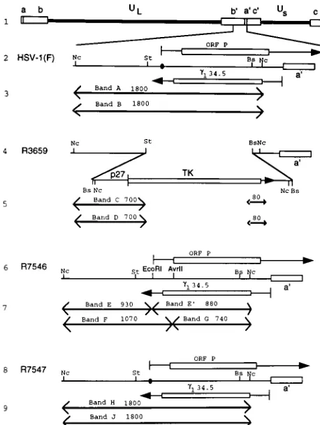
genes.tk2viruses were selected by plating the progeny of the cotransfection on 143TK2cells overlaid with Dulbecco modified Eagle medium containing 5% newborn calf serum and 40mg of bromodeoxyuridine per ml of medium. Plaque-FIG. 1. Schematic representations of sequence arrangements in the recom-binant viruses used in this study. Line 1, a representation of the HSV-1 (F) genome. Shown are the unique long (UL) and unique short (US) sequences
which are flanked by inverted repeatscabandb9a9c9. Line 2, domains of the ORF P andg134.5 genes in the inverted repeat sequenceb9a9flanking the UL
se-quences. The identical sequences in the inverted orientation map in the ab repeat. The coding domains (rectangular boxes) and transcripts (lines with ar-rows denoting direction) of the ORF P andg134.5 genes are shown. The closed
circle denotes a wild-type ICP4 binding site. Line 4, sequence arrangement of the relevant domains of R3659. TheStuI-BstEII sequences encoding ORF P and g134.5 were replaced in both repeats by the chimeric a27-tk gene. Line 6,
sequence arrangement of the relevant domains of recombinant R7546. The a27-tkgene of R3659 was replaced with sequences containing a mutated ICP4 binding site introducing a diagnosticEcoRI endonuclease site and a mutated ORF P initiation methionine introducing a diagnosticAvrII endonuclease site. Line 8, sequence arrangement of the relevant region of recombinant R7547. The mutations in the ORF P domain of R7546 were repaired by transfection with the NcoI fragment from the HSV-1(F)BamHI S fragment. Lines 3, 5, 7, and 9, expected sizes of fragments detected by hybridization of the 1,800-bpNcoI fragment with electrophoretically separated digests of viral DNAs withNco I-EcoRI (the first band set per virus), diagnostic of the ICP4 binding site mutation, orNcoI-AvrII (the second band set per virus), diagnostic of the replacement of the initiator methionine. The arrows in these lines denote restriction cleavage sites present in the respective viruses and therefore fragment boundaries. HSV-1(F) DNA would be expected to yield bands A and B, R3659 DNA would be expected to yield bands C and D, R7546 DNA would be expected to yield bands E, F, and G, and R7547 DNA would be expected to yield bands H and J. Abbreviations: St,StuI; Nc,NcoI; Bs,BstEII.
on November 9, 2019 by guest
http://jvi.asm.org/
[image:2.612.319.548.69.373.2]purified stocks were prepared as described elsewhere (26). Viral DNA was isolated from infected cells and purified on a 5 to 20% potassium acetate gradient as described elsewhere (18). Viral DNA from single plaques were analyzed for the presence of novelEcoRI andAvrII endonuclease restriction sites diagnostic of the ICP4 binding site and the initiator methionine mutations, respectively. Thetkgene of R7545 was repaired by plating the progeny of the cotransfection of rabbit skin cells with R7545 DNA and pRB103, which contains theBamHI Q fragment, on 143TK2cells overlaid in Dulbecco modified Eagle medium containing 5% fetal bovine serum, hypoxanthine, aminopterin, and thymidine. Virus was isolated, purified, and analyzed as described above. This process resulted in the isolation of R7546, which contains a wild-typeBamHI Q fragment, the ICP4 binding site mutation, and the ORF P initiator methionine mutation. The mutations in the ORF P gene of R7546 were repaired by cotrans-fection of rabbit skin cells with R7546 viral DNA and pRB4794, which contains the 1,800-bpNcoI fragment fromBamHI-S of HSV-1(F). Plaques were isolated, and viral DNA was analyzed for a wild-type restriction endonuclease pattern indicating the absence of the introducedEcoRI andAvrII sites withinBamHI-S. R7547 has a wild-type restriction endonuclease pattern and therefore has a wild-type genotype.
Analyses of viral DNAs.Viral DNAs were digested with appropriate restric-tion enzymes as detailed in the legend to Fig. 1. They were then subjected to electrophoresis on a 28-cm 0.85% agarose gel and transferred to a Zeta Probe membrane (Bio-Rad, Richmond, Calif.) by capillary action in 0.5 M NaOH. The membrane was rinsed in 23SSC (0.3 M NaCl, 0.015 M sodium citrate) and prehybridized in 30% formamide–63SSC–1% milk–1% sodium dodecyl sulfate– 100mg of single-stranded calf thymus DNA per ml for 30 min at 68°C; 106cpm
of denatured,32P-labelled pRB4794 was then added overnight, and the blot was
rinsed as recommended by the manufacturer. Autoradiographic images were obtained on Kodak XAR5 film.
Immunoblots.Immunoblot analyses were done as previously described (20). Briefly, infected cells were scraped into phosphate-buffered saline, pelleted un-der low-speed centrifugation, resuspended in disruption buffer containing 0.7 M b-mercaptoethanol, 2% sodium dodecyl sulfate, 50 mM Tris, and 2.75% sucrose, sonicated briefly, boiled, and electrophoretically separated on a denaturing poly-acrylamide gel cross-linked withN,N9-diallyltartardiamide (Bio-Rad). The elec-trophoretically separated, denatured proteins were electrically transferred to a nitrocellulose sheet, blocked, reacted with the appropriate antiserum, rinsed, reacted with either goat anti-rabbit immunoglobulin G (IgG) conjugated to alkaline phosphatase for rabbit polyclonal antisera or goat anti-mouse IgG con-jugated to alkaline phosphatase for mouse monoclonal antisera, rinsed again, and developed as recommended by the manufacturer (Bio-Rad).
RNA blots.RNA blot analyses were done as previously described (19). Briefly, roller bottles of Vero cells were exposed to 10 PFU of HSV-1(F), R7530, R7531, R7546, or R7547 per cell and incubated at 37°C for 13 h for the detection of g134.5 mRNAs, since this gene is expressed predominantly after the onset of viral
DNA synthesis. Vero cells grown in 150-cm2flasks were exposed to virus as
described above and incubated at 37°C for 6 h for the detection of ORF P mRNAs, since this RNA is made early in infection. Cells were collected, washed, treated with 0.1% Nonidet P-40 for plasma membrane solubilization, and then subjected to low-speed centrifugation for the isolation of cytoplasmic fractions. Nucleic acid extraction, precipitation, and DNase treatment followed. The amounts of RNAs which were loaded onto agarose-formaldehyde gels for the detection of different RNA species were as follows: (i) polyadenylated cytoplas-mic mRNAs were purified from 800mg of cytoplasmic RNA with the Promega PolyATtract system IV and loaded for the detection of cytoplasmicg134.5
mR-NAs; and (ii) 20mg of total cytoplasmic RNAs was loaded onto agarose-form-aldehyde gels for the detection of ORF P. Denatured RNAs were separated by electrophoresis in 1.5% agarose-formaldehyde gels and transferred to a Zeta Probe membrane by capillary action in 103SSC. Strand-specific32
P-radiola-belled riboprobes were generated by T7 transcription of pRB4794 for ORF P-specific riboprobes and SP6 transcription of pRB4794 forg134.5 riboprobes.
Hybridization and rinsing procedures were as recommended by the Zeta Probe manufacturer. Autoradiographic images were obtained on Kodak XAR5 film.
[35S]methionine labelling.Replicate SK-N-SH neuroblastoma cell cultures (American Type Culture Collection) in 25-cm2flasks were exposed to 10 PFU of
HSV-1(F), R3616, R7530, R7531, R7546, or R7547 per cell. At 14 h after infection, the medium was replaced with 199V lacking methionine but
supple-mented with 50mCi of [35S]methionine (Amersham). After 1 h of incubation at
37°C, cells were harvested, solubilized in disruption buffer, sonicated, boiled for 5 min, subjected to electrophoresis on a 10% denaturing polyacrylamide gel cross-linked withN,N9-diallyltartardiamide (Bio-Rad), transferred electrically to a nitrocellulose sheet, and exposed to Kodak XAR5 autoradiography film for 2 days.
Intracranial inoculation of mice.CBA/J mice (3.5 weeks old) from Jackson Laboratory were anesthetized with pentobarbital sodium (Nembutol) and in-jected intracerebrally with 10-fold serial dilutions of virus, seven mice per dilu-tion. Mice were monitored daily; mortality from 2 to 21 days after infection was attributed to the inoculated virus. The 50% lethal dose (LD50) ratios were
calculated by the method of Reed and Muench (27).
RESULTS
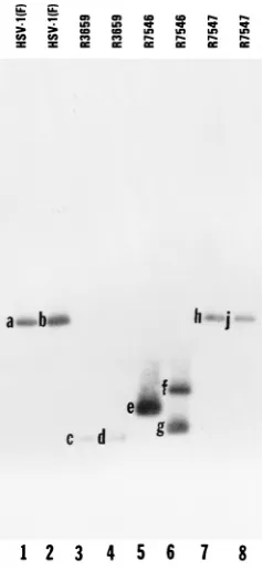
Construction and characterization of the recombinant virus R7546 (P11)2, expressing a derepressed, untranslated ORF P transcript, and the repaired recombinant virus R7547 (P11)2R. The procedures for the construction of recombi-nant viruses R7546 (P11)2 and R7547 (P11)2R are de-scribed in Materials and Methods, and the viral genotypes are presented in Table 1. The genotype of R7546 (P11)2was verified by the presence of theEcoRI restriction endonuclease site diagnostic of the ICP4 binding site mutation, the AvrII restriction endonuclease site diagnostic of the mutation of the ORF P initiator methionine, and a wild-typeBamHI Q frag-ment indicative of a repaired tkgene. The locations of the EcoRI and AvrII restriction sites within R7546 (P11)2 are shown in Fig. 1, line 6. R7546 (P11) DNA was incubated with restriction endonucleases NcoI-EcoRI orNcoI-AvrII, electro-phoretically separated on 0.85% agarose gels, transferred to Zeta Probe membranes, and hybridized with pRB4794, which contains the 1,800-bpNcoI fragment ofBamHI-S (Fig. 2, lanes 5 and 6). Restriction endonuclease cleavage of R7546 (P11) DNA withNcoI andEcoRI resulted in the predicted 880- and 930-bp NcoI-EcoRI doublet (Fig. 2, band e), confirming the presence of the ICP4 binding site mutation. Digestion of R7546 (P11)2viral DNA withNcoI andAvrII resulted in the predicted 1,070- and 740-bp NcoI-AvrII fragments (Fig. 2, bands f and g), confirming that the recombinant virus also contained the initiator methionine mutation. This pattern is distinct from that of the parental virus R3659 (Fig. 1, line 4), in that digestion withNcoI-EcoRI orNcoI-AvrII yielded a 700-bp fragment (Fig. 2, bands c and d) detected by hybridization with
32P-labelled pRB4794 DNA. The size of these fragments is
consistent with that predicted for the fragment upstream of the
a27-tkreplacement (Fig. 1, lines 4 and 5).
[image:3.612.63.557.83.157.2]As is necessary in all instances in which the phenotypes of deletion mutants are tested, the two mutations in the ORF P domain were repaired by cotransfection of rabbit skin cells with R7546 (P11)2viral DNA and pRB4794, which contains the 1,800-bp NcoI fragment from the BamHI S fragment of HSV-1(F). Viral DNAs from progeny were then screened for the wild-type pattern by restriction endonuclease cleavage. The repaired virus R7547 (P11)2R did not contain the novel EcoRI orAvrII sites, as indicated by the presence of 1,800-bp TABLE 1. Genotypes and phenotypes of viruses used in this study
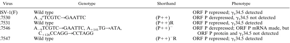
Virus Genotype Shorthand Phenotype
HSV-1(F) Wild type ORF P repressed;g134.5 detected
R7530 A29aTCGTC3GAATTC (P11) ORF P derepressed,g134.5 not detected
R7531 Wild type (P11)R ORF P repressed,g134.5 detected
R7546 A29TCGTC3GAATTC, A1141TG3ATA,
C1126CCAGG3CCTAGG
(P11)2 ORF P derepressed; ORF P mRNA made, but
ORF P protein andg134.5 not detected
R7547 Wild type (P11)2R ORF P repressed;g134.5 detected
aNucleotide number relative to transcriptional initiation site of ORF P (11).
on November 9, 2019 by guest
http://jvi.asm.org/
NcoI fragments (Fig. 2, bands h and j) which were also present in HSV-1(F) (Fig. 2, bands a and b). This result indicates that the mutations in the ORF P genes of the parental virus R7546 (P11)2have been replaced with the wild-type sequences.
Expression of ORF P andg134.5 proteins in cells infected with R7530 (P11), R7546 (P11)2, and R7547 (P11)2R recombinant viruses.Replicate 25-cm2flask cultures of Vero
cells were infected with 10 PFU of HSV-1(F), R7530 (P11), R7546 (P11)2, or R7547 (P11)2R per cell. After 18 h of incubation at 37°C, cells were harvested, lysed by sonication in disruption buffer, boiled for 5 min, subjected to electrophoresis in a denaturing 12.5% polyacrylamide gel, electrically trans-ferred to a nitrocellulose sheet, and probed with rabbit poly-clonal antiserum ORF Pc specific for ORF P. As shown in Fig. 3A, the antibody reacted with the typical, previously reported three ORF P polypeptide bands in electrophoretically sepa-rated lysates of cells infected with the recombinant R7530 (P11) containing an expressable, derepressed ORF P (19). These protein bands were not expressed in cells infected with the recombinant R7546 (P11)2carrying a derepressed ORF P but lacking the initiator methionine. Lastly, ORF P protein was also not expressed in cells infected with wild-type virus HSV-1(F) or the recombinant virus R7547 (P11)2R, in which both the ICP4 binding site and the initiator methionine codon were restored.
An earlier report from this laboratory showed that derepres-sion of ORF P resulted in undetectable amounts of g134.5
protein and mRNA (19). To determine whether this down-regulation was correlated with the synthesis of ORF P protein, a nitrocellulose sheet containing replicate samples from the experiment shown in Fig. 3A was probed with the rabbit
[image:4.612.327.546.68.244.2]poly-clonal antiserum BR4 to detect g134.5 protein. As shown in
Fig. 3B,g134.5 protein was present in lysates of cells infected
with HSV-1(F) and the repaired virus R7547 (P11)2R. The protein was undetectable in cells infected with R7530 (P11) or R7546 (P11)2. ICP0, which is unaffected by ORF P ex-pression in infected Vero cells (unpublished data), was used as a loading control. While the levels of ICP0 fluctuated slightly, the results presented in Fig. 3 indicate that the shutoff of expression of g134.5 protein correlates with derepression of
ORF P transcription rather than the expression of ORF P protein.
Expression of ORF P andg134.5 mRNAs in cells infected with R7530 (P11), R7546 (P11)2, and R7547 (P11)2R recombinant viruses. The synthesis of ORF P mRNA was tested in replicate 150-cm2flask cultures of Vero cells infected
with 10 PFU of HSV-1(F), R7530 (P11), R7531 (P11)R, R7546 (P11)2, or R7547 (P11)2R per cell. After 6 h of incubation at 37°C, cells were harvested, and the cytoplasmic RNAs were collected as described in Materials and Methods. Total cytoplasmic RNAs in 20-mg amounts per sample were subjected to electrophoresis in 1.5% agarose-formaldehyde gels, transferred to Zeta Probe membranes by capillary action, and hybridized with radiolabelled riboprobes generated by T7 transcription of pRB4794 specific for the ORF P transcript.
As shown in Fig. 4A, a specific 2.3-kb mRNA species cor-responding to the ORF P transcript was present in cytoplasmic RNAs extracted from cells infected with recombinants R7530 (P11) and R7546 (P11)2, in which transcription was dere-pressed by mutagenesis of the ICP4 binding site. This RNA species was not detected in cytoplasmic RNAs extracted from cells infected with wild-type virus or the recombinants R7531 (P11)R and R7547 (P11)2R, in which the wild-type se-quences were restored. Thus, as expected, the ICP4 binding site mutation derepressed the transcription of ORF P (Fig. 4A) but the initiator methionine mutation precluded ORF P pro-tein expression (Fig. 3A) in R7546 (P11)2-infected cells.
FIG. 2. Autoradiographic image of electrophoretically separated viral DNA fragments containing sequences in the domain of the ORF P andg134.5 genes.
Viral DNAs were digested with eitherNcoI andEcoRI (lanes 1, 3, 5, and 7) or NcoI andAvrII (lanes 2, 4, 6, and 8), electrophoretically separated on a 0.85% agarose gel, transferred to Zeta Probe membranes, hybridized to radiolabelled DNA of plasmid pRB4794 containing the 1,800-bpNcoI fragment of theBamHI S fragment, and exposed to Kodak XAR5 film. The expected sizes of the frag-ments generated by cleavages (bands a through j) are shown in Fig. 1.
FIG. 3. Photograph of infected cell proteins electrophoretically separated on a denaturing polyacrylamide gel and reacted with polyclonal rabbit antisera specific for ORF P (A) org134.5 (B). Replicate Vero cell cultures grown in
25-cm2flasks were infected with 10 PFU of HSV-1(F), R7530 (P11), R7546
(P11)2, or R7547 (P11)2R per cell, maintained at 37°C for 22 h, harvested, solubilized, electrophoretically separated on 12.5% polyacrylamide denaturing gels, transferred to a nitrocellulose sheet, and reacted with rabbit polyclonal antiserum (A) ORF Pc or (B) BR4, then goat anti-rabbit IgG conjugated to alkaline phosphatase, and finally alkaline phosphatase substrate. Both blots were then reacted with mouse monoclonal antiserum specific to ICP0, goat anti-mouse IgG conjugated to alkaline phosphate, and alkaline phosphatase.
on November 9, 2019 by guest
http://jvi.asm.org/
[image:4.612.119.238.70.327.2]To measure the accumulation ofg134.5 mRNA, Vero cells
grown in roller bottles were infected with 10 PFU of HSV-1(F), R7530 (P11), R7531 (P11)R, R7546 (P11)2, or R7547 (P11)2R per cell. After 13 h of incubation at 37°C, cells were harvested and processed as described above except that polyadenylated mRNA purified from 800mg of total cy-toplasmic RNA per sample was subjected to electrophoresis. The electrophoretically separated, denatured RNAs were transferred to Zeta Probe membranes and hybridized with radiolabelled riboprobes generated by SP6 transcription of pRB4794 specific for g134.5 mRNAs. In this instance, a27
mRNAs were used as a loading control (data not shown). As shown in Fig. 4B, a specific 1.2-kb mRNA species correspond-ing to g134.5 mRNAs was detected in cytoplasmic RNA
ex-tracted from cells infected with HSV-1(F), R7531 (P11)R, and R7547 (P11)2R but not in RNA extracted from cells infected with R7530 (P11) or R7546 (P11)2, which express ORF P transcripts.
The key conclusion to be derived from these results is that accumulations of detectable amounts of the two antisense mR-NAs, those of ORF P andg134.5, are mutually exclusive and
that transcription of ORF P prevented the accumulation of the antisense g134.5 mRNAs. It should be noted that the DNA
fragment used to construct the repair does not extend 59of the
g134.5 coding region, and therefore theg134.5 promoter has
not been altered in the repaired virus. The accumulation of
g134.5 mRNA in cells infected with R7547 (P11)2R indicates
that the control ofg134.5 mRNA accumulation resides at the
site of mutation, i.e., at the ICP4 binding site and not at some undefined site outside the domains of the ORF P andg134.5
genes.
Functional analyses of theg134.5 gene product in cells in-fected with wild-type and recombinant viruses.An earlier re-port from this laboratory showed that protein synthesis was shut off in human neuroblastoma SK-N-SH cells infected with
g134.52virus but not in cells infected with wild-type virus or
R7530 (P11) (19). These results suggested that small amounts ofg134.5 protein expressed in these cells were
suffi-cient to prevent the total shutoff of protein synthesis. To ex-clude the possibility that ORF P protein expression can com-pensate for the loss ofg134.5 protein synthesis, the following
experiment was done.
Replicate cultures of SK-N-SH cells in 25-cm2 flasks were
infected with 10 PFU of HSV-1(F), R3616 (g134.52/ORF P2),
R7530 (P11), R7531 (P11)R, R7546 (P11)2, or R7547 (P11)2R per cell. After 14 h of incubation at 37°C, the me-dium was replaced with 1 ml of 199V lacking methionine but supplemented with 50 mCi of [35S]methionine, and the cells
were reincubated for 1 h. The cells were then harvested, lysed in disruption buffer, sonicated, electrophoresed in 10% dena-turing polyacrylamide gels, transferred to nitrocellulose, and exposed to Kodak XAR5 film (Fig. 5). As predicted from earlier studies published elsewhere (12, 13), labelled proteins were essentially absent from lysates of cells infected with R3616 (g134.52/ORF P2). The lysates of all other infected cell
cultures accumulated approximately equivalent amounts of la-belled proteins. Inasmuch as protein synthesis was not shut off in cells infected with R7546 (P11)2, we may conclude that ORF P protein does not compensate for the decreased accu-mulation of the g134.5 protein by preventing the shutoff of
global protein synthesis.
A second function ofg134.5 is related to virulence, that is,
the loss of the capacity of null or deletion mutants in theg134.5
genes to destroy the CNS of animals used in experimental model systems (9, 30). This function differs from that which blocks the shutoff of protein synthesis inasmuch as (i) the chimeric gene formed by replacement of the carboxyl terminus ofg134.5 protein with the homologous domain from the
mu-rine GADD34 (MyD116) protein precluded the shutoff of pro-tein synthesis but did not restore the capacity of the
recombi-FIG. 4. Autoradiographic image of electrophoretically separated RNAs from cells infected with HSV-1(F), R7530 (P11), R7531 (P11)R, R7546 (P11)2, or R7547 (P11)2R and probed with sequences specific to ORF P (A) org134.5
(B) transcripts. (A) Twenty micrograms of total cytoplasmic RNA from Vero cells infected for 6 h at 37°C was separated by electrophoresis on 1.5% agarose-formaldehyde gels, transferred to Zeta Probe membranes, and hybridized with radiolabelled riboprobes specific for the ORF P transcript by T7 transcription of pRB4794; (B) 800mg of total cytoplasmic RNA from Vero cells infected for 13 h at 37°C was poly(A) purified, separated by electrophoresis on 1.5% agarose-formaldehyde gels, transferred to Zeta Probe membranes, and hybridized with radiolabelled RNA probes specific for theg134.5 transcript by SP6 transcription
[image:5.612.80.274.69.284.2]of pRB4794. The locations of the 28S and 18S ribosomal subunits are shown.
FIG. 5. Autoradiographic image of [35S]methionine-labelled infected cell
proteins electrophoretically separated on denaturing gels. Replicate SK-N-SH cells grown in 25-cm2flasks were infected with HSV-1(F), R3616 (g
134.52/ORF
P2), R7530 (P11), R7546 (P11)2, or R7547 (P11)2R (10 PFU per cell) and maintained at 37°C in medium 199V. At 14 h after infection, the medium was replaced with medium 199V lacking methionine but supplemented with 50mCi of [35S]methionine, and the cells were incubated at 37°C for 1 h. The cells were
then harvested, solubilized, electrophoretically separated on a denaturing 10% polyacrylamide gel, transferred to a nitrocellulose sheet, and exposed to Kodak XAR5 film.
on November 9, 2019 by guest
http://jvi.asm.org/
nant virus to replicate in the CNS of susceptible mice (4), and (ii) in-frame deletions at the 59domains of theg134.5 gene did
not affect protein synthesis but did attenuate virulence (9). The mechanism by which g134.5 renders the virus virulent is
un-clear. We have previously shown that derepression of ORF P caused a 2,000-fold increase in PFU/LD50of the recombinant
virus relative to that of its repair construct R7531 (P11)R or HSV-1(F) (19). A key unresolved question is whether the de-crease in the capacity of the virus to destroy the murine CNS is due to expression of ORF P protein or to the antisense effect of the transcription of ORF P. To address this question, mice were injected intracerebrally in groups of seven with 10-fold serial dilutions of HSV-1(F), R7530 (P11), R7546 (P11)2, or R7547 (P11)2R, and mortality was monitored from days 2 to 21. As shown in Table 2, HSV-1(F) and the repaired virus R7547 (P11)2R had PFU/LD50values of 150 and 250,
re-spectively whereas the PFU/LD50ratios of both R7530 (P11)
and R7546 (P11)2were reduced more than 200-fold (1.53 105and 6.23104, respectively). These results suggest that the
attenuated virulence of the ORF P derepressed virus was pri-marily due to antisense transcription. Given this effect, the contribution of the overexpressed ORF P cannot be evaluated properly.
ORF P protein synthesis is required for downregulation of theaproteins ICP0 and ICP22 which are derived from spliced mRNAs.ORF P has been shown to interact with p32, a com-ponent of the ASF/SF2 splicing factors. In a test of the signif-icance of this interaction, it was shown that the accumulation of ICP0 and ICP22 but not ICP27 or ICP4 was decreased in HeLa cells infected with R7530 (P11) (6). Whereas ICP0 and ICP22 are synthesized from spliced mRNAs, the mRNAs di-recting the synthesis of ICP4 and ICP27 are not spliced. To verify the hypothesis that the reduction in synthesis of ICP0 and ICP22 correlates with expression of ORF P protein and not merely to the transcription of the gene, 25-cm2 flasks of
HeLa cells were infected with 10 PFU of HSV-1(F), R7530 (P11), or R7546 (P11)2per cell. After 8 h of incubation at 37°C, cells were harvested, subjected to electrophoresis in 9% denaturing polyacrylamide gels, transferred to a nitrocellulose sheet, and reacted sequentially with mouse monoclonal anti-sera for ICP27 (H1113) and ICP0 (H1112) and with rabbit polyclonal antiserum for ICP22 (R77). The data in Fig. 6 show that ICP0 and ICP22 accumulated at lower levels in cells in-fected with R7530 (P11) than in cells infected with R7546 (P11)2. Although the levels of ICP27 fluctuated slightly, the results are consistent with the hypothesis that ORF P protein expression is required for the downregulation of ICP0 and ICP22 and that antisense transcription alone does not contrib-ute significantly to this phenotype.
DISCUSSION
The issues dealt with in this report arose from the observa-tion that ORF P andg134.5 genes are almost completely
an-tisense to each other. In cells infected with wild-type virus, ORF P is expressed only in the absence of functional ICP4; indeed, to define the function of ORF P, the ICP4 binding site at the transcription initiation site of ORF P had to be de-stroyed by mutagenesis (6, 19, 21). In contrast, theg134.5 gene
is expressed in productive infection before but especially after the onset of viral DNA synthesis (2). Although the two genes are expressed under entirely different physiological conditions, we were concerned that, because of their location, the pheno-type of the ORF P mutants may reflect a novel regulatory pathway—inhibition of gene expression by antisense transcrip-tion.
Specifically, theg134.5 protein appears to have at least two
functions. One, mapping in the carboxyl-terminal domain of the protein, prevents the shutoff of protein synthesis by binding to and diverting the protein phosphatase 1ato dephosphory-late theasubunit of eIF2, the substrate of the double-stranded RNA-activated protein kinase, PKR (8, 12, 13, 16, 17). The second function ofg134.5 appears to map throughout the gene
inasmuch as in-frame deletions of any portion of the gene reduced the capacity of mutant viruses to multiply and cause mortality of animals in experimental model systems (9). The studies on ORF P, albeit less advanced, also revealed three phenotypic manifestations. Specifically, (i) ORF P protein as-sociates with a splicing factor, localizes in spliceosomes, and causes a reduction of aproteins expressed from spliced mR-NAs but does not affect the accumulation of the products of intronless mRNAs in cells infected with a mutant carrying derepressed ORF P genes (6), (ii)g134.5 mRNA and protein
[image:6.612.388.483.73.227.2]do not accumulate in detectable cells infected with the dere-pressed ORF P mutant virus although their presence may be deduced from the phenotype of the virus in infected cells in vitro (19), and (iii) the mutant overexpressing ORF P is highly attenuated in assays involving intracerebral inoculation of mice (19). Although repair of the sequences mutagenized to dere-press ORF P rendered the recombinant virus virtually identical to its wild-type parent, the question remained as to whether TABLE 2. Pathogenicity of wild-type, mutant, and repaired viruses
in mice following intracerebral inoculation
Virus PFU/LD50a
HSV-1(F)...150 R7530 (P11)... 1.53105
R7546 (P11)2... 6.23104
R7547 (P11)2R ...250
aMortality was counted from days 2 to 21 after inoculation. PFU/LD 50was
[image:6.612.58.299.90.145.2]calculated by the Reed and Muench test.
FIG. 6. Photograph of infected cell proteins electrophoretically separated on denaturing gels and reacted sequentially with mouse monoclonal antiserum for ICP27, mouse monoclonal antiserum for ICP0, and rabbit polyclonal antiserum for ICP22. Replicate HeLa cell cultures in 25-cm2flasks were infected with 10
PFU of HSV-1(F), R7530 (P11), or R7546 (P11)2per cell. After 8 h of incubation at 37°C, cells were harvested, solubilized, electrophoretically sepa-rated on a denaturing 9% polyacrylamide gel, transferred to a nitrocellulose sheet, and reacted sequentially with mouse monoclonal antiserum for ICP27, goat anti-mouse IgG conjugated to alkaline phosphatase, and alkaline phospha-tase substrate. The same blot was reacted sequentially with mouse monoclonal antiserum for ICP0 and rabbit polyclonal antiserum for ICP22 and then pro-cessed as described above.
on November 9, 2019 by guest
http://jvi.asm.org/
any feature of the phenotype of the mutant R7530 (P11) carrying the derepressed ORF P could be due to inhibition of expression of the g134.5 gene. To resolve the question posed
above, we constructed two recombinant viruses. In the first, we doubly mutagenized the ORF P gene such that (i) transcription was derepressed by mutagenesis of the ICP4 binding site and (ii) the initiator methionine was mutagenized to preclude translation of the mRNA. In the second recombinant, we re-paired both mutations to wild-type sequence. Although in prin-ciple the technology for the introduction of mutations or whole genes into the HSV genome is well advanced, introduction of mutations into the ORF P and g134.5 genes poses special
problems because the viral genome contains two copies of these genes and because the inverted repeats tend to be un-stable. Extensive studies were done to verify the context and phenotype of each of the mutants and repaired viruses in-volved in these studies. The key conclusions of these studies and their implications are as follows.
(i) We have extended the results published earlier (19) that derepression of ORF P results in a decrease in the accumula-tion ofg134.5 protein (Fig. 3B) and mRNA (Fig. 4B). In the
studies presented here, we show that the decrease in the ac-cumulation ofg134.5 protein and mRNA correlates with the
transcription of ORF P rather than with the synthesis of ORF P protein. This conclusion is evident from the observation that the mutant carrying a derepressed, nontranslated ORF P is just as effective as the mutant which expresses the ORF P protein in reducing the synthesis ofg134.5 mRNA and protein.
In an earlier report, it was noted that ORF P andg134.5 are
the first antisense genes discovered within the HSV-1 genome and that the regulation by antisense transcription is a novel mechanism of regulation of gene expression (19, 20). Subse-quently this laboratory reported another set of antisense genes, UL43 and UL43.5, whose expression is also mutually exclusive
and apparently regulated by a similar pathway except that in this instance both UL43 and UL43.5 are expressed during
pro-ductive infection (7).
We have also extended the results published earlier showing that protein synthesis is not shut off in SK-N-SH cells infected with the recombinant virus expressing the derepressed ORF P genes even though we could not detect the accumulation of the
g134.5 protein (19). As reported earlier, at least two lines of
evidence suggest that the amount ofg134.5 protein required to
block the shutoff of protein synthesis is very small. First, anal-yses of clonal lines of SK-N-SH cells stably transformed with the g134.5 gene varied in the amount of g134.5 protein
pressed. Protein synthesis was unaffected in clonal lines ex-pressing undetectable amounts ofg134.5 and infected with the g134.52 virus (10). Second, whereas g134.5 deletion mutant
R3616 induced shutoff of protein synthesis in both SK-N-SH cells and in human foreskin fibroblasts (HFF), the mutant R4009 carrying a stop codon in theg134.5 gene induced shutoff
of protein synthesis in SK-N-SH cells but not in HFF cells (10). Althoughg134.5 protein could not be detected in HFF cells
infected with the R4009 mutant, stop codon suppression is not uncommon in eukaryotic cells and the small amount of intact
g134.5 protein made in those cells could have been sufficient to
block the shutoff of protein synthesis.
(ii) The experimental animal studies presented earlier (19) and extended in this report showed unambiguously that viruses carrying a derepressed ORF P are attenuated upon intracere-bral inoculation of mice. The results in this instance show that the derepressed mutant expressing the ORF P protein is only slightly more attenuated than the mutant expressing ORF P RNA but not the protein. These results suggest that the atten-uation is due largely to the suppression ofg134.5 gene
expres-sion and that the contribution of ORF P, if any, cannot be evaluated in these studies. While very small amounts ofg134.5
are sufficient to preclude the shutoff of protein synthesis, they may not be sufficient to promote wild-type virulence.
(iii) We verified and extended an earlier report (6) showing that the levels of accumulation of ICP0 and ICP22, but not those of ICP4 and ICP27, are reduced in cells infected with the virus expressing the derepressed ORF P. The significance of these results stems from the evidence that ICP0 and ICP22 are the products of spliced mRNAs whereas ICP4 and ICP27 are translated from intronless transcripts and that ORF P binds to a splicing factor and colocalizes with spliceosomes. In this study, we show that the reduction in accumulation of ICP0 and ICP22 correlated with synthesis of ORF P protein and is not the consequence of antisense transcription of ORF P. Specif-ically, ICP22 and ICP0 accumulated at a much lower level in cells infected with R7530 (P11) expressing the ORF P protein than in cells infected with the recombinant virus containing a derepressed ORF P expressing the RNA but not the protein (Fig. 6).
In essence, the studies presented in this report indicate that transcription of ORF P resulted in a decrease in the expression of theg134.5 gene and that some of the manifestations of the
mutant carrying a derepressed ORF P gene are the conse-quence of antisense inhibition of transcription of the g134.5
gene. A key finding, reinforced by results reported earlier (6), is that the reduction in the accumulation of ICP0 and ICP22 but not of otheraproteins is a phenotype directly related to the function of the ORF P protein, consistent with the affinity of the protein for p32 and its localization in spliceosomes.
ORF P mRNA and protein are not detectable during pro-ductive infection, and earlier studies from this laboratory in-dicate that it is expressed only under conditions of infection in which ICP4 is either not made or is nonfunctional (19–21, 31). As pointed out earlier, the mapping of ORF P in the domain of the genome transcribed during latency and the phenotype of the derepressed gene are consistent with the hypothesis that it is one of several genes which may be involved in the establish-ment of the latent state.
ACKNOWLEDGMENTS
We thank Michael Lagunoff for plasmids pRB4929 and pRB4930 and Lindsay Smith for expert technical assistance with the mouse studies.
This study was aided by Public Health Service grants from the National Cancer Institute (CA47451) and the National Institute for Allergy and Infectious Diseases (AI124009).
REFERENCES
1.Ackermann, M., D. K. Braun, L. Pereira, and B. Roizman.1984. Character-ization ofaproteins 0, 4, and 27 with monoclonal antibodies. J. Virol.
52:108–118.
2.Ackermann, M., J. Chou, M. Sarmiento, R. A. Lerner, and B. Roizman.1986. Identification by antibody to a synthetic peptide of a protein specified by a diploid gene located in the terminal repeats of the L component of the herpes simplex virus genome. J. Virol.58:843–850.
3.Ackermann, M., M. Sarmiento, and B. Roizman.1985. Application of anti-body to synthetic peptides for the characterization of the intact and trun-cateda22 protein specified by herpes simplex virus 1 and the R325a222 deletion mutant. J. Virol.56:207–215.
4.Andreansky, S. S., B. He, G. J. Gillespie, L. Soroceanu, J. Markert, J. Chou, B. Roizman, and R. J. Whitley.1996. The application of genetically engi-neered herpes simplex viruses to the treatment of experimental brain tumors. Proc. Natl. Acad. Sci. USA93:11313–11318.
5.Bohenzky, R. A., A. G. Papavassiliou, I. H. Gelman, and S. Silverstein.1993. Identification of a promoter mapping within the reiterated sequences that flank the herpes simplex virus type 1 ULregion. J. Virol.67:632–642.
6.Bruni, R., and B. Roizman.1996. Open reading frame P—a herpes simplex virus gene repressed during productive infection encodes a protein that binds a splicing factor and reduces synthesis of viral proteins made from spliced
on November 9, 2019 by guest
http://jvi.asm.org/
mRNA. Proc. Natl. Acad. Sci. USA93:10423–10427.
7.Carter, K., P. Ward, and B. Roizman.1996. Characterization of the products of the UL43 gene of herpes simplex virus 1: potential implications for
reg-ulation of gene expression by antisense transcription. J. Virol.70:7663–7668. 8.Chou, J., J.-J. Chen, M. Gross, and B. Roizman.1995. Association of a Mr
90,000 phosphoprotein with protein kinase PKR in cells exhibiting enhanced phosphorylation of translation initiation factor eIF-2aand premature shutoff of protein synthesis after infection withg134.52mutants of herpes simplex
virus I. Proc. Natl. Acad. Sci. USA92:10516–10520.
9.Chou, J., E. R. Kern, R. J. Whitley, and B. Roizman.1990. Mapping of herpes simplex virus-1 neurovirulence tog134.5, a gene nonessential for
growth in culture. Science250:1262–1266.
10. Chou, J., A. P. W. Poon, J. Johnson, and B. Roizman.1994. Differential response of human cells to deletions and stop codons in theg134.5 gene of
herpes simplex virus. J. Virol.68:8304–8311.
11. Chou, J., and B. Roizman.1990. The herpes simplex virus gene forg134.5,
which maps in inverted repeats, is conserved in several limited-passage isolates but not in strain 17syn1. J. Virol.64:1014–1020.
12. Chou, J., and B. Roizman.1992. Theg134.5 gene of herpes simplex virus I
precludes neuroblastoma cells from triggering total shutoff of protein syn-thesis characteristic of programmed cell death in neuronal cells. Proc. Natl. Acad. Sci. USA89:3266–3270.
13. Chou, J., and B. Roizman.1994. The herpes simplex virus Ig134.5 gene
function which blocks the host response to infection maps in the homologous domain of the genes expressed during growth arrest and DNA damage. Proc. Natl. Acad. Sci. USA91:5247–5251.
14. Ejercito, P. M., E. D. Kieff, and B. Roizman.1967. Characterization of herpes simplex virus strains differing in their effects on social behaviour of infected cells. J. Gen. Virol.2:357–364.
15. Faber, S. W., and K. W. Wilcox.1986. Association of the herpes simplex virus regulatory protein ICP4 with specific nucleotide sequences in DNA. Nucleic Acids Res.14:6067–6083.
16. He, B., J. Chou, D. A. Lieberman, B. Hoffman, and B. Roizman.1995. The carboxyl terminus of the murine MyD116 gene substitutes for the corre-sponding domain of theg134.5 gene of herpes simplex virus to preclude the
premature shutoff of total protein synthesis in infected human cells. J. Virol.
70:84–90.
17. He, B., M. Gross, and B. Roizman.1997. Theg134.5 protein of herpes
simplex virus I complexes with protein phosphatase 1ato dephosphorylate theasubunit of the eukaryotic translation initiation factor 2 and preclude the shutoff of protein synthesis by double-stranded RNA-activated protein kinase. Proc. Natl. Acad. Sci. USA94:843–848.
18. Igarishi, K., R. Fawl, R. J. Roller, and B. Roizman.1993. Construction and properties of a recombinant herpes simplex virus 1 lacking both S-compo-nent origins of DNA synthesis. J. Virol.67:2123–2132.
19. Lagunoff, M., G. Randall, and B. Roizman.1996. Phenotypic properties of
herpes simplex virus 1 containing a derepressed open reading frame P gene. J. Virol.70:1810–1817.
20. Lagunoff, M., and B. Roizman.1994. Expression of a herpes simplex virus 1 open reading frame antisense to theg134.5 gene and transcribed by an RNA
39coterminal with the unspliced latency-associated transcript. J. Virol.68:
6021–6028.
21. Lagunoff, M., and B. Roizman.1995. The regulation of synthesis and prop-erties of the protein product of open reading frame P of the herpes simplex virus 1 genome. J. Virol.69:3615–3623.
22. Leopardi, R., N. Michael, and B. Roizman.1995. Repression of the herpes simplex virus 1a4 gene by its gene product (ICP4) within the context of the viral genome is conditioned by the distance and stereoaxial alignment of the ICP4 DNA binding site relative to the TATA box. J. Virol.69:3042–3048. 23. Markovitz, N., D. Baunoch, and B. Roizman.1997. The range and
distribu-tion of murine central nervous system cells infected with theg134.52mutant
of herpes simplex virus 1. J. Virol.71:5560–5569.
24. Michael, N., and B. Roizman.1993. Repression of the herpes simplex virus a4 gene by its gene product occurs within the context of the viral genome and is associated with all three identified cognate sites. Proc. Natl. Acad. Sci. USA90:2286–2290.
25. Post, L. E., S. Mackem, and B. Roizman.1981. Regulation ofagenes of herpes simplex virus: expression of chimeric genes produced by fusion of thymidine kinase withagene promoters. Cell24:555–565.
26. Post, L. E., and B. Roizman.1981. A generalized technique for deletion of specific genes in large genomes:agene 22 of herpes simplex virus I is not essential for growth. Cell25:227–232.
27. Reed, L. J., and H. Muench.1938. A simple method of estimating fifty percent endpoints. Am. J. Hyg.27:493–497.
28. Spivack, J. G., and N. W. Fraser.1987. Detection of herpes simplex virus type 1 transcripts during latent infection in mice. J. Virol.61:3841–3847. 29. Stevens, J. G., E. K. Wagner, G. B. Devi-Rao, M. L. Cook, and L. T. Feldman.
1987. RNA complementary to a herpesvirusagene mRNA is prominent in latently infected neurons. Science235:1056–1059.
30. Whitley, R. J., E. R. Kern, S. Chaterjee, J. Chou, and B. Roizman.1993. Replication, establishment of latency, and induced reactivation of herpes simplex virusg134.5 deletion mutants in rodent models. J. Clin. Invest. 91:2837–2843.
31. Yeh, L., and P. A. Schaffer.1993. A novel class of transcripts expressed with late kinetics in the absence of ICP4 spans the junction between the long and short segments of the herpes simplex virus type 1 genome. J. Virol.67:7373– 7382.
32. Zwaagstra, J. C., H. Ghiasi, S. M. Slanina, A. B. Nesburn, S. C. Wheatley, K. Lillycrop, J. Wood, D. S. Latchman, K. Patel, and S. L. Wechsler.1990. Activity of herpes simplex virus type I latency-associated transcript (LAT) in neuron-derived cells: evidence for neuron specificity and for a large LAT transcript. J. Virol.64:5019–5028.