DNA Replication through Physical Interactions with Proteins in Viral
DNA Replication Complex
Mei-Tzu Su,aI-Hua Liu,aChia-Wei Wu,aShu-Ming Chang,aChing-Hwa Tsai,aPei-Wen Yang,aYu-Chia Chuang,aChung-Pei Lee,b Mei-Ru Chena
Graduate Institute and Department of Microbiology, College of Medicine, National Taiwan University, Taipei, Taiwana
; Center of General Education, National Taipei University of Nursing and Health Sciences, Taipei, Taiwanb
ABSTRACT
Epstein-Barr virus (EBV) BKRF3 shares sequence homology with members of the uracil-N-glycosylase (UNG) protein family and
has DNA glycosylase activity. Here, we explored how BKRF3 participates in the DNA replication complex and contributes to
viral DNA replication. Exogenously expressed Flag-BKRF3 was distributed mostly in the cytoplasm, whereas BKRF3 was
translo-cated into the nucleus and colocalized with the EBV DNA polymerase BALF5 in the replication compartment during EBV lytic
replication. The expression level of BKRF3 increased gradually during viral replication, coupled with a decrease of cellular
UNG2, suggesting BKRF3 enzyme activity compensates for UNG2 and ensures the fidelity of viral DNA replication. In
immuno-precipitation-Western blotting, BKRF3 was coimmunoprecipitated with BALF5, the polymerase processivity factor BMRF1, and
the immediate-early transactivator Rta. Coexpression of BMRF1 appeared to facilitate the nuclear targeting of BKRF3 in
immu-nofluorescence staining. Residues 164 to 255 of BKRF3 were required for interaction with Rta and BALF5, whereas residues 81 to
166 of BKRF3 were critical for BMRF1 interaction in glutathione
S
-transferase (GST) pulldown experiments. Viral DNA
replica-tion was defective in cells harboring BKRF3 knockout EBV bacmids. In complementareplica-tion assays, the catalytic mutant
BKRF3(Q90L,D91N) restored viral DNA replication, whereas the leucine loop mutant BKRF3(H213L) only partially rescued
vi-ral DNA replication, coupled with a reduced ability to interact with the vivi-ral DNA polymerase and Rta. Our data suggest that
BKRF3 plays a critical role in viral DNA synthesis predominantly through its interactions with viral proteins in the DNA
replica-tion compartment, while its enzymatic activity may be supplementary for uracil DNA glycosylase (UDG) funcreplica-tion during virus
replication.
IMPORTANCE
Catalytic activities of both cellular UDG UNG2 and viral UDGs contribute to herpesviral DNA replication. To ensure that the
enzyme activity executes at the right time and the right place in DNA replication forks, complex formation with other
compo-nents in the DNA replication machinery provides an important regulation for UDG function. In this study, we provide the
mechanism for EBV UDG BKRF3 nuclear targeting and the interacting domains of BKRF3 with viral DNA replication proteins.
Through knockout and complementation approaches, we further demonstrate that in addition to UDG activity, the interaction
of BKRF3 with viral proteins in the replication compartment is crucial for efficient viral DNA replication.
E
pstein-Barr virus (EBV) is a human gammaherpesvirus that
causes infectious mononucleosis and is associated with many
lymphoproliferative diseases, such as Burkitt’s lymphoma,
Hodg-kin’s disease, and posttransplantation lymphoproliferative disease
(PTLD), and epithelial cancers, including nasopharyngeal and
gastric carcinomas (
1
). After primary infection, the viral genome
persists in a latent form in memory B cells and is reactivated
peri-odically. Upon the induction of the lytic cycle, immediate-early
transactivators Zta and Rta turn on the expression of viral proteins
for viral DNA replication and, later, the structural components for
virion production. The core viral DNA replication complex
con-tains eight virus-encoded proteins, including BZLF1 (oriLyt
bind-ing protein), BRLF1 (immediate-early transactivator), BALF5
(DNA polymerase), BMRF1 (also called EA-D; polymerase
pro-cessivity factor), BALF2 (single-stranded-DNA binding protein),
BBLF4 (helicase), BSLF1 (primase), and BBLF2/3
(helicase-pri-mase-associated protein) (
2
,
3
). In addition, EBV encodes a viral
uracil DNA glycosylase (UDG), BKRF3, which is a homolog of the
human uracil-
N
-glycosylase (UNG) family (
4
).
The UDG superfamily comprises five protein families which
share a similar structural organization but lack sequence
homol-ogy at their active sites (
5
,
6
). Among them, the UNG family
(fam-ily 1) is the most ubiquitous and conserved across species (
7
).
Mammalian UNG2, one of the two isoforms encoded by the
UNG
gene through alternative splicing (
8
), is the major UNG localized
in the nucleus (
9
). The other isoform is the
mitochondrion-spe-cific UNG1. These two isoforms are encoded by the same gene and
differ in the first 35 and 44 residues on their N termini but are
identical in the remaining 269 residues (
8
). Under normal
physi-Received17 April 2014Accepted21 May 2014
Published ahead of print28 May 2014
Editor:R. M. Longnecker
Address correspondence to Mei-Ru Chen, [email protected].
Copyright © 2014, American Society for Microbiology. All Rights Reserved.
doi:10.1128/JVI.00950-14
on November 7, 2019 by guest
http://jvi.asm.org/
ological conditions, uracils possibly are introduced into DNA by
two major processes, including misincorporation of dUMP and
spontaneous deamination of cytosine within DNA. The
replica-tive incorporation of dUMP generates a U·A base pair (
10
).
Alter-natively, deamination of cytosine yields a G:U mismatch and leads
to a G·C-to-A·T transition if the lesion is not repaired before the
next round of replication. Normally, T-to-U or C-to-U nucleotide
changes are corrected by one of the two base excision repair (BER)
pathways, namely, the short- and long-patch pathways, which are
initiated following recognition of uracil by UNG (
11
). The
N-glycosylic bond between uracil and deoxyribose then is
hydro-lyzed by UNG, creating an apurinic/apyrimidinic (AP) site (
12
,
13
). The 5
=
end of the AP site is cleaved by AP endonuclease, and
the resulting single-strand break subsequently can be processed
via either a short-patch or a long-patch repair pathway (
14
,
15
).
To operate together with the DNA replication machinery,
dif-ferent UNG molecules use various strategies to translocate into
the nucleus. For example, human UNG2 is transported to the
nucleus by an unusual nuclear localization signal (NLS) in the N
terminus (
16
) and recruited to replication foci through the
phys-ical interactions of its N-terminal noncatalytic domains with
PCNA and replication protein A (RPA) to benefit DNA
replica-tion (
17
). UNG2 expression is highly regulated by the cell cycle,
with maximum levels and enzyme activities being detectable
dur-ing late G
1to early S phase (
18
). The cellular turnover, association
with RPA, and modulation of catalytic activity of UNG2 are
reg-ulated through distinct CDK-mediated phosphorylation (
19
).
The interactions of UNG2 with PCNA and RPA contribute to
efficient postreplicative repair of misincorporated uracils in newly
synthesized DNA (
20
). In addition, UNG2 also functions in
pre-replicative repair of U:G mismatch through direct interaction
with DNA repair protein XRCC1 (
21
). Previously, it was found
that overexpression of human UNG2 causes cell cycle delay and
increases DNA damage in fission yeast, suggesting uncoordinated
UNG2 activity induces DNA damage (
22
). Thus, specific
interac-tions with various DNA replication or repair proteins may provide
a sophisticated regulation of UDG function.
During herpesvirus infections, various cellular components of
the DNA repair machineries also participate in viral replication
compartments to either stimulate or inhibit viral DNA
replica-tion. Both nonhomologous end joining (NHEJ) and homologous
recombination repair (HRR) and chromatin remodeling factors
accumulate in herpes simplex virus type 1 (HSV-1) replication
compartments (
23
). Mismatch repair (MMR) and HRR factors
were found colocalized within EBV replication compartments
(
24
,
25
). Additionally, it was suggested that the modulation of the
cellular BER pathway plays an important role in human
cytomeg-alovirus (HCMV) replication (
26
). Depletion of UNG2 with a
short hairpin RNA (shRNA) approach attenuated the viral DNA
replication and virion production in Kaposi’s sarcoma-associated
herpesvirus (KSHV)-positive cells that were induced by
12-O-tetradecanoylphorbol-13-acetate (TPA) and sodium butyrate for
lytic replication (
27
). In our previous study, EBV replication was
dramatically reduced in the presence of Ugi, which can block both
cellular and viral UDG activities (
4
). These observations indicate
that cellular UNG2 contributes to viral DNA replication.
All human herpesviruses encode conserved UDGs that were
found in viral DNA replication compartments. Different UDG
mutant viruses also were generated to explore the exact function
of herpesviral UDG during replication. For example, the UDG of
HSV-1, UL2, was shown to be associated with the viral DNA
poly-merase UL30 and was required for efficient virus replication and
reactivation in neural cells (
28
,
29
). In a mouse infection model,
recombinant HSV-1 with a truncative mutant of UL2 replicates
less efficiently in the nervous system than the wild-type virus does
(
30
). HCMV UDG UL114 associates with viral polymerase
pro-cessivity factor UL44 for targeting the nucleus and forms
com-plexes with viral DNA polymerase UL54 (
31
,
32
). The association
with UL44 enhances the UDG activity of UL114 and increases the
efficiency of viral DNA synthesis (
33
). Deletion of HCMV UL114
in recombinant virus delayed viral replication in quiescent
fibro-blasts (
34
,
35
). However, the varicella-zoster virus (VZV) UDG
mutant virus, with a deletion of amino acids (aa) 160 to 282 of
ORF59, grew to titers similar to those of the parental virus
in vitro
(
36
). These observations suggest that viral UDGs are equipped for
viruses to replicate well in quiescent cells, where cellular UNG
expression is limited. Nevertheless, individual herpesviral UDG
may have unique features to promote viral DNA replication.
Pre-viously, we found EBV UDG BKRF3 colocalized with viral DNA
polymerase BALF5 by immunofluorescence microscopy (
4
).
However, the nuclear targeting mechanism of BKRF3 and the
function of BKRF3 in the viral replication compartment remain to
be elucidated.
BKRF3 first was shown to enhance EBV oriLyt-initiated
plas-mid replication in a transient cotransfection replication assay (
2
).
Crystal structure analysis of BKRF3 in complex with the UDG
inhibitor protein (Ugi) revealed that BKRF3 shares considerable
similarity in overall structure with proteins in the family 1 UNGs.
Four out of the five catalytic motifs are conserved completely,
whereas the fifth domain carries a seven-residue insertion in the
leucine loop, indicating that the leucine loop of BKRF3 plays a role
in viral replication (
37
). Previously, we characterized the
bio-chemical properties of BKRF3 with DNA glycosylase and mutator
assays. BKRF3 was able to complement the phenotype of an
Esch-erichia coli ung
mutant and showed a higher efficiency in removing
uracil from an artificial single-stranded DNA (ssDNA) probe than
double-stranded DNA (dsDNA)
in vitro
. Using short interfering
RNA (siRNA) to knock down BKRF3 expression caused an
ap-proximately 20% decrease in viral DNA replication in
EBV-posi-tive NA cells (
4
). In this study, we aimed to examine further the
role and functional domains of BKRF3 in EBV lytic replication.
We show that BKRF3 is translocated into the nucleus and
colocal-izes with the replication loci when EBV-positive cells are induced
into the lytic cycle. This observation is supported by the fact that
BKRF3 was immunoprecipitated with other EBV proteins within
the replication compartment. Using a bacmid system, we show
that a BKRF3 knockout recombinant EBV is defective for lytic
DNA replication, and this defect can be restored by the expression
of wild-type BKRF3 or the catalytic domain mutant of BKRF3 but
not the leucine loop mutant of BKRF3. In addition to UDG
activ-ity, the interaction of BKRF3 with the viral DNA replication
com-partment is critical for EBV lytic DNA replication.
MATERIALS AND METHODS
Plasmids.The full-length BKRF3 PCR products were amplified by prim-ers 5=-CGGGATCCATGGCATCGCGGGGGC-3=and 5=-CGGGATCCC TACAGCCTCCAATCTATC-3=using Akata cell lysates as the template. BKRF3 products were digested with BamHI and were cloned into the BamHI site of pCMV-Tag2B (Stratagene) to generate pFlag-BKRF3 (pNC1). To generate BKRF3 enzymatically dead plasmid
on November 7, 2019 by guest
http://jvi.asm.org/
BKRF3(Q90L,D91N) (pCYC11) or BKRF3 leucine loop mutant plas-mid pFlag-BKRF3(H213L) (pCYC12), single-primer mutagenesis (38) was performed using the primer 5=-TGGTTATTTTGGGCCTCAACCCC
TATCACGGGG-3=or 5=-TTCTGACCTCTCAGCTTCCCTCTCCCCTG
GC-3=. In addition, these three BKRF3 constructs were individually sub-cloned into the pHY25 plasmid, which is a pSG5 (Stratagene) derivative inserted with the hemagglutinin (HA) sequence and multiple cloning sites, to generate HA-BKRF3 (pIH4), HA-BKRF3(Q90L,D91N) (pIH5), and HA-BKRF3(H213L) (pIH6). For glutathione S-transferase (GST) pulldown assay, BKRF3 was subcloned into the EcoRI site of pGEX-4T1 (GE Healthcare) to generate 4T1-BKRF3 (pSMC8). The pGEX-4T1-BKRF3 mutants, including GST-d(1-30) (pSMC9), GST-d(28-83) (pSMC10), GST-d(81-166) (pSMC11), and GST-d(164-255) (pSMC12), were constructed by single-primer mutagenesis using primers LMRC654 TCCCCGGAATTCATGCTCCCCGACTTATGG-3=), LMRC655 (5=-GGTGTGAAAGGAGAAAATGACCCCTCTGATATTAAG-3=), LMRC656 (5=-CTGGGCCCGCTTTTGCTGGGCGTGGTTTACTG-3=), and LMRC657 (5=-GCCCGGCTCGCACGCATAGGAATTCCCGGGTC-3=). pMTS8, a pCR3.1 (Invitrogen)-based plasmid expressing HA-tagged BMRF1, was generated by inserting the HA sequence into pYPW88 (pCR3.1-BMRF1) (39). The Rta expression plasmid RTS15 and BALF5 expression plasmid pDH312 were gifts from Diane Hayward (40) and Gao et al. (41).
Cell lines and transfection.The HEK293T (293T) cell line is a deriv-ative of a human kidney epithelial cell line (CRL-1573; ATCC). The HeLa cell line was derived from human cervical epithelial cells (CCL-2; ATCC). The NA cell line, an EBV-positive cell line latently infected with recombi-nant Akata strain EBV (42), was selected from its parental cell line, NPC-TW01, an EBV-negative nasopharyngeal carcinoma (NPC) epithelial cell line (43). The EBV lytic cycle can be induced in NA cells with the treat-ment of 40 ng/ml 12-O-tetradecanoylphorbol-13-acetate (TPA) and 3 mM sodium butyrate (SB) (42). The 293TetER cell line, an Rta-inducible cell line constructed from the T-Rex 293 cell line (Invitrogen), carries an inducible Flag-Rta plasmid (44). The EREV8 cell line, constructed from the T-REx 293 cell line, carries inducible Flag-Rta plasmid DNA and also contains the Akata EBV genome. Doxycycline treatment of EREV8 cells induces Rta expression, which in turn triggers the EBV lytic cycle (45). DNA transfection was performed using Lipofectamine 2000 transfection reagent (Invitrogen) according to the protocol suggested by the manufac-turer. The cells were incubated for the indicated periods of time at 37°C with 5% CO2. Complementation of BKRF3 plasmids into 293TetER/ p2089BKRF3STOP cells was performed with the calcium phosphate– N,N-bis(2-hydroxyethyl)-2-aminoethanesulfonic acid (BES)-buffered solution (BBS) transfection protocol (46).
Immunofluorescence assay.Cells were cultured on fluorescence-neg-ative glass slides and transfected as required. At the harvest time points indicated, the slides were air dried and fixed with 4% paraformaldehyde in phosphate-buffered saline (PBS) at room temperature for 20 min. The slides were washed with PBS and incubated with anti-Flag (M2; Sigma-Aldrich), anti-HA (HA.11; Covance), or rabbit anti-BALF5 serum (DP-1) at 37°C for 1.5 h, washed, and subsequently incubated with a fluorophore-conjugated secondary antibody at 37°C for 1 h. Finally, the slides were stained with 100 ng/ml Hoechst 33258 at room temperature for 1 min, covered with H1000 mounting medium (Vector Laboratories), and ob-served by fluorescence (Zeiss, Axioskop 40 FL) or LSM 510 META con-focal microscopy (Zeiss).
Western blotting.Western blotting was performed as described pre-viously (47). To detect the EBV lytic proteins, the primary antibodies used were laboratory-made mouse anti-BKRF3 serum 3, mouse anti-Zta 1B4, anti-Rta 467, anti-BGLF4 2616, and anti-BMRF 88A9, as described pre-viously (4,48). The BALF2 antibody (OT13B) was kindly provided by J. M. Middeldorp (49). Other primary antibodies used were anti-gp350/ 220 (72A1; ATCC), anti-HA antibody (HA.11; Covance), anti-PARP (F-2; Santa Cruz Biotechnology), anti-␣-tubulin (DM1A; Calbiochem), anti-glyceraldehyde-3-phosphate dehydrogenase (GAPDH) (Biodesign),
anti-actin (Sigma-Aldrich), anti-myc (9E10) (50), and UNG polyclonal antibody (ab23926; Abcam).
Subcellular fractionation.The subcellular fractionation protocol was modified from a previous study (4). Briefly, trypsinized cells were har-vested, washed, and incubated with 1 ml hypotonic buffer (5 mM Tris-HCl, pH 7.4, 5 mM KCl, 1.5 mM MgCl2, 0.1 mM EGTA, 1 mM dithio-threitol [DTT], and 1 mM phenylmethylsulfonyl fluoride [PMSF]) with gentle shaking at 4°C for 1 h. The cell suspension then was passed 15 times through a 26-gauge needle and centrifuged at 500⫻gat 4°C for 5 min. The pellet was collected as the nuclear fraction. The supernatant was mixed with a 0.1⫻volume of 72% trichloroacetic acid and a 0.1⫻volume of 0.15% deoxycholic acid with gentle shaking at 4°C for 90 min and was centrifuged at 15,000⫻gat 4°C for 20 min. The resulting pellet was washed with 95% ethanol twice and air dried as the cytoplasmic fraction.
Coimmunoprecipitation assay.About 1⫻107cells were lysed with 1 ml of CSK buffer (48) with gentle shaking at 4°C for 2 h. Cell debris was precipitated by centrifugation at 16,000⫻gat 4°C for 10 min, and super-natant was collected. The supersuper-natant was precleaned with 100l of 20% protein A-Sepharose beads (GE Healthcare) for 1 h at 4°C. The precleaned lysate then was incubated with 1g of anti-Flag, anti-Rta, anti-GST, or anti-HA antibody for 2 h at 4°C, followed by incubation with 100l of 20% protein A-Sepharose beads for 1 h at 4°C. Immunocomplexes were collected and washed with CSK buffer once and with cold PBS three times. Immunocomplexes were disrupted by SDS-sample buffer, and the inter-acting proteins were detected by Western blot analysis.
GST pulldown assay.Recombinant protein expression inE. coli trans-formants was induced by adding isopropyl--D-thiogalactopyranoside
(IPTG) at a concentration of 0.1 mM to 100 ml of bacterial cultures of an optical density at 600 nm (OD600) of 0.6 to 0.8 and then incubating it at 25°C for 2 h. The bacteria then were harvested and resuspended in 2 ml of PBST (1⫻PBS, 1% Triton X-100, and 1⫻protease inhibitor) with 1 mg/ml lysozyme, incubated on ice for 1 h, and subjected to a⫺80 to 37°C freeze-thaw cycle. The bacteria were disrupted with sonication on ice, and the insoluble portion was removed by centrifugation at 12,000⫻gfor 10 min. The buffer-soluble supernatant was mixed with 150l of 50% glu-tathione-Sepharose beads (GE Healthcare) and incubated on a rotating platform with a speed of 5 rpm at 4°C for 2 h. The beads were washed three times with 1 ml of cold PBST with 10 rpm rotation at 4°C for 5 min. Cell lysates from Rta- or BALF5-transfected 293T cells were added and incu-bated with the beads in binding buffer (1% Triton X-100, 20 mM Tris-HCl, pH 8.0, 140 mM NaCl, 1 mM EGTA, 10% glycerol, 1.5 mM MgCl2, 1 mM DTT, 10g/ml PMSF, and 1⫻protease inhibitor) at 4°C for 18 h. The beads then were washed three times with 1 ml of washing buffer (20 mM Tris-HCl, pH 8.0, 150 mM NaCl, and 1% NP-40) and subjected to SDS-PAGE analysis.
UDG assay.The single-stranded oligonucleotide LMRC-U (5=-AGCT ACCATGCCTGCACGAAUTAAGCAATTCGTAATCATGGTCAT-3=) was labeled with [␥-32P]ATP at the 5=end, purified, and quantified as previ-ously described (4,51). Typically, labeled oligonucleotides equivalent to 4⫻105cpm were incubated withE. coliUNG enzyme (NEB) or cell lysates at 37°C for 10 min. A standard assay was carried out in 20l of the buffer containing 1 mM EDTA, 1 mM DTT, and 20 mM Tris-HCl, pH 8.0. The UDG activity was stopped by heating the reaction mixtures at 95°C for 5 min. After glycosylase cleavage, abasic sites were incised by 0.1 mM NaOH treatment at 95°C for 5 min. Reaction products were analyzed with electrophoresis on 15% (wt/vol) polyacrylamide denaturing gels (7 M urea, 1⫻Tris-borate-EDTA), and the gels were dried and subjected to autoradiography. The cleavage percentage of U probe was quantified with ImageQuant (GE Healthcare).
Construction of the BKRF3 knockout and revertant EBV bacmids and selection of doxycycline-inducible cells containing EBV bacmid DNA.The shuttle vector pGS284, donor strain S17pirE. coli(GS111), and recipient strain GS500 (recA⫹), used for EBV allelic exchange, were generous gifts from R. Sun (University of California, Los Angeles) (52). The 5=- and 3=-flanking regions of BKRF3 (B95.8 strain 110072-111072)
on November 7, 2019 by guest
http://jvi.asm.org/
were amplified by PCR using primers LMRC785 (5=-GAAGATCTCTTC TCGCGTTGGAAAACATTAGCGAC-3=) and LMRC786 (5=-AAGATCT TTAGCGAGGACAAAGTGGTTGTTGCCC-3=). The PCR product was digested and cloned into the BglII site of pGS284 to generate pIH1 (pGS284/BKRF2-3). Sequentially, the stop cassette containing triple-open reading frame (ORF) nonsense codons and an NheI site was inserted into pIH1 between nucleotides 110572 and 110573 of the B95.8 genome by double-primer PCR mutagenesis (53) using primers LMRC808 (5=-CATGTGCATGGCCGCTAGCTTGATTAATTGATGGGCCCGC TTTT-3=) and LMRC809 (5=-AAAAGCGGGCCCATCAATTAATC AAGCTAGCGGCCATGCACATG-3=). The resulting plasmid, pIH3 (pGS284/BKRF3STOP), was electroporated intoE. colistrain GS111 for allelic exchange. The EBV bacmid p2089 (54), a kind gift from H. J. De-lecluse (DKFZ unit F100, Heidelberg, Germany), was electroporated into E. colistrain GS500 (recA⫹). For allelic exchange, conjugation was per-formed by cross-streaking GS500/p2089 and GS111/pIH3 on LB agar at 37°C for 16 h, and recombinant bacmids were selected according to a previously described procedure (55). The incorporation of the stop codon in the BKRF3 open reading frame was determined by colony PCR using primers LMRC785 and LMRC786 and restriction enzyme diges-tion for the inserdiges-tion of the NheI site. Furthermore, the GS500/ p2089BKRF3STOP bacmid was cross-streaked with GS111/pIH1 to gen-erate the revertant bacmid of p2089BKRF3STOP by allelic exchange.
To select doxycycline-inducible EBV bacmid-positive cell lines, 293TetER cells (5⫻105cells/well) were seeded in a 6-well culture dish and transfected with 7g of p2089, p2089BKRF3STOP, or BKRF3 revertant (K3R) using T-Pro NTRII transfection reagent (T-Pro Biotechnology). At 72 h posttransfection, transfected cells were split into two 10-cm culture dishes and selected with hygromycin B (100g/ml) for 1 month. Four to 6 green fluorescent protein (GFP)-positive cell colonies were picked up to obtain pool clones. More than 10 pooled clones of individual transfected cells were selected. The selected 293TetER/p2089, 293TetER/p2089BKRF3STOP, or 293TetER/K3R pool clones were treated with doxycycline (50 ng/ml) to confirm successful lytic induction by Western blotting.
Immunoprecipitation-UDG assays.Cells were harvested and resus-pended in extraction buffer (10 mM Tris-HCl, pH 8.0, 200 mM KCl, 1 mM EDTA, 20% glycerol, 0.25% NP-40, 1 mM DTT, 1⫻protease inhib-itor). The cell mixture was rotated at 4°C for 3 h, and cell debris was removed by centrifugation at 16,000⫻gfor 5 min. Two hundred micro-grams of cell extract was incubated with 1g anti-HA antibody on a rotating shaker at 3 rpm at 4°C for 3 h. Subsequently, 100l of 20% protein A-Sepharose beads was added to the mixture, which then was incubated at 4°C for 1 h. Immunocomplexes were collected and washed three times with 10 mM Tris-HCl (pH 7.4) at 4°C for 15 min. The protein A bead-bound immunocomplexes that comprised 10% of the total im-munocomplexes were resuspended in 2l UDG extraction buffer (10 mM Tris-HCl, pH 8.0, 1 mM EDTA, 250 mM NaCl, and 1 mM DTT) for the UDG assay; the other 90% of immunocomplexes were analyzed by West-ern blotting.
Genomic DNA extraction and quantitative real-time PCR for EBV copy number.Cells were lysed in 400l digestion buffer (100 mM NaCl, 10 mM Tris-Cl, pH 8.0, 25 mM EDTA, pH 8.0, 0.5% SDS, 0.1 mg/ml proteinase K) and incubated at 55°C for 3 h. RNase A (0.5 mg/ml) then was added and incubated at 55°C for another 20 h. The genomic DNA was extracted with an equal volume of phenol-chloroform-isoamyl alcohol (25:24:1) and precipitated by adding 2 volumes of 100% ethanol and a 1/10 volume of 3 M sodium acetate (pH 5.2). Twenty-five ng of total DNA was evaluated for EBV genomic DNA by PCR or quantitative PCR (qPCR) using the following BamHI W primers: 5=-CCCTGGTATAAAGTGGTC CT-3=and 5=-AAGTCCACTTACCTCTGG-3=. The copy number of cel-lular-globin was detected for normalization using following primers:
5=-GGTTGGCCAATCTACTCCCAGG-3= and 5=-GCTCACTCAGTGG
CAAAG-3=. For qPCR, EBV DNA was quantified using a SensiFAST SYBR No-ROX kit (Bioline). The standard curve for qPCR was generated by a
10-fold serial dilution of a mixture of 104copies of genomic DNA of 293TetER cells and 2.5⫻106copies of purified EBV bacmid DNA.
DNA binding assay with single-stranded DNA cellulose. The [35S]methionine-labeled wild-type and mutant HA-BKRF3 proteins, pre-pared byin vitroTNT quick coupled transcription/translation systems (Promega), were used for ssDNA cellulose chromatography. The single-stranded calf thymus genomic DNA cellulose resin (Sigma-Aldrich) was equilibrated with the binding buffer (50 mM Tris-HCl, pH 8.0, 1 mM PMSF, and 10% glycerol). Thirtyl ofin vitrotranscription/translation products was diluted 1:10 in binding buffer containing 100g RNase A and applied to a 200-l bed volume of ssDNA cellulose resin in a Poly-Prep chromatography column (Bio-Rad). After washing with 300l binding buffer, the bound proteins were eluted with 300l serial gradi-ents of binding buffer containing 0.1, 0.2, 0.3, 0.4, and 0.5 M NaCl. Three l of transcription/translation products and 30l of eluents were sub-jected to electrophoresis in 15% SDS-PAGE gels and analyzed with a Ty-phoon Trio variable-mode imager (GE Healthcare).
RESULTS
BKRF3 was translocated into the nucleus during EBV lytic
rep-lication.
Previously, we showed that BKRF3 expression could be
detected in EBV-positive NA cells upon EBV reactivation in both
the nuclear and cytosolic fractions (
4
). However, when BKRF3
was transiently expressed alone in EBV-negative 293 cells, it was
located only in the cytoplasm, suggesting that interaction of
BKRF3 with other viral factors is required for BKRF3 nuclear
tar-geting. Here, we examined further the localization of BKRF3 with
or without EBV lytic cycle induction to gain a better
understand-ing of the expression dynamics of proteins interactunderstand-ing with BKRF3
in the cells. A Flag-BKRF3 expression plasmid was transfected into
an Rta-inducible EBV-positive 293-derived cell line, EREV8, and
the lytic cycle was induced with doxycycline at 24 h after
transfec-tion. At 48 h postinduction, protein expression was detected using
an immunofluorescence assay. As shown in
Fig. 1A
, without Rta
induction, BKRF3 was distributed mainly in the cytoplasm,
whereas with Rta induction, BKRF3 distribution moved into the
nuclei, as evidenced by the superimposed BKRF3 and
Hoechst-stained images. Moreover, BKRF3 showed a colocalized
distribu-tion with the EBV DNA polymerase, BALF5. In subcellular
frac-tionation and immunoblot analysis, most of the BKRF3 protein
was detected in the cytoplasmic fraction before Rta induction by
doxycycline and shifted to the nuclear fraction after induction
(
Fig. 1B
). Similarly, transfection of Flag-BKRF3 and Rta plasmids
into an EBV-positive NPC cell line, NA, resulted in a cytoplasmic
distribution of BKRF3 in the absence of Rta expression. When Rta
expression was induced by doxycycline, BKRF3 was translocated
into the nuclei and colocalized with BALF5 at discrete sites (
Fig.
1C
), possibly the replication compartments (
56
). More than 90%
of Flag-BKRF3-expressing cells showed a nuclear staining pattern
and colocalized with BALF5, suggesting that BKRF3 is recruited to
the nuclei through association with the viral DNA replication
ma-chinery.
To monitor the temporal expression profile of BKRF3 during
the stages of EBV replication, EBV-positive NA cells were induced
into lytic replication using TPA-SB. Protein expression of several
lytic-cycle proteins, Rta, Zta, BMRF1, BGLF4, and BKRF3, as well
as cellular UNG1 and UNG2, was determined at various time
points up to 60 h postinduction. As shown in
Fig. 1D
, expression
of BKRF3 in NA cells was detectable from 12 h postinduction and
increased significantly between 24 and 36 h postinduction. After
that, the expression of BKRF3 remained relatively stable until the
on November 7, 2019 by guest
http://jvi.asm.org/
end of the time course. Expression of UNG1, which is one of the
UDG
products located in the mitochondria, was not affected by
the expression of BKRF3 or other viral proteins. On the contrary,
expression of UDG2, which is the nucleus-localized UDG,
gradu-ally decreased over the time course of induction. To determine
whether this decrease of UDG2 expression caused an overall
re-duction of glycosylase activities, total cell lysates were collected at
each time point and subjected to a DNA glycosylase assay (
4
). As
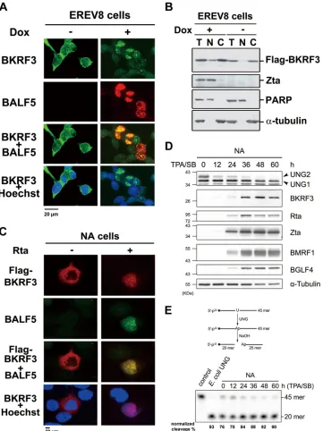
FIG 1Translocalization of BKRF3 during the lytic cycle. (A) In EREV8 cells, an Rta-inducible EBV-positive cell line, BKRF3 protein was mostly distributed in the cytoplasm before Rta expression is induced (left) and is transported into the nucleus and colocalized with BALF5 DNA polymerase after induction with 100 ng/ml doxycycline for 48 h (right). (B) Western blotting of fractionations of cell lysates indicates that in noninduced EREV8 cells, BKRF3 was mainly in the cytoplasmic (C) fraction, whereas after induction, a greater amount of BKRF3 was also detected in the nuclear (N) fraction. T, total; PARP, poly-ADP-ribose polymerase, a nuclear protein marker.␣-Tubulin serves as a cytoplasmic marker. (C) In EBV-positive NA cells cotransfected with vector control, Flag-BKRF3 was distributed in the cytoplasm at 48 h posttransfection (left) and was translocated to the nucleus when viral replication was induced by cotransfection of the Rta expression plasmid (right). The BKRF3 protein may interact with the viral DNA replication-associated proteins, as evidenced by its colocalization with BALF5 DNA polymerase in cells with virus replication. (D) To monitor temporal expression of EBV BKRF3 and cellular UNGs, lytic replication was induced in EBV-positive NA cells with TPA-SB. BKRF3 expression was detected at 12 h and significantly upregulated at 36 h postinduction. The protein expression levels of mitochondrial UNG1 remained stable, and nuclear UNG2 decreased through the time course. (E) The upregulated BKRF3 may compensate for the reduced amount of cellular UNG2 according to the results of the UDG activity analysis.
on November 7, 2019 by guest
http://jvi.asm.org/
[image:5.585.114.475.66.548.2]illustrated in
Fig. 1E
, incubating
32P-labeled 45-mer probes with
cell lysates containing DNA uracil glycosylases results in AP site
cleavage following alkaline treatment, generating
32P-labeled
20-mer fragments. The probe cleavage rate was about 76% before
induction and increased to 90% at 60 h after TPA-SB induction
(
Fig. 1E
). The time course assay indicated that, after the EBV lytic
cycle was induced, total DNA glycosylase activities increased
slightly, even though the expression of cellular UNG2 was
re-duced, and the gain of total UDG activities likely is from the
in-creased expression of BKRF3 during EBV lytic replication.
Physical interactions of BKRF3 with other EBV DNA
repli-cation proteins and the nuclear targeting of BKRF3.
Because
participation of UDG in the DNA replication core machinery was
seen for both mammalian UNG2 and some viral UDGs, we
per-formed coimmunoprecipitation assays to examine the physical
association between BKRF3 and EBV replication-associated
pro-teins. In a preliminary test with a doxycycline-induced EREV8
lysate and anti-Flag antibody, we observed that the viral DNA
polymerase and Rta, but not Zta, were coimmunoprecipitated
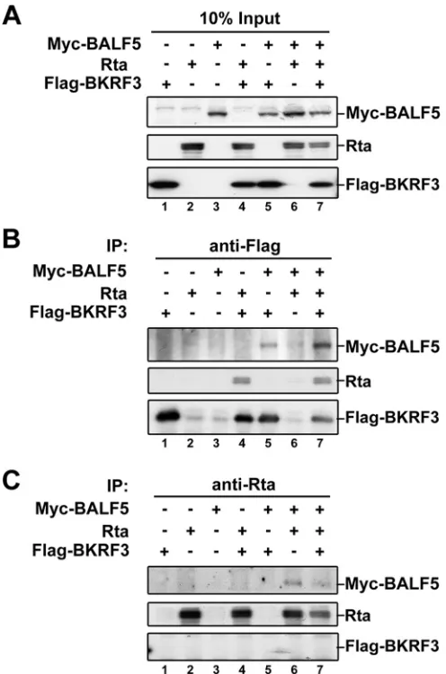
with Flag-BKRF3 (data not shown). To analyze the interactions
further, Myc-BALF5, Rta, and Flag-BKRF3 expression plasmids
were transfected into 293T cells, and protein expression was
de-tected by Western blotting (
Fig. 2A
). In the
coimmunoprecipita-tion reaccoimmunoprecipita-tion with anti-Flag antibody, Rta and Myc-BALF5 were
detected on the immunoblot with the respective antibodies,
indi-cating a complex comprised of at least these three proteins (
Fig.
2B
, lane 7). Moreover, Rta or Myc-BALF5 also formed a protein
complex with Flag-BKRF3 (
Fig. 2B
, lanes 4 and 5). Alternatively,
the immunocomplexes were captured with anti-Rta antibody and
immunoblotted, which indicates interaction between Rta and
Myc-BALF5 (
Fig. 2C
, lane 6). Together, these results suggest that
Flag-BKRF3, Rta, and Myc-BALF5 form a complex through direct
or indirect interactions. However, Flag-BKRF3 was not detected
in the Rta antibody-captured immunocomplex, possibly because
Rta antibody interferes with complex formation or the interaction
among Rta, BALF5, and BKRF3 prevents epitope recognition by
Rta antibody.
Since BKRF3-interacting proteins Rta and BALF5 localize to
replication compartments and are required for lytic EBV
replica-tion (
3
,
57
,
58
), we examined whether Rta or BALF5 can
translo-cate BKRF3 into the nucleus. To this end, HA-BKRF3 plasmid was
cotransfected with vector control or plasmid expressing Rta or
Myc-BALF5 into HeLa cells and examined with
immunofluores-cence staining. In Rta-expressing cells, BKRF3 distributed in the
cytoplasm predominantly the same as in BKRF3-only cells,
indi-cating expression of Rta did not change the cytoplasmic
distribu-tion of BKRF3 (
Fig. 3A
, upper). Interestingly, we found that
Myc-BALF5 displayed a partial colocalization pattern with BKRF3 in
the cytoplasm (
Fig. 3A
, lower). We searched for candidates that
can promote the nuclear targeting of BKRF3. Previously, UL114,
the UDG of HCMV, was found to associate with DNA polymerase
processivity factor UL44 in HCMV-infected cells (
33
). A recent
study further revealed that EBV DNA polymerase processivity
fac-tor BMRF1 interacts with BALF5 through the assistance of HSP90
to promote BALF5 nuclear targeting (
59
). Thus, we explored
whether BMRF1 can directly interact with and promote the
nu-clear targeting of BKRF3 or if the complex formation of
BMRF1-HSP90-BALF5 is required for nuclear targeting of BKRF3. In
immunofluorescence analysis, we found that coexpression of
BMRF1 can effectively promote the translocation of BKRF3 from
the cytoplasm into the nucleus in about 90% of coexpression cells
(
Fig. 3B
), suggesting the interaction with BALF5 is not required
for this process. In coimmunoprecipitation assays, BKRF3 was
detected in the immunocomplexes captured with HA
anti-body in HA-BMRF1- and Flag-BKRF3-coexpressed cells (
Fig. 3C
lane 5). Furthermore, BKRF3 and BALF5 also were detected in
BMRF1-associated immunocomplexes in the presence of all three
proteins (
Fig. 3C
, lane 7). Reciprocally, BMRF1 and BALF5 also
were detected in the anti-Flag antibody pulldown
immunocom-plexes (
Fig. 3D
, lanes 5 and 6). Interestingly, the amount of
HA-BMRF1 pulled down was less and Myc-BALF5 was not captured
by anti-Flag antibody in the presence of all three proteins,
suggest-ing the triple complex is detected by anti-Flag antibody less
effi-ciently (
Fig. 3D
, lane 7). Taken together, data here indicate that
BKRF3 interacts with viral replication-associated proteins,
in-FIG 2BKRF3 interacts with viral DNA replication-associated proteins. Im-munoprecipitation (IP) and Western blotting assays were used to examine protein interactions among BKRF3, BALF5, and Rta in a transient cotransfec-tion system. (A) Transfeccotransfec-tion of plasmid DNA expressing BALF5, Rta, or BKRF3 into 293T cells shows the expression of each protein. Vector plasmids were supplemented to ensure equal amounts of total DNA in each reaction. Cell lysates of transfected cells were displayed by Western blotting and immu-noprecipitated with Flag (B) or Rta (C) and detected with anti-BALF5, anti-Rta, or anti-Flag in the subsequent Western blotting.
on November 7, 2019 by guest
http://jvi.asm.org/
[image:6.585.298.546.66.441.2]cluding BALF5, Rta, and BMRF1, and the nuclear targeting of
BKRF3 is regulated by BMRF1.
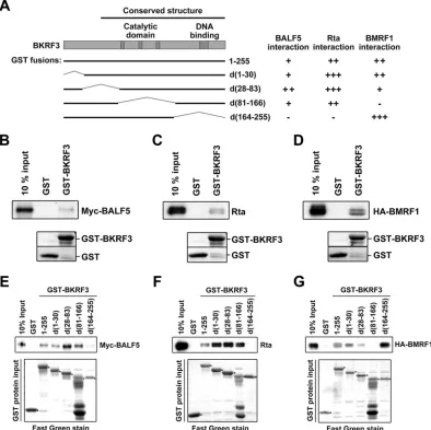
We then used a GST pulldown assay to map different domains
on BKRF3 responsible for interacting with different DNA
replica-tion proteins. The GST-BKRF3 wild type and a series of delereplica-tion
mutants were generated (
Fig. 4A
). Indeed, BALF5, Rta, or BMRF1
expressed in 293T cells was pulled down by bacterially expressed
GST-tagged wild-type BKRF3 (
Fig. 4B
to
D
). GST-fused wild-type
and mutant BKRF3 proteins were used in GST pulldown assays to
identify the domain important for interacting with BALF5 (
Fig.
4E
), Rta (
Fig. 4F
), or BMRF1 (
Fig. 4G
). BALF5 and Rta were
pulled down by GST-tagged wild-type BKRF3 and BKRF3
mu-tants d(1-30), d(28-83), and d(81-166) but not by d(164-255).
This suggests that residues 164 to 255 of BKRF3 are crucial for its
interaction with Rta and BALF5. On the other hand, BMRF1 was
pulled down by the GST-tagged wild-type BKRF3 and all BKRF3
mutants except d(81-166), suggesting BKRF3 forms complexes
with BMRF1 with a different interacting domain. The interacting
abilities of different GST deletion clones with BALF5, Rta, or
BMRF1 are shown in
Fig. 4A
. In summary, the C-terminal region
of BKRF3 is important for interaction with BALF5 and Rta,
whereas the middle region of BKRF3 is critical for interacting with
BMRF1.
Complex formation of BKRF3 with viral proteins correlated
with higher UDG activity of BKRF3 during EBV reactivation.
As
shown in
Fig. 1
, the reduced cellular UNG2 coupled with
upregu-lated BKRF3 during the EBV lytic cycle, implying BKRF3 restores
the UDG activity in virus-replicating cells. Because we have shown
that the UDG activity of purified recombinant BKRF3 is about
10-fold less than that of
E. coli
UDG in an
in vitro
UDG assay (
4
),
we wondered whether BKRF3 functions through interaction
with proteins in the viral DNA replication complex to achieve a
stronger UDG activity
in vivo
. To test this hypothesis,
Flag-BKRF3 proteins were enriched from Rta or vector-transfected
FIG 3BKRF3 is translocated into the nucleus in the presence of BMRF1. (A) HA-BKRF3 plasmid was transfected with vector control, Rta, or Myc-BALF5 plasmid into HeLa cells and detected with anti-HA, anti-Rta (467), or anti-Myc (9E10) antibody at 24 h posttransfection in an immunofluorescence assay. DNA was stained with Hoechst 33258. (B) Flag-BKRF3 was transfected into HeLa cells with HA-BMRF1 or control plasmid. At 24 h posttransfection, cells were fixed and stained for BKRF3, BMRF1, and DNA with anti-Flag antibody, anti-HA antibody, and Hoechst 33258, respectively. (C and D) 293T cells were transfected with the plasmids indicated for 24 h and harvested using radioimmunoprecipitation assay (RIPA) buffer. Cell lysates were incubated with anti-HA (C) or anti-Flag (D) antibody to precipitate BMRF1-associated or BKRF3-associated immunocomplexes and examined for BALF5, BMRF1, or BKRF3 by Western blotting. *, Ig light chain; **, Ig heavy chain.
on November 7, 2019 by guest
http://jvi.asm.org/
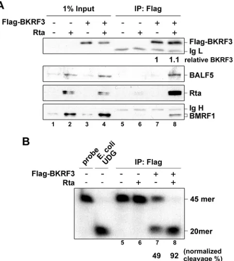
[image:7.585.103.485.67.437.2]NA cell lysates by immunoprecipitation with Flag
anti-bodies, and immunocomplexes were subjected to UDG assays.
The immunoprecipitation-Western blotting indicates that
BKRF3 forms a complex with BALF5, Rta, and BMRF1 as
de-scribed above (
Fig. 5A
, lane 8). Simultaneously, the
Rta-trans-fected immunocomplex associated with BKRF3 conferred a
significantly greater level of UDG activity (92% of probe
cleav-age) than the lysate without Rta transfection (49% of probe
cleavage) (
Fig. 5B
, lanes 7 and 8). The BKRF3 complexes
asso-ciated with viral replicating proteins, including BALF5, Rta,
and BMRF1, displayed enhanced UDG activity. Although it
cannot be excluded that the enhancement is attributed to
in-teraction with other viral proteins resulting in
posttransla-tional modification or enhanced stability of BKRF3, it is
evi-dent that the formation of BKRF3-associated complexes
enhanced BKRF3 UDG activity during EBV reactivation.
Generation of a BKRF3 knockout and the revertant EBV
bac-mid clones.
To investigate further the involvement of BKRF3 in
DNA replication during EBV lytic replication, we constructed the
p2089BKRF3STOP EBV bacmid (BKRF3STOP) by an allelic
ex-change procedure (
52
). A three-frame stop codon cassette was
inserted at amino acid 73 of BKRF3, and the incorporation of the
cassette in BKRF3 was confirmed by restriction enzyme digestion
of the inserted NheI site next to the stop codons (
Fig. 6A
). Using
BKRF3STOP bacmid as the backbone, BKRF3 revertant bacmid
(K3R) was constructed by allelic exchange with wild-type BKRF3
sequence (
Fig. 6B
). Compared to the wild-type bacmid p2089, the
24.6-kb DNA fragment containing BKRF3 was cut into two
frag-ments of 16.8 kb and 7.8 kb in the BKRF3STOP bacmid by NheI
digestion. The stop codon cassette was replaced with wild-type
sequence of BKRF3 in revertant K3R and resulted in the 24.6-kb
BKRF3-containing fragment (
Fig. 6B
and
C
, right). Furthermore,
the BamHI fragmentation analysis was performed to confirm that
no other recombination sites exist except the BKRF3 target site
(
Fig. 6C
, left). Wild-type p2089, BKRF3STOP, and K3R bacmids
were transfected into 293TetER cells, in which Rta expression can
FIG 4Mapping the interaction domains of BKRF3 using a GST pulldown assay. (A) Four GST-fused deletion mutants of BKRF3 were constructed, d(1-30), d(28-83), d(81-166), and d(164-255). The relative BKRF3-interacting abilities of individual GST fusion proteins are summarized on the right. (B to G) Bacterially expressed GST or GST-fused BKRF3 proteins were purified with glutathione beads and incubated with lysates of 293T cells transfected with Myc-BALF5, Rta, or HA-BMRF1 for 18 h. The beads were washed and examined with anti-Myc (B and E), anti-Rta (C and F), and anti-HA (D and G) antibodies by Western blotting.
on November 7, 2019 by guest
http://jvi.asm.org/
[image:8.585.95.489.69.462.2]be induced by tetracycline for the subsequent progression of
the lytic cycle genes (
44
), to establish 293TetER/p2089 (p2089),
293TetER/p2089BKRF3STOP (BKRF3STOP), and 293TetER/
K3R (K3R) stable clones.
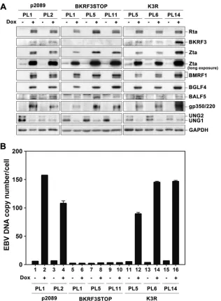
BKRF3 was required for EBV genome replication.
To
moni-tor the BKRF3 knockout effect on viral DNA replication, EBV
genome copy numbers were measured in doxycycline-treated
p2089, BKRF3STOP, and K3R cells at 72 h postinduction using
quantitative real-time PCR. After doxycycline induction,
immu-noblotting was performed to confirm that BKRF3 was detectable
in p2089 and K3R stable cells but not in BKRF3STOP cells (
Fig.
7A
). Simultaneously, induction of Rta successfully turned on viral
lytic protein expression in p2089, BKRF3STOP, and K3R cells,
including Zta, BMRF1, BGLF4, BALF5, and gp350/220 (
Fig. 7A
).
The EBV genome copy numbers increased 17- and 28-fold in
p2089 PL1 and PL2 cells, respectively, at 72 h postinduction,
whereas EBV DNA copy numbers did not significantly increase at
72 h postinduction in any of the BKRF3STOP pool clones (
Fig. 7B
,
lanes 1 to 10). In the BKRF3 revertant (K3R) clones, the EBV
genome copy number increased 18-, 44-, and 23-fold in K3R PL5,
PL6, and PL14 cells, respectively, at 72 h postinduction, indicating
that BKRF3 contributes to viral DNA replication and that there
are no additional mutations in BKRF3STOP (
Fig. 7B
, lanes 11 to
16). Notably, the expression of cellular UNG2 was significantly
reduced at 72 h postinduction in p2089, BKRF3STOP, and K3R
cells (
Fig. 7A
), very likely due to virus-mediated host shutoff
ef-fects. Thus, BKRF3 may compensate for cellular UNG2 to benefit
EBV replication at this time point.
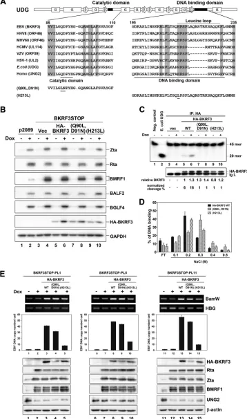
The interaction of BKRF3 with viral proteins in replication
complexes promoted EBV DNA replication through enzymatic
activity-independent pathways.
In this study, we found that the
sequence of aa 81 to 255 of BKRF3, which contains a catalytic
domain and DNA binding domain, was crucial for association
with other viral replicating components. To determine whether
BKRF3 plays other roles in addition to providing the UDG activity
in EBV DNA replication,
trans
-complementation assays were
per-formed in BKRF3STOP cells with wild-type or mutant BKRF3
expression plasmids. To this end, two Flag-BKRF3 mutants were
generated, one mutated at the catalytic site (Q90L,D91N) and the
other at the leucine loop (H213L) (
Fig. 8A
) (
37
). As indicated by
the immunoblot, viral lytic protein expression was comparable in
cells complemented with plasmids expressing wild-type or
mu-tant BKRF3 (
Fig. 8B
). In the immunoprecipitation-UDG assay,
the UDG activity of BKRF3 was normalized to the relative level of
immunoprecipitated HA-tagged BKRF3. The UDG activity of
wild-type BKRF3 increased after lytic induction (
Fig. 8C
, lanes 5
and 6), whereas the enzymatically dead BKRF3(Q90L,D91N)
failed to remove uracils (
Fig. 8C
, lanes 7 and 8). Interestingly, the
leucine loop mutant BKRF3(H213L), which contains a wild-type
catalytic domain, also was defective in UDG activity (
Fig. 8C
, lanes
9 and 10). According to the 3D structural analyses of BKRF3 and
human UNG (
37
,
60
), the leucine loop of UDG is within its
DNA-contacting region (resides 213 to 229 of BKRF3). Therefore,
sin-gle-stranded DNA cellulose chromatography was performed to
determine whether the replacement of histidine 213 with leucine
affects the general DNA binding ability of BKRF3. After binding of
in vitro
-transcribed/translated wild-type or mutant HA-BKRF3 to
the DNA cellulose, proteins were eluted with step gradients of
NaCl to indicate their relative DNA binding abilities (
Fig. 8D
).
The peaks of both wild-type BKRF3 and BKRF3(H213L) were
mostly detected in the 0.2 M NaCl fractions, indicating that the
general DNA binding affinity of BKRF3(H213L) is very close to
that of the wild type and that another mechanism is involved in the
inability of BKRF3(H213L) to remove uracil base. On the other
hand, BKRF3(Q90L,D91N) was eluted at a high level in the 0.3 M
NaCl fraction (
Fig. 8D
). According to PyMOL 3D structure
predic-tion, mutation sites of BKRF3(Q90L,D91N) are close to the

1 sheet
of BKRF3 and may create a conformational change of BKRF3 (data
not shown), leading to a slight increase in DNA binding affinity.
Consequently, the EBV genome copy number was
moni-tored to verify the biological function of BKRF3 in
BKRF3STOP cells after complementation. Compared to those
of vector control-transfected cells, EBV genome copy numbers
increased 29-, 34-, and 33-fold at 60 h postinduction in
BKRF3STOP PL1, PL5, and PL11 cells, respectively, with the
complementation of wild-type BKRF3, indicating that BKRF3
is crucial for EBV replication (
Fig. 8E
, lanes 3, 8, and 13).
Notably,
complementation
of
enzymatically
dead
BKRF3(Q90L,D91N) also increased viral DNA copies by 22-,
31-, and 18-fold in doxycycline-treated BKRF3STOP PL1, PL5,
and PL11 cells (
Fig. 8E
, lanes 4, 9, and 14), suggesting that
BKRF3 plays an essential role in the DNA replication process in
FIG 5Complex formation with viral DNA replication machinery in Rta-reactivated NA cells stimulates BKRF3 UDG activity. NA cells were seeded and transfected with vector or BKRF3-expressing plasmid, coupled with Rta or uncoupled, to induce lytic replication. At 60 h posttransfection, whole-cell lysates were harvested and Flag-BKRF3 was immunoprecipitated by anti-Flag (M2) antibody for coimmunoprecipitation assay and IP-UDG assay. (A) Ninety percent of Flag-BKRF3-associated immunocomplexes were examined for viral lytic proteins, including Rta, BALF5, and BMRF1 by immunoblotting assay. (B) Ten percent bed volumes of immunoprecipitated products were subjected to IP-UDG assay. The percentage of U-probe cleavage was quanti-fied by ImageQuant (GE Healthcare) and normalized to the relative fold of immunoprecipitated Flag-BKRF3, as shown in panel A. Data are representa-tive of two independent experiments.
on November 7, 2019 by guest
http://jvi.asm.org/
[image:9.585.45.286.67.336.2]addition to its UDG activity. In cells complemented with the
leucine loop mutant BKRF3(H213L), the level of viral DNA
increased
5-,
10-,
and
6-fold
in
doxycycline-treated
BKRF3STOP PL1, PL5, and PL11 cells, respectively (
Fig. 8E
,
lanes 5, 10, and 15). Leucine loops of gammaherpesviral UDGs
are seven amino acid longer than those of alpha- and
betaher-pesviruses (
Fig. 8A
). We found that the mutation of histidine
213 in BKRF3 attenuated its ability to rescue viral DNA
repli-cation in BKRF3STOP cells, indicating that the leucine loop of
BKRF3 is important for viral DNA replication. Thus, data here
FIG 6Construction and characterization of BKRF3 knockout and the revertant EBV bacmids. (A) Schematic summary of the BKRF3 mutant EBV bacmid cloning strategy. The termination cassette containing an NheI site and translation stop codons was inserted into nucleotide 110572 of the EBV B95.8 genome (GenBank accession no.V01555.2) by allelic exchange, as described in Materials and Methods. (B) Recombination of allelic exchange led to a size change of NheI fragments from 24.6 kb to 16.8 and 7.8 kb, resulting in the disruption of BKRF3 without interfering with the coding sequence of BKRF2. The BKRF3 revertant bacmid (K3R), which contains the same genomic pattern with which the wild-type bacmid was generated, used BKRF3STOP bacmid as the backbone. (C) The wild-type, BKRF3STOP, and K3R bacmids were digested with BamHI or NheI and displayed by agarose gel electrophoresis. The fragmented viral DNA of wild-type, BKRF3STOP, and K3R bacmids were identical in BamHI digestion (left). In addition, cleavage of the BKRF3STOP bacmid produced 16.8- and 7.8-kb fragments (right, lane 2) that replaced the wild-type 24.6-kb fragment (right, lane 1), and the NheI-STOP cassette was replaced with the wild-type genome by allelic exchange-generated K3R (right, lane 3).
on November 7, 2019 by guest
http://jvi.asm.org/
[image:10.585.133.446.65.557.2]suggest that the proper overall structure of BKRF3 contributes
more to EBV genome replication in our current system than
does enzyme activity.
To determine whether the recruitment of BKRF3 to the viral
replication compartment was affected in BKRF3 mutants,
immu-nofluorescence staining was performed to visualize the
colocaliza-tion of BKRF3 with the DNA polymerase BALF5 in NA cells (
Fig.
9A
). With the transfection of Rta expression plasmid, the
colocal-ization intensity of BKRF3(H213L) with BALF5 was weaker than
that of WT BKRF3 in reactivated NA cells in confocal analysis (
Fig.
9A
). Although BKRF3(H213L) displayed mainly intranuclear
dis-tribution, reduced colocalization with BALF5 was observed in
about 80% of NA cells with lytic cycle progression in the
immu-nofluorescence assay (data not shown). Consistent with this
find-ing, the amounts of HA-BKRF3(H213L)-associated Rta and
BALF5 were reduced (
Fig. 9B
, lanes 7 and 8). BKRF3(H213L) still
interacted with BMRF1 for its nuclear localization (
Fig. 9C
);
how-ever, its targeting to DNA replication complexes was attenuated.
Interestingly, the amount of BALF5 DNA polymerase being
coimmunoprecipitated with HA-BKRF3(Q90L,D91N) was about
2-fold higher than that of WT BKRF3 (
Fig. 9B
, lanes 3 to 6). In the
middle rows of
Fig. 9A
, more distinct colocalization signals of
HA-BKRF3(Q90L,D91N) and BALF5 also were observed. Because both
Rta and BALF5 were mapped to interact with the C terminus of
BKRF3 (
Fig. 4
), it is possible that mutation of H213L affects the ability
of BKRF3 to interact with these proteins. Overall, data here suggest
that the interaction of BKRF3 with viral proteins in replication
com-partments correlated with its ability to support viral DNA replication.
FIG 7EBV genome replication was deficient in cells containing BKRF3 knockout bacmids. (A) Wild-type p2089, BKRF3STOP, and K3R EBV bacmids were transfected into 293TetER cells, and stable clones were selected with hygromycin (50g/ml). EBV bacmid stable cells (p2089, BKRF3STOP, and K3R) were treated with doxycycline (50 ng/ml) to induce the EBV lytic cycle for 72 h, and viral lytic proteins were detected by immunoblotting. (B) The EBV genome copy number of wild-type p2089, BKRF3STOP, and K3R cells with or without doxycycline induction was determined by qPCR, using-globin as an internal control, and results were compared to those for a 10-fold serial dilution of standard DNA as described in Materials and Methods.
on November 7, 2019 by guest
http://jvi.asm.org/
[image:11.585.136.452.64.493.2]FIG 8Trans-complementation by wild-type or enzymatically dead BKRF3(Q90L,D91N), but not leucine loop mutant BKRF3(H213L), bacmids rescued lytic viral DNA replication in BKRF3STOP cells. (A) Sequence alignment of the catalytic domain and DNA binding domain of UDG within the herpesviruses,E. coli, and humans. Residues conserved within UNG (family 1) are marked in gray boxes. The domains of catalytic activity and DNA binding are indicated at the bottom, and the leucine loop involved in the DNA binding domain is indicated at the top. Secondary structures of EBV UDG are illustrated above the alignment.
on November 7, 2019 by guest
http://jvi.asm.org/
[image:12.585.112.470.76.682.2]DISCUSSION
Herpesviral DNA replication-associated enzymes have been
con-sidered good antiviral targets. The intranuclear viral DNA
repli-cation compartments contain multiple viral and cellular DNA
replication and repair enzymes and chromatin modifiers that can
coordinately function together for efficient virus replication. In
this study, we revealed how BKRF3 is recruited into the nucleus by
viral DNA polymerase processivity factor BMRF1. We also
showed the regulation of the enzyme activity of BKRF3 and its
interaction with viral DNA replication protein complexes and
vi-ral DNA replication. The inability of the BKRF3STOP bacmid to
replicate following lytic induction indicates that BKRF3 is crucial
for viral DNA replication. The distinct outcomes of
complemen-tation with BKRF3(Q90L,D91N) and BKRF3(H213L) suggest the
recruitment function of BKRF3 via its C-terminal leucine loop is
important for EBV replication.
Here, we observed that BKRF3 was translocated from the
cy-toplasm into the nucleus upon lytic induction and colocalized
with the DNA polymerase BALF5 in both NPC NA cells and
293-derived EREV8 cells (
Fig. 1A
to
C
). We also found, upon the
induction of lytic replication, that the expression levels of UNG2
decreased along with increasing expression of viral lytic
replica-tion proteins (
Fig. 1D
and
E
). The exact mechanism leading to
UNG decrease during virus replication is not clear. However, the
expression of UNG2 was reported to peak in and throughout S
phase and then decline to undetectable levels until the next S phase
through the ubiquitin-proteasome pathway (
61
). Taking into
ac-count that EBV replication causes cell cycle arrest at the G
1/S
tran-sition without cellular DNA replication (
62
,
63
), we suspect the
decrease of UNG2 is caused by downregulated transcription or
increased protein degradation.
Unlike cellular UNG2, which contains a unique NLS as well as
subnuclear targeting signals (
16
), we found that the interaction of
BKRF3 with viral DNA polymerase processivity factor BMRF1
promoted its nuclear transport (
Fig. 3
). The nuclear targeting of
BKRF3 is similar to that of HCMV UL114, which associates with
UL44, the homolog of BMRF1 (
31
), suggesting interactions with
nuclear replicating proteins are required for herpesviral UDGs to
participate in the viral replication compartment. The nuclear
tar-geting of BALF5 was recently reported to be dependent on BMRF1
and cellular chaperone protein Hsp90 (
59
). It is not clear whether
other cellular proteins are required for BMRF1-dependent
nu-clear targeting of BKRF3.
Interaction of cellular UNG2 with PCNA and RPA replicating
complex ensures postreplicative repair of misincorporated uracil
(U·A) in DNA (
64
). Similarly, the association between UDGs and
viral DNA polymerases to function in replication-coupled base
excision repair has been described for HSV-1 and HCMV (
29
,
32
).
HCMV UL44 facilitates loading of UL114 onto DNA and
pro-motes UL114 to remove uracil from DNA (
33
). In our study,
BKRF3 interacted with the immediate-early transactivator Rta,
DNA polymerase BALF5, and DNA polymerase processivity
fac-tor BMRF1 in coimmunoprecipitation and GST pulldown assays
(
Fig. 2
to
4
). We suggest the interaction with BALF5 and BMRF1
helps BKRF3 to form complexes with other replication proteins to
enhance UDG activity of BKRF3 and carry out repairs at DNA
replication loci. Because Rta was found to promote the nuclear
targeting of the DNA primase-associated factor BBLF2/3, which is
the linker for proteins targeting viral DNA replication origin (
65
),
it is possible that Rta also helps the stabilization of BKRF3 to the
DNA replication complex through network interactions with
other viral replication proteins. Furthermore, the UDG activity of
HA-BKRF3 was stimulated when it formed complexes with other
viral DNA replication proteins (
Fig. 5
). In this way, viral UDG
activity may be regulated to function along with viral DNA
repli-cation, avoiding nonspecific DNA damages.
A search through the literature found that the UDG homologs
are dispensable in alphaherpesviruses for infection of actively
growing fibroblasts but are essential for infection and reactivation
in the murine nervous system (
29
,
30
). The UDG of HCMV,
UL114, is required for full virus replication in a culture system.
Viral replication of the HCMV UL114 mutant is retarded in
quiescent fibroblasts but proceeded smoothly in actively
grow-ing fibroblasts (
35
). Additionally, using a random
signature-tagged mutagenesis approach to select the viral genes essential
for the murine gammaherpesvirus 68 life cycle, Song et al.
found viral UDG ORF46 is not essential for viral replication in
a mouse fibroblast cell line (
66
). These observations suggest
that cellular UDG activity is able to compensate for viral UDG
deficiency to a certain extent. Previously, we found viral DNA
replication was decreased about 10 to 20% at 48 h
postinduc-tion when BKRF3 was knocked down by siRNA (
4
). It is
possi-ble that residual BKRF3 was sufficient to support EBV DNA
replication complex formation in that setting. In this study, we
demonstrated cellular UNG2 was undetectable at 60 h
post-EBV reactivation, while expression of BKRF3 gradually
in-creased and the total UDG activity was sustained in NA cells
(
Fig. 1E
). Thus, the BKRF3 UDG activity may function in cells
with low levels of UNG2 expression.
Notably, the presence of BKRF3 in the viral DNA replication
compartment is correlated with its function in DNA replication.
In bacmid experiments, we found the EBV DNA copy number
The mutation sites of enzymatically dead BKRF3(Q90L,D91N) and leucine loop mutant BKRF3(H213L) are indicated. (B) p2089 and BKRF3STOP cells were transfected with 7, 14, or 4g of HA-tagged wild-type BKRF3 (WT), BKRF3(Q90L,D91N), or BKRF3(H213L) plasmid, respectively, and treated with doxycy-cline (50 ng/ml) at 12 h posttransfection. After Doxycydoxycy-cline (Dox) treatment for 60 h, cells were harvested for Western blotting of viral lytic proteins. (C) The UDG activities of HA-tagged BKRF3 WT, BKRF3(Q90L,D91N), and BKRF3(H213L) in BKRF3STOP cells treated with doxycycline or left untreated. The transfected cell lysates were prepared for IP-UDG assays as described in Materials and Methods.E. coliUDG (10 U) was used as a positive control. The cleavage percentage of input32P-labeled U probe was normalized to relative amounts of immunoprecipitated BKRF3. (D) The DNA binding activities ofin vitro
-transcribed/translated [35S]methionine-labeled BKRF3 WT, BKRF3(Q90L,D91N), and BKRF3(H213L) were measured in the binding buffer with ssDNA
cellulose chromatography, eluted with binding buffer with step gradients of NaCl, and subjected to electrophoresis with 15% SDS-PAGE. The DNA-bound [35S]methionine-labeled proteins in each eluent were measured by phosphorimaging and normalized to the total input. Data are representative of two
inde-pendent experiments. (E) BKRF3STOP pool clones (PL1, PL5, and PL11) were transfected with control vector (vec), HA-BKRF3 WT, BKRF3(Q90L,D91N), or BKRF3(H213L). At 24 h posttransfection, doxycycline (Dox; 50 ng/ml) was added to induce lytic replication for 60 h. Subsequently, intracellular EBV DNA copy numbers oftrans-complementation cells were quantified with qPCR for the EBV BamHI W fragment and human beta-globin (HBG) as described in Materials and Methods. Data are representative of three independent experiments.
on November 7, 2019 by guest
http://jvi.asm.org/
significantly increased about 20- to 40-fold following the
induc-tion of Rta by doxycycline in p2089 and in K3R revertant cells, but
not in BKRF3STOP cells, at 72 h postinduction (
Fig. 7
). In the
sequence alignment, the extended leucine loop at the
C-termi-nal region is conserved among gammaherpesviruses (
Fig. 8A
),
suggesting functions in facilitating viral DNA replication.
BKRF3STOP virus, which was inserted with a stop codon cassette
at aa 73 of BKRF3, did not replicate after lytic induction. However,
MHV-68 ORF46 mutant virus, which was inserted with a
trans-poson at aa 103 of the viral UDG, still produced infectious viruses
(
66
). We suspect that BKRF3 plays a unique role in EBV DNA
replication or the residual fragment of truncated UDG in
MHV-68 still facilitates viral DNA replication. Nevertheless,
ex-pression levels of UNG2 in virus-replicating cells may affect the
outcome of viral UDG defective viruses. Thus, it would be
inter-esting to address whether UNG2 also participates in the viral DNA
FIG 9Point mutation at His213 of BKRF3 attenuated its recruitment to the viral DNA replication compartment and its ability to interact with the viral DNA polymerase BALF5 and immediate-early gene, Rta. (A) NA cells were seeded at 3⫻106cells per 10-cm dish for slide culture and transfected with Flag-BKRF3 or
control plasmid combined with vector or Rta expression plasmid. At 48 h posttransfection, cells were fixed with 4% paraformaldehyde and stained for BKRF3, BALF5, and DNA with mouse anti-Flag antibody, rabbit BALF5 antiserum, and Hoechst 33258, followed by confocal microscopy analysis. (B) To measure the ability of wild-type or mutant BKRF3 to interact with viral DNA replication-associated proteins, NA cells transfected with 7, 14, or 4g of HA-BKRF3 WT, BKRF3(Q90L,D91N), or BKRF3(H213L) were induced into the lytic cycle by Rta transfection. At 60 h posttransfection, cell lysates were harvested for coimmu-noprecipitation assay. The HA-BKRF3-associated complexes were precipitated with anti-HA and detected with anti-Rta, anti-BALF5, and anti-HA antibodies by Western blotting. This is a representative result of triplicate experiments. (C) To verify the interacting abilities of BMRF1 with BKRF3 WT, BKRF3(Q90L,D91N), and BKRF3(H213L), 293T cells were cotransfected with expression plasmids as indicated for 48 h. Whole-cell lysates were subjected to a coimmunoprecipitation assay. The HA-BMRF1-associated protein complexes were precipitated with anti-HA and detected with anti-Flag by Western blotting. This is a representative result of duplicate experiments. *, Ig heavy chain; **, Ig light chain.
on November 7, 2019 by guest
http://jvi.asm.org/
[image:14.585.112.473.67.512.2]replication compartment and tethers other cellular DNA
replica-tion factors to enhance viral DNA replicareplica-tion.
Complementary expression of both the wild type and two
BKRF3 mutants in BKRF3STOP cells further demonstrated the
contribution of different functional domains of BKRF3 in viral
DNA replication. Previously, we showed that blocking cellular
and viral UDG activity by the inhibitor Ugi decreases 50% of
EBV DNA replication. Here, the fact that enzymatically dead
mutant BKRF3(Q90L,D91N) restored EBV genome replication
in BKRF3STOP cells at a slightly lower level than WT BKRF3
strongly suggests that BKRF3 has a function other than its
enzy-matic activity for EBV genome replication (
Fig. 8E
). With nuclear
distribution upon lytic induction, BKRF3(H213L) interacts with
proteins in the viral DNA replication complex less efficiently, as
revealed by immunofluorescence and coimmunoprecipitation
ex-periments (
Fig. 9
). Data here hint that precise associations with
other components in the complex affect the enzymatic activities
and function(s) of BKRF3. It is possible that BKRF3 promotes a
conformational change of BALF5/BMRF1 complexes to enhance
DNA synthesis. Such interactions also ensure viral UDG is
re-cruited to viral DNA replication forks before DNA synthesis.
Regulation through specific interaction with DNA binding
proteins were founded in cellular and viral UDGs. For example,
UNG2 directly interacts with XRCC1 to enhance the repair
effi-ciency of the XRCC1-mediated repair pathway (
64
,
67
). Likewise,
the HCMV UDG, UL114, was found to interact with the
chroma-tin remodeling factor SMARCB1 and to participate in the
recruit-ment of the chromatin remodeling complex onto replication foci
(
33
). Although no BKRF3-interacting cellular proteins were
iden-tified in two previous yeast-two hybrid screening studies (
68
,
69
),
it is still possible that BKRF3 also recruits the cellular machinery to
support viral DNA replication.
ACKNOWLEDGMENTS
We thank Ji-Ying Huang at the National Taiwan University Hospital Im-age Core Laboratory for confocal analysis and Hsiu-Ming Shih of the Institute of Biomedical Sciences, Academia Sinica, for helpful discussions. We appreciate the p2089 bacmid from H. J. Delecluse. We are grateful to Ren Sun and Ting-Ting Wu (Department of Molecular and Medical Phar-macology, University of California, Los Angeles, David Geffen School of Medicine, Los Angeles, CA, USA) for the protocol and reagents for gen-erating recombinant bacmids and Tim J. Harrison of University College London for critical reading and modification of the manuscript.
This study was supported by the Taiwan National Science Council (grant NSC 101-2320-B-002-031-MY3), the National Health Research Institute (grant NHRI EX103-10201BI), and National Taiwan University (intramural grant 103C101-A4).
REFERENCES
1.Longnecker R, Kieff E, Cohen J.2013. Epstein-Barr virus, p 1898 –1959. InKnipe DM, Howley PM, Cohen JI, Griffin DE, Lamb RA, Martin MA, Racaniello VR, Roizman B (ed), Fields virology, 6th ed, vol 2. Lippincott Williams & Wilkins, Philadelphia, PA.
2.Fixman ED, Hayward GS, Hayward SD.1995. Replication of Epstein-Barr virus oriLyt: lack of a dedicated virally encoded origin-binding pro-tein and dependence on Zta in cotransfection assays. J. Virol.69:2998 – 3006.
3.El-Guindy A, Ghiassi-Nejad M, Golden S, Delecluse HJ, Miller G.2013. Essential role of Rta in lytic DNA replication of Epstein-Barr virus. J. Virol.
87:208 –223.http://dx.doi.org/10.1128/JVI.01995-12.
4.Lu CC, Huang HT, Wang JT, Slupph