Klebsiella Phage
⌽
K64-1 Encodes
Multiple Depolymerases for Multiple
Host Capsular Types
Yi-Jiun Pan,aTzu-Lung Lin,bChing-Ching Chen,bYun-Ting Tsai,b
Yi-Hsiang Cheng,bYi-Yin Chen,bPei-Fang Hsieh,bYi-Tsung Lin,cJin-Town Wangb,d Department of Microbiology, School of Medicine, China Medical University, Taichung, Taiwana; Department of Microbiology, National Taiwan University College of Medicine, Taipei, Taiwanb; Division of Infectious Diseases, Department of Medicine, Taipei Veterans General Hospital, Taipei, Taiwanc; Department of Internal Medicine, National Taiwan University Hospital, Taipei, Taiwand
ABSTRACT The genome of the multihost bacteriophage⌽K64-1, capable of infect-ing Klebsiella capsular types K1, K11, K21, K25, K30, K35, K64, and K69, as well as
new capsular types KN4 and KN5, was analyzed and revealed that 11 genes (S1-1,
S1-2, S1-3, S2-1, S2-2, S2-3, S2-4, S2-5, S2-6, S2-7, and S2-8) encode proteins with
amino acid sequence similarity to tail fibers/spikes or lyases. S2-5 previously was
shown to encode a K64 capsule depolymerase (K64dep). Specific capsule-degrading activities of an additional eight putative capsule depolymerases (S2-4 against K1, S1-1 against K11, S1-3 against K21, S2-2 against K25, S2-6 against K30/K69, S2-3 against K35, S1-2 against KN4, and S2-1 against KN5) was demonstrated by expression and purifica-tion of the recombinant proteins. Consistent with the capsular type-specific
depoly-merization activity of these gene products, phage mutants ofS1-2,S2-2,S2-3, orS2-6
lost infectivity for KN4, K25, K35, or K30/K69, respectively, indicating that capsule de-polymerase is crucial for infecting specific hosts. In conclusion, we identified nine functional capsule depolymerase-encoding genes in a bacteriophage and correlated ac-tivities of the gene products to all ten hosts of this phage, providing an example of type-specific host infection mechanisms in a multihost bacteriophage.
IMPORTANCE We currently identified eight novel capsule depolymerases in a multi-hostKlebsiellabacteriophage and correlated the activities of the gene products to all hosts of this phage, providing an example of carriage of multiple depolymerases in a phage with a wide capsular type host spectrum. Moreover, we also established
a recombineering system for modification of Klebsiella bacteriophage genomes
and demonstrated the importance of capsule depolymerase for infecting specific hosts. Based on the powerful tool for modification of phage genome, further studies
can be conducted to improve the understanding of mechanistic details ofKlebsiella
phage infection. Furthermore, the newly identified capsule depolymerases will be of great value for applications in capsular typing.
KEYWORDS Klebsiella, bacteriophage, capsular type, capsule depolymerase, multiple host
T
he genusKlebsiella, especially the species Klebsiella pneumoniae, is an importanthuman pathogen that causes a wide range of diseases, including both community and hospital-acquired infections. It is associated with septicemia, pneumonia, and urinary tract infections (1, 2) and also is responsible for a globally emerging disease, pyogenic liver abscess complicated with metastatic meningitis and endophthalmitis (3, 4).
Klebsiella spp. typically display a layer of thick, polysaccharide-based capsule on their surfaces. The expression of diverse capsule structure caused by different sugar compositions and linkages divide them into distinct serotypes. In addition, genetic
Received20 December 2016Accepted22 December 2016
Accepted manuscript posted online11 January 2017
CitationPan Y-J, Lin T-L, Chen C-C, Tsai Y-T, Cheng Y-H, Chen Y-Y, Hsieh P-F, Lin Y-T, Wang J-T. 2017. Klebsiella phage ΦK64-1 encodes multiple depolymerases for multiple host capsular types. J Virol 91:e02457-16.https:// doi.org/10.1128/JVI.02457-16.
EditorRozanne M. Sandri-Goldin, University of California, Irvine
Copyright© 2017 American Society for Microbiology.All Rights Reserved. Address correspondence to Jin-Town Wang, [email protected].
crossm
on November 7, 2019 by guest
http://jvi.asm.org/
variation of capsular polysaccharide synthesis (cps) regions in various types was also indicated. At present, at least 81 capsular types have been defined, including 77 types from reference strains recognized by serological reactivity tests established during the
period 1926 to 1977 and 4 new types ofK. pneumoniae(KN1 to KN4) characterized by
molecular genotyping and phage typing in recent years (5–9). These polysaccharide coats confer resistance to host immune defenses and hostile environments (10, 11) and are associated with increased virulence (12, 13). Moreover, capsule could also act as a primary receptor for bacteriophage, which often possess tail fibers or tail spikes containing capsule depolymerization activities (14, 15). Degradation of bacterial cap-sule enables the phage to gain access to the host cell surface and bind to the secondary receptor. Given the specificity of capsule depolymerases, capsule type-specific phage or
depolymerases also have been used in capsular typing.Klebsiellaphages have been
reported since 1940 and have been used for determination of several K-types of
Klebsiella(16–21). However, little was known about the host specificity determinants of these phages until the recent characterization and identification of bacteriophage-borne depolymerases (6, 22). A KN2-specific phage, 0507-KN2-1, and its capsule de-polymerase were identified and used for capsular typing of the new KN2 capsular type (6). Another study documented a phage (NTUH-K2044-K1-1) and its capsule depoly-merase that exhibited specificity for capsular type K1 and showed therapeutic efficacy
in K1 K. pneumoniae-infected mice (22). We recently isolated a multihost Klebsiella
-infecting bacteriophage,⌽K64-1 (8), which appeared to belong to theMyoviridaefamily
of viruses based on the sequence similarity. This phage (with a genome size of 346,602
bp) has 541 probable protein-coding genes (⬎300 bp in length), including 11 tail
fiber/spike or lyase encoding genes (designatedS1-1,S1-2,S1-3,S2-1,S2-2,S2-3,S2-4,
S2-5,S2-6,S2-7, andS2-8in the present study), which may possess depolymerization
activity. In a previous study, we presented the capsule depolymerase activity of K64dep
encoded by S2-5 and demonstrated the efficacy for treatment of
multiple-drug-resistantK. pneumoniaeinfections.
Given the correlation between phage-encoded capsular depolymerases and phage
host specificity, we speculated that⌽K64-1 (which has been proved to infectKlebsiella
K1, K11, K21, K25, K30, K35, K64, and K69 reference strains [8] and is here shown to be able to infect new types KN4 and KN5 as well) could encode other capsule depoly-merases apart from K64dep. Therefore, we sought to explore the functions of the rest of tail fiber/spike or lyase encoding genes and better understand their contributions to the wide capsular type host spectrum. In the present study, we identified an additional
eight⌽K64-1-encoded capsule depolymerases by assessing the specificity of proteins
produced from the corresponding genes and of recombinant bacteriophage deleted for the respective loci. This result provided a correlation between the activities of the
nine capsule depolymerases encoded by⌽K64-1 and the host range of this virus. These
observations revealed that carriage of multiple depolymerases accounts for this phage’s multiple host-specific infective activities. The newly identified capsule depoly-merases are expected to facilitate the classification of these capsular types.
RESULTS
Host range of ⌽K64-1. Seventy-seven reference strains, four strains with docu-mented new capsular types (KN1, KN2, KN3, and KN4), and an additional strain (Ca0431)
with a newwzcsequence (9) (accession numberLC121097) that may correspond to a
novel capsular type (designated KN5) of Klebsiellawere used to determine the host
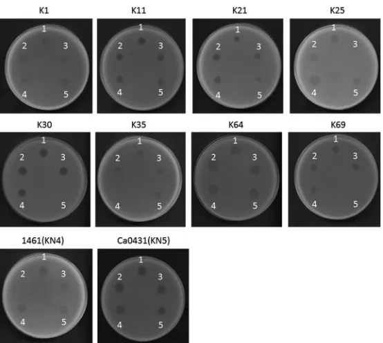
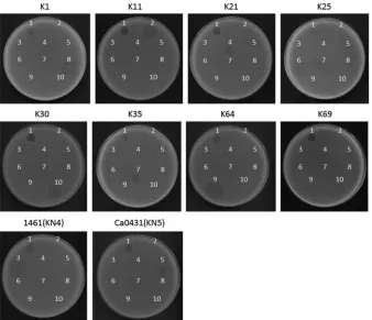
range of⌽K64-1. The results of spot tests indicated that⌽K64-1 can infect ten strains,
i.e., reference strains K1, K11, K21, K25, K30, K35, K64, and K69 and the new type strains 1461 (KN4) and Ca0431 (KN5) (Fig. 1). Additional K1 (NTUH-K2044, Canada PLA, A8126, and ATCC 35593), K21 (KCR74), K25 (KCR75), K30 (1353), KN4 (4565), and K64 (KCR2A, KCR3, KCR4, and KCR5) strains also were included in the spot tests. The results indicated
that these clinical strains were infected by⌽K64-1 (Fig. 2). We therefore hypothesized
that⌽K64-1 possessed capsule depolymerases able to recognize and digest at least 10
types of capsule.
on November 7, 2019 by guest
http://jvi.asm.org/
Analysis of the genome sequences and morphology of ⌽K64-1.The circularly
permutated genome of ⌽K64-1 is 346,602 bp (accession number AB897757) with a
G⫹C content of 31.7%. Analysis indicated the presence of 541 probable protein-coding
genes (⬎300 bp in length) and four functional tRNA genes (tRNAArg, tRNASer, tRNASer2,
and tRNAAsn) within the⌽K64-1 genome. Two genes code for RNA polymerase sigma
factors were predicted; however, no homologue of RNA polymerase was detected from
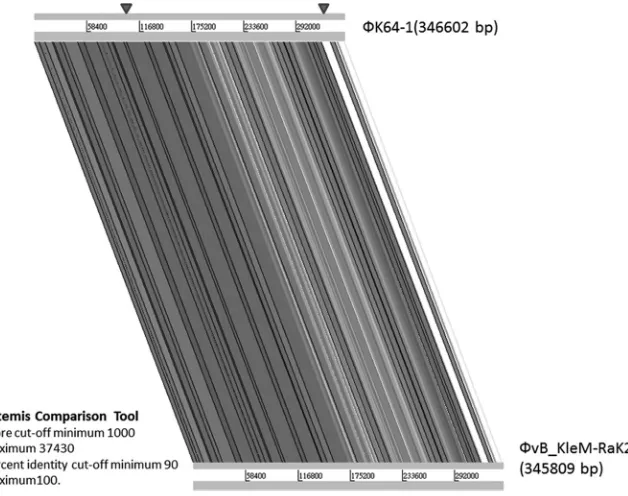
the genome. Nucleotide BLAST analysis revealed that ⌽K64-1 exhibited high DNA
similarity with a largeKlebsiella-infecting myovirus with a genome size of 345,809 bp,
bacteriophage vB_KleM-RaK2 (accession numberJQ513383); our sequence covered 94%
of the vB_KleM-RaK2 genome and showed 99% maximum DNA identity (144,392 bp/ 145,643 bp). Despite the high level of sequence similarity, some variable regions were
indicated by comparative genomic analysis (Fig. 3). We further screened the ⌽K64-1
genome for genes that encode tail fibers/spikes or lyases because these gene products were reported to have enzymatic activity against the bacterial capsule (23), which
deter-mine host range. In⌽K64-1, the products of 11 open reading frames (ORFs; S1-1 [GenBank
accession numberLC121100], S1-2 [LC121098], S1-3 [LC121099], S2-1 [LC121101], S2-2
[LC121102], S2-3 [LC121103], S2-4 [LC121104], S2-5 [AB897513], S2-6 [LC121105], S2-7 [LC121106], and S2-8 [LC121107]) exhibited similarity with tail fiber/spike or lyase proteins (Table 1). Among these 11 gene products, S2-2, S2-3, S2-6, and S2-8 showed a high degree of similarity to (are closely related to) proteins encoded by genes from
bacteriophage vB_KleM-RaK2 (⬎90% amino acid identity); the predicted S2-1, S2-4,
S2-5, and S2-7 proteins exhibited moderate sequence identity to gene products from bacteriophage vB_KleM-RaK2 (518/633 [82%], 231/259 [89%], 225/298 [76%], and 454/ 605 [75%], respectively). Notably, the S1-1, S1-2, and S1-3 proteins seemed to be unique
to ⌽K64-1, given that these three gene products showed only small regions of
sequence similarity to proteins encoded by bacteriophage vB_KleM-RaK2 (Table 2 and
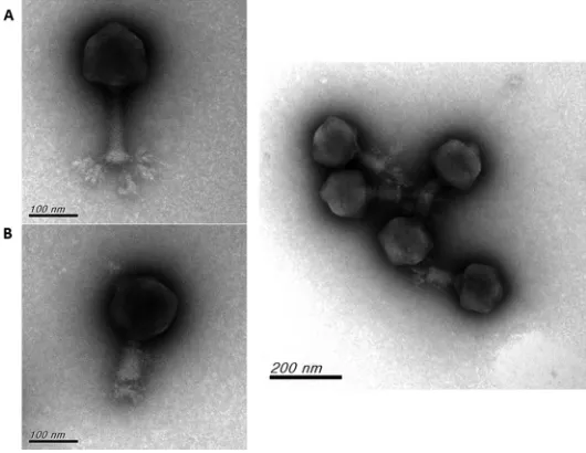
Fig. 4). The morphology of purified ⌽K64-1 phage particles was examined using
transmission electron microscopy (TEM) (Fig. 5). The phage is characterized by an
isometric head and a contractile tail and resembled Myoviridae family members.
FIG 1Spot test of wild-type andS1-2,S2-2,S2-3, andS2-6deletion mutants of⌽K64-1. The reference strains K1, K11, K21, K25, K30, K35, K64, K69, 1461 (KN4) and Ca0431 (KN5) were grown on LB plates. Phages (106PFU) were spotted on the plate, and the lysis zone could be observed after overnight incubation. Spots: 1, wild type; 2,S1-2deletion mutant; 3,S2-2deletion mutant; 4,S2-3deletion mutant; 5,S2-6deletion mutant.
on November 7, 2019 by guest
http://jvi.asm.org/
[image:3.585.68.345.68.316.2]Furthermore, this phage appeared to have six long tail fibers displaying spike-like structures, which is similar to bacteriophage vB_KleM-RaK2.
Expression of putative capsule depolymerases.In order to determine whether
these putative tail fibers/lyases had capsule-digesting activities, these ⌽K64-1 genes
FIG 2Spot test of⌽K64-1 and capsule depolymerases on additional clinical isolates ofKlebsiella. Clinical isolates with K1 capsule (A8126, NTUH-K2044, Canada PLA, and ATCC 35593), with K21 capsule (KCR74), with K25 capsule (KCR75), with K30 capsule (1353), with KN4 capsule (4565), and with K64 capsule (KCR2A, KCR3, KCR4, and KCR5) were grown on LB plates. Phages or capsule depolymerase were spotted on the plate, and a plaque- or capsule depolymerase-generated semiclear spot could be be observed after overnight incubation.
FIG 3Genome comparative analysis of⌽K64-1 and⌽vB_KleM-RaK2. Genome comparative analysis was performed using an Artemis comparison tool (score cutoffs: minimum, 1,000, and maximum, 37,430; percent identity cutoffs: minimum, 90, and maximum, 100). Arrowheads indicate the regions in which 11 putative capsule depolymerases were located (S2-8 is in the left site; the remaining 10 genes are in the right area).
on November 7, 2019 by guest
http://jvi.asm.org/
[image:4.585.73.338.67.314.2] [image:4.585.52.366.451.699.2]were cloned and expressed via a pET-28c or a pcold TF DNA expression system. Capsule depolymerase activities of the resulting purified proteins then were assessed by spot
tests against the ten known⌽K64-1 hosts (K1, K11, K21, K25, K30, K35, K64, K69, KN4,
and KN5). Results indicated that nine of the eleven putative tail fibers/spikes/lyases generated semiclear spots on individual lawns of K1, K11, K21, K25, K30, K35, K64, K69, KN4, or KN5 bacteria. Specifically, activities were observed as follows: S2-4 against K1, S1-1 against K11, S1-3 against K21, S2-2 against K25, S2-6 against K30/K69, S2-3 against K35, S2-5 against K64, S1-2 against KN4, and S2-1 against KN5 (Fig. 6), whereas no depolymerase activity was observed in S2-7 and S2-8. The specificities of these capsule depolymerases were further clarified by testing against all other documented capsular types and additional strains, including capsular types K1 (NTUH-K2044, Canada PLA, A8126, and ATCC 35593), K21 (KCR74), K25 (KCR75), K30 (1353), KN4 (4565), and K64 (KCR2A, KCR3, KCR4, and KCR5). These results revealed that each of the nine enzymes could digest only capsule from the respective unique capsular type (as indicated above), with the sole exception of S2-6, which was able to digest capsule from strains of both type K30 and type K69 (Fig. 2 and data not shown). Therefore, these enzymes appear to be capsule type-specific depolymerases, such that the nine enzymes
correspond to all ten hosts of⌽K64-1. To clarify whether proteins with activities for
[image:5.585.41.548.83.275.2]depolymerization of the same capsule exhibit sequence similarity, we compared the
TABLE 1Putative tail fibers/spikes/capsule depolymerases of phage K64-1a
ORF Type (location [nt])
Product
size (aa) Homologue Accession no
Sequence
identity (%)b Activityc S1-1 Complementary (325887–327995) 702 Tail fiber ofKlebsiellaphage K11 YP_002003830.1 360/590 (61) K11*
S1-2 Complementary (321593–323803) 736 Tail spike protein head-binding protein of
Klebsiellaphage 0507-KN2-1
YP_008532048.1 44/113 (39) KN4†
S1-3 Complementary (323855–325810) 651 Tail spike protein ofSalmonellaphage FSL SP-063 AGF88658.1 44/140 (31) K21*
S2-1 Complementary (328067–331648) 1193 Putative tail fiber protein ofPectobacterium
phage PP1
YP_007010682.1 40/133 (30) KN5*
S2-2 Complementary (331729–333483) 584 Phage T7 tail fiber protein ofKlebsiella pneumoniae
WP_020326882.1 177/496 (36) K25†
S2-3 Complementary (333493–335832) 779 K5 lyase ofEnterobacterphage K1-5 YP_654147.1 72/232 (31) K35†
S2-4 Complementary (335902–338568) 888 Tail fiber protein ofEnterobacterphage EcP1 YP_007003187.1 57/221 (26) K1*
S2-5 338917–341907 996 Putative tail fiber ofEnterobacterphage RTP YP_398994.1 171/579 (30) K64*
S2-6 341980–344283 767 Pectate lyase superfamily protein ofKlebsiella pneumoniae
WP_020801644.1 161/462 (35) K30, K69†
S2-7 344312–346471 719 Tail fiber domain protein ofPseudomonas savastanoipv.savastanoiNCPPB 3335
EFH99537.1 45/96 (47)
S2-8 90154–91941 595 Putative tail fiber protein ofCronobacterphage vB_CsaM_GAP32
YP_006987359.1 255/510 (50)
ant, nucleotide; aa, amino acids. bAs determined by BLAST-P.
c*, Evidenced by protein expression;†, evidenced by both protein expression and phage mutants.
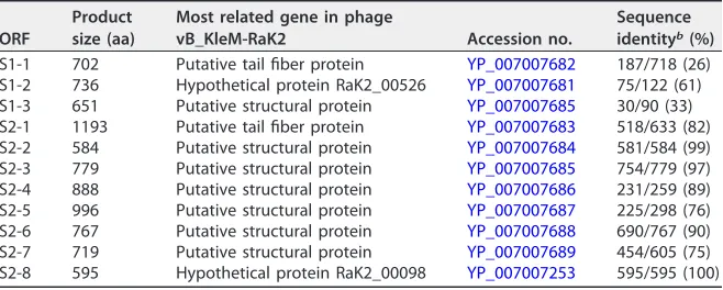
TABLE 2Related genes of phage K64-1 putative tail fibers/spikes/capsule depolymerases in phage vB_KleM-RaK2a
ORF
Product size (aa)
Most related gene in phage
vB_KleM-RaK2 Accession no.
Sequence identityb(%) S1-1 702 Putative tail fiber protein YP_007007682 187/718 (26) S1-2 736 Hypothetical protein RaK2_00526 YP_007007681 75/122 (61) S1-3 651 Putative structural protein YP_007007685 30/90 (33) S2-1 1193 Putative tail fiber protein YP_007007683 518/633 (82) S2-2 584 Putative structural protein YP_007007684 581/584 (99) S2-3 779 Putative structural protein YP_007007685 754/779 (97) S2-4 888 Putative structural protein YP_007007686 231/259 (89) S2-5 996 Putative structural protein YP_007007687 225/298 (76) S2-6 767 Putative structural protein YP_007007688 690/767 (90) S2-7 719 Putative structural protein YP_007007689 454/605 (75) S2-8 595 Hypothetical protein RaK2_00098 YP_007007253 595/595 (100)
aaa, amino acids. bAs determined by BLAST-P.
on November 7, 2019 by guest
http://jvi.asm.org/
[image:5.585.42.371.590.722.2]predicted amino acid sequence of S2-4 to that of a recently reported K1 capsule depolymerase, K1-ORF34 protein (22); the two gene products exhibited only limited amino acid sequence identity (205/612 [33%]) across the entire lengths of the two proteins. Moreover, we did not find any sequence conservation among the eleven
putative tail fibers/spikes/lyases encoded by⌽K64-1.
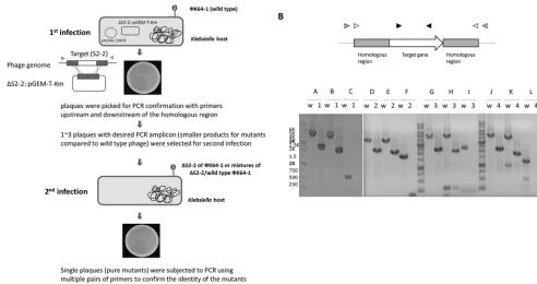
Determination of the functions of putative capsule depolymerases by deletion analysis in recombinant phage.We randomly selected four genes (the ORFs encoding
S1-2, S2-2, S2-3, or S2-6) to investigate their roles in infection of⌽K64-1 by generating
deletion mutants (deleted individually) (Fig. 7B). The positive rate of the desired mutants was shown in Table S1 in the supplemental material. As the results indicate, the frequency varied in different mutants (from 9 to 80% in the first infection). Since type/mutant mixtures existed when the first infection was carried out (i.e.,
wild-FIG 4Comparison of the coding regions of putative capsule depolymerases of⌽K64-1 and⌽ vB_KleM-RaK2. ORFsS1-1,S1-2,S1-3,S2-1,S2-2,S2-3,S2-4,S2-5,S2-6, andS2-7of⌽K64-1 and corresponding genes in⌽vB_KleM-RaK2 were compared, and the amino acid sequence identities are shown (results forS2-8
and its homolog in⌽vB_KleM-RaK2 are not shown in the diagram). Gene products that shared a high degree of similarity (ⱖ90% amino acid identity) are indicated by black arrows. Gene products that have no significant sequence similarity (and thus were unable to correlate to any genes in this region) are indicated by white arrows. Gray arrows indicate genes that exhibited⬍90% sequence identity to their corresponding genes.
FIG 5Electron micrographs of⌽K64-1. (A)⌽K64-1 phage particles with extended tail; (B)⌽K64-1 phage particles with contracted tail.
on November 7, 2019 by guest
http://jvi.asm.org/
[image:6.585.80.329.71.244.2] [image:6.585.72.337.511.716.2]type and mutant amplicons coexist in a single plaque [data not shown]), we selected one to three mutant-positive plaques and performed an additional, second infection to obtain single plaques again and finally confirmed the pure mutants with multiple primers. Having these mutants, we used spot tests and an efficiency-of-plating (EOP) assay to determine the effect of these deletions on phage infectivity toward different
hosts. Spot tests revealed that phage harboring ΔS1-2, ΔS2-2, ΔS2-3, and ΔS2-6lost
infectivity for KN4, K25, K35, and K30/K69, respectively, but retained infectivity for other
hosts (Fig. 1). The EOP assay also showed that ΔS1-2, ΔS2-2, ΔS2-3, and ΔS2-6lost the
ability to infect specific hosts, as spot tests revealed, and the ability to infect other types
was not significantly altered compared to that of the wild type (Table 3, Pⱖ 0.05
[Student t test]).These data are consistent with the protein expression and activity
experiment (above) that demonstrated that S1-2, S2-2, S2-3, and S2-6 are capsule depolymerases with specificity for KN4, K25, K35, and K30/K69, respectively. These results further demonstrated that these capsule depolymerases are crucial for the
infection of specific hosts by⌽K64-1.
Each⌽K64-1 virion possesses nine tail fiber/capsule depolymerase proteins.In
order to clarify whether each of the⌽K64-1 particles contains nine tail fiber proteins or
whether different populations of phage particles containing different depolymerases are produced after infection, we performed an adsorption assay by preincubating
⌽K64-1 with the ten hosts individually, and then we determined the decreased titer for
the ten hosts (23). For example, since phage particle containing K1 tail fiber would attach to K1 bacteria, the reduced titer would be similar on different hosts when each viral particle contains nine tail fiber proteins. Our results indicated that preincubation
of⌽K64-1 with its hosts for 5 min resulted in an⬃100-fold decrease of virions when
titered on different strains (see Table S2 in the supplemental material), whereas no
FIG 6Spot test of capsule depolymerase. The reference strains K1, K11, K21, K25, K30, K35, K64, and K69, as well as the new type strains 1461 (KN4) and Ca0431 (KN5), were grown on LB plates. Phage (106PFU) or capsule depolymerase (100 ng) were spotted on the plates. Spots: 1, K64-1 phage; 2, S1-1 protein; 3, S1-2 protein; 4, S1-3 protein; 5, S2-1 protein; 6, S2-2 protein; 7, S2-3 protein; 8, S2-4 protein; 9, S2-5 protein; 10,
S2-6 protein.
on November 7, 2019 by guest
http://jvi.asm.org/
[image:7.585.42.380.70.361.2]phage particle loss was observed when⌽K64-1 preincubated with a non-⌽K64-1 host (A4528-K2 strain) (data not shown). The results of adsorption also revealed that the reduced titers on different strains has no significant difference after preincubation with
K1 (Pⱖ0.05 [Studentttest]), indicating that each of the phage particles contained all
tail fiber/capsule depolymerase proteins. Similar results were observed when the phage was preincubated with K11, K21, K25, K30, K35, K64, K69, KN4, and KN5 strains, and the virus titers were determined for these strains.
DISCUSSION
The large (346,602-bp) genome of bacteriophage K64-1 exhibited homology to a
giantKlebsiella-infecting myovirus, bacteriophage vB_KleM-RaK2 (24). DNA sequences
and virion morphology revealed that ⌽K64-1 is a member of the Myoviridae family,
which belongs to theCaudovirales, an order of viruses also known as tailed
bacterio-phages with double-stranded DNA genomes.⌽K64-1 encodes 11 proteins that exhibit
sequence similarity to tail fiber/spike or lyase. Interestingly, with the exception ofS2-8,
these genes are clustered in a region of⬃25 kb. The phenomenon is consistent with
a previous observation that genes with similar functions are usually arranged in a modular or cassette configuration in phage genomes (25–27). The corresponding
region (⬃23 kb) of bacteriophage vB_KleM-RaK2 also contains multiple tail fiber/spike
encoding genes. It is worth noting that some genes within the region of⌽K64-1 exhibit
a high level of sequence similarity to genes from the region of bacteriophage vB_KleM-RaK2, while others share very limited sequence identity. Therefore, the variety of this region between the two phages may result from the occurrence of recombination events. Since it appeared that this region determines host specificity, we speculated that bacteriophage vB_KleM-RaK2 is also a multispecificity phage with a host spectrum
different from that of⌽K64-1, although the capsular types ofKlebsiellahosts of
bacterio-phage vB_KleM-RaK2 are still unknown.
In extension of our previous work (8) showing that K64dep (S2-5 in our genome sequence) is a K64 capsule depolymerase, we demonstrated here the capsule-degrading activities of another eight proteins, including K1 capsule depolymerase (S2-4), K11 capsule depolymerase (S1-1), K21 capsule depolymerase (S1-3), K25 capsule depolymerase (S2-2), K30/K69 capsule depolymerase (S2-6), K35 capsule depolymerase (S2-3), KN4 capsule depolymerase (S1-2), and KN5 capsule depolymerase (S2-1) among
the 11 putative tail fibers/spikes or lyases. The products of two genes,S2-7andS2-8, did
[image:8.585.42.371.84.209.2]not exhibit capsule depolymerization activity in the expression and assay systems used here. Even expression using the pcold TF DNA vector (known to facilitate efficient production of soluble proteins) did not permit definition of a capsule depolymerase activity. However, we cannot rule out the possibility that these two proteins may exhibit
TABLE 3EOPs of wild-typeФK64-1, ΔS1-2, ΔS2-2, ΔS2-3, and ΔS2-6on different hostsa
Host
EOP (%)
WT ⌬S1-2 ⌬S2-2 ⌬S2-3 ⌬S2-6
NTUH-K2044 (K1) 100 100 100 100 100
K11 113 113 106 114 125
K21 113 107 100 129 125
K25 82 93 0 121 125
K30 94 113 88 93 0
K35 113 88 100 0 133
K64 88 106 82 114 133
K69 114 88 100 107 0
KN4 93 0 76 107 125
KN5 131 94 100 93 117
aWT, wild type. Phage preparations were made, and the titers were determined using NTUH-K2044 (K1); the EOP was 100%. The infectivity of wild-type or mutant phages toward different hosts are shown by the EOP value, a ratio indicating how well the phage infects different hosts compared to NTUH-K2044 (K1). Three independent experiments were performed, and the averages are shown. The strains used in this experiment were NTUH-K2044 (K1), reference strain K11, 6668E (K21), VGHN4 (K25), reference strain K30, reference strain K35, reference strain K64, reference strain K69, 4565 (KN4), and Ca0431 (KN5).
on November 7, 2019 by guest
http://jvi.asm.org/
enzyme activities if we optimize expression conditions. According to genomic analysis,
⌽K64-1 exhibits high similarity with bacteriophage vB_KleM-RaK2. S2-7 protein shows
75% amino acid identity with ORF534, and S2-8 is identical to ORF098 from bacterio-phage vB_KleM-RaK2. Both ORF534 and ORF098 were proved to be structural proteins in bacteriophage vB_KleM-RaK2 by tandem mass spectrometry analysis, suggesting that S2-7 and S2-8 should be structural proteins. Therefore, whether these genes encode structural proteins without a relevant enzymatic function, or whether these proteins possess depolymerization activities toward other unknown capsular types
await further analyses. Moreover, the 11 putative tail fibers/spikes/lyases from⌽K64-1
exhibited no sequence conservation, suggesting that these capsule depolymerases seem to be structurally distinct. Even for proteins that are able to digest capsule of the same type (S2-4 and a previously reported K1 capsule depolymerase, the K1-ORF34 protein), only limited sequence identity was observed. It is possible that these enzymes depolymerize the polysaccharides via different mechanisms or target different cleavage sites. Future work will be needed to determine the characteristics of these enzymes.
According to the adsorption results, we proposed that the nine tail fiber/spike proteins coexist in each phage particle. In addition, although depolymerase activities have not yet been determined in S2-7 and S2-8, the two tail fiber proteins are
presumably present in ⌽K64-1 virion as well. Compared to bacteriophage
vB_KleM-RaK2, which has 10 tail spike/tail fiber proteins in its virus particle,⌽K64-1 may contain
11 tail spike/tail fiber proteins, and we have proved that 9 of them possess depoly-merase activities. Moreover, among the 11 proteins, 8 (i.e., S1-1, S1-2, S1-3, S2-1, S2-4, S2-5, S2-6, and S2-8) that exhibit similarity to tail spike proteins may be associated with the presence of spike-like structure studded on tail fibers. However, how these proteins
are assembled and how they are arranged on the tail structure of⌽K64-1 remains to
be clarified.
Recombineering is a powerful tool for modification of bacteriophage genomes. Notably, deletion of specific genes can be used to clarify the role of individual loci in phage biology. Some methods have been described for recombineering lytically grow-ing phages. One technique, which uses phage lambda as a model system, involves phage infection, competent cell preparation, and electroporation of recombineering DNA substrates (28, 29). The second technique is bacteriophage recombineering of electroporated DNA (BRED), which was first described for mycobacteriophage engi-neering (30) and subsequently applied to construction of coliphage mutants (31). In BRED, bacterial cells inducibly expressing recombination functions are electroporated with a combination of phage DNA template and a targeting substrate. Moreover, CRISPR-Cas system is also exploited to edit the genome of phages by increasing recombination efficiencies (32) or used to counterselect nonedited phage genomes
(33). In the present work, we modified the reported methods to generate Klebsiella
bacteriophage mutants for further examination. Briefly, genetic engineering of⌽K64-1
was conducted using aKlebsiellahost carrying two plasmids: one plasmid contained
the mutated genes, and the second plasmid (pKD46) expressed-Red recombinases
under inducing conditions; production of the recombinases increased the recom-bination efficiency. After infection of the host by the target phage, homologous recombination can occur, resulting in the generation of the desired mutant phage.
In the present study, we successfully constructed deletion mutants of⌽K64-1 using
this modified approach. To the best of our knowledge, this work represents the first
example of the generation ofKlebsiellaphage mutants by recombineering.
To clarify the role of these genes in phage infection, purified mutant phages were
subjected to host range testing. ΔS1-2, ΔS2-2, ΔS2-3, and ΔS2-6mutations resulted in a
loss of infectivity for KN4, K25, K35, and K30/K69, respectively. These results were consistent with the specificity of the capsule depolymerization activities of S1-2, S2-2, S2-3, and S2-6 for the respective hosts. These results further demonstrated the impor-tance of specific capsule depolymerase activities for infecting bacteria with the corre-sponding capsule types. Notably, K30 and K69 are known to share very similar capsule
structures, differing only in the linkage between-D-Gal-p and the pyruvyl group (34,
on November 7, 2019 by guest
http://jvi.asm.org/
35). Thus, we presume that S2-6’s capsule depolymerase activity recognizes a structure shared between the K30 and K69 capsules; thus, the loss of S2-6 would result in the inability to infect both K30 and K69 type strains.
Since degradation of the bacterial capsule by depolymerases enables phage to penetrate the capsule and to gain access to receptors on the cell surface, variation in the capsular structure may be a mechanism for host evasion of bacteriophage infection. However, bacteriophage also have developed strategies to overcome these defenses. Previous studies documented a dual-specificity coliphage (K1-5) that can infect and
grow on either K1 or K5 strains ofEscherichia coli;⌽K1-5 encodes two different tail fiber
proteins that confer this extended host spectrum (23). The present work provides an
extreme example of a multihost bacteriophage: ⌽K64-1 encodes multiple capsule
depolymerases that contribute to the observed capsule type-specific host spectrum. Acquisition of various capsule depolymerases may confer an evolutionary advantage for bacteriophage that grows in an environment with a mixture of bacteria that possess different capsular types.
The difficulties in determining capsular types inKlebsiellaby serological diagnosis
have been noted in several studies (36–38). Consequently, several techniques for molecular capsular typing were developed to circumvent these problems (7, 9, 39, 40). In addition, phage-borne capsular polysaccharide depolymerases recently were used in
capsular typing of Klebsiella (6, 22). The identification of capsule depolymerases in
⌽K64-1 with specificity for K1, K11, K21, K25, K30/K69, K35, K64, KN4, and KN5 could
serve as the basis for further rapid and simple approaches for the characterization of these capsular types.
In conclusion, we identified eight capsule depolymerases in the multihost bacterio-phage K64-1. Together with the K64dep (S2-5), which was characterized in our previous study, these enzymes represent a total of nine capsule depolymerases, with activities
consistent with the ten known hosts of⌽K64-1. Our study not only provides an example
of carriage of multiple depolymerases in a phage with a wide capsular type host spectrum
but also establishes a recombineering system for modification ofKlebsiellabacteriophage
genomes. Based on this powerful tool for modification of phage genome, more studies can be conducted to improve our understanding of the mechanistic details of phage infection in this genus. In addition, we expect to find practical applications for the newly identified
capsule depolymerases inKlebsiellacapsular typing.
MATERIALS AND METHODS
Bacterial strains.Strains representing 82 capsular types were used for host range determination, which include 77Klebsiellareference strains purchased from the Statens Serum Institute (Copenhagen, Denmark), four new type strains reported previously (A1517, Ca0507, N386, and 1461 represent KN1, KN2, KN3, and KN4, respectively) (6–9), and another strain, Ca0431, exhibited a novel type (currently identified and designated KN5). Additional K1 (NTUH-K2044, Canada PLA, A8126, and ATCC 35593), K21 (KCR74, 6668E), K25 (KCR75, VGHN4), K30 (1353), KN4 (4565), and K64 (KCR2A, KCR3, KCR4, and KCR5) strains were also used (Table 4) (8, 9, 22).
EOP assay.An efficiency-of-plating (EOP) assay was used to quantitate the ability of phage to infect different hosts as previous described (18). The EOP value is a ratio indicating how well a bacteriophage plates on different strains compared to the host which the phage preparation made from.
Determination of host range of phage and capsule depolymerase activity.Spot tests (41) were performed to observe whether bacteria are permissive for phage infection; the assay also was applied to verify the activity of capsule depolymerases. Briefly, a 9-cm-diameter Luria-Bertani (LB) agar plate was overlaid with top agar that had been inoculated with 200l of a fresh bacterial culture. Aliquots of 106PFU phage or 100 ng of a suspension of purified recombinant capsule depolymerase were spotted onto the plate after the top agar layer had solidified. After overnight incubation at 37°C, lytic or semiclear spots were observed.
Genome sequence analysis.Coding sequences were predicted by MolGen bioinformatics webtools (http://www.molgenrug.nl/index.php/bioinformatics) and annotated by NCBI-protein BLAST. Compara-tive genomic analysis was performed using the Artemis comparison tool (Sanger Institute, Hinxton, United Kingdom) using the score cutoffs of a minimum of 1,000 and a maximum of 37,430, and percent identity cutoffs of a minimum of 90 and a maximum of 100. tRNAscan-SE 1.21 (http:// lowelab.ucsc.edu/tRNAscan-SE/) was used to search for tRNAs (42).
Expression and purification of putative capsule depolymerases.To yield N-terminally (His)6 -tagged proteins, ORFs (including stop codons) were inserted into a pET-28c expression vector (Novagen, Madison, WI) or a pcold TF DNA expression system (TaKaRa, Tokyo, Japan; this system can increase protein solubility) via flanking NheI and XhoI (S1-1,S1-2,S1-3,S2-1,S2-3,S2-4,S2-6, andS2-8) or NdeI and
on November 7, 2019 by guest
http://jvi.asm.org/
TABLE 4Klebsiellastrains used in this study
Capsular type Strain Species Source or referencea
K1 A5054 K. pneumoniae Reference strain
NTUH-K2044 K. pneumoniae 22
Canada PLA K. pneumoniae 22
A8126 K. pneumoniae 22
ATCC 35593 K. pneumoniae 22
K2 B5055 K. pneumoniae Reference strain
K3 C5046 K. pneumoniae Reference strain
K4 D5050 K. pneumoniaesubsp.ozaenae Reference strain K5 E5051 K. pneumoniaesubsp.ozaenae Reference strain K6 F052 K. pneumoniaesubsp.ozaenae Reference strain
K7 Aerogenes 4140 K. pneumoniae Reference strain
K8 Klebsiella 1015 K. pneumoniae Reference strain
K9 Klebsiella 1056 K. pneumoniae Reference strain
K10 Klebsiella 919 K. pneumoniae Reference strain
K11 Klebsiella 390 K. pneumoniae Reference strain
K12 Klebsiella 313 K. pneumoniae Reference strain
K13 Klebsiella 1470 K. pneumoniae Reference strain
K14 138 K. (Raoultella)planticola Reference strain
K15 Mich. 61 K. pneumoniae Reference strain
K16 2069/49 K. pneumoniae Reference strain
K17 2005/49 K. pneumoniae Reference strain
K18 1754/49 K. pneumoniae Reference strain
K19 293/50 K. pneumoniae Reference strain
K20 889/50 K. pneumoniae Reference strain
K21 1702/49 K. pneumoniae Reference strain
KCR74 K. pneumoniae 8
6668E K. pneumoniae NTUH
K22 1996/49 K. pneumoniae Reference strain
K23 2812/50 K. pneumoniae Reference strain
K24 1680/49 K. pneumoniae Reference strain
K25 2002/49 K. pneumoniae Reference strain
KCR75 K. pneumoniae 8
VGHN4 K. pneumoniae VGH
K26 5884 K. oxytoca Reference strain K27 6613 K. pneumoniae Reference strain K28 5758 K. pneumoniae Reference strain
K29 5725y K. oxytoca Reference strain
K30 7824 K. pneumoniae Reference strain 1353 K. pneumoniae NTUH
K31 6258 K. pneumoniae Reference strain K32 6837 K. (Raoultella)ornithinolytica Reference strain K33 6168 K. pneumoniae Reference strain K34 7522 K. pneumoniae Reference strain K35 7444 K. (Raoultella)planticola Reference strain K36 8306 K. pneumoniae Reference strain K37 8238 K. pneumoniae Reference strain K38 8414 K. pneumoniae Reference strain K39 7749 K. pneumoniae Reference strain K40 8588 K. pneumoniae Reference strain K41 6177 K. michiganensis Reference strain K42 1702 K. pneumoniae Reference strain K43 2482 K. pneumoniae Reference strain K44 7730 K. (Raoultella)ornithinolytica Reference strain K45 8464 K. pneumoniae Reference strain K46 5281 K. pneumoniae Reference strain K47 9682 K. pneumoniae Reference strain K48 1196 K. variicola Reference strain K49 6115 K. variicola Reference strain
(Continued on next page)
on November 7, 2019 by guest
http://jvi.asm.org/
XhoI (S2-2andS2-7) restriction sites. The primers used for construction of the expression vectors are listed in Table 5. PCR amplifications were performed with the Long and Accurate PCR system (TaKaRa). The cycling program was 96°C for 3 min, followed by 30 cycles of 96°C for 30 s, 50°C for 15 s, and 72°C for 3 min. The products were ligated into expression vectors after restriction enzyme digestion. The resulting plasmids were transformed intoE. coliBL21(DE3), and expression was induced by incubation with 0.1 mM IPTG (isopropyl--D-thiogalactopyranoside) at 15°C overnight. The resulting His-tagged proteins were purified using nickel beads (GE Healthcare, Uppsala, Sweden) according to the manufac-turer’s instructions.
Phage deletion mutant construction.The genome of⌽K64-1 was modified based on previously described methods (28, 30). First, in order to increase the recombination rate of homologous regions in
Klebsiella, a pKD46-DHFR plasmid was constructed. In brief, a dihydrofolate reductase-encoding gene,
dhfr, which confers trimethoprim resistance, was amplified from EZ-Tn5⬍DHFR-1⬎Tnp transposome (Epicentre, Madison, WI) with the Long and Accurate PCR system. Cycling conditions were as follows: 96°C for 3 min, followed by 30 cycles of 96°C for 30 s, 54°C for 15 s, and 72°C for 2 min. The amplified products were ligated to ApaLI-digested pKD46, a plasmid that encodes components of the-Red recombinase system (31). The resulting plasmid was electroporated into the hosts of⌽K64-1 (e.g., K1
Klebsiellastrains) and selected on LB agar supplemented with trimethoprim at 75g/ml at 30°C. Second, each target depolymerase-encoding ORF (along with flanking regions) was amplified by PCR and cloned into a modified pGEM-T Easy vector (modified by insertion of a kanamycin resistance encoding cassette into the NdeI site [7]), followed by inverse PCR and self-ligation. Each resulting plasmid carried a selective deletion of the ORF while leaving the flanking regions intact (e.g., ΔS2-2::pGEM-T-Km). The resulting plasmids were purified and transformed into the hostKlebsiellastrains carrying pKD46-DHFR. Transfor-TABLE 4(Continued)
Capsular type Strain Species Source or referencea
K50 1303/50 K. pneumoniaeII-B Reference strain
K51 4715/50 K. pneumoniae Reference strain
K52 5759/50 K. pneumoniae Reference strain
K53 1756/51 K. variicola Reference strain
K54 Stanley K. variicola Reference strain
K55 3985/51 K. pneumoniae Reference strain
K56 3534/51 K. variicola Reference strain
K57 4425/51 K. variicola Reference strain
K58 636/52 K. variicola Reference strain
K59 2212/52 K. michiganensis Reference strain
K60 4463/52 K. pneumoniaeII-B Reference strain
K61 5710/52 K. pneumoniae Reference strain
K62 5711/52 K. pneumoniae Reference strain
K63 5845/52 K. pneumoniae Reference strain
K64 NCTC 8172 K. pneumoniae Reference strain
KCR2A K. pneumoniae 8
KCR3 K. pneumoniae 8
KCR4 K. pneumoniae 8
KCR5 K. pneumoniae 8
K65 SW4 K. (Raoultella)terrigena Reference strain
K66 438(3a) K. michiganensis Reference strain
K67 264(1) K. (Raoultella)terrigena Reference strain K68 265(1) K. (Raoultella)terrigena Reference strain K69 889 K. (Raoultella)terrigena Reference strain K70 167 K. michiganensis Reference strain K71 4349 K. variicola Reference strain K72 1205 K. (Raoultella)ornithinolytica Reference strain K74 371 K. oxytoca Reference strain K79 325 K. (Raoultella)planticola Reference strain K80 708 K. pneumoniaeII-B Reference strain K81 370 K. pneumoniae Reference strain
K82 3454-70 K. pneumoniae Reference strain
KN1 A1517 K. pneumoniae 7
KN2 Ca0507 K. pneumoniae 6
KN3 N386 K. pneumoniae 8
1461 K. pneumoniae 9
KN4 4565 K. pneumoniae NTUH
KN5 Ca0431 K. pneumoniae 6
aNTUH, National Taiwan University Hospital; VGH, Taipei Veterans General Hospital.
on November 7, 2019 by guest
http://jvi.asm.org/
[image:12.585.43.376.85.493.2]mants harboring both the deletion plasmid (e.g., ΔS2-2::pGEM-T-Km) and pKD46-DHFR were selected on LB agar supplemented with kanamycin at 50g/ml and trimethoprim at 75g/ml at 30°C. The modified host was cultured in LB medium containing 1 mM arabinose, which induced expression of the recom-binase system. Log-phase cultures of the induced bacteria (optical density at 600 nm ⫽0.5) were coincubated with various titers of⌽K64-1 for 30 min at 30°C. Mixtures of phage and bacteria were inoculated into top agar and then overlaid on LB agar plates. After overnight incubation at 30°C, single plaques were picked from the plate with moderate number of plaques (⬃30) for PCR confirmation to detect the presence of phage-borne mutant DNA resulting from recombination (first infection). Because wild-type/mutant mixtures may exist in a single plaque when the first infection was carried out, ca. 1 to 3 mutant-positive plaques were selected for the second infection to obtain single plaques again. The recombinant phage were further coincubated withKlebsiellafor 30 min at 37°C, followed by use of the agar overlay method for isolation of a pure phage (second infection). The single plaques were subjected to PCR to confirm the identity of the mutant phage-forming plaques. Subsequently,⌽K64-1 deletion mutants were isolated and validated by PCR using multiple pairs of primers (Table 5 and Fig. 7). TABLE 5Primers used in this study
Primer Sequence (5=–3=) Purpose
S1-1-Nhe1-Sac1-F TGGGCTAGCGAGCTCGCAAATAAATTAACACAGCCAAAAGG S1-1 expression
S1-1-Xho1-R(stop) GGCTTTATTCTCGAGTTATCCAGCTAATATAAAAGAAACC S1-1 expression
S1-2-NheI-PF ATATGAGGTTAAGGCTAGCACAAATAGTTTAATACAACC S1-2 expression
S1-2-XhoI-PR ATATGGAGGCTCTCGAGTTAGCTATTGAATGATATTAC S1-2 expression
S1-3-NheI-PF TATATTTGGAGAAGCTAGCTAATGTCTACTGAATTAACAC S1-3 expression
S1-3-XhoI-PR TCATCAAAACTCGAGTTATATTAAAAATAGTCTAATATAAC S1-3 expression
S2-1-Nhe1-Sac1-F AGGGCTAGCGAGCTCGCATTTAAATTTAAAGGCTCACTATC S2-1 expression
S2-1-Xho1-R(stop) GGGGCTTTTTCTCGAGTTAACCAGACACTTGAATATTAAATG S2-1 expression
S2-2-Nde1-F TAGGATTAACATATGGGAAATTTTATACAACCTAAAG S2-2 expression
S2-2-Xho1-R(stop) GCTTTATTTTTCTCGAGTTATGCACCTCTAATATAAG S2-2 expression
S2-3-Nhe1-Sac1-F GAGGCTAGCGAGCTCATAAACGGATTAATTCAACCAAAAGGC S2-3 expression
S2-3-Xho1-R(stop) TCCCATATTCTCGAGCTATTTTTGTAATTGTTTTTC S2-3 expression
S2-4-NheI-PF ACAGCAAATTAAGCTAGCAAATGGAAACAGAGGGTTTAAC S2-4 expression
S2-4-XhoI-PR AGGAGGCTTCTCGAGTTATAATGATATTTGCCAATATATAG S2-4 expression
S2-5-NheI-PF TGCAAACTAAGAGGCTAGCACATGTCTTTAAGTAATTTAAG S2-5 expression
S2-5-XhoI-PR ACCCGAAGGTGCTTCTCGAGTTACTGTAAATAAATTCCTG S2-5 expression
S2-6-Nhe1-Sac1-F GAGGCTAGCGAGCTCTCATTAATTCAACTTTCACCAAGTAATG S2-6 expression
S2-6-Xho1-R(stop) CCTATTTATTTCTCGAGTTACCAAGTATTTATAGATAC S2-6 expression
S2-7-Nde1-F GATTTATCACATATGTCATTAACTAATTTAAACTC S2-7 expression
S2-7-Xho1-R(stop) GGCTTTTTTCTCGAGTTATATAGTTAAGAAACTTAC S2-7 expression
S2-8-Nhe1-Sac1-F AGGGCTAGCGAGCTCAGTATTAGTTTTAATCACCCCCAGAATAC S2-8 expression
S2-8-XhoI-PR TTATTTTCGTTCAACTCGAGTTATCGACCGATTGCTTGCC S2-8 expression
S1-2 201F1 GGCATATTACGTATGCGTTC S1-2 mutant construct and check
S1-2 4434R2 GTTTCTGGATATGCAGTTCG S1-2 mutant construct and check
S1-2 gF GGTTTTACACATTTCACAACTG S1-2 mutant check
S1-2⫹48 inverse F AAAACAAATTTTATCCGGCG S1-2 mutant check
S1-2 A2 TGGTGGTCAAATACATAGAC S1-2 mutant check
S1-2 PR TTAGCTATTGAATGATATTAC S1-2 mutant check
S2-2⫹1034F TGCTTATTCAAGTACTGACG S2-2 mutant construct and check
S2-2⫹1007R CTATCCAAGAAAACTAATACTTC S2-2 mutant construct and check
S2-2-125IF GTACAAATACACCTACTGGG S2-2 mutant construct
S2-2-35IR GAAGTTGAACCTTTAGGTTG S2-2 mutant construct
S2-2_g⫹1061F GCCGCCAAGTAACAACGAAT S2-2 mutant check
S2-2_g⫹1208R ATCACCTTCTTGTAGTGGTG S2-2 mutant check
11_S2_ORF2F PF ATATGGGAAATTTTATACAAC S2-2 mutant check
11_S2_ORF2R PR GCACCTCTAATATAAGCTTG S2-2 mutant check
S2-3⫹1079F TCAGGGGTGGTATGGTGTAG S2-3 mutant construct and check
S2-3⫹1005R CCGTTACGGTCAATCAATTCCC S2-3 mutant construct and check
S2-3_inverseF TAGGATTAAAATATGGGAAATTTTATAC S2-3 mutant construct
S2-3_inverseR TACATTAACCTCAATTTACAATATAC S2-3 mutant construct
S2-3_g⫹1159F CACAGGTGATGTTAGCGGAA S2-3 mutant check
S2-3_g⫹1087R GAGGATATTCTTCCGTTTCG S2-3 mutant check
11_S2_ORF3F PF TAAATGATAAACGGATTAATTC S2-3 mutant check
11_S2_ORF3R PR TTTTGTAATTGTTTTTCAATTTC S2-3 mutant check
S2-6⫹793F TTGTTCATTTGTGGGGTTAG S2-6 mutant construct and check
S2-6⫹965R TACATTATCACCTGCTGGCC S2-6 mutant construct and check
S2-6 inverseF CACACTGAGAACCAAATATG S2-6 mutant construct
S2-6 inverseR TTAATAAACCTCGTTATAAAG S2-6 mutant construct
S2-6⫹1068gF TCGAACCGTGGGGATTTAAAG S2-6 mutant check
S2-6⫹1050gR ACCAAGTGTCAAACTACCAC S2-6 mutant check
S2-6 5B CAACTATATGTCCTCGACCA S2-6 mutant check
S2-6 5A GTATGGAGTGGTGTTGGTGT S2-6 mutant check
on November 7, 2019 by guest
http://jvi.asm.org/
TEM.Phages were purified by CsCl density gradient centrifugation. The phage suspension was layered on the top of CsCl step gradient (densities, 1.1 and 1.7 g/ml) and centrifuged using a SW 41 Ti swinging bucket rotor at 66,000⫻gfor 16 h at 4°C. After ultracentrifugation, the phages were collected from visible hazy blue/white bands using syringe with a 23G needle, and the majority of the CsCl was removed by buffer exchange in double-distilled H2O using Amicon Ultra centrifugal filter (100,000 MWCO; Millipore). Purified phage samples were applied on the carbon-coated nitrocellulose grids, followed by negative staining with 2% uranyl acetate, and then examined in a Hitachi H-7100 transmis-sion electron microscope.
Preincubation (adsorption) assay to determine whether all⌽K64-1 phage particles contain nine tail fibers/depolymerases.A phage preparation of⬃108PFU was made using NTUH-K2044 (K1) strain, and the titer was determined on the 10 hosts (K1, K11, K21, K25, K30, K35, K64, K69, KN4, and KN5). Adsorption assay (23) was performed by preincubating⌽K64-1 with 5⫻107CFU of the 10 hosts and A4528-K2 strain (as a control) individually. After preincubation for 5 min at room temperature, the mixture was filtered using a 0.45-m-pore-size hydrophilic polyethersulfone membrane. The filtrate titers were then determined for all 10 hosts. The phage titer was quantified by a spot titer culture assay (43). Accession number(s).Newly determined sequences were deposited in GenBank under accession numbersLC121097toLC121107.
SUPPLEMENTAL MATERIAL
Supplemental material for this article may be found at https://doi.org/10.1128/
JVI.02457-16.
TEXT S1,PDF file, 0.05 MB.
FIG 7Construction of theS1-2,S2-2,S2-3, andS2-6deletion mutants. (A) Schematic depiction of the process for the generation of phage mutants. Two plasmids were transformed into theKlebsiellahost, one is pKD46-DHFR, which can increase the recombination rate, the other is a plasmid that carried a selective deletion of the ORF while leaving the flanking regions intact (ΔS2-2::pGEM-T-Km is an example for the deletion ofS2-2). After phage infection (refer to as the first infection), single plaques were picked from the plate for PCR confirmation to detect the presence of mutant DNA. Furthermore, a second infection was performed to obtain pure mutants because wild-type/mutant mixtures may exist in a single plaque when the first infection was performed. Finally, the single plaques from the second infection were subjected to PCR using multiple pairs of primers to confirm the identity of the mutant phage. (B) PCR confirmation ofS1-2,S2-2,S2-3, andS2-6deletion mutants. The upper panel shows a diagram of the genome region of target gene and primers. Three pairs of primers for each mutant were used in PCR confirmation. The primer pair indicated by black arrowheads was used to check that the target gene was absent in the mutant bacteriophage. White and gray arrowheads denote the two primer pairs used to confirm the gene alignment of this region after deletion. The lower panel shows the PCR results for the wild type and mutants using different pairs of primers. Lanes: w, wild type; 1, S1-2 mutant; 2, S2-2 mutant; 3, S2-3 mutant; 4, S2-6 mutant. A, D, G, and J: gray arrowhead primers were used (S1-2 gF and S1-2⫹48 inverse F, S2-2_g⫹1061F and S2-2_g⫹1208R, S2-3_g⫹1159F and S2-3_g⫹1087R, and S2-6⫹1068gF and S2-6⫹1050gR for the corresponding target genesS1-2,S2-2,S2-3, andS2-6, respectively); B, E, H and K: white arrowhead primers were used (S1-2 201F1 and S1-2 4434 R2, S2-2⫹1034F and S2-2⫹1007R, S2-3⫹1079F and S2-3⫹1005R, and S2-6⫹793F and S2-6⫹965R for the target genesS1-2,S2-2,
S2-3, andS2-6, respectively); C, F, I, and L: black arrowhead primers were used (S1-2 A2 and S1-2 PR, 11_S2_ORF2F PF and 11_S2_ORF2R PR, 11_S2_ORF3F PF and 11_S2_ORF3R PR, and S2-6 5B and S2-6 5A for the target genesS1-2,S2-2,S2-3, andS2-6, respectively). The expected size of the PCR amplicons were (in bp) 4,554, 2,343, 4,253, 2,042, 553, none, 4,021, 2,429, 3,791, 2,199, 1,753, none, 4,583, 2,244, 4,421, 2,082, 2,340, none, 4,230, 2,116, 3,869, 1,755, 1,255, and none for A-w, A-1, B-w, B-1, C-w, C-1, D-w, D-2, E-w, E-2, F-w, F-2, G-w, G-3, H-w, H-3, I-w, I-3, J-w, J-4, K-w, K-4, L-w, and L-4, respectively.
on November 7, 2019 by guest
http://jvi.asm.org/
[image:14.585.47.539.69.329.2]ACKNOWLEDGMENTS
This study was supported by grants from Ministry of Science and Technology, National Taiwan University, National Taiwan University Hospital, China Medical Univer-sity, and the Liver Disease Prevention and Treatment Research Foundation in Taiwan.
REFERENCES
1. Abbot SL. 2003.Klebsiella,Enterobacter,Citrobacter,Serratia,Plesiomonas, and otherEnterobacteriaceae, p 684 –700.InMurray PR, Baron EJ, Jor-gensen JH, Pfaller MA, Yolken RH (ed), Manual of clinical microbiology, 8th ed. ASM Press, Washington, DC.
2. Podschun R, Ullmann U. 1998.Klebsiellaspp. as nosocomial pathogens: epidemiology, taxonomy, typing methods, and pathogenicity factors. Clin Microbiol Rev 11:589 – 603.
3. Ohmori S, Shiraki K, Ito K, Inoue H, Ito T, Sakai T, Takase K, Nakano T. 2002. Septic endophthalmitis and meningitis associated withKlebsiella pneumoniae liver abscess. Hepatol Res 22:307–312. https://doi.org/ 10.1016/S1386-6346(01)00153-X.
4. Tsai FC, Huang YT, Chang LY, Wang JT. 2008. Pyogenic liver abscess as endemic disease, Taiwan. Emerg Infect Dis 14:1592–1600. https:// doi.org/10.3201/eid1410.071254.
5. Ørskov I, Fife-Asbury MA. 1977. NewKlebsiellacapsular antigen K82 and the deletion of five of those previously assigned. Int J Syst Bacteriology 27:386 –387.https://doi.org/10.1099/00207713-27-4-386.
6. Hsu CR, Lin TL, Pan YJ, Hsieh PF, Wang JT. 2013. Isolation of a bacterio-phage specific for a new capsular type ofKlebsiella pneumoniaeand characterization of its polysaccharide depolymerase. PLoS One 8:e70092.
https://doi.org/10.1371/journal.pone.0070092.
7. Pan YJ, Fang HC, Yang HC, Lin TL, Hsieh PF, Tsai FC, Keynan Y, Wang JT. 2008. Capsular polysaccharide synthesis regions inKlebsiella pneumoniae
serotype K57 and a new capsular serotype. J Clin Microbiol 46: 2231–2240.https://doi.org/10.1128/JCM.01716-07.
8. Pan YJ, Lin TL, Lin YT, Su PA, Chen CT, Hsieh PF, Hsu CR, Chen CC, Hsieh YC, Wang JT. 2015. Identification of capsular types in carbapenem-resistantKlebsiella pneumoniaestrains bywzcsequencing and implica-tions for capsule depolymerase treatment. Antimicrob Agents Che-mother 59:1038 –1047.https://doi.org/10.1128/AAC.03560-14. 9. Pan YJ, Lin TL, Chen YH, Hsu CR, Hsieh PF, Wu MC, Wang JT. 2013.
Capsular types ofKlebsiella pneumoniaerevisited bywzcsequencing. PLoS One 8:e80670.https://doi.org/10.1371/journal.pone.0080670. 10. Roberts IS. 1995. Bacterial polysaccharides in sickness and in health: the
1995 Fleming Lecture. Microbiology 141(Pt 9):2023–2031.
11. Cross AS. 1990. The biologic significance of bacterial encapsulation. Curr Top Microbiol Immunol 150:87–95.
12. Chuang YP, Fang CT, Lai SY, Chang SC, Wang JT. 2006. Genetic deter-minants of capsular serotype K1 ofKlebsiella pneumoniaecausing pri-mary pyogenic liver abscess. J Infect Dis 193:645– 654.https://doi.org/ 10.1086/499968.
13. Fang CT, Chuang YP, Shun CT, Chang SC, Wang JT. 2004. A novel virulence gene inKlebsiella pneumoniaestrains causing primary liver abscess and septic metastatic complications. J Exp Med 199:697–705.
https://doi.org/10.1084/jem.20030857.
14. Stummeyer K, Schwarzer D, Claus H, Vogel U, Gerardy-Schahn R, Muhlen-hoff M. 2006. Evolution of bacteriophages infecting encapsulated bacteria: lessons from Escherichia coli K1-specific phages. Mol Microbiol 60: 1123–1135.https://doi.org/10.1111/j.1365-2958.2006.05173.x.
15. Rakhuba DV, Kolomiets EI, Dey ES, Novik GI. 2010. Bacteriophage recep-tors, mechanisms of phage adsorption and penetration into host cell. Pol J Microbiol 59:145–155.
16. Adams MH, Park BH. 1956. An enzyme produced by a phage-host cell system. II. The properties of the polysaccharide depolymerase. Virology 2:719 –736.
17. Gaston MA, Ayling-Smith BA, Pitt TL. 1987. New bacteriophage typing scheme for subdivision of the frequent capsular serotypes ofKlebsiella
spp. J Clin Microbiol 25:1228 –1232.
18. Pieroni P, Rennie RP, Ziola B, Deneer HG. 1994. The use of bacterio-phages to differentiate serologically cross-reactive isolates ofKlebsiella pneumoniae. J Med Microbiol 41:423– 429. https://doi.org/10.1099/ 00222615-41-6-423.
19. Rieger-Hug D, Stirm S. 1981. Comparative study of host capsule depoly-merases associated with Klebsiella bacteriophages. Virology 113: 363–378.https://doi.org/10.1016/0042-6822(81)90162-8.
20. Thurow H, Niemann H, Rudolph C, Stirm S. 1974. Host capsule depoly-merase activity of bacteriophage particles active onKlebsiella K20 and K24 strains. Virology 58:306 –309. https://doi.org/10.1016/0042 -6822(74)90166-4.
21. Rakieten ML, Eggerth AH, Rakieten TL. 1940. Studies with bacterio-phages active against mucoid strains of bacteria. J Bacteriol 40:529 –545. 22. Lin TL, Hsieh PF, Huang YT, Lee WC, Tsai YT, Su PA, Pan YJ, Hsu CR, Wu MC, Wang JT. 2014. Isolation of a bacteriophage and its depolymerase specific for K1 capsule ofKlebsiella pneumoniae: implication in typing and treatment. J Infect Dis 210:1734 –1744.https://doi.org/10.1093/infdis/ jiu332.
23. Scholl D, Rogers S, Adhya S, Merril CR. 2001. Bacteriophage K1-5 encodes two different tail fiber proteins, allowing it to infect and replicate on both K1 and K5 strains ofEscherichia coli. J Virol 75:2509 –2515.https:// doi.org/10.1128/JVI.75.6.2509-2515.2001.
24. Simoliunas E, Kaliniene L, Truncaite L, Zajanckauskaite A, Staniulis J, Kaupinis A, Ger M, Valius M, Meskys R. 2013.Klebsiellaphage vB_KleM-RaK2: a giant singleton virus of the family Myoviridae. PLoS One 8:e60717.https://doi.org/10.1371/journal.pone.0060717.
25. Brussow H, Hendrix RW. 2002. Phage genomics: small is beautiful. Cell 108:13–16.https://doi.org/10.1016/S0092-8674(01)00637-7.
26. Kaliniene L, Klausa V, Zajanckauskaite A, Nivinskas R, Truncaite L. 2011. Genome of low-temperature T4-related bacteriophage vB_EcoM-VR7. Arch Virol 156:1913–1916.https://doi.org/10.1007/s00705-011-1084-y. 27. Nolan JM, Petrov V, Bertrand C, Krisch HM, Karam JD. 2006. Genetic
diversity among five T4-like bacteriophages. Virol J 3:30.https://doi.org/ 10.1186/1743-422X-3-30.
28. Marinelli LJ, Hatfull GF, Piuri M. 2012. Recombineering: a powerful tool for modification of bacteriophage genomes. Bacteriophage 2:5–14.
https://doi.org/10.4161/bact.18778.
29. Oppenheim AB, Rattray AJ, Bubunenko M, Thomason LC, Court DL. 2004. In vivo recombineering of bacteriophage lambda by PCR fragments and single-strand oligonucleotides. Virology 319:185–189. https://doi.org/ 10.1016/j.virol.2003.11.007.
30. Marinelli LJ, Piuri M, Swigonova Z, Balachandran A, Oldfield LM, van Kessel JC, Hatfull GF. 2008. BRED: a simple and powerful tool for con-structing mutant and recombinant bacteriophage genomes. PLoS One 3:e3957.https://doi.org/10.1371/journal.pone.0003957.
31. Feher T, Karcagi I, Blattner FR, Posfai G. 2012. Bacteriophage recom-bineering in the lytic state using the lambda red recombinases. Microb Biotechnol 5:466 – 476. https://doi.org/10.1111/j.1751-7915.2011 .00292.x.
32. Martel B, Moineau S. 2014. CRISPR-Cas: an efficient tool for genome engineering of virulent bacteriophages. Nucleic Acids Res 42: 9504 –9513.https://doi.org/10.1093/nar/gku628.
33. Kiro R, Shitrit D, Qimron U. 2014. Efficient engineering of a bacterio-phage genome using the type I-E CRISPR-Cas system. RNA Biol 11:42– 44.
https://doi.org/10.4161/rna.27766.
34. Hackland PL, Parolis H, Parolis LA. 1988. A structural investigation of the capsular polysaccharide ofKlebsiellaK69. Carbohydr Res 172:209 –216.
https://doi.org/10.1016/S0008-6215(00)90855-3.
35. Lindberg B, Lindh F, Lonngren J, Sutherland IW. 1979. Structural studies of the capsular polysaccharide of Klebsiella type 30. Carbohydr Res 76:281–284.https://doi.org/10.1016/0008-6215(79)80031-2.
36. Fung CP, Hu BS, Chang FY, Lee SC, Kuo BI, Ho M, Siu LK, Liu CY. 2000. A 5-year study of the seroepidemiology ofKlebsiella pneumoniae: high prevalence of capsular serotype K1 in Taiwan and implication for vaccine efficacy. J Infect Dis 181:2075–2079.https://doi.org/10.1086/315488. 37. Jenney AW, Clements A, Farn JL, Wijburg OL, McGlinchey A, Spelman DW,
Pitt TL, Kaufmann ME, Liolios L, Moloney MB, Wesselingh SL, Strugnell RA. 2006. Seroepidemiology ofKlebsiella pneumoniaein an Australian tertiary hospital and its implications for vaccine development. J Clin Microbiol 44:102–107.https://doi.org/10.1128/JCM.44.1.102-107.2006.
38. Tsay RW, Siu LK, Fung CP, Chang FY. 2002. Characteristics of bacteremia between community-acquired and nosocomialKlebsiella pneumoniae
on November 7, 2019 by guest
http://jvi.asm.org/
infection: risk factor for mortality and the impact of capsular serotypes as a herald for community-acquired infection. Arch Intern Med 162: 1021–1027.https://doi.org/10.1001/archinte.162.9.1021.
39. Brisse S, Issenhuth-Jeanjean S, Grimont PA. 2004. Molecular serotyping of Klebsiella species isolates by restriction of the amplified capsular antigen gene cluster. J Clin Microbiol 42:3388 –3398.https://doi.org/ 10.1128/JCM.42.8.3388-3398.2004.
40. Brisse S, Passet V, Haugaard AB, Babosan A, Kassis-Chikhani N, Struve C, Decre D. 2013.wzigene sequencing, a rapid method for determination of capsular type forKlebsiella strains. J Clin Microbiol 51:4073– 4078.
https://doi.org/10.1128/JCM.01924-13.
41. Verma V, Harjai K, Chhibber S. 2009. Characterization of a T7-like lytic bacteriophage ofKlebsiella pneumoniaeB5055: a potential therapeutic agent. Curr Microbiol 59:274 –281.https://doi.org/10.1007/s00284-009 -9430-y.
42. Lowe TM, Eddy SR. 1997. tRNAscan-SE: a program for improved detec-tion of transfer RNA genes in genomic sequence. Nucleic Acids Res 25:955–964.https://doi.org/10.1093/nar/25.5.0955.
43. Beck NK, Callahan K, Nappier SP, Kim H, Sobsey MD, Meschke JS. 2009. Development of a spot-titer culture assay for quantifying bacteria and viral indicators. J Rapid Methods Automation Microbiol 17:455– 464.
https://doi.org/10.1111/j.1745-4581.2009.00182.x.