“A STUDY ON FUNCTIONAL OUTCOME ANALYSIS
OF UNCEMENTED TOTAL HIP REPLACEMENT”
THE TAMILNADU DR.M.G.R. MEDICAL
UNIVERSITY, CHENNAI
In partial fulfilment of regulations for the award of the degree of
M.S.(ORTHOPAEDIC SURGERY)
GOVERNMENT KILPAUK MEDICAL COLLEGE
“A STUDY ON FUNCTIONAL OUTCOME ANALYSIS
UNCEMENTED TOTAL HIP REPLACEMENT”
W|ááxÜàtà|ÉÇ áâuÅ|ààxw
W|ááxÜàtà|ÉÇ áâuÅ|ààxw
W|ááxÜàtà|ÉÇ áâuÅ|ààxw
W|ááxÜàtà|ÉÇ áâuÅ|ààxw
gÉ
gÉ
gÉ
gÉ
THE TAMILNADU DR.M.G.R. MEDICAL
UNIVERSITY, CHENNAI
In partial fulfilment of regulations for the award of the degree of
M.S.(ORTHOPAEDIC SURGERY)
BRANCH-II
GOVERNMENT KILPAUK MEDICAL COLLEGE
CHENNAI
APRIL-2015
“A STUDY ON FUNCTIONAL OUTCOME ANALYSIS
UNCEMENTED TOTAL HIP REPLACEMENT”
THE TAMILNADU DR.M.G.R. MEDICAL
In partial fulfilment of regulations for the award of the degree of
DECLARATION BY THE CANDIDATE
I hereby declare that this dissertation tittled“A STUDY ON
FUNCTIONAL OUTCOME ANALYSIS OF UNCEMENTED
TOTAL HIP REPLACEMENT” is a bonafide and genuine research
work carried out by me under the guidance of PROF. DR.
R.BALACHANDRAN.M.S.(Ortho).,D.Ortho., in the Department of
Orthopaedics, Government Royapettah Hospital, Chennai.
This dissertation is submitted to THE TAMILNADU
DR.M.G.R.MEDICAL UNIVERSITY, CHENNAI in the partial
fulfilment of the university regulations of the Tamil Nadu Dr M.G.R
Medical University, Chennai for M.S. Orthopaedic Surgery examination
to be held in April 2015.
DR.V.SRIMOHANAKANNA
Date:
CERTIFICATE
This is to certify that the dissertation entitled “A STUDY ON FUNCTIONAL
OUTCOME ANALYSIS OFUNCEMENTED TOTAL HIP
REPLACEMENT” is the bonafide work done by
DR.V.SRIMOHANAKANNA in partial fulfilment of the university regulations of the Tamil Nadu Dr. M.G.R. medical university, Chennai for M.S. Orthopaedic surgery (Branch II) examination to be held in April 2015.
PROF.DR.R.BALACHANDRAN.M.S.ORTHO.D.ORTHO
Professor Of Orthopaedics,
Department Of Orthopaedics,
Govt. Royapettah Hospital,
PROF.N.NAZEER AHMED,M.S.ORTHO.D.ORTHO., Chennai- 14 Professor & Head Of The Department,
Department Of Orthopaedics,
Govt.Kilpauk Medical College,
Chennai- 10
PROF.Dr.N.GUNASEKARAN, M.D, D.T.C.D
DEAN
GOVT.KILPAUK MEDICAL COLLEGE
CERTIFICATE BY THE GUIDE
This is to certify that the dissertation titled “A STUDY ON
FUNCTIONAL OUTCOME ANALYSIS OF UNCEMENTED
TOTAL HIP REPLACEMENT” is a bonafide research work done by
Dr.V.SRIMOHANAKANNA Post Graduate in M.S.Orthopaedic Surgery,
Government Royapettah Hospital, Kilpauk Medical College, Chennai
under my direct supervision and guidance, in my satisfaction, and in
partial fulfilment of the requirements for the degree of M.S.Orthopaedic
Surgery.
PROF.DR.R.BALACHANDRAN.M.S.ORTHO.D.ORTHO
Professor Of Orthopaedics,
Department Of Orthopaedics,
Govt. Royapettah Hospital,
Chennai-14
Date:
ACKNOWLEDGEMENTS
I would like to thank God for the things he has bestowed upon me.
I would like to thank my parents and sisters for making me who I am today and for supporting me in every deed of mine.
I thank each and every person involved in making this manuscript from inception to publication.
I am most thankful to PROF .Dr.N.GUNASEKARAN, M.D, D.T.C.D, Dean, Kilpauk Medical College and Hospital for the opportunity to conduct this study in the Department of Orthopaedics, Government Royapettah Hospital, Kilpauk Medical College, Chennai.
My deepest gratitude to my guide and mentor
Prof.R.BALACHANDRAN.M.S. ORTHO.,D.ORTHO., Professor of Orthopaedics, Department of Orthopaedics,GovtRoyapettah Hospital, Chennai who has inspired me immeasurably during my training as a post graduate student.
I am very grateful to Prof. S.ANBAZHAGAN, M.S.ORTHO.D.ORTHO, Professor of Orthopaedics, Department of Orthopaedics, GovtRoyapettah Hospital, chennai for the encouragement and unrestricted permission to use the Department of Orthopaedics.
I am very grateful to Prof. N. NAZEER AHMED,
I also acknowledge the invaluable advice and counseling received from
Dr.P.KOSALARAMAN, Dr.A.SRINIVASAN, Dr.FAKHRUDDIN,
Dr.VIKRAM.
This study would have not been possible without the support of my fellow post graduates and interns who have been a source of help in need.
The most important part of any medical research is patients. I owe great deal of gratitude to each and every one of them.
I thank each every one particularly teachers who guided and supported
TOPIC
:
FUNCTIONAL OUTCOME ANALYSIS OF UNCEMENTED TOTAL
HIP REPLACEMENT
AIM:
To analyze the functional and radiological outcomes of total hip
Replacement.
BACKGROUND
Cemented total hip arthroplasty was the ideal mode of joint
replacement. but bone cement is the weakest link between the implant
and bone. Subsequently failures of cemented total hip arthroplasty were
seen due to various reasons like micro fractures of cement mantle under
torsional loading, loosening due to particulate induced osteolysis, bone
loss with difficulty in future revision. The above mentioned adverse
effects of bone cement led to the popularity of uncemented total hip
arthroplasty. Here porous and hydroxy apatite coated components are
used. This creates a biological interface called bone in growth. Thus
uncemented total hip arthroplasty today has become, the main mode of
MATERIALS AND METHODS
This study was conducted at Government Royapettah Hospital,
Chennai, consisting of 30 patients
for various indications in the age
group of 20 -60 yrs. Appropriate preoperative planning was done and
correct acetabular and femoral component was selected. A standard
protocol was used in the post operative period. Follow up visits are made
at 3 month, 6 month, 1 year and periodically thereafter. Routine X-rays
are taken at 1-2 year . Post operatively Radiological assessment done
for femoral component and acetabular component. Functional
assessment done with Modifed Harris Hip Score.
RESULTS
Radiologically Majority of the femoral stem had shown good osteo
integration with bony in growth. In majority of cases acetabular cup
seated correctly without polar gaps and in optimal inclination. Anterior
thigh pain in 2 cases and limb length discrepancy in 5 cases and intra
operative femur fracture in 1 case were the encountered complications.
CONCLUSION and DISCUSSION
Restoration of the biomechanics of the hip is important for the good
outcome and longevity of the prosthesis . Patients with chronic arthritis
are incapacitated by pain and restricted motion and thus the relief of
these two factors greatly determines the satisfactory outcome of the
surgery. Uncemented Total hip arthroplasty is mainly indicated in
young patients with adequate bone stock. Careful patient selection along
with pre op and post op evaluation of both patients and radiographs is
CONTENTS
Chapter Title Page No
No.
1. INTRODUCTION 1
2. AIM OF THE STUDY 3
3. REVIEW OF LITERATURE 4
4. MATERIALS AND METHODS 32
5. ANALYSIS& RESULTS 56
6. DISCUSSION 71
7. CONCLUSION 104
8. BIBLIOGRAPHY 105
9. ANNEXURES 115
PROFORMA
MASTER CHART
ETHICAL CLEARANCE CERTIFICATE
INTRODUCTION
Total hip replacement is commonly performed adult reconstructive
surgery. The contributions of Charnley in total hip replacement have
improved the quality of life for many patients . Research about total
hip replacement continues for the benefit of needed patients.
The goals of total hip replacement are to provide motion and to
relieve pain and to correct the deformity while maintaining stability.
Total hip replacement can either be uncemented or cemented.
In earlier days cemented total hip replacement was commonly done.
But failures were encountered due to loosening of the implant because of
micro fractures of cement mantle and osteolysis induced by cement
particles. The noted complications of bone cement used include sudden
myocardial depression and hypotension and pulmonary embolism.
In response to the aforementioned disadvantages of using cement,
uncemented total hip replacement has been popularized. Here porous and
hydroxyl apatite coated components have been used for durable skeletal
fixation. In this method biological interface known as bone in growth is
created and so this fixation becomes strong and permanent.
Absence of micro motion and intimacy of porous surface and the
Uncemented total hip replacement has some disadvantages of excessive
wear, periprosthetic bone loss and inadequate initial fixation. Techniques
to overcome these disadvantages have been investigated and evolved
including use of highly cross linked polyethylene to reduce wear and use
of super alloys.
The paramount importance for the success of total hip replacement
mainly depends on appropriate patient selection and the use of correct
implants and the methodological performance of the surgery.
Hence in Orthopedic surgery department, Government Royapettah
Hospital, Chennai this study was conducted to evaluate and to analyse
AIM OF THE STUDY
The aim of the study was to analyse the clinical, radiological and
HISTORICAL REVIEW
1840 -Carnochan, New York used wooden block between the damaged
ends of hip joint
1860 -AugusteStanislasVerneuil, Paris performed the first soft tissue hip
interposition
1890 -Gluck introduced an Ivory ball and socket joint fixed to bone with
Nickel-plated screws
1919 -Delbet used Rubber femoral head for femoral neck fractures
1925 -Marius N Smith Peterson, Boston introduced the Moldarthroplasty
1936 -Vitallium, an alloy of cobalt-chromium introduced
1938 -Philip Wiles - first Total Hip Arthroplasty with a metal-on-metal
prosthesis made of stainless steel
1939 -Frederick R. Thompson of New York – Thompson prosthesis
1952 -Gaenslen introduced metallic acetabular cup
1958 -John Charnley developed Low Friction Arthroplasty (LFA) using
1964 -Ring prosthesis – Acetabular cup with a long threaded stem and a
modified Moore’s prosthesis as femoral stem
1972 -Pierre Boutin - Femoral component entirely made of ceramic
1980 -Silane used cross-linked HDPE – Wrightington Hospital
1995 -Muller – Cobalt chrome alloy pairings
EVOLUTION OF CEMENTLESS TOTAL HIP REPLACEMENT
Sir John Charnley in 1958 pursued methods of replacing both the femoral
head and the acetabulum of the hip joint and he developed a concept of
low friction arthroplasty (LFA) after analysing animal joint lubrication.
He realized that a cartilage substitute was necessary in order to allow
artificial joints to function at extremely lowfriction levels as seen in
nature. He first used Teflon shells on the surface of the femoral head and
acetabular components. The rapid failure of Teflon parts led to
thedevelopment of a new design with a small diameter metallic femoral
head attached toacrylic-fixed stem, which articulated with a thick walled
Teflon shell. This new design failed quickly due to poor wear
characteristics and also led to generation of huge amount of wear debris.
These wear debris promoted massive inflammatory reactions in the
joints.This led to the development of a socket made of High Molecular
Teflon. Failures of cemented total hip replacement were reported due to
many reasons. Toovercome this situation cement less components were
developed. Since its launch on the international market in 1985, after
having been implanted for thefirst time in 1983 the CLS stem designed by
Prof. L. Spotorno hasproven itself as one of the most successful
uncemented stem. Thestem is made up of a high strength Ti6 Al TNb
forged alloy(PROTASUL-100) and has a rough corundum – blasted
finish. Theprosthesis is used with a modular head of Co Cr Mo
alloy(PROTOSUL-1) or Al2O3 Ceramic (Biolox) with necks of
variouslength.
SURGICAL ANATOMY OF HIP JOINT
Hip joint is a stable ball and socket type of synovial joint with multi axial
movements.
ARTICULAR SURFACES
The spherical shaped femoral head articulates with cup like
acetabulum of hip bone. Except at the fovea all of the head is covered
with hyaline cartilage. The acetabulum has acetabular rim consists of
lunate articular surface and acetabular notch. Transverse acetabular
ligament bridges the acetabular notch.
acetabulum. Acetabular fossa is the central deep non articular part formed
by thinner part of ischium.
Factors increase hip joint stability
1. Depth of the acetabulum is increased by acetabular labrum
2. Tension and strength of ligaments
3. Strength of the surrounding muscles
4. Length and obliquity of neck of femur
LIGAMENTS
Has outer fibrous layer and inner synovial membr
acetabulum fibrous layer is attached to the acetabular rim and transverse
acetabular ligament. In the femur fibrous layer attached is at the
intertrochanteric line anteriorly. Fibers of this fibrous layer
two layers the outer longitudinal and inner circular
Extension of hip joint winds and tightens
increases stability of the joint but restricts extension beyond 20
Synovial membrane lines
covers the ligamentumteres. Distal reflection upon neck of femur raises
the synovium as ridges called retinacula encloses cervical arteries to
femoral head.
fibrous layer and inner synovial membrane. In the
acetabulum fibrous layer is attached to the acetabular rim and transverse
acetabular ligament. In the femur fibrous layer attached is at the
intertrochanteric line anteriorly. Fibers of this fibrous layer
two layers the outer longitudinal and inner circular (orbicular zone).
Extension of hip joint winds and tightens the fibers of fibrous layer and
increases stability of the joint but restricts extension beyond 20
Synovial membrane lines the inner part of the fibrous capsule and
covers the ligamentumteres. Distal reflection upon neck of femur raises
the synovium as ridges called retinacula encloses cervical arteries to ane. In the
acetabulum fibrous layer is attached to the acetabular rim and transverse
acetabular ligament. In the femur fibrous layer attached is at the
intertrochanteric line anteriorly. Fibers of this fibrous layer arranged in
(orbicular zone).
the fibers of fibrous layer and
increases stability of the joint but restricts extension beyond 20o.
the inner part of the fibrous capsule and
covers the ligamentumteres. Distal reflection upon neck of femur raises
2) Ileofemoral ligament (ligament of Bigelow)
Strong Y shaped ligament arises from anterior inferior iliac spine and
inserted to intertrochanteric line. It prevents hyperextension of hip joint.
3) Pubofemoral ligament
It arises from obturator crest of pubic bone and merges with fibrous
capsule. It gets tightened during extension and abduction and it prevents
over abduction of hip.
4) Ischiofemoral ligament
Weak ligament arises from ischium and attached medial to the
greater trochanter.
5) Ligamentumteres
Flat and fan shaped ligament. It narrow end is attached to fovea and
wide end attached to acetabular notch. It carries a small artery, which
contributes major blood supply to femoral head before epiphysis fusion.
6) Acetabular labrum
It is also called cotyloid ligament. It is a fibrocartilagenous ring
which deepens the cavity of acetabulum.
Bridges the acetabular notch and completes the acetabular rim. Foramen
present beneath this ligament traverses vessels and nerves to the hip joint.
BLOOD SUPPLY
Medial and lateral circumflex femoral arteries are primary arterial
supply. They form extra capsular arterial ring around the base of the neck.
The lateral and medial Ascending cervical arteries derived from this ring
pass beneath the synovium and then form sub synovial anastomotic ring
at the junction of femoral head and neck. Epiphyseal and Metaphyseal
branches arise from this ring and supply respectively. Lateral epiphyseal
arteries arise from medial circumflex femoral artery is the predominant
arterial supply. In the normal condition the intraosseous pressure of
femoral head is about 30mmHg. When pressure is increased more than
NERVE SUPPLY
The nerves supply the hip joint includes femoral nerve and obturator
nerve and the nerve to the quadratusfemoris.
MOVEMENTS
Flexion and Extension - Around transverse Axis
Flexion by iliopsoas
Extension by Gluteal Maximus
Hamstrings
Abduction and Adduction - Around AP axis
Adduction by Adductor longus,
Adductor brevis,
Adductor Magnus
Abduction by Gluteus medius,
Gluteus minimus
Medial and lateral rotation - Around vertical axis
Gluteus medius
Gluteus minimus
Lateral rotation by internal obturator
External obturator
Superior gemellus
Inferior gamellus
Quadratusfemoris.
Total hip Replacement biomechanics are not same as of the screws, plates
used in fracture fixation. The latter gives only partial support and used in
anticipation of the bone union. Total hip prosthesis should withstand
cyclical loading as a minimum of 3- 5 times of body weight for many
years at times subjected to 10-12 times the body weight(1) (2).
Forces acting on the hip
The lever arm of the body weight extends from the center of femoral
head to body’s center of gravity. Abductor lever arm extends from center
of femoral head to lateral tip of greater trochanter and the ratio between
this is about 2.5:1. In one legged stance phase, to maintain pelvis level
abductor muscles should exert force of about 2.5 times weight of the
body. The femoral head is subjected to the estimated load equal to sum
of abductor lever arm force and body weight lever arm force which is
same during straight leg rising.
Crown in shield et al. (1) found forces across the hip joint estimated to
3.5 to 6 times body weight in normal gait and increased up to 10 times
when lifting and jumping. These increased forces can cause loosening
and bending of the femoral component.
The charnley hypothesis of total hip replacement is to lengthen the
abductor lever arm and shorten the body weight lever arm (1) (6) which can
be done by deepening the acetabulum and reattaching the ostetomized
greater trochanter laterally (1) (6). In arthritic hip and dysplastic hip the
abductor lever arm is shortened due to various reasons like hip
dislocation and destructed head. The ratio between the above mentioned
two lever arms is about 4:1 in arthritic hip. Reconstruction to make the
ratio to approach 1:1 is essential to reduce the load on hip.
The principle of centralization preserves subchondral bone in the
acetabulum as much possible and encloses the implant to bone (10) (11).The
joint reaction force is reduced when center of rotation is kept in
anatomical position. Isolated superior placement produces small
increments in forces on the surrounding bone. This principle is used in
the treatment of dysplastic hip where superior bone stock is deficient. In
spite of this, several clinical studies documented migration of component
and higher incidence of radiolucencies are noted in DDH patients and in
revision surgery done in patients where center of rotation of hip was
AXIS OF HIP JOINT WITH CENTER OF GRAVITY
The body’s center of gravity is not exactly on the axis of the hip. The
forces acting on both in two planes bend the stem (1) (2). During normal
walking, forces act with the angle of 150 to 250 anterior to prosthesis.
These forces applied in more increased polar angle during stair climbing
FORCES PRODUCING TORSION OF THE STEM
Rotational Stability
Freeman et al. (1) (4) found that torsional stability can be increased by
increasing the width of the femoral stem so that it fills completely in the
proximal metaphyseal region. Rotational stability can be increased by
various methods include retaining the femoral neck with longer segment
and also by using stem with distal tip rounded or rectangular. Extensive
porous coating and using stem with longitudinal cutting flukes also
APPLIED BIOMECHANICS
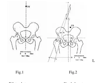
R L
Fig.1 Fig.2
Bilateral stance Single leg stance
The body weight shared equally on both hips when person stands on both
legs (fig .1). The force (R) exerted is equal in both hip joint and is 50% of
the total body weight. When person stands on his one leg (Right) his
weight of the lifted leg (Left) adds to the body weight. So the centre of
gravity shifted to the left at CG as in fig.2.This is marked as vector K in
fig.2. Abductor muscles of the standing leg should exert downward pull
to keep the pelvis level through vector (M) at the level (B) as in fig.2.
Total amount of force on the fulcrum of the hip at the level (O) is the sum
of the vectors M and vector K marked as R in fig.2. The amount of force
[image:27.612.140.464.103.387.2]OB and OC respectively. So the total force at femoral head (O) is sum of
M and B that is four times K.
In coxavalga, small abductor lever arm needs more abduction force
to maintain the pelvic level such as 7 to 8 times of body weight. The
pressure exerted on femoral head is increased. To decrease the pressure
and pain patient lists towards the affected hip. This is specific waddling
gait of coxavalga. This causes back pain due to secondary strain on
lumbar spine. To decrease pressure and pain over femoral head and
femoral neck (abductor lever arm) should be adequately maintained
particularly in replacement surgeries so that the durability of the
prosthesis is increased.
When supporting cane used on the normal side, decreases the body
weight (K) and so abductor pull decreased. The cane acts through long
lever arm so that slight pressure given through the cane will reduce the
pressure on hip. 60% of pressure can be reduced when 10% pressure
given through cane.
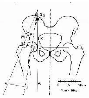
Waddle hip Biomechanics
When the trunk lists on the affected side, the center of gravity (S5) shifted
to affected side as in fig.3. The body weight lever arm (OC) is reduced
The force exerted on femoral head (R) is slightly higher than body
weight(9) (10).
Fig.3
Waddle hip Biomechanics
Neck lengths and offsets
a) Vertical offset - is the distance from a fixed bony point such as lesser
trochanter to the center of the femoral head. It is essential to restore this
distance to prevent limb length discrepancy. It is determined by length
obtained with the modular head plus base length of the prosthetic neck
used.
b) Horizontal offset - simply known as offset is the distance between line
through the axis of the stem and the center of the femoral head.
[image:29.612.238.409.134.323.2]arm results in future limp, bone impingement and dislocation. Excessive
offset results in future stem loosening and stem breakage. Offset can be
increased without limb lengthening by reducing the neck stem angle or
c) Version - is referred to neck orientation in reference to the coronal plane.
It can be denoted as anteversion or retroversion. It is important in
achieving stability of the implant.
“Jump distance” refers to the distance the head should travel to come
out of the rim of socket which can be increased with use of large diameter
head. Increased jump distance results in increased movements of hip(1).
BIOMECHANICS RELATED TO IMPLANTS
Basic Requisites of implant materials
1. Biocompatibility of Materials
2. Optimal strength and wear resistance
4. Implant materials resistant to environment changes ( corrosion
resistant)
Tissues response to corrosion of implants can be minimum response
of only fibrous tissue formation to maximum response of more thickened
avascular tissue barrier formation. Corrosion produces osteolysis and
osteoclasis so implants become loose. Chemical Osteomyelitis produces
mild sclerosis and new bone formation.
Friction and Wear:-
Contact between bear surfaces occur at particular points which can
be seen at microscopic level. Wear and particle production due to
corrosion occur at contact area. The different types of wear are:-
1. Adhesive Wear: depends on molecular bonding at the contact area. So
wear is reduced if surface finish improved.
2. Abrasive wear: due to rough area at the contact points.
3. Corrosive wear
4. Surface Fatigue
Factors reducing wear:
1. Materials: Alloys which resist corrosion should be used, such as cobalt –
linked polyethylene liner because it has more co efficient of friction. High
molecular weight polyethylene has low friction coefficient and high wear
resistance. (7) (8)
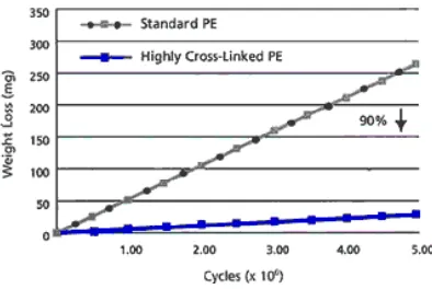
Degree of cross linking is related to wear resistance of polyethylene.
Crown et al (7) (8) in his study found 90% reduction in the polyethylene
wear when high molecular weight cross linking used. This can be
[image:33.612.224.421.286.418.2]measured as amount of weight loss in the polyethylene.
Fig.4. Graphic representation comparing standard and high cross linking.
Measures polyethylene weight loss in mg per million cycles
2. Reduction of vertical loads and friction torque: Amount of forces act on
femoral head reduced by lateralizing greater trochanter and deepening of
acetabulum.
3. Optimal design and made: Spherical design causes close fit and produces
more adhesive wear. If contact more towards the polar area frictional
4. Metal on poly combination: The frictional torque is more on metal on
metal combination than metal on poly combination. So wear will be less
in metal on poly combination.
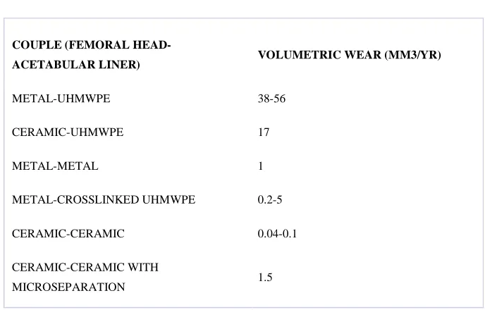
COUPLE (FEMORAL
HEAD-ACETABULAR LINER) VOLUMETRIC WEAR (MM3/YR) METAL-UHMWPE 38-56
CERAMIC-UHMWPE 17
METAL-METAL 1
METAL-CROSSLINKED UHMWPE 0.2-5 CERAMIC-CERAMIC 0.04-0.1 CERAMIC-CERAMIC WITH
[image:34.612.124.468.195.415.2]MICROSEPARATION 1.5
Fig.5. Comparison of various alternative bearing surfaces
5. Reduction in the radius of femoral head: Torque Friction (Q) is directly
proportional to the head radius: Q = rF.
6. Environment around Implant: PH of environment is acidic when infection
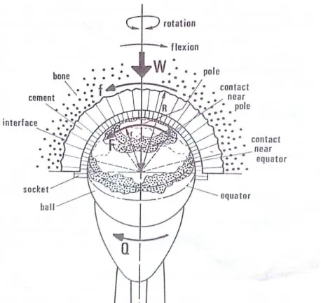
Fig.6. Geometric representation of frictional Torque viewed from medial
side at section with acetabulum. Here
Q- Magnitude of torque. r – Radius of head. F- Frictional force. W- Load
on the joint. R – Radius of bone implant interface.
Frictional torque is defined as resistance to rotatory motion of femoral
head with in the acetabulum. Frictional Torque varies at different axis of
motion with maximum during flexion and extension. The magnitude of
torque is the product of radius and frictional force which acts tangential to
the joint that is Q=rF. If the radius of bone implant interface is R and
tangential force is f, then Q= Rf. (8) (9) so f= rF/R that is frictional force is
directly proportional to the radius of femoral head. When small diameter
of head is used contact occur more in the polar region so less frictional
contact more in the equator and so frictional force will be higher.
Frictional torque is less when contact occurs within 450 of polar region.
Fate of wear particles:
Debris produced due to metallic wear present in the joint fluid
giving the appearance of blackish discoloration. These particles are
ingested by phagocytes. But debris due to polyethylene wear is
transparent in surrounding tissues which can be seen under polar
microscope. These also ingested by phagocytes. Cement particles are
extracellular and birefringent, multifaceted. (7)(8)On long term effects
these particles act like carcinogen in humans is not yet known. But
investigation on animals found it produces carcinoma.(9) (10)
INDICATIONS AND CONTRAINDICATIONS
INDICATIONS
1) Arthritis
2) Avascular Necrosis
3) Pyogenic arthritis /Osteomyelitis
4) Failed Reconstruction
5) Hereditary Disorders
CONTRAINDICATIONS
Absolute
o Active infection of hip joint
o Unstable medical illness
Relative
o Neuropathic joint disease
o Absent or insufficient abductor mechanism
METHOD OF SELECTION OF IMPLANT
To decide on which stem (cemented or uncemented) to be implanted four
parameters are used. Each parameter is given one point. A value obtained
for the given patient may be used to select proper implantation. The
parameters were:-
1. Sex
Osteoporosis begins around 40 yrs of age in females. Further it is
increased during menopause by hormonal changes.
In patients with less than 60yrs of age an uncemented prosthesis is
indicated. Revision surgery if required it will allow easy removal of the
implants. In patients with 70 yrs of age or more than that cemented
prosthesis is used.
3. Singh’s index
This classification system measures the degree of Osteoporosis
depends on changes in the trabecular pattern in proximal end of femur
Stage 6 - (normal) all trabeculae present
Stage5 - loss of trochanteric and secondary tensile group
Stage4- loss of secondary compressive trabeculae
Stage3- definite osteopenia break in primary tensile group
Stage2- complete loss of primary tensile group
4. Morphological cortical index
Three types of shapes of femur can be seen on radiographs. These
are trumpet, cylindrical and dysplastic. Of which trumpet shape suitable
for cementless fixation.
Morphological index is the ratio of the distance between two outer
cortical surfaces at the level of lesser trochanter(AB) to the distance
between the inner cortical surfaces at the level of isthmus(CD).
Evaluation of patients
Gender Age Singh’s index MCI
Score Score Score Score
MALE 0 <50 0 6 0 >3 0
FEMALE 1 <51-60 1 5 1 3-2.7 1 <61-70 2 4-3 2 2.6- 2.3 2 >70 4 2-1 4 <2.3 4
Total score plan
0-4 Uncemented
5 Possible
≥ 6 Cemented
FIXATION OF CEMENTLESS IMPLANTS
Biological fixation is paramount for the success and durability of
uncemented total hip replacement. Immediate primary fixation should be
stable so that secondary bone growth can occur. Formation of woven
bone surrounding the implant without cartilaginous intermediary occurs
first followed by lamellar bone remodeling that contributes to bone
Based on radiographic findings engh and bobyn provided radiological
classification of implant fixation.(1) (2)
1. Fixation by bone in growth is established as when no subsidence and
no or minimal radio opaque line around the implant. Hypertrophy of
cortex at the distal end of the stem and “spot welds” between stem and
periosteum may present. Proximal stress shielding of varying degree may
present.
2. Fixation by fibrous growth is defined as extensive radiopaque lines
around the stem and without migration of the implants. The radio opaque
lines lying in parallel fashion around the stem. These lines separated from
the stem with 1mm wide radiolucent areas. No local cortical hypertrophy
of femur suggested uniform load transfer function.
3. Unstable implant is said when there is presence of either migration of
the stem or subsidence present and also there is widely separated
divergent radio opaque lines surrounding the stem present. Localized
cortical hypertrophy around the distal end of the stem and collar present
MATERIALS AND METHODS
In our institution 30 cases of uncemented total hip arthroplasty
were done for various
1year to three years.
AGE DISTRIBUTION IN OUR STUDY
S.No 1. 2. 4. 5. 20-29 yrs
MATERIALS AND METHODS
In our institution 30 cases of uncemented total hip arthroplasty
were done for various indications. Follow up period ranges from
1year to three years.
AGE DISTRIBUTION IN OUR STUDY
Age group No.cases % cases
20- 29 4 13.3%
30-39 7 23.3%
40-49 15 50%
50-60 4 13.3%
40-49 yrs
50-60 yrs 30-39 yrs
AGE DISTRIBUTION
In our institution 30 cases of uncemented total hip arthroplasty
indications. Follow up period ranges from
SEX DISTRIBUTION IN OUR STUDY
S.No Gender
1. Male
2. Female
I.
SIDE DISTRIBUTION
S. No side
1. Right
2. Left
FEMALES 43% 0%
SEX DISTRIBUTION
SEX DISTRIBUTION IN OUR STUDY
Gender No. of cases % of cases
Male 17 56.3%
Female 13 43.3%
side No. of cases % of cases
Right 16 53.3%
Left 14 36.6%
MALES 57% FEMALES
43%
0%
VARIOUS INDICATIONS
S.No DIAGNOSIS No. cases % of cases
1. Chronic Arthritis 16 53.3
2. AVN of head of femur 7 23.3
3. Fracture Neck of femur with
implant failure
4 13.3
4. Fracture neck of femur 2 6.6
5. Dysplastic Hip 1 3.3
Most common indication is chronic arthritis.
0 2 4 6 8 10 12 14 16 18
arthritis AVN NUI NON UNION DDH
DATA COLLECTION METHODS
Collection of the data as per the proforma with consent from
the patients admitted in the Orthopaedic department, Govt.
Royapettah Hospital, Kilpauk Medical College, Chennai.
INCLUSION CRITERIA
- Patients in the age group < 60 years
- Patients with hip pain and Arthritic Changes in the Radiographs
EXCLUSION CRITERIA
- Active Infection of the Hip joint
- Unstable Medical Illness
METHODS
PREOPERATIVE EVALUATION OF THE
PATIENTS AND RADIOGRAPHS
All patients were assessed with the following protocols.
• Medical condition of the patient is assessed for DM, HT,
Anemia, cardiac diseases, thromboembolism.
• Aspirin and other anticoagulants are stopped 1 week before
surgery.
• Skin lesions like pyogenic infection should be eradicated.
• Purulent discharge of hip joint should be investigated for
culture and sensitivity.
• Transurethral resection of prostate to be done for urethral
obstruction before elective procedure.
• Soft tissue of hip to be investigated for scarring and
inflammation.
• Abductors strength is evaluated before surgical procedure.
• Any fixed deformity and limb length discrepancy is
assessed.
• In case of ipsilateral involvement of hip and knee arthritic
hip should be operated first.
• Hip joint status should be assessed pre operatively by Harris
hip score.
• About 1000- 1500 ml of blood loss is expected during
perioperative period of total hip arthroplasty.
• To reduce transfusion related reaction and infection banking
of autologous blood prior to surgery to be done.
• Transfusion of 4to 5 units of blood for revision surgery and
3units of blood for primary procedure is required.
• In bilateral involvement the most painful hip is operated first
3months after the other hip should be operated.
PREOPERATIVE ASSESSMENT OF RADIOGRAPHS
• Anteroposterior view of pelvis with proximal femur and hip
lateral view with proximal femur are required. X ray knee and
spine is needed in special cases.
• Radiographs are reviewed to check the adequacy of bone
stock for the acetabular component fixation, the need for bone graft
and amount of reaming required and to check whether osteophytes
present or protrusion can make the dislocation difficult.
• When old previous fractures occurred special views are
• The diameter of medullary canal is measured as in DDH and
dwarfs it may be small in diameter. In these situations femoral
stem with small diameter and short length is required.
• Presence of Anterior bowing in situations like Paget’s
disease and old fracture shaft of femur make the medullary canal
reaming difficult. In these cases femoral osteotomy is indicated.
DESCRIPTION OF IMPLANT
Femoral Component
In all cases “Taper Loc” hip system used. Primary type 1 taper loc
stem has the advantages of excellent rotational stability and smooth
load transfer to the femur and with the lateral offset design that
allows stability without lengthening of leg. Taper loc stem is made
with high strength titanium alloy. Plasma spray porous coating
with titanium alloy powder generates a random distribution of pore
size between 100 to 1000 microns providing a large contact area
between substrate and particles. The stem is used with a modular
A flat wedge shaped design used in the typical ovoid femoral canal
provides better rotational stability. Sharkey et al. found in his study
taper loc stem to have excellent stability. The tapered titanium
geometry in the taper loc stem design, allows for a gradual
transition in stiffness from the upper end of femur to the middle of
femur. The use of collarless stem design in the taper lochip tends to
allow for the self-seating of the implant and achievement of
optimal rotational stability.
‘Ring loc’ acetabular component was used in all cases. The ring
locacetabular component redefines the standard acetabular
technology. The components provide an unparalleled liner locking
mechanism, maximum polyethylene thickness and congruity. The
taper loc system is compatible with all ring locacetabular
components. Fixation achieved by forcing the implant into the
under reamed acetabulum.
The components of this are:-
Titanium shell
It consists of hemispherical acetabular shell and polyethylene
liner.
Screw sockets
Openings for cancellous bone screws present in proximal half
of the shell. Three dome holes present in the 11 clock 3clock and 1
clock.
Pyramids
On the edge of shell sharp pyramid like elevation is present
which are impacted into cancellous bone on insertion.
Polyethylene liner
We used 10o polyethylene liner which shifts the center of
restores the center of rotation of acetabular components which are
vertically placed.
SURGICAL PROCEDURE
Preoperative Templating
Femoral stem and acetabular cup size is measured by
template.Templating was done before surgical procedure to know
the approximate neck length, offset and version. The following
procedure of templating was used.
• AP view pelvis and lateral view of affected hip was taken in
proper position and 15 degrees internal rotation. The amount of
magnification is estimated correctly.
• Amount of limb shortening was measured by drawing a line
parallel to the level of ischial tuberosity that intersects the lesser
trochanter on each side.
• To measure acetabular cup size the template with its medial
position was placed at the level of tear drop and its inferior tip at
the level of obturator foramen. The center of acetabular component
was noted, it is the new center of rotation of hip.
• Femoral component size was selected that matches with
proximal canal. To restore limb length appropriate neck length was
selected.
Spinal anaesthesia with epidural anaesthesia is given in all cases.A
third generation intravenous antibiotics like ceftriaxone 1gm
administered in the operative room ½hr before surgery. Peak serum
concentration is obtained 20 min after administration. The infection
rate decreased from 11% to 1%.
Surgical approach
Hardinge direct lateral approach is used in all except 3cases in
which Moore’s posterior approach is used. Charnley used
anterolateral approach with greater trochanteric osteotomy.
Nowadays this approach used less commonly because of problem
related to nonunion of greater trochanter. Surgeon preferred over
the choice of approach.
Hardinge lateral approach
Patient in lateral position bony landmarks are marked, a lazy J
shaped incision centered over greater trochanter made. Fascia lata
incised in line with skin incision and retracted anteriorly and
gluteus maximus retracted posteriorly to expose the gluteal medius
insertion and vastuslateralis origin.
Gluteus medius tendon incised at post 2/3 and ant 1/3 junction
of muscle. Incision extended distally along the vastuslateralis and
dissection not to be extended 5cm proximally in gluteus medius.
Gluteus medius retracted to expose the gluteus minimus.
Insertion of gluteal medius and vastuslateralis are elevated and
hip abduction exposes the anterior capsule to be incised. Gluteus
medius and minimus splitting allow dislocation of hip anteriorly
and provide good exposure of acetabulum.
The dall variation of this approach is elevation of thin plate
of bone along with anterior portion of abductor muscles from the
anterior edge of greater trochanter so that repair can be done easily.
TECHNIQUES OF TOTAL HIP REPLACEMENT
Preparation of acetabulum
Femoral head is dislocated with the above one of the
mentioned approach. After femoral head dislocation appropriate
amount of neck resection done. Amount of neck resection depends
on type of the prosthesis implanted.
Insertion of acetabular cup:
1. Patient in true lateral position is checked again.
2. Acetabulum is exposed by rotating the femur internally or
externally so that proximal femur part will lie posterior to the
acetabulum. Superior and inferior lip of acetabulum is seen well
3. Progressive reaming is done while maintaining the
congruency of acetabular surface. A component selected is 1 to
2mm more than the last reamer size used to give more stability.
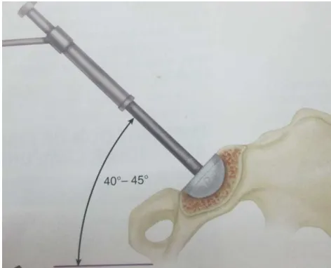
Fig.1.Anteversion
4. Arranging the acetabular component with the positioning
device. To decide correct angle of inclination a bar extending from
the positioning device oriented parallel or vertical to the floor is
used. One more extension bar determines the degree of
anteversion. The correct angle of inclination is 40 to 45 degrees.
[image:54.612.222.458.475.666.2]The correct anteversion is 20 degrees.(Fig.1,2)
5. Positioning device is maintained in alignment until the
impaction completed. Once the subchondral bone reached a change
in sound is heard. After that the positioning device is removed if
position is satisfactory. If gap is still present further tapping done
till the gap close.
6. Fixation of augmentation screws in the posterosuperior
region is ideal. Drill hole should be made in center of hole.
Improper placement of screw resulting in lifting of acetabular cup
and difficulty in positioning the liner can happen. Sciatic nerve can
be prevented by placing finger in the sciatic notch. Stability of the
cup should be checked. Excess osteophytes and debris should be
removed.
7. Soft tissue interposition should be cleared off from the cup
before inserting polyethylene liner. Center of offset should be
placed in superior or posterosuperior region.
Insertion of cementless femoral stem
1. After exposing the femur entry reaming done at a point
posterior and lateral to pyriformis fossa. Lateralization is done by
forward the reamer initially towards the greater trochanter then
aiming down towards the medial femoral condyle. Reaming
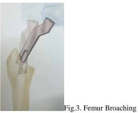
Fig.3. Femur Broaching
2. Femoral broaching done till size of less than 2 of the
measured stem size. Initially broaching done towards laterally to
avoid medialisation of the stem. Version aligned and maintained
while broaching. Progressive broaching with larger diameter
should be done. If progression is difficult lateralization should be
checked again. Fit of the broach assessed for contact with cortical
surfaces. Stability of fixation assessed by rotating the broach and
observed for any motion. If motion is seen broaching proceeded
Fig.4 Femoral anteversion and rotational stability
3. If stability is adequate final neck preparation to be done.
Neck resection pre
lesser trochanter. If needed necessary neck resection can be done at
this stage. Trial neck inserted and center of femoral head and tip of
trochanter distance assessed.
4. If neck length is adequate thorough deb
hip reduced. If reduction is difficult reassess the offset and neck
size and polyethylene liner.
5. Impingement of neck and stability of joint are checked. Hip
joint should be stable in extension and flexion and adduction. Suck
test to be done if it is positive use longer size neck. If limb length Femoral anteversion and rotational stability
If stability is adequate final neck preparation to be done.
Neck resection pre operatively assessed at what level from the
lesser trochanter. If needed necessary neck resection can be done at
this stage. Trial neck inserted and center of femoral head and tip of
trochanter distance assessed.
If neck length is adequate thorough debris removal done and
hip reduced. If reduction is difficult reassess the offset and neck
size and polyethylene liner.
Impingement of neck and stability of joint are checked. Hip
joint should be stable in extension and flexion and adduction. Suck
be done if it is positive use longer size neck. If limb length If stability is adequate final neck preparation to be done.
operatively assessed at what level from the
lesser trochanter. If needed necessary neck resection can be done at
this stage. Trial neck inserted and center of femoral head and tip of
ris removal done and
hip reduced. If reduction is difficult reassess the offset and neck
Impingement of neck and stability of joint are checked. Hip
joint should be stable in extension and flexion and adduction. Suck
appears to be increased use stem with more offset so that stable hip
acceptable limb lengthening can be achieved.
6. Trial components are exchanged with implants if acceptable
reduction and range of motion with stability achieved. If intra
operative femur fracture occurs the stem should be removed first
and exposed till full extension of the fracture visible. Cerclage wire
applied at appropriate level and stem reinserted. Tension in wire
applied is checked. Stability of hip joint is checked.
POSTOPERATIVE PROTOCOL
Antibiotics are given for 12 days till suture removal. Drain
removed on 2nd post op day. A triangular pillow is used to
maintain the hip in 150abduction so that dislocation can be
prevented in the immediate postoperative period. Pre operatively
patient is advised about dos and don’ts like to avoid strenuous
activity and not to sit on floor with cross legged and not to squat
and to maintain ideal body weight.
During the 1st postoperative day limited mobilization and bed
exercises begin. Quadriceps and gluteal isometrics and deep
breathing exercises begin. Straight leg rising is not beneficial in
On the 2nd postoperative day patient can sit on the chair in
semi recumbent position.
During 1st postoperative period or after drain removal gait
training started using a walker for balance and stability. Amount of
weight bearing allowed depends on presence of stress risers in
femur and bone grafts used and method of fixation of components
and trochanteric osteotomy. Limited weight bearing for 6 to 12
weeks is recommended in cases of cement less fixation.
Patient is encouraged to walk on the 3rd postoperative day
with crutches. Limited weight bearing should not exceed more than
1/3 of body weight. Patient can be discharged under normal
conditions during 10th to 12th postoperative day after suture
removal. Follow up examination is done at 7 weeks after surgery
and advised gradual increase in weight bearing for the subsequent
7 weeks. Hip extension exercises are advised if preoperative
flexion deformity co exists. Patient is advised to use western type
toilet for toilet purposes. Sexual activity may be resumed.
During 3 to 6 months of postoperative period nearly half of
muscle strength is regained. After 3 months patient can return to
FOLLOW UP PERIOD PROTCOL
A standard protocol was used in the postoperative period. Follow
up visits are made at 1 month, 3months, 6 months, 1 year and
periodically thereafter. Routine plain radiographs were taken and
assessed for loosening, migration, osteolysis during follow up
visits.
Postoperative Radiological Assessment
Femoral Component
1. By using Moss Template Center of Rotation of hip
compared is to normal side.
2. Fixation of femoral component is assessed by presence of
optimal contact of stem with both lateral and medial endosteal
cortical surfaces for about 5cm.
3. Tip of the stem positioned in Neutral without any varus
valgus angulation.
4. Level of the Greater Trochanter tip which corresponds to the
center of femoral Head
5. Level of the both sides lesser trochanter for any limb length
discrepancies.
6. Seating of collar of the stem correctly over the
7. Restoration of both vertical and medial offset compared to
normal side.
8. Orientation of Neck to the vertical offset. In valgus hip the
medial offset is smaller than vertical offset so the center of head
lies superior to the level of trochanter tip. In varus hip medial offset
larger than vertical offset so the center of head lies inferior to level
[image:61.612.271.408.277.491.2]of trochanter tip.
Fig.5 femoral loosening zones as d
9. Gruen et al Zonal analysis for loosening
The Femoral stem is separated into 7 zones, Each zone is analyzed
for radiolucency and radiopaque lines and for bony apposition Restoration of both vertical and medial offset compared to
Orientation of Neck to the vertical offset. In valgus hip the
is smaller than vertical offset so the center of head
lies superior to the level of trochanter tip. In varus hip medial offset
larger than vertical offset so the center of head lies inferior to level
of trochanter tip.
femoral loosening zones as described by Gruen
Gruen et al Zonal analysis for loosening
The Femoral stem is separated into 7 zones, Each zone is analyzed
for radiolucency and radiopaque lines and for bony apposition Restoration of both vertical and medial offset compared to
Orientation of Neck to the vertical offset. In valgus hip the
is smaller than vertical offset so the center of head
lies superior to the level of trochanter tip. In varus hip medial offset
larger than vertical offset so the center of head lies inferior to level
escribed by Gruen
The Femoral stem is separated into 7 zones, Each zone is analyzed
10. Bending of stem is assessed by angle made between a line
drawn along the lateral surface of distal stem and another line
drawn through the center of neck and head.
11. EBRA- FCA Method for measuring the migration of stem.
Einzel –Bild–Roentgen-Analyse method is used to assess the
femoral component migration. Compared with Roentgen stereo
photogrammetric analysis this method has the specificity of 100%.
This is accurate method to assess stability with in particular period.
Migration of prosthetic implants early is predicted to later failure.
This method gives information regarding about subsidence and the
lateral and medial distance between prosthetic margin and bone
margin.
There are four Different reference lines to describe the migration.
a - Tip of greater trochanter to stem shoulder
b - Tip of greater trochanter to center of head
c - Tip of lesser trochanter to shoulder head
d - Tip of lesser trochanter to center of head
Acetabular component
Assessed by following parameters:-
1. Optimal size and correct seating of cup without polar gap
2. Correct inclination of the cup to the tear drop level.
adducting the limb. Horizontal inclination results in posterior
dislocation and early impingement on flexion.
3. Degree of anteversion – normal anteversion of acetabulum is
about 150 to 200. The version is determined by position of the
anterior and posterior half of circumferential wires in the cup.
4. Polyethylene wear- is measured by the distance of migration
of femoral head into polyethylene. It is assessed by superolateral
penetration of femoral head of more than 2.5mm.
5. The position of transacetabular screws
The acetabulum is divided into four quadrants by two lines which
are antero superior, antero inferior, postero superior, postero
inferior. Screws placed in the anterosuperior quadrant may injure
the external iliac arteries and vein. Screws placed in the
anteroinferior quadrant may injure the obturator vessels and nerves.
Screws placed in the postero superior and postero inferior quadrant
may not emerge within the pelvis. But it may pass through the
sciatic notch and injure the sciatic nerve and superior gluteal
vessels. However the screw threads and drill bit can be felt and
palpated in the sciatic notch, so that injury to sciatic nerve can be
avoided. The anterosuperior quadrant can be avoided and postero
6. The acetabular component loose
and Charnley.
[image:64.612.236.445.158.418.2]Loosening is measured in three zones of acetabulum.
Fig.6 loosening zones as described by DeLee and charnley
7. Reinforcement of acetabulum with rings, cages, and
impaction grafting are noted.
8. Protrusion of
of hip is the migration of the cup medially beyond the ilio
line (kholer’s line).
Radiologically patients were classified into
Group I - No evidence of loosening (migration, Osteolysis)
The acetabular component loosening is described by DeLee
Loosening is measured in three zones of acetabulum.
loosening zones as described by DeLee and charnley
Reinforcement of acetabulum with rings, cages, and
impaction grafting are noted.
Protrusion of cup- The Radio graphic hall mark of protrusion
migration of the cup medially beyond the ilio
line (kholer’s line).
Radiologically patients were classified into
No evidence of loosening (migration, Osteolysis)
ning is described by DeLee
loosening zones as described by DeLee and charnley
Reinforcement of acetabulum with rings, cages, and
rk of protrusion
migration of the cup medially beyond the ilio- ischial
Group II - Evidence of loosening present on radiographs
but the patient is asymptomatic.
Group III - Evidence of loosening in a symptomatic patient
Functional outcome assessment
Assessment is done by using modified Harris Hip Score. Harris
Hip score has the following components:-
1. Pain – (44 Points Maximum)
2. Gait (walking maximum distance) (33 points Maximum)
3. Functional activity(14 points Maximum)
4. Absence of Deformity (4 points Maximum)
5. Range of motion (5 points Maximum)
6. Total – 100 points.
The Harris Hip Score is Graded as follows:-
Score Grade
<70 poor
70-79 Fair
80-89 good
ANALYSIS AND RESULTS
In our study majority of patients (53.3%) had chronic arthritis
of hip and 23.3% of patients had avascular necrosis of femoral
head.
13.3% of patients were treated with cancellous screws fixation
for fracture neck of femur, subsequently patient developed
avascular necrosis of femoral head which are treated by
uncemented total hip replacement.
6.6% of patients had fracture nonunion neck of femur.
3.3% of cases had developmental dysplasia of hip with
secondary arthritis.
All patients had severe pain with limitation of daily activities
of living. Preoperative assessment with Harris hip score was done
in all cases.
The surgical approach used depends on the preference of the
operating surgeon. Hardinge’s direct lateral approach was used in
73.3% cases and Moore’s posterior approach used in 26.6% cases.
“Ring Loc” standard cup with highly cross linked polyethylene
liner was used in all cases. “Taper Loc” porous coated femoral
stem was used in all cases. Follow up period ranges from 1 to 3
All patients were radiologically assessed for the following
parameters.
Femoral Component
• Prosthesis level above the lesser trochanter averages about
1.5 cm
• Optimal position of the prosthesis
Neutral - 76.6%
Varus - 13.3%
Valgus - 10%
• Canal fill of the stem in anteroposterior diameter averages
about 80%.
• Intra operative femoral fracture is seen in two cases which
were treated by cerclage.
• Aseptic loosening of femoral stem in Gruen zones 3, 4,5 is
seen in one case.
• Majority of the stem had shown good osteointegeration.
• There was no evidence of calcarresorption indicating no
subsidence in any of the case.
• There is no evidence of osteolysis around femoral stem as
indicated by radiopaque line formation in any of the case.
• Heterotopic ossification was noted in one case around the
Acetabular component
• Correct positioning with 45 degrees inclination and 15
degrees anteversion is seen in 83.3% cases.
• Acetabular component with overhanging margins beyond the
superolateral rim is seen in 2 cases.
• Correct seating without any polar gaps and intimate contact
with subchondral bone is seen in 93.3% cases.
• Transacetabular screws to secure the cup firmly to the
acetabulum used in all cases.
• Acetabular loosening in Charnley and DeLee zone2 is seen
in one case.
• Pelvic Osteolysis is seen in one case.
• No protrusion of the cup into the acetabulum was
encountered in our study.
• In a case of dysplastic hip acetabular reconstruction was
done using autologous iliac crest bone graft. In the subsequent
follow up period the graft was found to be incorporated
completely.
• Postoperative sciatic nerve palsy was encountered in
dysplastic hip case.
• Deep vein thrombosis is seen in two cases. It was treated
Preoperative hip score in our study is as follows
30- 39 3 cases
40- 49 23 cases
50- 60
Postoperative Harris Hip score as follows
90-100 excellent 3 cases(10%)
80-89 good 19 cases (63.3%)
70-79 fair 6 cases (20%)
<70
Preoperative hip score in our study is as follows
39 3 cases
49 23 cases
60 4 cases
Postoperative Harris Hip score as follows
100 excellent 3 cases(10%)
89 good 19 cases (63.3%)
79 fair 6 cases (20%)
The poor results are seen in 2 cases. In one case poor result is due
to postoperative dislocation. This patient had suffered from severe
arthritis of hip. In another case poor result is due to noncompliance
to follow up postoperative instructions and mobilization.
The fair to poor results are due to
- Severe arthritis with restriction of daily activities of living.
- Intraoperative femoral fracture in 2 cases.
- Deep vein thrombosis in 2 cases.
- Postoperative sciatic nerve palsy in one case.
- Pelvic osteolysis in one case.
- Persistent anterior thigh pain in 2 cases.
- Limb length discrepancy with shortening of 1cm in one case
RESULTS
- Uncemented total hip arthroplasty has a definitive role in the
treatment of chronic arthritis in young patients.
- In our study most of the patients belongs to the age group of
40- 50 yrs (53.3%).
- Males (56.6%) predominate in our study.
- Chronic arthritis is the most common indication in our study.
- Uncemented total hip arthroplast was done in all cases. In
bilateral affected cases severely operated limb was operated.
- Ring locacetabular cup and Taper loc porous coated femoral
stem is used in all cases.
- 76.6% cases had femoral stem in neutral position with osteo
integration.
- Optimal cup size with 45 degrees inclination at the tear drop
level is seen in 83.3% cases.
- 63.3% cases had good functional outcome with Harris hip
Incidence of various complications
- Postoperative dislocation in one case (3.3%)
- Intraoperative femoral fracture in two cases (6.6%).
- Postoperative sciatic nerve palsy in one case (3.3%).
- Postoperative deep vein thrombosis in two cases (6.6%).
- Femoral stem aseptic loosening in one case (3.3%).
- Acetabular cup loosening in one case (3.3%).
- Pelvic osteolysis in one case (3.3%).
- Limb length discrepancy in 4 cases (13.3%).
- An