1
DISSERTATION ON
STUDY ON PULMONARY HYPERTENSION
&DIASTOLIC DYSFUNCTION IN SCLERODERMA
Submitted in partial fulfilment of Requirements for
M.D. DEGREE BRANCH I GENERAL MEDICINE
of
THE TAMILNADU DR.M.G.R. MEDICAL UNIVERSITY, CHENNAI.
MADRAS MEDICAL COLLEGE
3
CERTIFICATE
This is to certify that this dissertation entitled “STUDY ON PULMONARY HYPERTENSION &DIASTOLIC DYSFUNCTION IN SCLERODERMA” submitted by Dr. BALAMURUGAN S. appearing for Part II M.D. Branch I General Medicine Degree examination in September 2006 is a bonafide record of work done by him under my direct audience and supervision in partial fulfillment of regulations of the Tamil Nadu Dr. M.G.R. Medical University, Chennai. I forward this to the Tamil Nadu Dr.M.G.R. Medical University, Chennai, Tamil Nadu, India
Prof. V. SUNDARAVADIVELU M.D. Director In-charge, Institute of Internal Medicine, Government General Hospital, Chennai – 600 003.
DEAN,
Madras Medical College, Government General Hospital,
4
ACKNOWLEDGEMENT
I am extremely grateful to the Dean Dr.Kalavathy Ponniraivan M.D. for granting me permission to do the dissertation in Madras Medical
College and Government General Hospital, Chennai.
I am very grateful to our Professor and Director in charge of Institute of Internal Medicine Prof.V.Sundaravadivelu M.D. for his valuable suggestions in preparing the dissertation.
I am greatly indebted to Cardiology Chief Prof.S. Shanmugasundaram M.D., D.M., who inspired, encouraged and guided
me in every step of this dissertation.
I express my heartiest thankfulness and wholehearted indebtness to Dr. Balasubramanian M.D, D.M (Cardiology), Dr. J. Ravishankar
M.D., D.M., who helped me in doing echocardiography and without whose
effort the study would not have been possible.
Words may not suffice to express my gratitude to Prof. C.P.Rajendiran M.D, D.M (Rheumatology), the Chief of
5 guiding me in each and every step and by taking much pain to give this dissertation its complete form.
I am really thankful to Dr. K. Sivasubramaniam M.D and Dr. G. Subbaragavulu M.D. and all the Assistant Professors for their
guidance and valuable suggestions throughout the study.
I am also thankful to my family members for their cooperation in finishing my dissertation.
6
CONTENTS
S.NO TITLE PAGE NO
1.
INTRODUCTION
1
2.
AIM
3
3.
REVIEW OF LITERATURE
4
4.
MATERIALS AND METHODS
38
5.
STATISTICAL ANALYSIS
49
6.
RESULTS AND OBSERVATION
50
7.
DISCUSSION
60
8.
CONCLUSION
65
9.
LIMITATION OF THE STUDY
68
10. BIBLIOGRAPHY
69
11. PROFORMA
79
8
INTRODUCTION
Systemic sclerosis (scleroderma) is a generalized disorder of connective tissue characterized clinically by thickening & fibrosis of skin & by distinctive forms of involvement of internal organs notably heart, lungs, kidneys, gastrointestinal tract. The etiology & pathogenesis are unknown(7).
It is characterized by fibrotic arteriosclerosis of peripheral & sytemic vasculature
• variable degrees of extracellular matrix accumulation (mainly collagen) occur in both skin & viscera.
• associated with specific antibodies most notably anti-centromere & anti-scl-70 (anti topoisomerase) (1). various subsets with specific clinical features & variable involvement of various organs.
9 phenomenon, esophageal dysmotility, sclerodactyly, telangiectasia) upto 60% have pulmonary hypertension (3,4,5).
Pulmonary hypertension is defined as a mean pulmonary artery pressure exceeding 25mmHg at rest or 30 mm Hg in exercise. When it occurs as a manifestation of SSc it is particularly severe & one year survival is approximately 55%. (2)
10
AIM OF STUDY
• To assess the prevalence of pulmonary hypertension in scleroderma patients with limited & diffuse forms
• To differentiate between those pulmonary hypertension due to interstitial lung disease (secondary PulmHT) from those without ILD (isolated PulmHT)
11
REVIEW OF LITERATURE
EPIDEMIOLOGY;
Its estimated that new cases of scleroderma(SSc) occur at a rare of approximately 18-20 per million of general population per year(9). The prevalence & severity of SSc varies among different racial & ethnic subgroups
The limited variant is found more commonly in caucasions while African-American females appear to have increased risk for diffuse cutaneous type & younger age of onset(10). The average age of onset is approximately 50 years(9).
GENITIC FACTORS
12 pregnancy or from their parents, after which the patients develop a graft versus host-like reaction that results in scleroderma.
Conversely, the work of Reveille et al(60) Suggests that DQ7 and DQ5 are more common. It was shown that patients with limited scleroderma and anticentromere antibodies were associated with HLA-DR-1, and diffuse scleroderma and antitopoisomerase antibody were associated with HLA-DR-5.
DIAGNOSTIC CRITERIA;
American college of rheumatology proposed preliminary criteria for classification based on clinical & lab assessments
Major:
Sclerodermatous skin change in any location proximal to metacarpophalangeal joints was the single major criterion
Minor:
13
CLASSIFICATION;
Patients with scleroderma are classified into subsets of disease by the degree of clinically involved skin.
1. diffuse; skin thickening present on trunk in addition to face, proximal & distal extremities.
2. limited: skin thickening limited to sites distal to elbow & knee also involving face & neck
3. sine scleroderma: characteristic internal organ manifestations, vascular, serologic, abnormalities but without clinically detectable skin change
4. In overlap.
DIFFUSE CUTANEOUS SCLERODERMA:
14 There is also a significant risk of internal organ involvement particularly in first 3 years(12).
The higher the skin nscore the greater the risk of renal disease& higher mortality. About 10-20% of patients with diffuse disease will develop life threatening internal organ Disease
The later stages are characterised by gradual softening of skin(11). Predictors of higher mortality include higher skin scores (Rodnanscore >20) reduced lung diffusing capacity, elevated ESR & evidence of heart or renal involvement.
LIMITED SCLERODERMA;
The clinical course, survival & clinical features of limited disease are quite different from than in diffuse form. The overall 5 year survival is 80-85% which is significantly better than in diffuse form(13).
15 The major cause of morbidity & mortality in limited form are severe Raynauds Phenomenon with occlusive digital vascular disease.& Pulm.HT.
PulmHT is the leading cause of mortality in limited disease patients. This complication occurs in approximately 10-15% of cases with limited disease & often occurs in the absence of interstitial lung disease(ILD)
PATHOPHYSIOLOGY;
The three key pathologic features are
- a unique vascular disease
- abnormal accumulation of extracellular matrix components,
- autoimmunity
RAYNAUDS PHENOMENON;
In SSc over 90% of patients have intense Raynauds phenomenon associated with tissue fibrosis, digital ulceration & on occasion ischemic demarcation , digital amputation.
16 Compared to normals ,SSc patients have a more pronounced decrease of both nutritional & thermoregulatory blood flow skin in response to cold temperatures. The reactive hyperemia which normally follows periods of digital ischemia is absent in SSc.(14)
There is good evidence that a generalized vasospastic disorder exists in SSc ( systemic Raynauds phenomenon) involving kidney , heart, lungs & other viscera. The renal crisis of SSc is a clear example of reversible vasoconstriction pf the cortical blood flow to kidney.(15)
CUTANEOUS MANIFESTATION;
The skin disease of scleroderma follows a course characterized by three phases:
• inflammatory edematous phase,
• indurative phase and
• atrophic phase.
17 with a moon-like fullness of the fingertip and there is loss of normal digital creases. Loss of the finger pad and tapering of the finger due to fingertip ischemia is often seen during this early phase of SSc..
The face can appear expressionless because of reduced capacity to smile or move the eyelids or cheeks. The opened mouth becomes circular with a remarkable reduction in the maximum oral aperture. Vertical lines or furrowing of the skin around the lips gives a pursed-lip The nose becomes pinched and the facial creases smooth so that the face has a mouse-like appearance (mauskopf) Pigmentary changes of the skin also occur during
18 changes are usually limited to the fingers(sclerodactyly). More prominent in limited disease are small and large mat-like telangiectasias that appear on the face, upper chest, palms, fingertips and mucous membranes. Subcutaneous calcinosis, composed of calcium hydroxyapatite deposits at sites of trauma such as the forearms, elbows or fingers.
Pathology in Dermis ;
The pathologic hallmark of systemic sclerosis is an excessive accumula-tion of extracellular matrix in the dermis, which leads to taut skin. Monotonously similar collagen fibers are present in the reticular dermis and there is thinning of the papillary dermis. Capillary loss can be seen in vivo in the skin by the technique of wide-field nailfold capillaroscopy
MUSCULOSKELETAL INVOLVEMENT
Pain on motion of the ankle, wrist, knee or elbow may be accompanied by a coarse 'friction rub caused by fibrinous deposits on the tendon sheath. These rubs are detected in approximately 30% of patients with diffuse scleroderma(16).
19 weakness can result from deconditioning secondary to restricted mobility and contractures.
GASTROINTESTINAL INVOLVEMENT
Gastrointestinal tract involvement is found in almost every patient with scleroderma and is characterized by abnormal motility secondary to dysfunctions caused by abnormal innervation, smooth muscle atrophy and tissue fibrosis(19).
OROPHARYNX
. Many patients with scleroderma have a sensation of dry eyes and mouth (the sicca complex). In addition, the majority of scleroderma patients with sicca complaints usually do not have antibodies against Ro/SSA or La/SSB, suggesting that the mechanism of dry membranes in scleroderma is a secondary process different from that seen in Sjogren's syndrome(21).
ESOPHAGUS
20 the esophagus. Substernal burning pain may be coupled with a feeling of indigestion or nausea. These reflux symptoms are typically worse after meals, with exercise and after bedtime. Severe esophageal reflux and esophagitis occur equally in patients with either limited or diffuse scleroderma..
Low or absent primary and secondary peristalsis of the distal esopha-gus and low lower esophageal sphincter pressure in the presence of a normal proximal (striated muscle) esophagus are typical manometric findings in scleroderma(22). Excessive air in the esophagus is commonly detected on routine chest X-ray. Although symptoms are a poor guide to the degree of esophageal disease, patients who are asymptomatic on or off treatment are unlikely to have significant complications. Esophagitis appears to occur only in patients with impaired peristalsis and delayed acid clearance(23)
21
STOMACH AND SMALL BOWEL
Delayed emptying of the stomach with retention of solid foods aggra-vates reflux and is a frequent cause of bloating. Antral vascular ectasia (watermelon stomach) can be a cause of gastrointestinal bleeding in scleroderma patients. Endoscopy of these patients reveals antral gastritis and prominent longitudinal vascular folds that resembles the surface of a watermelon (24).
.
LARGE BOWEL
The large intestine and rectum are also affected by scleroderma. Scleroderma patients have decreased distensibility of the colon that does not necessarily correlate with symptoms(25). Because of muscular atrophy of the bowel wall, asymptomatic wide mouthed diverticula unique to scleroderma are commonly found in the transverse and descending colon .
PULMONARY INVOLVEMENT
22 fibrosis & pulmonary vascular are present in the lungs of patients with SSc but one pathologic process will be dominant cause of clinical problems.
Interstitial fibrosis is more likely to be severe in diffuse form while pulmonary vascular disease & PulmHT can be dominant in LC form. Approximately 80% of patients will have abnormal PFT’s.(26)
Patients with ILD commonly have a rapid decline in pulmonary function in conjunction with progressive lung disease. Dyspnea on exertion without chestpain is the most common presenting complaint with a dry cough being a late manifestation Of ILD.
The most common changes in PFT are either a reduced diffusion capacity, or a reduced lung volumes( FVC,) typical of restrictive ventilatory defect with associated reduction on gas exchange.
23 Broncho-alvelolar lavage is used to detect inflammation & active alveolitis. BAL demonstrates an increased percentage of neutrophils, esinophils or CD 8 cells. When disease progresses, there is worsening interstitial fibrosis & honey combing of lung parenchyma (27,28).
Pulmonary arterial vascular disease with PulmHT is one the most difficult clinical problems in SSc. The pulmonary vascular process can be indolent & remain clinically undetectable until severe irreversible PulmHT & signs of right sided heart failure develops.
Pulmonary hypertension can be detected early and non-invasively by measuring the pulmonary artery pressure with two-dimensional Doppler echocardiography. Pulmonary function testing often reveals an isolated decrease in diffusion capacity when pulmonary vascular disease is present.
24 patients with CREST syndrome usually do not experience clinical symptoms until more than 5 years after diagnosis. Risk factors for serious restrictive lung disease are African-American or Afro-Caribbean race and antitopoisomerase antibodies (29). A low diffusing capacity (less than 40% predicted) (30) or rapidly declining DLco and/or lung volumes predict a high mortality rate.
In predicting the histological pattern, CT, although useful, has not replaced lung biopsy as the ‘gold standard’ investigation. As yet, patients who appear to have early changes on CT should still be considered for a thoracoscopic biopsy for staging of the disease. In systemic sclerosis the predominant histological pattern is NSIP whereas in IPF the majority show UIP.
25 Studies are inconclusive about the association between pulmonary function impairment and abnormal esophageal function(31). There is likely an increased risk of lung cancer in patients with ILD(32).
HISTOLOGY;
The histology of fibrotic lung disease is that of expansion of the nor-mally thin alveolar wall interstitial space with the deposition of collagenand other connective tissue components. Progressive collagen accumulation with diminution of the alveolar air space volume, so that ultimately there is more fibrous tissue than air space a gas diffusion blockade, an increase in the alveolar—arterial Po2 gradient and ventilation-perfusion inequalities.
PULMONARY HYPERTENSION IN SSC;
Pulmonary arterial hypertension is a life threatening complication of both diffuse & limited scleroderma (including CREST syndrome). pulmonary vascular disease has a particularly adverse effect on prognosis.
26 phenomenon, esophageal dysmotility, sclerodactyly, telangiectasia) upto 60% have pulmonary hypertension
While not all patients. have clinically significant pulmonary hypertension, two thirds of patients with scleroderma will have pathologic evidence of pulmonary vascular disease(33 ,34)
Stupi et al reported two year survival un patients with in patients without pulmonary hypertension to be greater than 80% while patients with pulmonary hypertension had a two year survival of 40%(59).
Sacks et al reported two year survival of patients with pulmonary hypertension & either diffuse or limited forms to be approx. 50%(36).
Koh et al reports 40% survival in patients with scleroderma & pulmonary hypertension compared with higher survival in scleroderma patients without organ failure or with other lung involvement (i.e interstitial lung disease) at two years.(37)
PATHOGENESIS:
27 pulmonary circulation with direct involvement of pulmonary circulation with intimal proliferation & medial hypertrophy similar to that seen in primary pulmonary hypertension(38). Some cases may also be related to severe pulmonary parenchymal disease, such as interstitial disease with hypoxemia.
Additionally diastolic dysfunction of right &left ventricles has been seen in patients with scleroderma & may contribute to pulmonary hypertension(39)
Autoimmune processes have been implicated in the pathogenesis of pulmonary hypertension although the mechanism is not known. Positive antinuclear antibodies are frequently found in pulmonary hypertension patients without a diagnosis of connective tissue disease & pulmonary hypertension can occur before the onset of an identifiable connective disease.
AUTO-ANTIBODIES IN SCLERODERMA & PULMONARY HYPERTENSION;
28 surprising that anti-centromere antibodies associated with a higher incidence of pulmonaryhypertension Anti-fibrillarin antibodies (anti-u3- RNP) are frequently found in diffuse form associated with pulm.HT(40). Anti-endothelial Ab’s (aECA) are present in 40% & 13% of diffuse form & CREST respectively & are associated with pulm.HT & digital infarcts (41). In scleroderma & pulm.HT ,when accompanied by HLA-B 35 antigen, anti-topoisomeraseII Ab’s & antibodies to fibrin bound tissue type plasminogen are more common.(42)
RAYNAUDS & PULM.HT;
Raynauds phenomenon , vasospasm of arterioles in distal systemic circulation, is commonly reported in scleroderma. In one report all patients with Pulm.HT & CREST had raynauds while 68% without Pulm.HT had raynauds .Raynauds is also common in patients with SLE & MCTD & Pulm.HT. but only 10-14% of patients with primary pulmonary hypertension have Raynauds(45). This observation has led to the Pulmonary Raynauds hypothesis that vasospasm contributes to the development of Pulm.HT(46)
30
DIASTOLIC DYSFUNCTION IN SCLERODERMA
Diastolic dysfunction is defined as the deterioration of the ventricular filling capacity without any compensatory increase in the left atrial pressure. (74) Another definition is the abnormal ventricular filling defect causing cardiac output inadequacy. (75) In patients with diastolic dysfunction, the deterioration of ventricular dilatation (early diastole), decrease in compliance (early late diastole) or an external pressure in pericardium can lead to problems in ventricular filling. Cardiomyopathies, constructive pericarditis, ischemic heart diseases, volume overload (mitral insufficiency, arteriovenous fistulae), mitral and tricuspid valve stenosis may cause diastolic dysfunction.
31 An abnormal right ventricular filling is detected in 40% of SSc patients. Such alteration was detected in many without clinically evident cardiac disease & resulted to be correlated with both left ventricular diastolic abnormalities & Pulmonary hypertension.
Scleroderma heart disease is subclassified into primary & secondary formsPrimary cardiac disease in SSc depends on involvement of myocardium & or pericardium &small intramyocardial vessels by SSc itself. Secondary cardiac involvement develops either in patients with systemic hypertension induced by renal scleroderma (LV disease) & in those with pulmonary vascular & or interstitial lung disease.
SSc myocardial fibrosis is different from that occurring in patients with coronary atherosclerosis. Actually SSC myocardial fibrosis is equally distributed throughout the right & left ventricle, does not involve the immediate subendocardial layers, is not related to the distribution of epicardial coronary vessel & is not associated with hemosiderin deposits.
32 dysfunction Candell-Riera et al detected a significantly low Tr E/A ratio in 63 % of patients. (56)
PATHOGENESIS:
The pathogenesis of the cardiac lesion in scleroderma is controversial. An intriguing concept is one of repetitive vascular insults secondary to cold induced perfusion changes. Classic pathological changes of contraction band necrosis seen in scleroderma are similar to the findings in hearts subjected to prolonged ischemia and subsequent reperfusion.
MYOCARDIAL INVOLVEMENT:
Myoccardial lesions and fibrosis which are found in up to 80 percent of patients upon autopsy, may be patchy, may present in both ventricles and may be patchy may present in both ventricles and may bear no relationship to myocardial perfusion..
Myocardial dysfunction occurs often although clinical congestive heart failure occurs in less than 5 per cent of patients with progressive systemic sclerosis.
33 Ischemic chest pain and myocardial infarction are uncommon clinical problems in scleroderma. Thallium perfusion defects reflecting vascular disease of the endomyocardial vessels (not larger coronary arteries) are seen among scleroderma patients both at rest and with exercise. Because of this, the scleroderma patient with angina-like chest pain may need angiographic studies to rule out coronary arteriosclerosis because thallium scans are likely be abnormal as a result of the microvascular disease of scleroderma heart.
In fact, cold provocation of Raynaud's phenomenon can temporarily increase the number of thallium scan defects and induce local abnormalities in ventricular wall motion, supporting the notion that reversible vasospasm of the myocardial microcirculation occurs in scleroderma. Thallium scan perfusion defects predict more severe myocardial disease and poor outcome.
34 patients, and approximately 15% of diffuse scleroderma patients have abnormalities at rest. Echocardiographic studies suggest that both right and left ventricular dysfunction is common in scleroderma and that diastolic left ventricular dysfunction may occur independent of systolic dysfunction. Diastolic dysfunction may be secondary to hypertension (with or without renal disease) or myocardial fibrosis and may manifest with abnormal left ventricular compliance and pulmonary vascular congestion. Unexplained dyspnea on exertion may be the initial clinical manifestation of unappreciated diastolic dysfunction.
PERICARDIAL INVOLVEMENT
Autopsy series report pericardial involvement in up to 50 per cent of patients with systemic sclerosis. Pericardial involvement includes fibrinous pericarditis, pericardial adhesions and pericardial effusions
ECG changes in SSC
35 supraventricular tachycardia, AV or intraventricular conduction disorders are seen less commonly(57).
RENAL INVOLVEMENT;
The most important clinical manifestation of scleroderma kidney is accelerated hypertension and/or rapidly progressive renal failure: the scleroderma renal crisis(58) Surveys suggest that only about 10-15% of all scleroderma patients develop a crisis. The majority of patients who develop renal crisis have diffuse cutaneous disease and approximately 80% of cases of renal crisis occur within 4 years of disease onset. Risk factors for renal crisis include rapidly progressing diffuse skin disease, tendon friction rubs, new unexplained anemia and the presence of anti-RNA polymerase III antibody. Antecedent use of corticosteroids is also associated with a higher risk of developing renal crisis. Non-malignant hypertension, abnormalities on urinalysis, plasma renin level and the presence of anticentromere or antitopoiso-merase antibodies are not predictors of a scleroderma renal crisis.
36 The typical vasculopathy of scleroderma is present in the renal vessels of patients with or without renal crisis. This suggests that other factors, such as vasospasm, probably contribute to the development of renal crisis. Scleroderma patients may have a reduced creatinine clearance, proteinuria, microscopic hematuria and non-malignant hypertension, but often another cause for these abnormalities is found. For example, an immune complex process may be the cause for glomerulonephritis in patients with an overlap syndrome of SLE and scleroderma. A reversible proteinuria or even a crescentic glomerulonephritis may occur secondary to treatment with D-penicillamine
CLINICAL PRESENTATION & EVALUATION:
37 at a time when the skin may be improving or thinning. In limited scleroderma, the skin fibrosis is minimal and does not parallel the vascular disease such as pulmonary hypertension and digital loss.
Recently, a quantitative measure of disease severity has been developed that documents Degree of severity of involvement of each major organ graded from 0 (normal) to 4 (end-stage) . This scale has been externally validated on a large group of scleroderma patients and may be helpful in comparing groups of patients in clinical trials and for following disease in individual patients.
Dyspnea is the most common presenting symptom of SSc associated pulmonary hypertension. the clinical evaluation is similar to that of primary pulmonary hypertension. History & physical examination often reveal findings of underlying connective tissue disease.
38 Screening for PAH is certainly worthwhile in these high risk patients since it’s the only way to identify PAH earlier & begin treatment earlier. Although PAH is incurable, it can now be contained for many years through use of new effective treatments for this condition .We now have a greater incentive to identify PAH patients & manage them through combined clinics with rheumatologist working together with cardiologist, pulmonologist & pulmHT nurses(2). Doppler echo has emerged .as a reliable & reproducible means of non invasively assessing pulmonary artery systolic pressure(PASP) & has been employed to detect Pulm.Ht in patients with connective tissue disease.
39 carbon monoxide diffusing capacity as a non- invasive measure of PAH is less certain.
Stupi et al (59) & Ungerer et al (6) have demonstrated an association between DLCO <40-55% & isolated SSc PAH in a total of 89 patients.
Burke et al (60)& Jezek & widimsky etal(62) were unable to confirm a clear link between low DLCO & PAH.
Mukerjee (76)et al suggested echo estimated TG or PASP & DLCO, most commonly used screening tools for SScPAH, perform well only in the diagnosis of advanced pulm.HT.
Echo perfomed better in identifying patients who require urgent referral & treatment for advanced disease. Patients suspected of advanced disease in SSc & Pah have an annual mortality of 40%. Relying on echo alone to identify these patients results in a high false negative rate of 42% .however combining 3D echo & clinical findings ( modified NYHA dyspnea grade ) identifies > 90% of patients with advanced PAH.
40 ventricular cavity. This technique utilizes digitalization of the borders of left ventricular cavity, with which the left ventricular dimension increases in early diastole being noted
Doppler echo currently is the primary technique used for evaluating ventricular diastolic function.. With normal pressures the early diastolic mitral velocity (E) exceeds that following atrial systole or late mitral(A) velocity (E/A greater than 1)
Under several conditions one of which is normal aging , the velocity in early diastole decreases & the late velocity increases. Among the pathologic states that. produce this change are LV hypertrophy. & myocardial ischemia.both conditions produce abnormal relaxation & decreased early velocity into LV. This situation will commonly produce elevated LV diastolic pressures unusually low filling pressures in lefty atrium may also have a similar pattern.
41 The atrial velocity is now reduced. This type of mitral flow may also occur if there is a restrictive pattern of filling of LV as may occur with restrictive cardiomyopathy.
ABNORMAL PATTERNS
Impaired Myocardial Relation Pattern
In nearly all types of cardiac disease, the initial abnormality of diastolic filling is slowed or impaired myocardial relaxation , include LV hypertrophy, hypertrophic cardiomyopathy, and myocardial ischemia/infarction. The Isovolumic relaxation time(IVRT) is prolonged. Mitral E velocity is decreased and A velocity is increased, roducing and E/A ratio<1, with prolonged DT.
Whenever the E/A ratio is below 1, impaired relaxation is usually present.
RESTRICTIVE FILLNG (or Decreased Compliance) PATTERN:
42 congestive heart failure, advanced restrictive cardiomyopathy, severe coronary artery disease, acute severe aortic regurgitation, and consrtrictive pericarditis.
The increase in LA pressure result in earlier opening of the mitral valve, shortened IVRT, and a greater initial transmitral gradient(high E velocity). Early diastolic filling into a noncompliant LV cause a rapid (increase in early LV diastolic pressure, with rapid equalization of LV and LA pressures producing a shortened DT, Atrial contraction increased LA pressure increases even more rapidly, when LV diastolic pressure is markedly increased, there sure is markedly increased, there may be diastolic mitral regurgitation during mid-diastole or with atrial relaxation. Therefore, restrictive physiology is characterized by mitral flow velocities that show increased E velocity, decreased A velocity (<<E), and shortened DT(<160 msec) and IVRT (<7 msec). Typically, the E/A ratio is greater than 2...
PSEUDONORMALIZED PATTERN:
43 to as the pseudonormalized filling pattern and it represents a moderate stage of diastolic dysfunction. The pseudonormal pattern can be distinguished from a true normal pattern by Doppler Tissue Imaging(DTI)
GRADING OF DIASTOLIC DYSFUNCTION:
. Therefore, diastolic dysfunction can be graded as follows according to the diastolic filling pattern.
Grade 1 = impaired relaxation
Grade 2 = pseudonormalized pattern
Grade 3 = reversible restrictive pattern
Grade 4 = irreversible restrictive pattern.
As previously discussed, patients with scleroderma should be considered an “at risk” group for the development of pul-monary hypertension, and echocardiography may reveal right ventricular hypertrophy and dilatation even before the onset of symptoms
45
MATERIALS AND METHODS
STUDY POPULATION
All patients were prospectively identified from rheumatology department of our college. There were 40 patients of SSc defined according to previously mentioned ACR criteria were studied. Of them 36 were females and 4 were males. Of them thirty patients satisfied the criteria for limited disease & ten had diffuse disease.
Mean age of forty patients is 38.5 years. The average duration of illness is 3.5 years. All patients except three had raynauds phenomenon. Twenty patients were affected with digital pitting scars & ulcers , & two had digital amputation.
For comparison twenty normal persons were selected( seventeen females, three male) of the same age group. All patients & controls gave consent for the study.
46 disease, pulmonary tuberculosis or pulmonary thromboembolism as assessed by history, physical examination
CLINICAL EVALUATION:
A questionnaire prepared noted the duration of SSc, extra-cutaneous complications, the use of current and previous disease-modifying drugs, . Questions were asked relating to previous chest disease, cough, dyspnea, sputum production, chest pain, weight loss and risk factors for respiratory disease, including smoking, medications, and occupation( exposure to silica,solvent industry) .A detailed clinical examination was performed. All patients had venous blood taken for full blood count, renal and liver function, C-reactive protein and antinuclear antibodies. Patients also underwent skin biopsy.
47
ELECTROCARDIOGRAM: .
Patients also had 12 lead ECG that was reviewed for presence of left or right atrial enlargement and left or right ventricular hypertrophy.
ECHOCARDIOGRAM;
All echocardiograms were performed by two Cardiologists. Whenever possible, these cardiologists, who were blinded to clinical details, determined pulmonary artery pressure All studies were performed using a phased array ultrasonoscope (ALOKA) with a combined 2.5 MHz. Imaging/continuous wave Doppler and colour Doppler transducer.
Doppler recordings were made from the parasternal, apical and subcostal positions using a modified views when appropriate. A systematic search was performed using two dimensional and colour flow Doppler to identify the most complete tricuspid regurgitant jet followed by continuous wave Doppler acquisition of spectral envelopes of greatest maximal velocity and density. The systolic transtriuspid gradient was calculated using the modified Bernoullie equation
48 Where V represents maximal regurgitant velocity in metres per second
TWO DIMENSIONAL ECHOCARDIOGRAPHY
PulmHT is easily recognized when the following M-mode and 2D echocardiographic features are present( )
Diminished or absent “a”(atrial) wave of the pulmonary valve
Midsystolic closure or notching of the pulmonary valve
Enlarged chambers on the right side of the heart
D-shaped left ventricular (LV)caviry caused by a flattened ventricular septum
Estimated Right Atrial Pressure(RAP):
49
Estimated Pulmonary Artery Systolic Pressure(PASP):
PASP was calculated as the sum of transtricuspid gradient and the estimated RAP. This method is highly accurate over a wide range of Pulmonary Artery Pressure.
Pulmonary hypertension is defined as a PASP of 25 mm Hg.or greater.. In the absence of pumonic stenosis or RV outflow obstruction, RV systolic pressure is equal to pulmonary artery systolic pressure. The normal TR velocity is 2.0 to 2.5 m/sec. A higher velocity indicates pulmonary hypertension, RV outflow tract obstruction, or pulmonic stenosis.
Mitral Flow Velocities;
50 DECELERATION TIME;
The diastolic filling pattern is characterized further by measuring deceleration time(DT), the interval from the peak of the E velocity to its extrapolation to baseline. DT is prolonged in patients with a relaxation abnormality as the predominant diastolic dysfunction, because it takes longer for LA and LV pressures to be equilibrated with a slower and continued decrease in LV pressure until mid to late diastole.
Tricuspid Flow Velocities
Just as mitral flow velocity variables characterize the LV diastolic filling pattern, so tricuspid flow velocity recordings characterize the RV diastolic filling pattern, using the same criteria. Left and right diastolic filling patterns are not necessarily the same in a patient. The main difference between mitral and tricuspid velocities is a respiratory variation in tricuspid flow velocities in normal subjects.
51
Mitral&Tricuspid Annulus Velocities by Doppler Tissue Imaging(DTI):
Doppler tissue imaging (DTI), or tissue Doppler, has been applied to evaluate diastolic function by measuring mitral & tricuspid annulus velocity during diastole. The mitral & tricuspid annulus velocity profile during diastole reflects the rate of changes in the long axis dimension and in LV & RV volume respectively. When myocardial relaxation is abnormal, the ratio of mitral & tricuspid annulus motion during atrial systole to the total diastolic annular motion is increased. Sohn et al(77) demonstrated that mitral annulus velocity determined by DTI is relatively preload-independent and is useful in differentiating pseudonormal(grade 2 diastolic dysfunction) from normal mitral inflow velocity pattern.
Left Ventricular Ejection Fraction(LVEF):
LVEF was used as an index of LV systolic pump function and was calculated by Simonsons formula
Right Ventricular Accelerated Fractional Shortening( RVAFS):
The percentage of shortening of RV area during systole was used as an index of RV systolic function and was calculated.Fractional shortening was calculated as the difference between diastolic and systolic diameters
52 The following variables were also assessed:
The left atrial (LA) and right atrial (RA) areas were traced manually
and measured at end-systole from the two-dimensional apical four-chamber
view.& ,right ventricular dimension
Pulmonary artery pressure control group:
For the normal population, limited data are available on pulmonary artery pressure estimated by Doppler echocardiography. A study of 20 normal healthy adults by Vachiery et al. (65) using Doppler echocardiography found that the maximum estimated pulmonary artery pressure was 24 mmHg
To assess the diagnostic validity of the results of the echocardiogram, a control group of 20 normal persons were subjected for echocardiogram & readings were incorporated into the study. Echocardiography was undertaken with the same ALOKA echocardiogram machine by the same cardiologists who performed the echocardiography in the patients with SSc.
53
DEFINITIONS
Pulmonary hypertension:
The gold standard for pulmonary artery pressure measurement is invasive right-heart catheterization. Pulmonary hypertension, defined by right-heart catheterization of the pulmonary artery is a pressure of 25 mmHg or greater at rest and at least 30 mmHg during exercise (2).
Echocardiography has now been used widely in patients with cardiac disease. Reported correlations between Doppler and catheter measurements range from 0.89 to 0.97; the average standard error for systolic pulmonary artery pressure ranges from 5 to 9 mmHg, and interobserver variability is <3% (83). We have taken Denton et al.'s definition of pulmonary hypertension on Doppler echocardiography as an estimated PASP of 25 mmHg or greater .
Pulmonary Function Tests;
Of the 40 patients, 18 patients (9 with limited-type SSc, and 9 with
diffuse-type SSc) underwent lung function testing within period of 1 week
after the echocardiography examination. Of these 18 patients, none were
being treated for pulmonary disease. . Pulmonary function testing was
54
Wuerzburg, Germany) according to the recommendations of the American
Thoracic Society. Values are presented as a percentage of the predicted
value.
Significant lung disease:
Significant lung disease that could be causing pulmonary hypertension was defined as pulmonary function measurements outside the normal range: a forced expiratory volume in 1 s/forced vital capacity (FEV1/FVC) ratio of less than 65% or a vital capacity lung volume of less than 80% of the predicted value (80,81)
High-resolution CT (HRCT) of lungs:
When PFT is abnormal patients were advised to undergo HRCT of lungs to see whether they have radiographic evidence of ILD. High resolution CT was performed in eighteen of the patients (Philips Tomoscan LX; Philips; Eindhoven, the Netherlands).
Scans were performed at full inspiration in the supine position with
120 kV and 175 mA, including continuous scans through the lungs with
10-min thickness followed by scans with 1.5-mm thickness with a slice spacing
56
STATISTICAL ANALYSIS
57
RESULTS & OBSERVATION
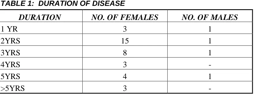
[image:57.612.89.530.163.328.2]There were forty cases with mean age of 35.58+10 years.
TABLE 1: DURATION OF DISEASE
DURATION NO. OF FEMALES NO. OF MALES
1 YR 3 1
2YRS 15 1
3YRS 8 1
4YRS 3 -
5YRS 4 1
>5YRS 3 -
Adequate images & Doppler spectral envelopes of tricuspid regurgitatin were obtained in 37 of 40 patients. The calculated pulmonary artery pressure ranged from 16-72 mm of Hg with a mean of 24.41(SD 15.30)
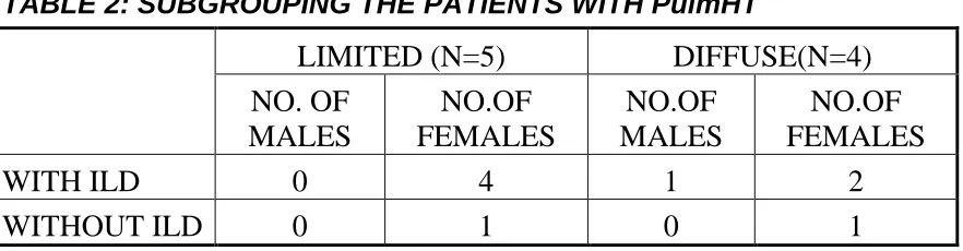
58 Chest HRCT showed that 4(10%) (1 of limited: 3 of diffuse) patients had interstitial lung disease with a fibrosing alveolitis pattern. All the 4 patients, had lung disease was sufficiently severe to cause significant volume loss (as defined above, under significant lung disease) on pulmonary function testing.& hence had secondary pulmonary hypertension . So the remaining (5) 12.5 % of patients had isolated pulmonary hypertension without lung disease evident on pulmonary function testing. [Table 2]
[image:58.612.89.530.459.574.2]The One remaining female patient in diffuse category had no evidence of ILD ( had a moderately high pulmonaryartery pressure). For this patient a second echo was done three months later on the advice of rheumatologist & was found to have a tricuspid gradient which had become lower ( reversible)
TABLE 2: SUBGROUPING THE PATIENTS WITH PulmHT
LIMITED (N=5) DIFFUSE(N=4)
NO. OF MALES
NO.OF FEMALES
NO.OF MALES
NO.OF FEMALES
WITH ILD 0 4 1 2
WITHOUT ILD 0 1 0 1
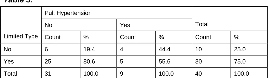
59 The clinical features of the SSc patients with pulmonary hypertension were compared with those of SSc patients who had a pulmonary artery pressure below 25 mmHg. (The findings are shown in the tables3,4,5,6).| There was No statistically Significant difference found between the two SSc groups in type of SSc, age,sex and disease duration(table ). . Also the acute phase response as assessed by C-reactive protein did not differ significantly between the two groups.
In the control group, none of them had pulmonary artery pressure above 25 mm of Hg. . The pulmonary artery systolic pressure was higher in patients with SSc (24.18+ 15.51 mm Hg) than in controls (18.2+3.55 mm) (P =0.003).There was also a weak correlation between the pulmonary artery pressure and the age of the patient (r=0.02,). [Figure 1]
Correlation between age and PASP
y = 0.2006x + 19.922 R2 = 0.0217
0 20 40 60 80
0 20 40 60 80
Age in years
P
A
S
P
60
Table 3:
Pul. Hypertension
No Yes Total
Limited Type Count % Count % Count %
No 6 19.4 4 44.4 10 25.0
Yes 25 80.6 5 55.6 30 75.0
Total 31 100.0 9 100.0 40 100.0
[image:60.612.84.532.80.210.2]Not significant
Table 4:
Pul. Hypertension
No Yes Total
Diffused Type Count % Count % Count %
No 25 80.6 5 55.6 30 75.0
Yes 6 19.4 4 44.4 10 25.0
Total 31 100.0 9 100.0 40 100.0
Not significant
Table 5:
Pul. Hypertension
No Yes Total
Sex Count % Count % Count %
Female 28 90.3 8 88.9 36 90.0
Male 3 9.7 1 11.1 4 10.0
Total 31 100.0 9 100.0 40 100.0
[image:60.612.83.531.463.626.2]61
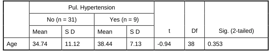
Table 6:
Pul. Hypertension
No (n = 31) Yes (n = 9)
Mean S D Mean S D t Df Sig. (2-tailed)
Age 34.74 11.12 38.44 7.13 -0.94 38 0.353
Not significant
There is no statistically significant differences at baseline in patients with or without elevated PASP.
Echocardiographic and Doppler Differences Between Patients
With SSc and Controls:
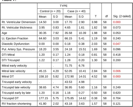
There was a significant difference between the two groups regarding the right ventricular cardiac chamber dimensions (17.7+2.9), compared to controls(14.5+3.1)(p< 0.05). But there was no difference between the ejection fraction of left ventricle and fractional shortening of the right ventricle.[table 7]
62 Moreover in the group of patients we found a weak correlation between the deceleration time and disease duration (r= -0.1, ), late diastolic tricuspid filling velocity A cm/s and disease duration (r= -0.2, ) correlation_0 .2
Correlation between duration of diseases and TRa
y = -0.8539x + 39.725 R2 = 0.0652
0 20 40 60
0 2 4 6 8 10
Duration of disease in years
T ri c u s p id e a rl y v e lo c ity Correlation -0.1
Correlation between Disease duration and MDT
y = -1.3973x + 177.44 R2 = 0.0279
0 100 200 300
0 2 4 6 8 10
Disease duration in years
64
Table 7:
TYPE
Control (n = 20) Case (n = 40)
Mean S D Mean S D T df Sig. (2-tailed)
Rt. Ventricular Dimension 14.50 3.00 17.70 2.90 3.98 58 0.000
Rt. Vetricular thickness 3.95 0.83 4.65 1.61 1.82 58 0.073
Age 30.35 7.92 35.58 10.39 1.98 58 0.053
Lt. Ejection Fraction 64.60 3.03 66.15 5.41 1.19 58 0.240
Diastolic Dysfunction 0.00 0.00 0.18 0.38 2.03 58 0.047
Pul. Artery Sys. Pressure 18.20 3.55 24.18 15.51 1.69 58 0.096
DTI Mitral 1.20 0.17 1.24 0.18 0.81 58 0.418
DTI Tricuspid 1.22 0.17 1.28 0.20 1.30 58 0.200
Mitral early velocity . . 71.75 6.76
Mitral late velocity 45.15 4.20 52.05 10.41 2.84 58 0.006
Mitral DT 158.10 5.82 172.90 14.01 4.52 58 0.000
Tricuspid early velocity . . 43.53 4.98
Tricuspid late velocity 38.65 4.74 36.95 5.60 1.16 58 0.249
Tricuspid early by late 1.20 0.16 1.16 0.27 0.50 58 0.620
Tricuspid DT 177.15 5.75 175.85 14.62 0.38 58 0.704
RV fraction shortening 41.80 2.02 43.18 3.63 1.57 58 0.121
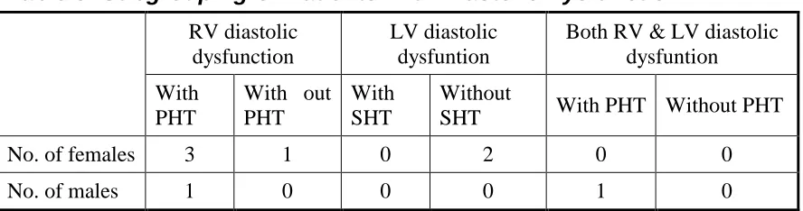
7 patients had diastolic dysfunction.one male & six females. The male patient had severe Pulm.HT & diastolic dysfunction of both RV & LV.
Of the 6 female patients 4 had right ventricular diastolic function (3 had associated Pulm.HT), 2 had left ventricular diastolic dysfunction (no associated systemic hypertension).[table8]
65
Table 8: Subgrouping Of Patients With Diastolic Dysfunction
RV diastolic dysfunction
LV diastolic dysfuntion
Both RV & LV diastolic dysfuntion
With PHT
With out PHT
With SHT
Without
SHT With PHT Without PHT
No. of females 3 1 0 2 0 0
No. of males 1 0 0 0 1 0
Differences between Patients with SSc with and without Ventricular Diastolic Dysfunction
To further investigate the implication of the ventricular diastolic dysfunction in patients with SSc without clinically evident cardiovascular disease, we assessed whether patients with SSc who had left ventricular diastolic dysfunction had some clinical or investigational peculiarities that might help identify these patients.
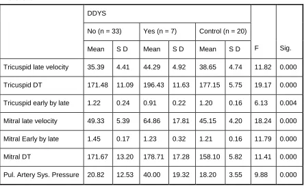
However, no statistically significant differences in sex, age were found (data not shown). But there was a significant difference in the estimated pulmonary artery systolic pressure between patients with and without ventricular diastolic dysfunction (40.+19.32vs 20.82+12.53,P=0.00).[table9]
66
Table 9:
DDYS
No (n = 33) Yes (n = 7) Control (n = 20)
Mean S D Mean S D Mean S D F Sig.
Tricuspid late velocity 35.39 4.41 44.29 4.92 38.65 4.74 11.82 0.000
Tricuspid DT 171.48 11.09 196.43 11.63 177.15 5.75 19.17 0.000
Tricuspid early by late 1.22 0.24 0.91 0.22 1.20 0.16 6.13 0.004
Mitral late velocity 49.33 5.39 64.86 17.81 45.15 4.20 18.24 0.000
Mitral Early by late 1.45 0.17 1.23 0.32 1.21 0.16 11.79 0.000
Mitral DT 171.67 13.20 178.71 17.28 158.10 5.82 11.41 0.000
67
DISCUSSION
As per study by Fredrick M Wigley & Laura K Hummers et al(1) the prevalence of SSc is higher in females than in males(3:1), & the difference is greater in younger age group(7:1) .(9)
In my study also there is a female preponderance with a female :male ratio of 12:1.The average age of onset is 50 years.(1). This is in contrast to my cohort of patients where age of onset is much younger(mean age is 35.8 years+SD 10)
The results indicate that unrecognized elevation of PASP is present in significant (22.5%) of patients in my study. Only 3 patients in total cohort had evidence of Pulm. HT by physical examination & ECG or both .the prevalence of pulmonary symptoms were similar in patients with or without Pulm.HT. Thus clinical assessment did not have Discriminant power with regard to presence or absence of Pulm.HT.
68 In the same study the prevalence rate of Pulm.HT in their cohort of 34 patients was 12 (35%). In my study the prevalence was lower (22.5%).
The gold standard for assessing pulmonary artery pressure is right heart catheterization. However as catheterization is invasive & As Doppler echocardiography and cardiac catheterization have been reported to have a correlation of between 0.89 and 0.97 in cardiac causes of pulmonary hypertension (83), we have not undertaken catheterization of our SSc patients.
Six other patients who had undergone PFT & HRCT for symptoms of dyspnea Had ILD but no Pulm.HT.
In a study by Ungerer RG, Tashkin et al(6), indicate that specific noninvasive studies are helpful in assessing the likelihood of normal or definitely elevated pulmonary artery pressures in patients with progressive systemic sclerosis, but patients with mild pulmonary hypertension are not likely to be identified by these noninvasive studies.
69 Echo assessment of TG is traditionally regarded as accurate technique. However under or overestimation are well known to occur. Overestimates are rare. underestimation is are due to either absence of TR Jet or inability to obtain full alignment with regurgitation jet.(83)
Rich etal(45) & Richards et al(68) have demonstrated variations upto 30 % within 24 hours in pulmonary artery pressure.
One patient had reversible elevation of pulmonary artery pressure.Its probably due to raynauds phenomenon occurring in pulmonary vasculature.
Pulmonary parenchymal abnormalities are recognized with increased with increased frequency as a complication of SSc patients. In patients with CREST syndrome as many as 60-70% develop abnormal results in PFT’s despite significantly lower incidence of either symptoms of dyspnea or radiographic abnormalities.
70 Of these isolated Pulmonary vascular injury is most common histologic feature occurring in upto 50 % of autopsy in CREST syndrome. Typically patients present with exertional dyspnea . Early identification of PulmHT at a potentially reversible stage can modify the natural course of disease.
Because primary diastolic dysfunction is an important cause of heart failure, as it often is a silent alteration preceding systolic dysfunction (39), knowledge of this complication in patients with SSc without clinically evident cardiac disease may be important to improve patient survival. The early detection of cardiopulmonary involvement in SSc is clearly desirable
both for optimal treatment and for implementation of preventive measures in
the early stages of the disease.
The extent of RV diastolic dysfunction was not related to the duration
of the disease or to the SSc skin score. in the present group of patients with
SSc, LV function was normal, RV systolic function was preserved, but BV
diastolic function was disturbed. This was evidenced by abnormal relaxation
71
These abnormalities were defined using both conventional Doppler
echocardiography and Doppler tissue imaging (DTI). The hallmarks of SSc
heart disease are myocardial fibrosis and ischemia. Studies (55) have
demonstrated that intermittent coronary vasospasm, similar to Raynaud
phenomenon, is prevalent in patients with SSc. The pattern of RV &LV
diastolic disturbance seen in the present study could therefore be related to
myocardial fibrosis and/or ischemia, both of which are known to affect
ventricular relaxation and filling and are characterized by reduced E-wave
velocity.
Detection of abnormalities in RV diastolic function might provide a
72
CONCLUSIONS
Scleroderma is higher in females than in males.
Pulmonary hypertension occurs in a significant proportion of patients with scleroderma. More often its is asymptomatic.
Symptoms consistent with PAH have no Discriminant power to differentiate those with or without elevated PASP.
Its more commonly associated with ILD in diffuse disease, but can occur without lung or heart abnormalities in limited form (isolated PulmHT).
The ratio of patients with limited to diffuse is 3:1.
Mild or early intermittent pulmonary hypertension are not likely to be
identified by these non-invasive studies & hence mild PulmHT cannot be
excluded in other patients.
There is no statistically significant difference between patients with or
without PulmHT when compared by type of SSc ,age ,sex ,duration of
73 When comparing Echo and Doppler differences between Patients With SSc and controls there was a significant difference between the two groups regarding the right ventricular cardiac chamber dimensions.
There was a significant difference in the estimated pulmonary artery systolic pressure between patients with and without ventricular diastolic dysfunction.
Raynaud phenomenon within the lung is a possibility.
Diastolic dysfunction of RV, LV or both can occur in SSc, which can occur with or without the presence of PulmHT or systemic hypertension. More often it is asymptomatic.
The extent of RV diastolic dysfunction was not related to the duration
of the disease or to the SSc skin score.
The hallmarks of SSc heart disease are myocardial fibrosis and
ischemia. Intermittent coronary vasospasm, similar to Raynaud
phenomenon, is prevalent in patients with SSc. The pattern of RV &LV
diastolic disturbance seen in the present study could therefore be related to
74
Doppler echocardiography is a sensitive and non-invasive method of detecting cardiac abnormalities and systolic and/or diastolic function and for detecting pulmonary hypertension.
Since so many therapeutic options are in the offing for management of PulmHT early detection is useful in altering the natural course of disease & in decreasing morbidity & mortality & to monitor the progression of disease.
Detection of abnormalities in diastolic function might provide a
75
LIMITATION OF THE STUDY
• In my study, I did not do diffusing capacity of carbon monoxide (DLCO) because of its limited availability and cost.
76
BIBLIOGRAPHY
1. Fredrick M wigley,Laura k Hummers ; clinical features of SSc: Textbook of rheumatology Hochberg.144-152
2. Pulm.arterial hypertension ;C.Black Editorial Rheumatology journal 2005;44;141-142
3. Battle RW, Davitt MA, Cooper SM, et al. Prevalence of pulmonary hypertension in limited and diffuse scleroderma. Chest1996;110 (6):1515-9.
4. MacGregor AJ, Canavan R, Knight C, et al. Pulmonary hypertension in systemic sclerosis: risk factors for progression and consequences for sur-vival. Rheumatology (Oxford) 2001;40(4):453-9.
5. Sacks DG, Okano Y, Steen VD, et al. Isolated pulmonary hypertension in systemic sclerosis with diffuse cutaneous involvement: association with serum anti-U3RNP antibody. J Rheumatol1996;23(4):639-42.
6. Ungerer RG, Tashkin DP, Furst D, et al. Prevalence and clinical corre-lates of pulmonary arterial hypertension in progressive systemic sclerosis. Am J Med1983;75(1):65-74.
77 8. Valentini G,Vitale DF;et al diastolic abnormalities in SSc , evidence
for associated defective cardiac functional reserve.Annsl of Rheumatism 1996;55;451-456
9. Medsger TA. Epidemiolgy of SSc.Clin.dermatology 1994;12 ;207-209
10. Tan EM,Rodnan GP ,diversity of aantinuclear Ab,s in SSc: arthritis rhlaing
11. Ciements Pi, Hurwitz EL, Wong Wl< eta!. Skin thickness score as a predictor and correlate of outcome in systemic sclerosis. Arthritis Rheum 2000; 43:2445-2454.
12. Steen VD, Medsger TA Jr. Severe organ involvement in systemic sclerosis with diffuse scleroderma. Arthritis Rheum 2000; 43: 2437-2444.
13. Jacobsen 5, Halberg P UlIman S. Mortality and causes of death of 344 Danish patients with systemic sclerosis Iscieroderma). Br J Rheumatol 1998; 37:750-755.
14. Wigley FM, Wise RA, Miller R eta!. Anti-centromere antibody predicts the ischemic loss of digits in patients with systemic sclerosis. Arthritis Rheum 1992; 35: 688-693.
78 16. Steen VD, Medsger TA Jr. The palpable tendon friction rub. Arthritis
Rheum 1997; 40: 1146-1151..
17. Olsen NJ, King LE, ParkJH. Muscle abnormalities in scieroderma. RheUm Dis din North Am 1996; 22: 783-796.
18. Clements PJ, Furst DE. Campion DS eta!. Muscle disease in progressive systemic sclerosis: diagnostic and therapeutic considerations. Arthritis Rheum 1978; 21:62-71.
19. Young MA, Rose S, Reynolds JC. Gastrointestinal manifestations of scieroderma. Rheum Dis Clin North Am 1996; 22:797-823.
20. Cameron AJ, Payne WS. Barrett’s esophagus occurring as a complication of scleroderma. Mayo Clin Proc 1978; 53:612.
21. Clements PJ, Furst DE. Systemic sclerosis. Baltimore, MD: Williams & Wilkins; 1996.
22. Bassotti G, Battaglia E, Debernardi Vet a!. Esophageal dysfunction in scieroderma. Arthritis Rheum 1997; 40:2252-2259.
23. Basiilsc&G, Barbera R, Molgora Metal. Acid clearance and oesophageal sensitivity in patients with progressive systemic sclerosis. Gut 1993; 34: 1487-1491.
79 25. Whitehead ME, Taitelbaum G, Wigley FM, Schuster MM.
Rectosigmoid motility and myoelectric activity in progressive systemic sclerosis. Gastroenterology 1989; 96: 428-432.
26. Schneider P, Hochberg MC, Wise RA, Wigley FM. Serial pulmonary function in patients with systemic sclerosis. Am J Med 1982; 73: 385-394.
27. Steen VD, Conte C, Owens GR. Medsger TA Jr. Severe restrictive lung disease in systemic sclerosis. Arthritis Rheum 1994; 37: 1283-1289.
28. Steen VD. Conte G, Owens GR eta!, isolated diffusing capacity reduction in systemic sclerosis. Arthritis Rheum 1992; 35: 765-770. 29. Greidinger EL, Flaherty KT, White Beta!. African-American race and
antibodies to topoisomerase I are associated with increased severity of scieroderma lung disease. Chest 1998; 114: 801-807.
30. Peters-Golden M, WiseR, Hochberg M eta Clinical and demographic predictors of loss of pulmonary function in systemic sclerosis. Medicine 1984; 63: 221-232.
31. Troshinsky MB, Kane GC, Varga J eta!. Pulmonary function and gastroesophageal reflux in systemic sclerosis. Ann intern Med 1994; 121:6-10.
80 33. Salerni RG, Rodnan P, Leon DF, et al. Pulmonary hypertension in
theCREST Syndrome variant of progressive system sclerosis(scleroderma). Ann Intern Medl 1977;86(4):394-9.
34. Young RH, Mark GJ.Pulmonary vascular changes in scleroderma. Am J Med 1978;64(6):998-1004.
35. Asherson RA, Higenbottam TW, Dinh Xuan AT, et al. Pulmonary hypertension in a lupus clinic: experience with twenty – fourty patients. J heumato11990;17(10):1292-8.
36. Sacks DG, Okano Y, Steen VD, et al. Isolated pulmonary hypertension in systemic sclerosis:risk factors for progression and consequences for survival.
37. Koh et al.Pulmonary hypertension in sys-temic sclerosis: an analysis of 17 patients. Br J Rheumatol 1996;35 (10):989-93.
38. Yousem SA. The pulmonary pathologic manifestations of the CREST syndrome. Hum pathol 1d990;21(5):467-74.
39. Giunta A, et al: Right ventricular diastolic abnormalities in SSc:Ann rheum disease 2000;59;948-950
81 41. Negi VS, et al: Antiendothelial cell antibodies in sclerderma correlate
with severe digital ischemia and pulmonary arte-rial hypertension. J Rheumato11998;25(3)
42. Yoshio T, et al: Antiendothelial cell antibodies and their relation to pulmonary hypertension in systemic lupus erythematosus.
43. Rubin et al Bosentan therapy in PulmHt NEJM 2002; 36
44. Reville et al Association of HLA DQ B1with anti scl-70 ab in SSc J clinical inv 992;90;
45. Rich et al: Primary pulmonary HT. A national prospective study. Ann Intern Med 1987.
46. Fahey PJ , et al: Raynaud’s phenomenon of the lung. Am J Med 1984;76(2).
47. Morgan JM, Grifiths M, et al: Hypoxic pulmonary vasoconstriction in SSc and PPH. Chest 1991;99(3)
48. Shuck JW, et al: Pulmonary vascular response during Raynaud’s phenomenon in SSc.Am J Med 1985;78(2).
49. Romero LI, et al; Differentioa expression of nitric oxide by Dermal microvascular endothelial cells from patients with SSc. Vasc med 2000;5(3)
82 51. Galie N , et al: Relation of endothelin –1 to survival in patients with
PPH. European Journal of clinical Investgation 1996;26.
52. Maione S, et al: Evaluation of cardiac Structures and functions in SSc by Doppler. Cardiaology 1991; 79.
53. Kazzam, et al: Non invasive assessment of LV diastolic function in patients with SSc. J Intern Med 1990,228.
54. Valentini G, et al: Deastolic abnormalities in SSc. Ann Rheum Dis 1996;55.
55. Owens GR,et al:Cardio pulmonary manifestations of SSc. Chest 1987;91.
56. Candell-Riera J, et al: Non Invasisve assessment of cardiac involvement in limited SSc.Arthritis Rheum 1996;39.
57. Deswal A, Follansbee WP. Cardiac involvement in scleroderma. Rheum Dis Clin North Am 1996; 22:841-860,
58. Steen VD. Renal involvement in systemic sclerosis. Clin Dermatdl 1994; 12:253-258.
59. Stupi AM, Steen VD, Owens OR, Barnes EL, Rodnan GP, Medsger TA Jr. Pulmonary hypertension in the CREST syndrome variant of systemic sclerosis. Arthritis Rlieum 1986;29:515-24.
83 61. Denton CP, Cailes JB, Phillips GD, Wells AU, Black CM, [ Bois RM.
Comparison of Doppler echocardiography and right heart catheterization to assess pulmonary hypertension in systemic sclerosis. Br J Rheumatol l997;36:239-43.
62. Jezek V. Widimsky J. Noninvasive diagnosis of pulmonary hyperten sion and activities pursued by a working group of the WHO. Cor Vasa 1990;32:178-82.
63. Gibbs JSR,Higgenbottom T. recommendations on the management of PulmHT in clinical practice. Heart 2001 ;86
64. Medsger TA,Masi AT ; epidemiology of SSc; Annals of internal medicine 1971;714-721.
65. Vachiery JL et al Doppler assessment of Hypoxic Pulm, vasoconstriction in high altitude pulmonary edema ;Thorax 1995;50 22-7
66. Yock PG, Popp RL non invasive estimation of RV systolic pressure by Doppler ultrasound in patients with TR; circulation 1984 ;4;657-662
67. Rich S ,D Alonzo et al magnitude of spontaneous hemodynamic variability in primary pulmonary hypertension. Am.J. Cardiology 1985; 55; 159-163