0022-538X/06/$08.00⫹0 doi:10.1128/JVI.02212-05
Copyright © 2006, American Society for Microbiology. All Rights Reserved.
Functional Correlation between a Novel Amino Acid Insertion at
Codon 19 in the Protease of Human Immunodeficiency Virus
Type 1 and Polymorphism in the p1/p6 Gag Cleavage Site
in Drug Resistance and Replication Fitness
Terrence W. Brann,
1Robin L. Dewar,
2Min-Kan Jiang,
2Akram Shah,
2Kunio Nagashima,
3Julia A. Metcalf,
4Judith Falloon,
4H. Clifford Lane,
4and Tomozumi Imamichi
1*
Laboratory of Human Retrovirology,1Virus Isolation and Serology Laboratory,2and Image Analysis Laboratory,3Science Applications International Corporation—Frederick, Inc., Frederick, Maryland, and Laboratory of Immunoregulation, National Institute of Allergy and Infectious Diseases,
National Institutes of Health, Bethesda, Maryland4
Received 20 October 2005/Accepted 22 March 2006
Population-based sequence analysis revealed the presence of a variant of human immunodeficiency virus type 1 (HIV-1) containing an insertion of amino acid Ile in the protease gene at codon 19 (19I) and amino acid substitutions in the protease at codons 21 (E21D) and 22 (A22V) along with multiple mutations associated with drug resistance, M46I/P63L/A71V/I84V/I93L, in a patient who had failed protease inhibitor (PI) therapy. Longitudinal analysis revealed that the P63L/A71V/I93L changes were present prior to PI therapy.
Polymor-phisms in the Gag sequence were only seen in the p1/p6 cleavage site at the P1ⴕposition (Leu to Pro) and the
P5ⴕposition (Pro to Leu). To characterize the role of these mutations in drug susceptibility and replication
capacity, a chimeric HIV-1 strain containing the 19I/E21D/A22V mutations with the M46I/P63L/A71V/I84V/ I93L and p1/p6 mutations was constructed. The chimera displayed high-level resistance to multiple PIs, but not to lopinavir, and grew to 30% of that of the wild type. To determine the relative contribution of each mutation to the phenotypic characteristic of the virus, a series of mutants was constructed using site-directed mutagen-esis. A high level of resistance was only seen in mutants containing the 19I/A22V and p1/p6 mutations. The E21D mutation enhanced viral replication. These results suggest that the combination of the 19I/E21D/A22V mutations may emerge and lead to high-level resistance to multiple PIs. The combination of the 19I/A22V mutations may be associated with PI resistance; however, the drug resistance may be caused by the presence of a unique set of mutations in the p1/p6 mutations. The E21D mutation contributes to replication fitness rather than drug resistance.
Treatment of human immunodeficiency virus type 1 (HIV-1)-infected individuals with a combination of anti-human immu-nodeficiency virus (HIV) dugs is highly effective in controlling HIV-1 replication and decreases AIDS-related morbidity and mortality (29, 43). Treatment reduces viral load in plasma below 50 copies per ml and increases total CD4⫹T-cell counts. Treatment failure, however, does occur and is typically defined as an increase in viral load, a decrease in CD4⫹T-cell counts, and the emergence of drug-resistant variants.
Drug-resistant variants in the genetic pool of HIV-1 in pa-tients emerge as the predominant variants under the selective pressure of anti-HIV drug. The character of the highly error-prone HIV-1 reverse transcriptase (RT) results in the emer-gence of a high level of mutations and increasing genetic di-versity (32). This characteristic of RT makes it possible for a number of amino acid substitutions to occur in the protease (PR), RT, and envelope of HIV-1. A number of amino acid substitutions in the PR and RT have been identified as muta-tions conferring drug resistance (34). Recently, the emergence
of variants containing amino acid insertions in RT or PR has also been reported. The insertions in RT emerge between codons 69 and 70 or near codon 103 (7, 21, 22, 27, 37, 38, 39). The insertion of 2, 5, 8, 11, or 15 amino acids at these positions has been reported. Nearly all variants containing insertions also possess well-described mutations associated with nucleo-side RT inhibitors. The T215Y/F change is associated with insertions between codons 69 and 70, while the Q151M or M184V mutation is seen with insertions near codon 103.
The insertions in PR appear between codons 17 and 18, 21 and 25, 31 and 38, 70 and 71, and 95 and 96 of the PR (18, 35, 36, 40). Approximately two-thirds of the PR insertions are also contained with well-described mutations associated with PR resistance, e.g., I84V and L90M. The PR insertions by them-selves have not shown to be associated with drug resistance. Mutants containing the insertions in association with known protease inhibitor (PI)-resistant mutations show reduced sus-ceptibility to PIs comparable to that of drug-resistant variants without the insertions; however, the range of the reduction is not significant. Thus, it appears that the PR insertions do not contribute directly to drug resistance (18).
We have identified the presence of a variant containing a novel insertion of the amino acid Ile in the PR between codon 19 and 20 (19I), amino acid substitutions Glu to Asp at codon * Corresponding author. Mailing address: Laboratory of Human
Retrovirology, Applied and Development Research Directorate, Clin-ical Services Program, Building 550, Room 126, SAIC—Frederick, Inc., 1050 Boyles Street, Frederick, MD 21702. Phone: (301) 846-5450. Fax: (301) 846-6762. E-mail: [email protected].
6136
on November 8, 2019 by guest
http://jvi.asm.org/
21 (E21D), and Ala to Val at 22 (A22V) with multiple well-described drug-resistant mutations in a patient who had been treated with a combination of PIs. We have also determined that the variant contained mutations in the p1/p6 cleavage site at the P1⬘and P5⬘positions. In this study, we demonstrated the impact of the mutations in the PR and the p1/p6 cleavage sites on drug susceptibility and replication fitness and showed that the insert could contribute to inducing the high level of mul-tiple PI resistance in the presence of a unique set of circum-stances.
MATERIALS AND METHODS
Cells and reagents.MT-2 cells (12, 13) and the HIV-1 proviral DNA clone pNL4.3 (1) were obtained from the AIDS Research and Reference Reagent Program, NIAID, NIH, MT-2 was obtained from Douglas Richman, and pNL4.3 was obtained from Malcolm Martin. RD cells (a human embryonal rhabdomyo-sarcoma cell line) were provided by the American Type Culture Collection (Rockville, MD). Peripheral blood mononuclear cells (PBMCs) were isolated from heparinized whole blood from healthy donors using lymphocyte separation medium (ICN Biomedicals, Inc., Aurora, OH) (15) and then stimulated with 5
g/ml of phytohemagglutinin (Sigma, St. Louis, MO). MT-2 and PBMCs were
cultured in RPMI 1640 supplemented with 10% fetal calf serum (HyClone
Laboratories, Logan, UT), 10 mML-glutamine, 100 U of penicillin/ml
(Invitro-gen, Carlsbad, CA), and 100g of streptomycin/ml (Invitrogen) (RPMI-10). RD
cells were maintained in Eagle’s minimum essential medium supplemented with
10% fetal calf serum, 10 mML-glutamine, 100 U of penicillin/ml, and 100g of
streptomycin/ml as previously described (15).
DNA sequencing of HIV protease andgag.HIV RNA was isolated from 130l of plasma by using the QIAamp viral RNA mini kit (QIAGEN, Valencia, CA) as
previously described (15). HIV-1 RNA corresponding to full-lengthgagor part
ofgag, full-length PR, and part of RT (p7-PR-RT) was amplified by PCR as
described before. DNA sequencing was performed using an ABI Prism genetic analyzer 3130x (Applied Biosystems, Foster City, CA). The nucleotide sequences
ofgag, protease, and the reverse transcriptase gene were translated and aligned
with Sequence Navigator (Applied Biosystems). Changes in thegag, protease,
and reverse transcriptase region were compared with HIV-1 consensus B se-quence as a reference sese-quence. Population-based sese-quence analysis was per-formed using the TruGene system by following the manufacturer’s protocol (Bayer Diagnostic, Berkeley, CA).
Generation of viral stocks.A series of chimeric HIV clones was constructed by replacing the region from p7 to PR in a modified cloned proviral DNA, pNL4.3PFB (wild type [WT]) (15) with the equivalent region derived from a
clinical isolate. To generate recombinant infectious HIV-1NL4.3virus stocks,
pNL4.3 was transfected into RD cells using TransIT LT-1 (PanVera, Madison, WI) as previously described (15). After 24 h, the transfected RD cells were cocultured with fresh MT-2 cells for an additional 24 h. The MT-2 cells thus infected were collected and cultured for 3 days. Cell-free culture supernatants
were then obtained and stored at⫺80°C. The 50% tissue culture infectious dose
of each stock was determined as previously described (15).
Drug resistance assay.Drug resistance assays were performed using MT-2 or PBMCs as previously described (15). HIV replication on day 7 was monitored by the p24 amount in culture supernatants using a p24 antigen capture assay kit (Beckman-Coulter, Miami, FL). Each assay was performed in quadruplicate.
3⬘-Azido-3⬘-deoxythymidine (AZT) was purchased from Sigma; ritonavir (RTV),
saquinavir (SQV), nelfinavir (NFV), lopinavir (LPV), atazanavir (AZV), and amprenavir (APV) were obtained through the AIDS Research and Reference Reagent Program, Division of AIDS, NIAID, NIH. Indinavir (IDV) was pro-vided by Merck Research Laboratory (West Point, PA). Sensitivities were re-ported as the concentrations of the drugs that inhibited p24 production by 50%
(IC50s) (15).
Comparative replication assay and competitive replication assay.To compare the level of HIV-1 replication capacity, a comparative replication assay was performed as previously described (15). Briefly, HIV-infected MT-2 cells were
cultured in 2 ml of RPMI-10 in the absence of PIs at a density of 0.1⫻106/ml
in 24-well plates. Each assay was performed in triplicate. The p24 levels in day 7 culture supernatants were measured by p24 antigen assays; results were ex-pressed as a percentage of growth of the WT, NL4.3. To determine the growth advantage between two strains, a competitive assay was performed as described previously (15). To assess the viral population changes in each passage, HIV-1 RNA was isolated from culture supernatants, reverse transcribed to cDNA, and
amplified by PCR, followed by direct DNA sequencing. The relative peak heights of each nucleotide at codon 19 were compared by using the 4Peaks software package (version 1.5; The Netherlands Cancer Institute) (14).
Western blotting.A series of mutant plasmids of HIV-1NL4.3was transfected
into RD cells (2.5⫻106cells) in a T75 flask as described above and then cultured
for 3 days. Culture supernatants were clarified by low-speed centrifugation (500⫻g
for 5 min) and filtered through a Millex-GV, 0.45-m polyvinylidene difluoride
membrane (Millipore, Billerica, MA) to remove cellular debris. To pellet HIV-1
virions in the supernatants, ultracentrifugation (10,000⫻g, 2 h at 4°C) was
performed using 20% sucrose (wt/vol) in 150 mM sodium chloride (NaCl) and 50 mM HEPES, pH 7.4. The pelleted virions were resuspended in radioimmuno-precipitation assay lysis buffer (50 mM Tris-HCl, pH 7.5, 150 mM NaCl, 1% NP-40 [Roche Molecular Biochemical], 0.5% sodium deoxycholate [Sigma],
0.1% sodium dodecyl sulfate [Sigma], 1⫻protease inhibitor cocktail [Sigma]).
The total p24 amount in the viral lysate was determined using the BCA protein
assay kit (Pierce, Rockford, IL). The lysate (1g of total protein) was loaded
onto a 4 to 12% Bis-Tris gel (Invitrogen) and transferred to a nitrocellulose membrane (Invitrogen). Blots were then probed with diluted mouse anti-p24 or RT monoclonal antibodies (Advanced Biotechnologies, Inc., Columbia, MD): goat anti-p17, rabbit anti-p7, or rabbit anti-p6 sera (kindly provided by R. J. Gorelick, SAIC-Frederick, Inc.). The primary antibody was detected with a horseradish peroxidase-conjugated anti-mouse immunoglobulin (Ig) or anti-rab-bit IgG (Amersham Life Science, Piscataway, NJ) or anti-goat Ig (Santa Cruz Biotechnology, Santa Cruz, CA). A positive signal was detected with the ECL Plus Western blotting detection system (Amersham Life Science). The mem-brane was stripped with the Restore Western blot stripping buffer (Pierce) and then reprobed with other antibodies.
Transmission EM. RD cells were transfected with plasmids as described above. The transfected cells were cultured for 2 days at 37°C and then washed with warmed phosphate-buffered saline. The transfectants were fixed using 2% glutaraldehyde in cacodylate buffer (0.1 M sodium cacodylate trihydrate [Sigma], pH 7.0) and processed for electron microscopy (EM) analysis, as previously described (11).
Statistical analysis.Differences between HIV variants in comparative
repli-cative ability were calculated by using the unpairedttest using the StarView
program (Abacus Concepts, Berkeley, CA).
Nucleotide sequence accession number.The GenBank accession numbers of the protease sequences containing the 19L/A22V/L63P/A71V/I93L and 19I/
E21D/A22V/M46I/L63P/A71V/I84V/I93L mutations are DQ431463 and
DQ431464, respectively.
RESULTS
Response of a patient to therapy.Figure 1 shows changes in
HIV-1 viral load and CD4⫹T-cell number in the blood of a patient who transiently has taken combination therapy using RT inhibitors and PIs with one cycle of intermittent interleukin 2 administration (20). The patient was initially treated with AZT plus stavudine (D4T) plus didanosine (ddI) therapy prior to a PI-containing therapy. PI therapy was started on the pa-tient from April 1995, with IDV monotherapy for 2 weeks. The therapy was subsequently switched to a combination of D4T plus ddI plus AZT plus lamivudine (3TC) followed by combi-nation of SQV plus RTV plus D4T. In October 1998, the regimen was switched to a combination of APV plus NFV plus efavirenz plus 3TC, used until October 2000. After a 1-month cessation of drug therapy, the regimen was resumed in Decem-ber 2000 with a combination of ddI plus D4T plus LPV plus RTV. During the course of therapy, particle-associated HIV-1 RNA levels in plasma were determined by version 3.0 of the branched-DNA signal amplification assay (detection limit, 50 copies per ml; Bayer Health Care, Tarrytown, NY) (9). The HIV copy numbers in blood had consistently been higher than 1,000 copies/ml. The regimen containing LPV plus RTV plus D4T plus ddI led the viral load to be below the sensitivity of the assay. Thus, it was speculated that clinical isolates derived from
VOL. 80, 2006 19I INSERTION ASSOCIATES WITH MULTIPLE PI RESISTANCE 6137
on November 8, 2019 by guest
http://jvi.asm.org/
the patient might have acquired resistance to some PIs but remained sensitive to LPV.
To elucidate the nucleotide substitutions in the HIV se-quence, a population-based sequence analysis was first per-formed using a plasma sample from December 2000. The ge-notypic analysis showed that the PR contained resistance mutations at codons 46 (M46I), 63 (L63P), 71 (A71V), 84 (I84V), and 93 (I93L) with several polymorphisms in the amino acid sequence at codons 15 (I15V), 62 (I62V), and 74 (T74P) (42). Of note, the PR contained a novel insertion of Ile at codon 19 (19I) and a novel amino acid change at codon 21 (E21D) with a recently reported amino acid change at 22 (A22V) (42). Results from genotyping of RT showed that the virus contained drug resistance mutations at codons 67 (D67N), 69 (T69D), 70 (K70R), 103 (K103N), 215 (T215V/F), 219 (K219Q), and 225 (P225H). Polymorphisms were noted at codons 122 (E122K), 123 (E123E), 135 (I135L), 207 (Q207E), 214 (L214F), and 228 (L228H). No novel amino acid
substitu-tions were seen in the RT sequence. The HIV db:drug resis-tance algorithm (beta test) (http://hivdb.stanford.edu/index .html) predicted that the variant might show high-level resistance to APV, NFV, 3TC, abacavir, AZT, D4T, dideoxy-cytosine, ddI, and nonnucleoside RT inhibitors; intermediate resistance to AZV, IDV, RTV, and SQV; and low-level resis-tance to LPV and tenofovir.
Evolutionary path of the emergence of 19I, D21E, and A22V
changes.To define when and how the 19I, E21D, and A22V
[image:3.585.69.513.66.435.2]mutations were selected under therapy, we performed DNA se-quencing using longitudinally stored plasma samples (Table 1). Amino acid substitutions of I15V/I62V/L63P/A71V/I93L in PR existed prior to the initiation of PI therapy in April 1995. The amino acid insertion at codon 19 and A22V changes emerged under the selective pressure of IDV, D4T, and ddI in July 1995. The inserted amino acid was Leu (19L). The inserted DNA sequence was a duplicate of the nucleotide sequence of codon 19, CTA. Subsequently, M46I/I84V mutations and FIG. 1. Virological and immunological responses of the patient to antiretroviral therapy and HIV PR genotype. Particle-associated HIV RNA levels in plasma were determined by the branched-DNA signal amplification assay; the detection limit was 50 copies per ml. The drug treatment history is shown above the graph. Sequence data were obtained from RT-PCR-amplified HIV RNA. Boxed numbers 15, 21, 22, 46, 62, 63, 71, 74, 84, and 93 refer to amino acid changes at the indicated codons, as follows: 15, Ile to Val; 21, Glu to Asp; 22, Ala to Val; 46, Met to Ile; 62, Ile to Val; 63, Leu to Pro; 71, Ala to Val; 74, Thr to Pro; 84, Ile to Val; 93, Ile to Lys. 19L and 19I refer to Leu and Ile insertion at codon 19, respectively. Short arrows indicate time points when interleukin-2 was administrated. The drug treatment history is shown in boxes above the graph. EFV, efavirenz.
on November 8, 2019 by guest
http://jvi.asm.org/
E21D emerged under drug pressure of IDV plus 3TC plus AZT in August 1995. The nucleotide sequence, CTA changed to ATA; the resulting change 19L to 19I was seen under a treatment using SQV plus RTV plus D4T plus 3TC in Oc-tober 1995.
As we first reported, PI resistance in clinical isolates can be caused by accumulation of mutations in PR and amino acid changes in the Gag cleavage sites (45). To define the correla-tion between emergence of the mutacorrela-tions in PR and changes in gag sequence, a full-length gag was amplified from clinical isolates from the longitudinal plasma samples. Genotypic anal-yses revealed the presence of polymorphisms in the p1/p6 cleavage site prior to PI therapy. The amino acid changes of Leu to Pro at the P1⬘position (LP1⬘P) and Pro to Leu at the P5⬘position (PP5⬘L) in p1/p6 were observed. These changes have been reported as polymorphisms (2, 5, 8). The polymor-phisms were preserved during the course of therapy without changing. No amino acid substitutions were seen in other Gag cleavage sites (data not shown).
Impact of 19I/E21D/A22V and polymorphisms in p1/p6 on
drug susceptibility.Since a variant containing an insertion at
codon 19 and the A22V changes emerged prior to the selection of drug-resistant mutations and persisted during therapy, it was speculated that a combination of these mutations might be associated with drug resistance. To define the impact of the combination of 19I/E21D/A22V to drug susceptibility and rep-lication capacity, a recombinant chimeric HIV-1 isolate was constructed by replacing the region from p7 to PR in a
mod-ified cloned proviral DNA, pNL4.3: pNL4.3PFB (WT) (15), with the equivalent region derived from the clinical isolate from December 2000. The resulting chimeric HIV, HIV3116 contained 19I/E21D/A22V with the M46I/L63P/A71V/I84V/ I93L and p1/p6 mutations. The chimera also contained natural polymorphisms of I15V/I62V/T74P in PR. In addition, to as-sess the impact of the 19I/E21D/A22V mutations in the phe-notypic assay, a variant lacking the motif but otherwise iden-tical to HIV3116 was constructed (resulting clone, HIV3121). 19I was deleted, and the consensus B sequence was induced at codons 21 and 22 in HIV3116 using site-directed mutagenesis. Results of phenotypic analyses of these constructs expressed as mean IC50s from at least three independent assays are
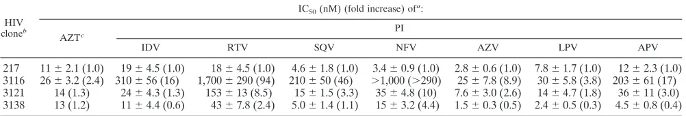
shown in Table 2 and Fig. 2. In these assays, a nucleoside RT inhibitor, AZT, was used as an internal control. The presence of the 19I/E21D/A22V motif (HIV3116) led to 2- to⬎ 10-fold-higher IC50s to PIs than those lacking the motif (HIV3121).
These substitutions had no impact on susceptibility to AZT. A similar drug resistance profile for HIV3116 was observed in a culture system using phytohemagglutinin-stimulated PBMC (data not shown); thus, the resistant profile for HIV3116 is not associated with host cell factors. HIV3121 showed susceptibil-ity to nearly all tested PIs but resistance to RTV and NFV; therefore, the increased IC50 in the variants containing the
19I/E21D/A22V motif is associated with drug resistance rather than with either genetic effect.
A viral replication assay was performed in the absence of drugs to determine the replication competence of each vari-TABLE 1. Longitudinal analysis of p1/p6 cleavage site and HIV PRa
Time point
p1/p6 sequence Mutation(s) atb:
RPGNF LQSRP I15 L19 Insertion K20 E21 A22 M46 I62 L63 A71 T74 I84 I93
April 1995 --- P---L V — X — — — — V P V — — L
June 1995 --- P---L V — X — — — (6)/V (4) — V P V — — L
July 1995 --- P---L V — L (6)/X (2) — — V — V P V — — (6)/V (2) L
August 1995 --- P---L V — L — D V I V P V — V L
October 1995 --- P---L V — I — D V I V P V — V L
April 1996 --- P---L V — I — D V I V P V — V L
October 1998 --- P---L V — I — D V I V P V P V L
December 2000 --- P---L V — I — D V I V P V P V L
a
HIV RNA was isolated from plasma, and then RT-PCR was performed to amplify the full-lengthgagregion. All data were obtained from 8 to 10 clones at each
time point.
b
[image:4.585.42.545.81.188.2]Major mutations in HIV PR are listed first, followed by slashes (/) and minor mutations. Numbers in parentheses indicate the number of clones that contained the specific polymorphism. Dashes (—) denote identity with the HIV-1 consensus B sequence; X denotes absence of insertion.
TABLE 2. Drug resistance profile of HIV mutants
HIV
cloneb
IC50(nM) (fold increase) ofa:
AZTc PI
IDV RTV SQV NFV AZV LPV APV
217 11⫾2.1 (1.0) 19⫾4.5 (1.0) 18⫾4.5 (1.0) 4.6⫾1.8 (1.0) 3.4⫾0.9 (1.0) 2.8⫾0.6 (1.0) 7.8⫾1.7 (1.0) 12⫾2.3 (1.0) 3116 26⫾3.2 (2.4) 310⫾56 (16) 1,700⫾290 (94) 210⫾50 (46) ⬎1,000 (⬎290) 25⫾7.8 (8.9) 30⫾5.8 (3.8) 203⫾61 (17) 3121 14 (1.3) 24⫾4.3 (1.3) 153⫾13 (8.5) 15⫾1.5 (3.3) 35⫾4.8 (10) 7.6⫾3.0 (2.6) 14⫾4.7 (1.8) 36⫾11 (3.0) 3138 13 (1.2) 11⫾4.4 (0.6) 43⫾7.8 (2.4) 5.0⫾1.4 (1.1) 15⫾3.2 (4.4) 1.5⫾0.3 (0.5) 2.4⫾0.5 (0.3) 4.5⫾0.8 (0.4)
aData show means⫾standard errors of IC
50s. Numbers in parentheses show relative increases compared with wild-type HIV217. The values were derived from at
least three independent assays.
b217, wild type; 3116, chimeric HIV containing 19I/E21D/A22V plus M46I/L63P/A71V/I84V/I93L; 3121, chimeric HIV containing M46I/L63P/A71V/I84V/I93L;
3138, recombinant HIV containing 19I/E21D/A22V.
cAZT is an RT inhibitor.
VOL. 80, 2006 19I INSERTION ASSOCIATES WITH MULTIPLE PI RESISTANCE 6139
on November 8, 2019 by guest
http://jvi.asm.org/
[image:4.585.43.544.598.683.2]FIG. 2. Drug susceptibility to IDV and replication properties of HIV-1 constructs. Recombinant HIV-1 mutants were constructed using site-directed mutagenesis. MT-2 cells were infected with 2,500 50% tissue culture infectious dose per 4⫻106cells. In the drug resistance assay, the infected cells were cultured for 7 days in the presence of different concentrations of IDV. IC50s were determined by at least three independent assays. Data are means⫾standard errors (SE). Numbers in parentheses show relative increases compared with those for WT HIV217. In the replication capacity assay, the infected cells were cultured for 7 days in the absence of drug. Results are expressed as mean percentages of growth ⫾SE compared to WT growth. In this assay, the p24 concentration of the WT was 1,566⫾281 ng/ml. N.D.*, not done; N.D.**, not detected.
on November 8, 2019 by guest
http://jvi.asm.org/
ant. The results are expressed as mean percentages of growth of variants compared with that of WT (HIV217) (Fig. 2). In the absence of 19I/E21D/A22V, a variant con-taining only the drug-resistant mutations (HIV3121) showed growth activity similar to that of WT; however, the presence of 19I/E21D/A22V with the drug resistance mutations (HIV3116) showed 30% of the replication activity of the WT. These results suggest that the presence of the combi-nation of 19I/E21D/A22V leads to higher-level drug resis-tance at the expense of replication capacity.
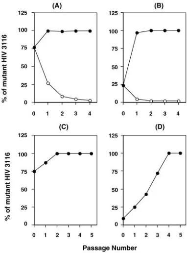
To further compare the replication fitness of the mutant to that of the WT, a competitive fitness assay was performed. WT and HIV3116 were mixed at different ratios and cultured in the presence or absence of 1M IDV. In the absence of IDV, the WT became the dominant species by the second tissue culture passage. In the presence of 1M IDV, however, the mutant virus rapidly became the predominant species within the first passage (Fig. 3A and B). These results suggested that HIV3116 has a disadvantage in viral growth in the absence of drug.
As described above, longitudinal analysis showed that the 19L insertion was selected by a duplication of DNA sequence of codon 19, and then the amino acid was changed to 19I. This evolutional change implicated that 19I may have advantages in replication and/or PI susceptibility. To address the impact of the change, a site-directed mutagenesis was performed on
HIV3116 to induce a 19L change from 19I. The resulting clone, HIV3173, showed drug susceptibility similar to that of HIV3116 (data not shown) but less replication capacity (11% of WT, one-third of the growth of HIV3116). To demonstrate that HIV3116 possesses a relatively better replication capacity than HIV3173, a competitive replication assay was performed with HIV3116 and HIV3173 at the ratios 1:1 or 1:10 and cultured in the absence of drug. The results from the assays showed that the 19I mutant (HIV3116) outgrew the 19L mu-tant (Fig. 3C and D). These results suggest that the evolution from 19L to 19I leads to enhanced replication fitness rather than an increase in drug resistance.
Characterization of mutations at codons 19, 21, and 22 and
gagcleavage sites.To further characterize the impact of each
mutation of 19I, E21D, and A22V on drug susceptibility and replication activity, a series of mutants were constructed by site-directed mutagenesis on HIV3116. Drug susceptibility was analyzed using IDV (Fig. 2). Of note, variants containing known PI resistance mutations and A22V but not 19I (HIV3117 and HIV3127) were defective. Variants containing 19I with the context of PI-resistant mutations but without A22V (HIV3121, HIV3122, and HIV3132) had modest changes in drug susceptibility and showed a reduced replica-tion activity; however, the presence of both 19I and A22V in the context of PI-resistant mutations (HIV3131) caused a high level of drug resistance with a low level of replication activity. The presence of E21D by itself in the context of the PI-resis-tant mutations (HIV3128) had no effect on either drug suscep-tibility or replication activity. These results suggest that 19I or A22V alone in the context of PI-resistant mutations is not associated with drug resistance; however, a combination of the two mutations is associated with a high level of multiple-PI resistance but induces impaired replication. It appears that the addition of E21D to the impaired variant compensates repli-cation. Since a variant containing A22V alone with PI-resistant mutations (HIV3127) was noninfectious, it was postulated that the A22V mutation by itself induced immature, nonviable par-ticles. To address the impact of A22V on replication activity, A22V was induced in NL4.3. The resulting strain was defective (data not shown). Thus, the A22V change by itself leads to a defective protease.
Mutations at the P1⬘ position in p1/p6 were reported as compensational mutations for viral fitness in the activity of PI-resistant mutations in PR. It is reported that a polymor-phism of PP5⬘L in p1/p6 may be associated with drug resis-tance through the I84V mutation (2). To address the roles of each mutation in drug susceptibility and replication fitness, an additional series of recombinant variants was constructed using HIV3116. Variants lacking both mutations at the P1⬘and P5⬘ positions (HIV3118, HIV3119, HIV3124, and HIV3133) were nonviable (Fig. 2). The presence of a single mutation at the P1⬘ (HIV3129) or P5⬘(HIV3130) position restored replication ac-tivity to 9% and 30%, respectively. These results suggest that the presence of the p1/p6 mutations restores replication activ-ity in the context of 19I/E21D/A22V with other PI resistance mutations. Thus, the p1/p6 mutations are required to allow emergence of the 19I/E21D/A22V mutations.
[image:6.585.67.258.69.327.2]To define whether the complex of 19I/E21D/A22V muta-tions alone induces PI resistance, the mutamuta-tions were induced in NL4.3 using site-directed mutagenesis. The resulting variant FIG. 3. Competitive replication assay of WT and mutant virus. WT
and HIV3116 virus were mixed at 1:1 (A) or 10:1 (B). Infected cells were cultured in the presence (closed circles) or absence (open circles) of 1M IDV. To assess the fitness impact of the amino acid change in the insert, HIV3116 and a newly described mutant, HIV3173, in which the 19I was changed to 19L, were mixed at a ratio of 1:1 (C) or 1:10 (D) and then used for infection. The infected cells were cultured in the absence of drug. Data were generated based on relative peak heights of electropherogram produced from direct DNA sequencing of viral RNA from tissue culture supernatant at the end of each passage.
VOL. 80, 2006 19I INSERTION ASSOCIATES WITH MULTIPLE PI RESISTANCE 6141
on November 8, 2019 by guest
http://jvi.asm.org/
was nonviable (data not shown); however, addition of the LP1⬘P and PP5⬘L mutations to the variant restored replication activity. A variant containing 19I/E21D/A22V with PP5⬘L (re-sulting strain, HIV3138) replicated comparably to the WT, was
susceptible to all drugs tested (Table 2), and showed hyper-sensitivity to AZV, LPV, and APV.
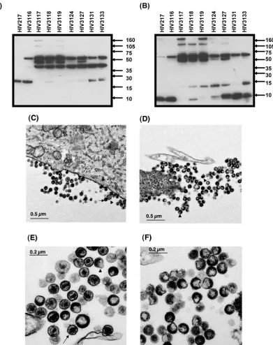
Impact of mutations on cleavage of Gag precursor and
[image:7.585.100.491.69.563.2]structure of HIV-1 virion.The HIV-1 PR cleaves the Gag and
FIG. 4. Western blot and EM pictures of a series of defective HIV mutants. RD cells were transfected with a series of plasmids encoding nonviable viruses (Fig. 2) and cultured for 3 days. HIV-1 virions were isolated from culture supernatants of RD cells using ultracentrifugation as described in Materials and Methods. For each blot, 1g of total protein was loaded on 4 to 12% bis-Tris sodium dodecyl sulfate gels, and Western blotting was performed using an anti-p24 antibody (A) and antisera against p7 (B). The tested variants were HIV217 (WT), HIV3116 (containing the p1/p6 mutations and 19I/E21D/A22V), HIV3117 (lacking 19I), HIV3118 (lacking the p1/p6 mutations), HIV3119 (lacking 19I and the p1/p6 mutations), HIV3124 (lacking 19I, E21D, and the p1/p6 mutations), HIV3127 (lacking 19I and E21D), HIV3131 (lacking E21D), and HIV3133 (lacking E21D and the p1/p6 mutations). Molecular size markers (in kilodaltons) are indicated to the right of the panels. RD cells were transfected with plasmid encoding HIV3116 (C and E) or HIV3117 (D and F) and cultured for 2 days, followed by fixation. Arrows indicate mature virions, and arrowheads indicate immature forms of virions. The electron microscopic pictures were taken as described in Materials and Methods (11).
on November 8, 2019 by guest
http://jvi.asm.org/
the Gag-Pol precursor in the virion to compose the mature form of the infectious virions (19). As described above, absent the 19I insertion or the p1/p6 mutations in the chi-meric HIV, the virus demonstrates a defect in replication. The absence of E21D in the chimeric HIV also led to a decrease in replication activity of the chimeric HIV. Thus, it was speculated that the nonviable mutants might be due to the presence of nonfunctional PR. To address the correla-tion between the PR mutacorrela-tions and the p1/p6 mutacorrela-tions in the cleavage site, Western blotting was performed using antibodies against p24 or RT and antisera against p6, p7, or p17 with virus lysate of isolated HIV virions, as described in Materials and Methods.
Figure 4A and B show results from the Western blot using anti-p24 antibody and anti-p7 serum, respectively. All variants tested showed uncleaved Gag precursors (57 kDa) in virions. Since the partially cleaved products were also seen, the mu-tated PR in these nonviable viruses retained some degree of enzymatic function.
All mutants possessed a 38-kDa fragment, which is equiva-lent to the molecular size of uncleaved p17/p24/p2. HIV3117, lacking the 19I insertion, possessed fragments associated with full-length Gag (57 kDa) and Gag-Pol (110 kDa) but not the p24 and p7 fragments, and an unexpected 50-kDa fragment was seen. The 50-kDa fragment also cross-reacted with anti-p6 and anti-p17 antisera but not to the anti-RT antibody (data not shown). HIV3118 lacking the p1/p6 mutations possessed frag-ments associated with p24, p7, and p7/p1/p6 and the 50-kDa fragment. HIV3131 lacking E21D was infectious (Fig. 2) and contained fragments associated with p24, p24/p2, p7, and p7/p1 and a low amount of p7/p1/p6 but not a significant amount of the 50-kDa fragment. HIV3133, lacking both E21D and the p1/p6 mutations, was defective and contained fragments of p24, p24/p2, p7, p7/p1, and p7/p1/p6 but not the 50-kDa ment. Overall, the defective viruses contain the 50-kDa frag-ment or the p7/p1/p6 fragfrag-ment.
To clarify the morphology of the defective HIV mutants, electron micrographic analyses were performed using RD cells transfected with a series of cloned proviral DNAs, as described in Materials and Methods. The infectious chimeric virus, HIV3116, demonstrated many normal budding, immature, and mature viral particles. Mature particles contained a condensed core of electron-dense material in the center of the virions (Fig. 4C and E). The mutant lacking 19I (HIV3117) demon-strated normal budding and only immature teardrop viral par-ticles lacking the core material (Fig. 4D and F). High-power magnifications revealed that HIV3117 only demonstrated a double layer membrane, likely due to lack of complete Gag processing (Fig. 4F). The same results were seen with other defective viruses, HIV3118, HIV3119, HIV3124, HIV3127, and HIV3133 (data not shown).
DISCUSSION
The emergence of 1 to 6 amino acid insertions in PR at codons near 18, 25, 36, 70, and 95 in clinical isolates has been reported previously (18, 35, 36, 40). The reports indicate that (i) most variants containing amino acid insertions also have one or more well-described PI resistance mutations, (ii) vari-ants containing the insertions in the absence of drug resistance
mutations have been shown to be fully susceptible to all ap-proved PIs, (iii) the presence of the insertions in the context of drug resistance leads to a modest decrease in PI susceptibility, and (iv) the presence of the insertions in the context of PI resistance confers a replication advantage rather than an in-crease in drug resistance (18, 35). In our study, we demon-strated that an amino acid insertion may be selected at codon 19, but the insertion by itself may not contribute to drug sus-ceptibility. We, however, showed that a combination of the insertion and the A22V mutation in the context of drug tance synergistically induced a high level of multiple-PI resis-tance. To our knowledge, this is the first report that the amino acid insertion may contribute to high-level PI resistance, al-though only in the presence of a set of unique circumstances between PR and Gag.
Longitudinal analysis using stored samples showed that the mutations at the p1/p6 cleavage sites existed prior to PI ther-apy, and the 19L insertion initially emerged followed by the 19I change. It is reported that PR-containing insertions appear in nearly 0.1% of patients (16, 18, 35), and the prevalence of the mutation at the P1⬘position with the P5⬘position at the p1/p6 cleavage site is nearly 5% (2). We demonstrated that the com-plex of 19I/E21D/A22V lacking the p1/p6 mutations was non-viable; therefore, it appears that the emergence of the complex in PR may be regulated by the Gag sequence prior to PI therapy, and thus, the prevalence of the complex of 19I/A22V mutations is most likely only lower than suggested by previous reports regarding insertions in PR. Since it has been reported that the emergence of mutations in Gag is concomitant with the development of PI resistance (3, 4, 6, 10, 17, 24, 26, 28, 31, 33, 44, 45), further studies of the correlation between Gag sequences and insertions may provide more insights regarding the possible mechanism of an evolutionary pathway in the emergence of insertions in PR.
Longitudinal analysis showed that the nucleotide sequence of the insertion was initially a duplicate of the sequence of codon 19, CTA, which encodes Leu; subsequently, a C-to-A change led to a Leu-to-Ile change in the insertion. The com-parative replication assay showed that replication capacity in the19I variant was higher than that of the 19L. Since the drug susceptibility was not seen as significantly different, it appears most likely that the Leu-to-Ile change was selected because of an advantage in growth rather than because of drug suscepti-bility.
It has been reported that, in vitro, PR cleaves the Gag precursor (p17/p24/p2/p7/p1/p6) in the order of p2/p7, p17/ p24, and p1/p6; p7/p1; and p24/p2. The cleavages of the p7/p1 and p24/p2 fragments are the two slowest steps (30). There-fore, after initial cleavage of the Gag precursor, the resulting fragments are p17/p24/p2 (43 kDa) and p7/p1/p6 (14 kDa). A chimeric infectious clone, HIV3116, contained lower amounts of noncleaved Gag precursor and 43-kDa fragments than de-fective clones. Dede-fective viruses accumulated the 43-kDa frag-ment with low amounts of the p24 fragfrag-ment. Thus, the mutated PR studied here may inefficiently cleave p17/p24 and subse-quently lead to an accumulation of the 43-kDa fragment.
Virions lacking 19I or the Gag mutations contained an un-expected 50-kDa fragment as well as partially cleaved Gag-Pol fragments. The 50-kDa fragment cross-reacted with anti-p6, -p7, -p17, and -p24 antibodies but not with anti-RT antibody.
VOL. 80, 2006 19I INSERTION ASSOCIATES WITH MULTIPLE PI RESISTANCE 6143
on November 8, 2019 by guest
http://jvi.asm.org/
Even though further study is needed to identify the compo-nents of the fragment, the mutated PR may have a different substrate specificity than WT PR and produce the unexpected fragment from the Gag or Gag-Pol precursors.
The variants lacking the p1/p6 mutations contained the non-cleaved p7/p1/p6 fragment. The p7/p1/p6 fragments were also observed in virions derived from a variant lacking 19I and E21D (HIV3127 retaining A22V). Thus, the p1/p6 mutations and/or A22V may affect the cleavage of p7/p1/p6 fragment.
The crystal structure of multiple-drug-resistant PR has been compared with that of WT PR (23, 25). These studies demon-strated that the presence of drug-resistant mutations in PR led to an expansion of the active site. The same principle may be applied here. Since the codons 19, 21, and 22 are located near the active site in the PR (41), the insertion at codon 19 and the A22V mutation add additional pressure outside the active site, presumably compressing it such that there is an inability of drug to bind (high level of drug resistance), and decrease the replication capacity due to this compromise. The subsequent E21D change helps to relieve this compromise and increases replication competency. Further molecular study may need to address the role of each mutation in the context of a unique set of mutations.
In this study, we have shown that emergence of a novel insertion (19I), a novel amino acid change (E21D), and the A22V mutation may be selected under PI pressure in a clinical isolate. A combination of the 19I and A22V changes are asso-ciated with drug resistance, and the E21D mutation plays a role in compensation of replication fitness. The complex of 19I/E21D/A22V mutations may emerge in the presence of mutations in the p1/p6 cleavage site at P1⬘and P5⬘positions. Thus, a low overall prevalence of this variant is predicted; however, viruses with this genotype demonstrate a high level of multiple drug resistance to PIs but remain sensitive to LPV.
ACKNOWLEDGMENTS
We thank M. Baseler, R. Stevens, and A. Rupert for providing the CD4⫹ T-cell counts. We also thank M. Jason de la Cruz for EM technical support, H. Imamichi and S. Berg for DNA sequencing, and R. Lempicki for a critical reading. The antisera against p6, p7, and p17 were obtained from the AIDS Vaccine Program, SAIC-Frederick, Inc., NCI-Frederick.
This project has been supported by NIAID, contract no. N01-CO-12400.
REFERENCES
1.Adachi, A., H. E. Gendelman, S. Koenig, T. Folks, R. Willey, A. Rabson, and M. A. Martin.1986. Production of acquired immunodeficiency syndrome-associated retrovirus in human and nonhuman cells transfected with an
infectious molecular clone. J. Virol.59:284–291.
2.Bally, F., R. Martinez, S. Peters, P. Sudre, and A. Telenti.2000. Polymor-phism of HIV type 1 gag p7/p1 and p1/p6 cleavage sites: clinical significance and implications for resistance to protease inhibitors. AIDS Res. Hum.
Retrovir.16:1209–1213.
3.Carrillo, A., K. D. Stewart, H. L. Sham, D. W. Norbeck, W. E. Kohlbrenner, J. M. Leonard, D. J. Kempf, and A. Molla.1998. In vitro selection and characterization of human immunodeficiency virus type 1 variants with
in-creased resistance to ABT-378, a novel protease inhibitor. J. Virol.72:7532–
7541.
4.Clavel, F., E. Race, and F. Mammano.2000. HIV drug resistance and viral
fitness. Adv. Pharmacol.49:41–66.
5.Cote, H. C., Z. L. Brumme, and P. R. Harrigan.2001. Human immunodeficiency virus type 1 protease cleavage site mutations associated with protease inhibitor
cross-resistance selected by indinavir, ritonavir, and/or saquinavir. J. Virol.75:
589–594.
6.Croteau, G., L. Doyon, D. Thibeault, G. McKercher, L. Pilote, and D. Lamarre.1997. Impaired fitness of human immunodeficiency virus type 1
variants with high-level resistance to protease inhibitors. J. Virol.71:1089–
1096.
7.de Jong, J. J., J. Goudsmit, V. V. Lukashov, M. E. Hillebrand, E. Baan, R. Huismans, S. A. Danner, J. H. ten Veen, F. de Wolf, and S. Jurriaans.1999. Insertion of two amino acids combined with changes in reverse transcriptase containing tyrosine-215 of HIV-1 resistant to multiple nucleoside analogs.
AIDS13:75–80.
8.de Oliveira, T., S. Engelbrecht, E. Janse van Rensburg, M. Gordon, K. Bishop, J. zur Megede, S. W. Barnett, and S. Cassol.2003. Variability at human immunodeficiency virus type 1 subtype C protease cleavage sites: an
indication of viral fitness? J. Virol.77:9422–9430.
9.Dewar, R. L., H. C. Highbarger, M. D. Sarmiento, J. A. Todd, M. B. Vasudevachari, R. T. Davey, Jr., J. A. Kovacs, N. P. Salzman, H. C. Lane, and M. S. Urdea.1994. Application of branched DNA signal amplification to monitor human immunodeficiency virus type 1 burden in human plasma.
J. Infect. Dis.170:1172–1179.
10.Doyon, L., G. Croteau, D. Thibeault, F. Poulin, L. Pilote, and D. Lamarre.
1996. Second locus involved in human immunodeficiency virus type 1
resis-tance to protease inhibitors. J. Virol.70:3763–3769.
11.Gonda, M. A., S. A. Aaronson, N. Ellmore, V. H. Zeve, and K. Nagashima.
1976. Ultrastructural studies of surface features of human normal and tumor cells in tissue culture by scanning and transmission electron microscopy.
J. Natl. Cancer Inst.56:245–263.
12.Haertle, T., C. J. Carrera, D. B. Wasson, L. C. Sowers, D. D. Richman, and D. A. Carson.1988. Metabolism and anti-human immunodeficiency virus-1
activity of 2-halo-2⬘,3⬘-dideoxyadenosine derivatives. J. Biol. Chem.263:
5870–5875.
13.Harada, S., Y. Koyanagi, and N. Yamamoto.1985. Infection of HTLV-III/ LAV in HTLV-carrying cells MT-2 and MT-4 and application in a plaque
assay. Science229:563–566.
14.Harrigan, P. R., B. Stuart, and B. A. Larder.1998. Relative replicative fitness of zidovudine-resistant human immunodeficiency virus type 1 isolates in
vitro. J. Virol.72:3773–3778.
15.Imamichi, T., S. C. Berg, H. Imamichi, J. C. Lopez, J. A. Metcalf, J. Falloon, and H. C. Lane.2000. Relative replication fitness of a high-level 3⬘
Azido-3⬘-deoxythimidien resistant variant of human Immunodeficiency virus type-1
possessing an amino acid deletion at codon 67 and a novel substitution (Thr
to Gly) at codon 69. J. Virol.74:10958–10964.
16.Kagan, R., M. Winters, T. Merigan, and P. Heseltine.2004. HIV type 1 genotypic resistance in a clinical database correlates with antiretroviral
uti-lization. AIDS Res. Hum. Retrovir.20:1–9.
17.Kaufmann, D., M. Munoz, G. Bleiber, S. Fleury, B. Lotti, R. Martinez, W. Pichler, P. Meylan, and A. Telenti.2000. Virological and immunological
characteristics of HIV treatment failure. AIDS14:1767–1774.
18.Kim, E. Y., M. A. Winters, R. M. Kagan, and T. C. Merigan.2001. Functional correlates of insertion mutations in the protease gene of human
immuno-deficiency virus type 1 isolates from patients. J. Virol.75:11227–11233.
19.Kohl, N. E., E. A. Emini, W. A. Schleif, L. J. Davis, J. C. Heimbach, R. A. Dixon, E. M. Scolnick, and I. S. Sigal.1988. Active human immunodeficiency virus protease is required for viral infectivity. Proc. Natl. Acad. Sci. USA
85:4686–4690.
20.Kovacs, J. A., S. Vogel, J. M. Albert, J. Falloon, R. T. Davey, Jr., R. E. Walker, M. A. Polis, K. Spooner, J. A. Metcalf, M. Baseler, G. Fyfe, and H. C. Lane.1996. Controlled trial of interleukin-2 infusions in patients infected
with the human immunodeficiency virus. N. Engl. J. Med.335:1350–1356.
21.Larder, B. A., S. Bloor, S. D. Kemp, K. Hertogs, R. L. Desmet, V. Miller, M. Sturmer, S. Staszewski, J. Ren, D. K. Stammers, D. I. Stuart, and R. Pauwels.1999. A family of insertion mutations between codons 67 and 70 of human immunodeficiency virus type 1 reverse transcriptase confer
multinucleo-side analog resistance. Antimicrob. Agents Chemother.43:1961–1967.
22.Lobato, R. L., E. Y. Kim, R. M. Kagan, and T. C. Merigan.2002. Genotypic and phenotypic analysis of a novel 15-base insertion occurring between codons 69 and 70 of HIV type 1 reverse transcriptase. AIDS Res. Hum.
Retrovir.18:733–736.
23. Logsdon, B. C., J. F. Vickrey, P. Martin, G. Proteasa, J. I. Koepke, S. R. Terlecky, Z. Wawrzak, M. A. Winters, T. C. Merigan, and L. C. Kovari.2004. Crystal structures of a multidrug-resistant human immunodeficiency virus
type 1 protease reveal an expanded active-site cavity. J. Virol.78:3123–3132.
24.Mammano, F., C. Petit, and F. Clavel.1998. Resistance-associated loss of viral fitness in human immunodeficiency virus type 1: phenotypic analysis of protease and gag coevolution in protease inhibitor-treated patients. J. Virol.
72:7632–7637.
25.Martin, P., J. F. Vickrey, G. Proteasa, Y. L. Jimenez, Z. Wawrzak, M. A. Winters, T. C. Merigan, and L. C. Kovari.2005. “Wide-open” 1.3 A structure
of a multidrug-resistant HIV-1 protease as a drug target. Structure13:1887–
1895.
26.Martinez-Picado, J., A. V. Savara, L. Sutton, and R. T. D’Aquila.1999. Replicative fitness of protease inhibitor-resistant mutants of human
immu-nodeficiency virus type 1. J. Virol.73:3744–3752.
27.Masquelier, B., E. Race, C. Tamalet, D. Descamps, J. Izopet, C. Buffet-Janvresse, A. Ruffault, A. S. Mohammed, J. Cottalorda, A. Schmuck, V. Calvez, E. Dam, H. Fleury, F. Brun-Vezinet, and ANRS AC11 Resistance
on November 8, 2019 by guest
http://jvi.asm.org/
Study Group.2001. Genotypic and phenotypic resistance patterns of human immunodeficiency virus type 1 variants with insertions or deletions in the reverse transcriptase (RT): multicenter study of patients treated with RT
inhibitors. Antimicrob. Agents. Chemother.45:1836–1842.
28.Nijhuis, M., R. Schuurman, D. de Jong, J. Erickson, E. Gustchina, J. Albert, P. Schipper, S. Gulnik, and C. A. Boucher.1999. Increased fitness of drug resistant HIV-1 protease as a result of acquisition of compensatory
muta-tions during suboptimal therapy. AIDS13:2349–2359.
29.Palella, F. J., K. M. Delaney, A. C. Moorman, M. O. Loveless, J. Fuhrer, G. A. Satten, D. J. Aschman, S. D. Holmberg, et al.1998. Declining morbidity and mortality among patients with advanced human immunodeficiency virus
infection. N. Engl. J. Med.338:853–860.
30.Pettit, S. C., N. Sheng, R. Tritch, S. Erickson-Viitanen, and R. Swanstrom.
1998. The regulation of sequential processing of HIV-1 Gag by the viral
protease. Adv. Exp. Med. Biol.436:15–25.
31.Picchio, G. R., H. Valdez, R. Sabbe, A. L. Landay, D. R. Kuritzkes, M. M. Lederman, and D. E. Mosier.2000. Altered viral fitness of HIV-1 following failure of protease inhibitor-based therapy. J. Acquir. Immune Defic. Syndr.
25:289–295.
32.Rambaut, A., D. Posada, K. A. Crandall, and E. C. Holmes.2004. The causes
and consequences of HIV evolution. Nat. Rev. Genet.5:52–61.
33.Robinson, L. H., R. E. Myers, B. W. Snowden, M. Tisdale, and E. D. Blair.
2000. HIV type 1 protease cleavage site mutations and viral fitness: impli-cations for drug susceptibility phenotyping assays. AIDS Res. Hum.
Retro-vir.16:1149–1156.
34.Schinazi, R. F., B. A. Larder, and J. W. Mellors.2000. Mutations in retroviral genes associated with drug resistance: 1999–2000 update. Int. Antivir. News
7:46–69.
35.Sturmer, M., S. Staszewski, H. W. Doerr, and K. Hertogs.2003. A 6-base pair insertion in the protease gene of HIV type 1 detected in a protease inhibitor-naive patient is not associated with indinavir treatment failure.
AIDS Res. Hum. Retrovir.19:967–968.
36.Tramuto, F., F. Bonura, S. Mancuso, N. Romano, and F. Vitale.2005. Detection of a new 3-base pair insertion mutation in the protease gene of human immunodeficiency virus type 1 during highly active antiretroviral
therapy (HAART). AIDS Res. Hum. Retrovir.21:420–423.
37.van der Hoek, L., N. Back, M. F. Jebbink, A. de Ronde, M. Bakker, S. Jurriaans, P. Reiss, N. Parkin, and B. Berkhout.2005. Increased multinucleo-side drug resistance and decreased replicative capacity of a human
immunode-ficiency virus type 1 variant with an 8-amino-acid insert in the reverse
transcrip-tase. J. Virol.79:3536–3543.
38.van Vaerenbergh, K., K. Van Laethem, J. Albert, C. A. Boucher, B. Clotet, M. Floridia, J. Gerstoft, B. Hejdeman, C. Nielsen, C. Pannecouque, L. Perrin, M. F. Pirillo, L. Ruiz, J. C. Schmit, F. Schneider, A. Schoolmeester, R. Schuurman, H. J. Stellbrink, L. Stuyver, J. Van Lunzen, B. Van Remoortel, E. Van Wijngaerden, S. Vella, M. Witvrouw, S. Yerly, E. De Clercq, J. Destmyer, and A. M. Vandamme.2000. Prevalence and characteristics of multinucleoside-resistant human immunodeficiency virus type 1 among Eu-ropean patients receiving combinations of nucleoside analogues.
Antimi-crob. Agents Chemother.44:2109–2117.
39.Winters, M. A., K. L. Coolley, Y. A. Girard, D. J. Levee, H. Hamdan, R. W. Shafer, D. A. Katzenstein, and T. C. Merigan.1998. A 6-basepair insert in the reverse transcriptase gene of human immunodeficiency virus type 1
confers resistance to multiple nucleoside inhibitors. J. Clin. Investig.102:
1769–1775.
40.Winters, M. A., R. M. Kagan, P. N. Heseltine, and T. C. Merigan.2005. New two-amino acid insertion near codon 70 of the HIV type 1 protease gene.
AIDS Res. Hum. Retrovir.21:311–313.
41.Wlodawer, A., and J. W. Erickson.1993. Structure-based inhibitors of HIV-1
protease. Annu. Rev. Biochem.62:543–583.
42.Wu, T. D., C. A. Schiffer, M. J. Gonzales, J. Taylor, R. Kantor, S. Chou, D. Israelski, A. R. Zolopa, W. J. Fessel, and R. W. Shafer.2003. Mutation patterns and structural correlates in human immunodeficiency virus type 1
protease following different protease inhibitor treatments. J. Virol.77:4836–
4847.
43.Yeni, P. G., S. M. Hammer, C. C. Carpenter, D. A. Cooper, M. A. Fischl, J. M. Gatell, B. G. Gazzard, M. S. Hirsch, D. M. Jacobsen, D. A. Katzenstein, J. S. Montaner, D. D. Richman, M. S. Saag, M. Schechter, R. T. Schooley, M. A. Thompson, S. Vella, and P. A. Volberding.2002. Antiretroviral treatment for adult HIV infection in 2002: updated recommendations of the International
AIDS Society-USA Panel. JAMA288:222–235.
44.Zennou, V., F. Mammano, S. Paulous, D. Mathez, and F. Clavel.1998. Loss of viral fitness associated with multiple Gag and Gag-Pol processing defects in human immunodeficiency virus type 1 variants selected for resistance to
protease inhibitors in vivo. J. Virol.72:3300–3306.
45.Zhang, Y. M., H. Imamichi, T. Imamichi, H. C. Lane, J. Falloon, M. B. Vasudevachari, and N. P. Salzman.1997. Drug resistance during indinavir therapy is caused by mutations in the protease gene and in its Gag substrate
cleavage sites. J. Virol.71:6662–6670.
VOL. 80, 2006 19I INSERTION ASSOCIATES WITH MULTIPLE PI RESISTANCE 6145