0022-538X/95/$04.0010
Copyrightq1995, American Society for Microbiology
Mutations within a Putative Cysteine Loop of the
Transmembrane Protein of an Attenuated
Immunodeficiency-Inducing Feline Leukemia Virus Variant Inhibit Envelope
Protein Processing
CARA CARTHEL BURNS, MARY L. POSS, ELAINE THOMAS,†
ANDJULIE OVERBAUGH*
Department of Microbiology, University of Washington, Seattle, Washington 98195
Received 17 October 1994/Accepted 5 January 1995
A replication-defective feline leukemia virus molecular clone, 61B, has been shown to cause
immunodefi-ciency in cats and cytopathicity in T cells after a long latency period when coinfected with a minimally
pathogenic helper virus (J. Overbaugh, E. A. Hoover, J. I. Mullins, D. P. W. Burns, L. Rudensey, S. L.
Quackenbush, V. Stallard, and P. R. Donahue, Virology 188:558–569, 1992). The long-latency phenotype of 61B
has been mapped to four mutations in the extracellular domain of the envelope transmembrane protein, and
we report here that these mutations cause a defect in envelope protein processing. Immunoprecipitation
analyses demonstrated that the 61B gp85 envelope precursor was produced but that further processing to
generate the surface protein (SU/gp70) and the transmembrane protein (TM/p15E) did not occur. The 61B
precursor was not expressed on the cell surface and appeared to be retained in the endoplasmic reticulum or
Golgi apparatus. Two of the four 61B-specific amino acid changes are located within a putative cysteine loop
in a region of TM that is conserved among retroviruses. Introduction of these two amino acid changes into a
replication-competent highly cytopathic virus resulted in the production of noninfectious virus that exhibited
an envelope-protein-processing defect. This analysis suggests that mutations in a conserved region within a
putative cysteine loop affect retroviral envelope protein maturation and viral infectivity.
Retroviral extracellular surface glycoprotein (SU) and
trans-membrane protein (TM) envelope polypeptides are
synthe-sized as a single polyprotein precursor that is proteolytically
cleaved to produce the mature functional envelope proteins.
The envelope precursor enters the secretory pathway during
translation, the signal sequence is cleaved, and the precursor is
glycosylated in the endoplasmic reticulum. After glycosylation,
the envelope precursor protein oligomerizes and the
com-plexes are transported to the Golgi apparatus. In the Golgi
apparatus, high-mannose oligosaccharides are modified to
complex and hybrid carbohydrate side chains, and the
glyco-protein precursor is cleaved by a host cell protease to generate
SU and TM. The mature envelope proteins are then
trans-ported to the cell surface as an oligomeric complex (for a
review, see reference 27).
The envelope protein has been implicated as the
determi-nant of disease specificity for variants of feline leukemia virus
(FeLV) that cause immunodeficiency (12, 45). Viral chimeras
that encode the SU of immunodeficiency-inducing
replication-defective variants 61C or 61B in the context of a mildly
patho-genic, replication-competent provirus, 61E, cause cytopathic
effects in feline T cells and immunodeficiency disease in cats
(12, 39, 45). However, 61B is delayed compared with 61C in its
ability to cause cytopathic effects in T cells and
immunodefi-ciency in cats as a result of four amino acid differences in the
envelope TM protein (40, 53). Two of the changes should
introduce positively charged residues in place of negatively
charged residues in a region of TM that is highly conserved in
FeLV and among other retroviruses (10, 41, 50).
In this study, we have investigated the effect of the predicted
amino acid differences in 61B and 61C TM on envelope
pro-tein synthesis and processing using stable cell lines expressing
61C, 61B, or chimeric envelope proteins. This analysis shows
that the 61B envelope precursor protein is not processed to SU
and TM and that 61B-specific TM sequences between two
highly conserved cysteine residues confer the
envelope-pro-cessing defect. The 61B envelope precursor protein cannot be
detected on the cell surface and appears to be sequestered in
the endoplasmic reticulum or Golgi apparatus. In addition, the
two 61B-specific amino acid changes in TM abolish the
infec-tivity of an infectious molecular clone.
MATERIALS AND METHODS
Plasmid constructions and mutagenesis.The plasmids p61C, p61B, pEECC, and pCCC([CB]C) have been described previously (39, 53) and are diagrammed in Fig. 1. Chimera CCC([CB]C]) consists of 61C proviral DNA that contains 154 amino acids of the 61B TM.
The plasmid pEECC-envK536K537was constructed by the following procedure:
two nucleotide changes were introduced into p61C by a ligase-mediated PCR technique (35) using a phosphorylated mutagenic primer, 61B-env24 (59GAAG CAACATTTTTTTTTTAATGCGGCACAG39). This primer contained 61B-specific sequences at env nucleotides 1606 and 1609 (i.e., amino acids 536 and 537 of the envelope precursor), which introduced T changes (underlined and in boldface for the primer) relative to 61C. Primers that hybridize to the 39end of SU (61C [GenBank M18246] env nucleotides 1040 to 1060) and to the 39end of TM (61C env nucleotides 1904 to 1929) were used to generate a 890-bp fragment containing the mutagenic primer.
PCR was performed as described by Michael (35). The desired 890-bp product was purified after gel electrophoresis and then cloned into a 3961C subclone (p39CC [39]) using conserved NcoI and RsrII sites. To reconstruct a full-length provirus, the EcoRI-XhoI long terminal repeat (LTR)-gag-pol from 61E (6.5 kb) was ligated into p39CC-envK536K537. The structure of the plasmid
(pEECC-envK536K537) was verified by restriction fragment analysis. The PCR-amplified
portion of the genome was sequenced by using the Sanger dideoxy chain
termi-* Corresponding author. Mailing address: Department of Microbi-ology, SC-42, University of Washington, Seattle, WA 98195. Phone: (206) 543-3146. Fax: (206) 543-8297. Electronic mail address: [email protected].
† Present address: Department of Medicine, University of New Mexico, Albuquerque, NM 87131.
2126
on November 9, 2019 by guest
http://jvi.asm.org/
nation method (48) to verify that the desired changes were present and that there were no additional mutations introduced during amplification.
Transfection and isolation of stable cell lines.AH927 feline embryonic fibro-blasts and 3201 T cells were cultured as described previously (40). Stable AH927 cell lines that contain 61C and 61B proviral DNA have been described previously (39). We used a similar strategy to establish cell lines that contained various FeLV chimeras. Briefly, FeLV proviral DNA plus pMOneo (20) was transfected into AH927 cells at a 3:1 molar ratio using calcium phosphate (Stratagene, La Jolla, Calif.) (6). Single-cell clones were isolated after selection in 0.6 mg of G418 (Gibco/BRL, Grand Island, N.Y.) per ml for 2 to 3 weeks. Individual colonies were analyzed for the presence of proviral DNA by Southern blotting (40) and for the expression of p27gag
by enzyme-linked immunosorbent assaying (ELISA) (Virachek/FeLV; Synbiotics, San Diego, Calif.). Total cellular RNA, prepared by
the guanidinium isothiocyanate procedure (8), was analyzed by Northern (RNA) blotting using the LTR-specific exU3 probe (37) to ensure that the expected full-length and subgenomic mRNAs were expressed.
3201 cells were transfected using Lipofectamine (Gibco/BRL) and 2mg of pEECC or pEECC-envK536K537plasmid DNA according to the supplier’s
in-structions. Cultures were monitored by FeLV p27gagELISA for virus replication
and spread at 3- to 4-day intervals for 5 weeks after transfection.
Metabolic labeling and radioimmunoprecipitation of cellular lysates. Radio-immunoprecipitation analysis (RIPA) was performed with minor modification of a method described previously (43). Cells were labeled for lengths of time specified in the figure legends in methionine- and cysteine-deficient Dulbecco’s modified Eagle’s medium supplemented with 100mCi of [35S]methionine plus
[35S]cysteine per ml (Trans-Label; ICN, Irvine, Calif.). After labeling, cells were
washed twice in cold phosphate-buffered saline and then lysed in cold lysis buffer A (25 mM Tris hydrochloride [pH 8.0], 0.15 M NaCl, 1% Triton X-100, 1% deoxycholate, 1 mM phenylmethylsulfonyl fluoride [56]). Cellular lysates were clarified by centrifugation at 15,0003g at 48C for 5 min, which was followed by preclearing with protein A-Sepharose beads overnight with rocking at 48C. In-corporated radioactivity was determined by precipitation using trichloroacetic acid. An equal number of trichloroacetic acid-precipitable counts was added to each RIPA reaction mixture.
Antibodies were obtained from Custom Monoclonals, Sacramento, Calif. (an-ti-SU monoclonal antibody C11D8 and anti-TM monoclonal antibody PF6J-2A) or Quality Biotech Inc., Resource Lab, Camden, N.J. (serum no. 77S000301). Serum no. 77S000301 is a goat polyclonal antiserum that was made using dena-tured FeLV virion as immunogen.
Antibody was mixed with protein A-Sepharose beads for 30 min at 48C, which was followed by washing once in 50 mM Tris (pH 8.0)–150 mM NaCl. Cell lysates were added to antibody-protein A beads and incubated with rocking for 3 h at 48C. RIPA reaction mixtures were washed five times in 1 ml of cold wash buffer (0.05 M Tris [pH 7.2], 0.15 M NaCl, 0.1% sodium dodecyl sulfate [SDS], 0.1% Triton X-100) on ice, and then the beads were resuspended in SDS-polyacryl-amide gel electrophoresis (PAGE) sample buffer and analyzed by SDS-PAGE (30) and fluorography.
For endo-b-N-acetylglucosaminosidase H (endo H) digestions, beads contain-ing immunoprecipitated proteins were boiled for 3 min and then subjected to digestion using conditions described by the supplier (Genzyme, Cambridge, Mass.). Endo H digestion was performed at 378C for 1 h, additional enzyme was added, and digestion was continued overnight.
Metabolic labeling and radioimmunoprecipitation of culture supernatant.For analysis of proteins present in the culture supernatant, subconfluent cells were labeled in methionine- and cysteine-deficient medium that contained 300mCi of [35S]methionine plus [35S]cysteine per ml for 1 h, which was followed by the
addition of an equal volume of complete medium and incubation for 22 h. Culture supernatant was clarified by centrifugation, and 53detergent mix (56) was added before RIPA was performed.
FACS.Immunostaining and fluorescence-activated cell sorting (FACS) were performed by standard methods. Cells were incubated with primary antibody (C11D8) at a concentration of 34mg/ml at 378C. Cells were incubated at 48C with 50mg per ml of secondary antibody, an anti-mouse immunoglobulin G2B-fluo-rescein isothiocyanate (Fisher, Pittsburgh, Pa.).
RESULTS
Comparison of envelope protein processing in cell lines
ex-pressing 61C and 61B.
To define the biochemical defect
asso-ciated with a replication defect encoded by the 61B envelope
gene, we examined the envelope protein produced by the 61B
provirus. For this purpose, we used stable cell lines that
con-tain 61B or 61C replication-defective proviral genomes. In
these cell lines, full-length viral RNA and subgenomic
enve-lope RNA are expressed but no infectious virus is produced,
because of a mutation(s) in the 5
9
LTR, gag, or pol (39).
Envelope proteins were analyzed by RIPA of lysates of cells
labeled with [
35S]methionine and [
35S]cysteine for 7 h (Fig.
2A). Immunoprecipitation was performed using C11D8, an
antibody that recognizes an epitope (MGPNL) found in the
FeLV SU protein (25). This antibody did not recognize any
AH927 cellular proteins in an immunoprecipitation reaction
(Fig. 2A, lane 1). The gp85 envelope precursor and the mature
SU protein, gp70, were precipitated from the lysates of cells
expressing 61C (lane 3). The 61C lysate contained
approxi-mately equal amounts of gp70 and gp85. Similar results were
obtained for 61E-infected cells (Fig. 2A, lane 2). However, in
cell lines expressing the 61B envelope protein (lanes 4 and 5),
only the gp85 envelope precursor was detected. Mature 61B
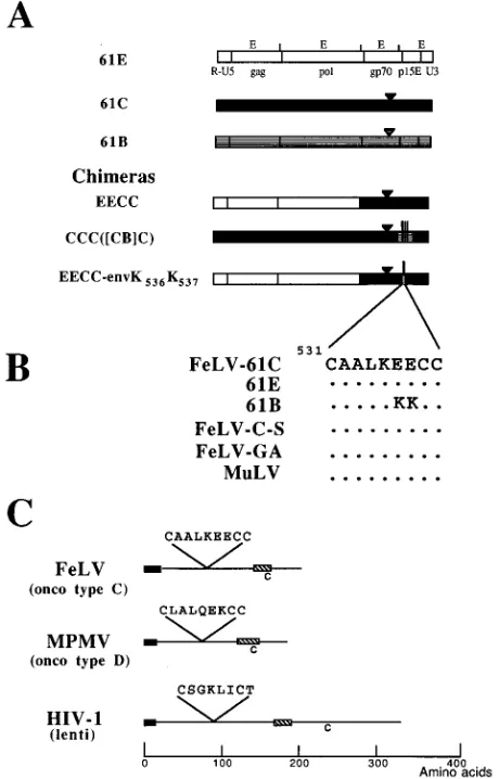
FIG. 1. (A) Schematic diagram of 61E, 61C, and 61B viral genomes and chimeras. The chimeras are named according to the system used previously (12, 40, 46). For the chimeras with four-letter names, the letters designate, respec-tively, the 59LTR-gag segment; the pol segment; the XhoI-NcoI fragment en-coding a small segment of 39pol and most of env; and the NcoI-EcoRI fragment
encoding the C-terminal end of gp70, all of p15E, and the 39LTR. Parentheses in the names of subsequent chimeras indicate a subdivision of the fourth seg-ment, and brackets denote a further subdivision of part of that segment. The triangle denotes the location of the six-amino-acid insertion that is a primary determinant of pathogenicity (12, 45), and the vertical lines indicate the locations of amino acid differences in 61B relative to 61C or 61E in TM (53). (B) Amino acids identical to those in the 61C reference sequence are denoted by dots. The 61C sequence starts at amino acid 531 of the gp85 coding region. Shown are sequences for FeLV 61E, 61C, and 61B (39, 40), FeLV-C-Sarma (FeLV-C-S) (47), FeLV-GA (subgroup B FeLV, Gardner-Arnstein isolate) (18), and murine leukemia virus (MuLV; the sequence is identical for Moloney murine leukemia virus [49], Akv murine leukemia virus [31], and Friend murine leukemia virus [28]). (C) Diagram of representative TM proteins. The black boxes denote the fusion peptides, and the cross-hatched boxes represent the membrane-spanning regions. onco, MPMV oncovirus (50); lenti, HIV-1 lentivirus (Bru isolate) (55).
VOL. 69, 1995 FeLV 61B ENVELOPE PROCESSING 2127
on November 9, 2019 by guest
http://jvi.asm.org/
[image:2.612.62.292.72.431.2]SU protein was not detected by Western blot (immunoblot)
analysis or in pulse-chase experiments, even at times when
most of the 61C envelope protein had been processed to gp70
(data not shown).
Figure 2B shows the results of RIPA of cellular lysates using
an anti-TM antibody, PF6J-2A, which recognizes an epitope in
the amino terminus of FeLV TM, upstream of the region
where 61B and 61C sequences differ (Custom Monoclonals);
however, this antibody reacts poorly with the gp85 envelope
precursor. PF6J-2A antibody did not precipitate any labeled
proteins from uninfected AH927 cells (lane 1). The expected
TM protein (p15E) was detected in lysates of cells expressing
61C envelope (lane 3) and in 61E-infected cells (lane 2). In
contrast, no p15E was immunoprecipitated in the cells
express-ing 61B envelope (lanes 4 and 5), and a longer exposure of the
gel did not show any detectable p15E in these lysates (data not
shown). Taken together, the absence of mature 61B SU and
TM protein in cellular lysates suggests that 61B envelope
pro-cessing is defective.
Localization of 61B envelope protein.
Because mature 61B
envelope proteins could not be detected, we wanted to
deter-mine which step in the processing pathway was defective. Endo
H analysis was used to determine if the envelope precursor
contained the types of oligosaccharides found on proteins at
early steps in the oligosaccharide-processing pathway or types
of oligosaccharides characteristic of completely processed
pro-teins. Figure 3 shows the products of immunoprecipitation of
61B envelope using C11D8 anti-SU antibody and endo H
di-gestion. Incubation of the immunoprecipitate in the absence of
endo H did not change the mobility of the gp85 precursor
protein (lanes 1 and 4). The 61B gp85 was sensitive to endo H,
as indicated by a change in the mobility of the 61B envelope
precursor protein (lanes 2 and 3). The size of the digested
protein is consistent with the predicted size of the fully
degly-cosylated protein, which suggests that the envelope precursor
has not been exposed to the processing enzymes in the Golgi
apparatus that convert high-mannose residues to complex and
hybrid residues. Therefore, the 61B envelope precursor is
likely located in the endoplasmic reticulum or proximal Golgi
apparatus.
[image:3.612.351.515.71.320.2]To test for envelope protein on the cell surface, we
per-formed indirect immunofluorescence using anti-SU primary
antibody, incubation with a fluorescein
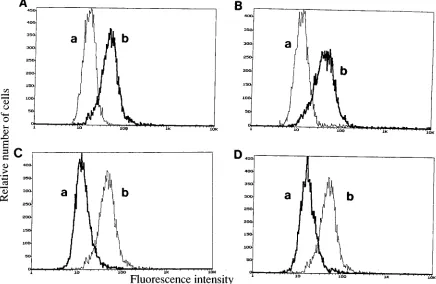
isothiocyanate–anti-mouse secondary antibody, and FACS analysis (Fig. 4). As
shown in Fig. 4A, 61C envelope could be detected on the
surface of cells transfected with 61C proviral DNA. Envelope
protein was detected on the surface of FeLV-infected AH927
cells but not on uninfected AH927 cells (Fig. 4B). In contrast,
cells expressing 61B envelope did not exhibit fluorescence
un-der similar conditions (Fig. 4C). The absence of 61B envelope
on the cell surface is consistent with the endo H analysis, which
FIG. 2. Immunoprecipitation of envelope proteins from AH927 cell clones expressing defective 61C, 61B, or chimeric viral genomes. AH927 cells were labeled with [35S]methionine plus [35S]cysteine for 7 h. The viral genome
ex-pressed in each cell line is indicated above each lane. 61B nos. 1 and 2 are independent cell lines expressing 61B. Molecular weight markers (in kilodaltons) are labeled adjacent to the bands, as are gp85, gp70, and p15E. (A) Cellular lysates immunoprecipitated with the anti-gp70 and -gp85 antibody C11D8 and analyzed by SDS-9% PAGE; (B) Cellular lysates immunoprecipitated with the anti-p15E antibody PF6J-2A and analyzed by SDS-12.5% PAGE.
FIG. 3. Endo H analysis of 61B envelope protein. AH927 cells expressing 61B envelope protein were labeled with [35S]methionine plus [35S]cysteine for 7
h. Cellular lysates were immunoprecipitated using C11D8 antibody. Precipitated proteins were incubated in duplicate overnight in the presence (lanes 2 and 3) or absence (lanes 1 and 4) of endo H and analyzed by SDS-9% PAGE.
on November 9, 2019 by guest
http://jvi.asm.org/
suggests that the envelope precursor protein was trapped in the
endoplasmic reticulum or Golgi apparatus.
Mapping the envelope-processing defect of 61B.
We were
interested in the role of specific 61B envelope determinants in
defective envelope processing because the long-latency
pheno-type and a replication defect colocalized to 61B TM. To
inves-tigate this, we analyzed envelope expression of several
repli-cation-defective 61C/61B chimeras encoding 61B TM (see Fig.
1 for a diagram of the clones.) Chimera CCC([CB]C) contains
61C proviral DNA with an 154-amino-acid segment of 61B TM
and has been shown to exhibit the long-latency phenotype and
a replication defect (53). Figure 2A, lane 6, shows that anti-SU
antibody precipitated gp85, but not gp70, in lysates of AH927
cell lines expressing this chimera. Similarly, no p15E was
im-munoprecipitated by anti-TM antibody (Fig. 2B, lane 6). In
addition, no envelope protein was detected on the surface of
cells that contain chimeric FeLV genomes, as depicted in Fig.
4D (graph a). These experiments indicate that the
envelope-processing defect was, like the replication defect and the
long-latency phenotype, conferred by 61B TM sequences.
Site-specific mutagenesis of 61C TM to test the infectivity of
EECC-envK
536K
537.
The region of 61B TM that confers the
long-latency phenotype and a defect in envelope protein
pro-cessing contains four predicted amino acid differences relative
to 61C. Two of the mutations encode nonconservative changes
in a conserved region of the TM protein. In this region, the 61B
sequence contains a positively charged lysine in place of a
negatively charged glutamate at two adjacent positions
be-tween two cysteines that are present in the extracellular
do-main of all retroviral TMs (Fig. 1) (23, 24, 38, 41). Thus, we
predicted that these two changes confer the
defective-enve-lope-processing phenotype, and we introduced them into the
TM protein of the EECC chimera, a clone that encodes the 5
9
LTR, gag, and pol genes of 61E and the env gene and 3
9
LTR
of 61C (39). The mutations were introduced into EECC
be-cause EECC virus is replication competent and highly
infec-tious for feline T cells; thus, the effect of the mutations on
infectivity could be examined. The resulting chimera,
EECC-envK
536K
537, contained 61B-specific sequences at two
posi-tions, which introduced lysine residues at amino acids 536 and
537 in the envelope precursor.
In order to determine if the mutations affected replication
and/or infectivity, we established stable AH927 cell lines that
contained EECC-envK
536K
537provirus and tested for
infec-tious virus by transferring cell-free culture supernatants to
3201 cells. No p27
gagwas detected in the 3201 recipient cells
during a 7-week period, whereas the EECC positive control
produced high levels of p27
gagand exhibited cytopathic effects
in 3201 cells at 1 week postinfection. This suggests that the
glutamate-to-lysine mutations in TM made the virus
replica-tion defective. In addireplica-tion, transfecreplica-tion of EECC-envK
536K
537plasmid into 3201 T cells did not result in the production of
infectious virus during a 5-week period, although replication of
[image:4.612.88.524.75.359.2]EECC was detected by p27
gagELISA at 10 days posttransfection.
FIG. 4. FACS analysis of cells expressing 61C, 61B, and chimeric 61C/61B envelope proteins. AH927 cells were incubated sequentially with C11D8 antibody (primary antibody) and mouse anti-immunoglobulin G2B–fluorescein isothiocyanate (secondary antibody) and then FACS analyzed. In each panel, graph b represents cells expressing envelope (61C or 61E) incubated with primary and secondary antibody. (A) AH927 cells expressing 61C envelope incubated with secondary antibody only (graph a) or with primary and secondary antibody (graph b); (B) uninfected AH927 cells incubated with primary and secondary antibody (graph a) and AH927 cells infected with 61E incubated with primary and secondary antibody (graph b); (C) AH927 cells expressing 61B envelope incubated with primary and secondary antibody (graph a) and AH927 cells expressing 61C envelope incubated with primary and secondary antibody (graph b, included for comparison); (D) AH927 cells expressing CCC([CB]C) chimeric envelope incubated with primary and secondary antibody (graph a) and AH927 cells expressing 61C envelope incubated with primary and secondary antibody (graph b, included for comparison).
VOL. 69, 1995 FeLV 61B ENVELOPE PROCESSING 2129
on November 9, 2019 by guest
http://jvi.asm.org/
Envelope protein processing of EECC-envK
536K
537.
In order
to evaluate the effect of the two glutamate-to-lysine mutations
on envelope protein processing, we performed RIPA of the
AH927 cell lines expressing EECC-envK
536K
537. In each of the
control extracts, including cells expressing 61E, 61C, and
EECC, both gp70 and gp85 were precipitated as expected (Fig.
5A, lanes 5 to 7). Similarly, p15E was detected in these lysates
(Fig. 5B, lanes 5 to 7). Immunoprecipitates of cells expressing
EECC-envK
536K
537contained gp85 but did not contain any
detectable gp70 or p15E protein (Fig. 5A and B, lanes 2 to 4).
Cell lines expressing EECC-envK
536K
537were examined in
order to determine whether they produced virus particles that
contain envelope proteins. RIPA of the culture supernatant
was performed using an anti-virion antiserum that precipitated
Gag proteins (p27
gagand p15
gag) and Env protein (gp70) in
culture supernatant of cells infected with 61E (Fig. 6A, lane 3)
or EECC (lane 4). In culture supernatant from stable cell lines
expressing EECC-envK
536K
537, Gag proteins were detected,
but no gp70 was present. The level of Gag proteins in
EECC-envK
536K
537culture supernatant was approximately threefold
lower than the level of Gag proteins in the culture supernatant
of 61E-infected cells. However, a longer exposure of the gel
did not reveal any gp70 in immunoprecipitates of envK
536K
537culture supernatant (data not shown), demonstrating that the
absence of gp70 in cell lysates is not due to shedding from the
cell surface or from virus particles. RIPA using SU
anti-body yielded similar results, except that a small amount of
protein, approximately the size of gp70, was observed in
im-munoprecipitates of the culture supernatant of envK
536K
537FIG. 5. Immunoprecipitation of envelope proteins from AH927 cell clones expressing EECC-envK536K537. Experimental procedures and the layout of the
figure are as described in the legend to Fig. 2. (A) Cellular lysates immunopre-cipitated with the anti-gp70 and -gp85 antibody C11D8; (B) cellular lysates immunoprecipitated with the anti-p15E antibody PF6J-2A.
FIG. 6. Immunoprecipitation of viral proteins in culture supernatant from AH927 cells expressing EECC-envK536K537. AH927 cells were labeled with
[35S]methionine plus [35S]cysteine for 22 h. (A) Clarified culture supernatants
immunoprecipitated with anti-FeLV antibody and analyzed by SDS-12.5% PAGE; (B) clarified culture supernatants immunoprecipitated with anti-SU an-tibody and analyzed by SDS-9% PAGE.
on November 9, 2019 by guest
http://jvi.asm.org/
cell lines (Fig. 6B). It is possible that processing occurs very
slowly and that some gp70 accumulates in the culture
super-natant during the long labeling period.
DISCUSSION
Retroviral TM proteins consist of a large extracellular
do-main, a membrane-spanning dodo-main, and a cytoplasmic tail of
variable length. A fusion domain is located in the extracellular
domain of TM (1, 29), as are sequences involved in
oligomer-ization (15–17) and interaction with SU (3–5, 29). Within the
extracellular domain of TM there is a region that is conserved
among retroviral TM proteins (10, 41, 47, 50) that includes
both the conserved cysteines depicted in Fig. 1 and an
up-stream region that has been referred to as the
immunosup-pressive peptide (9) or as a leucine zipper (14). The function of
the conserved region of TM is not completely understood. The
position of these cysteines, relative to the fusion peptide and
the membrane-spanning domain, is conserved in TM proteins
from widely divergent retroviruses (human T-cell leukemia
virus, human immunodeficiency virus [HIV], Rous sarcoma
virus, murine leukemia virus, and Mason-Pfizer monkey virus
[MPMV]) (10, 11, 41, 51). More extensive alignments of
ret-roviral TM proteins, which involved comparison of sequences
for several retroviral TMs in the region spanning the cysteine
residues, have been presented previously (11, 23, 41). Analysis
of the immunogenicity and structure of this region of HIV TM
has suggested that disulfide bonds form between these
cys-teines (24, 38), and investigators have inferred that a similar
structure exists for FeLV (23). The present study defines
amino acids between the cysteines that play an important role
in envelope protein processing.
In the oncovirus MPMV, deletion analyses implicated a
35-amino-acid region including the cysteine loop as important in
envelope protein processing (3). Recent studies have shown
that mutation of the conserved cysteine residues to glycine or
serine in the HIV type 1 (HIV-1) TM protein disrupts
enve-lope protein processing and abolishes infectivity (11, 51). Our
data suggest that the putative cysteine loop is important for
proper envelope protein processing for FeLV as well as HIV-1,
even though there are few amino acid similarities within the
loop sequences of these viruses (Fig. 1C). More importantly,
our experiments demonstrate that not just the cysteines that
form the disulfide bridge but also specific sequences within the
loop are required for proper envelope protein maturation.
The absence of 61B envelope on the cell surface and the
endo H analysis of the 61B envelope precursor suggest that the
processing defect is probably at the level of oligomerization or
subsequent transport from the endoplasmic reticulum to the
Golgi apparatus. A similar phenotype was observed for the
MPMV envelope containing a 35-amino-acid deletion in the
extracellular domain of TM (3). Similar results have also been
observed in other retroviral systems due to changes in SU, not
TM (19, 33, 36, 52). Analysis of these mutant precursors
sug-gests that when the block in the envelope processing pathway
occurs in the endoplasmic reticulum, the precursor is not
trans-ported to the cell surface. On the other hand, amino acid
changes that affect later steps in the processing pathway, such
as cleavage site mutations, usually result in uncleaved envelope
precursors that are transported to the cell surface and, in some
cases, incorporated into virions (2, 13, 21, 26, 34, 42). In all
cases, cleavage of the envelope precursor is required for virus
infectivity (2, 7, 11, 13, 22, 26, 32, 34, 54). The studies
pre-sented here demonstrate that point mutations in TM can result
in a block in envelope processing in the endoplasmic reticulum
or proximal Golgi apparatus and prevent subsequent transport
of the precursor to the cell surface.
Previous studies have shown that envelope processing is
delayed in the immunodeficiency-inducing, highly cytopathic
FeLV variant 61C (43, 44). The defect in 61B envelope protein
processing, in contrast, is more pronounced than that for 61C,
with no detectable gp70 produced. The envelope-processing
phenotypes of 61C and 61B are further distinguished by the
fact that the delayed-processing phenotype of the 61C
enve-lope is conferred by SU sequences, whereas the
defective-processing phenotype of the 61B envelope is conferred by TM
sequences. The present study suggests that a recombinant virus
encoding the 61B SU and 61E TM, such as was observed late
in 61B (61E) mixed infections (53), would express functional
SU and TM proteins. This recombinant virus would have a
selective advantage in T cells because the 61B SU, like the 61C
SU, confers on the virus the ability to replicate to high levels in
T cells. The high level of replication, then, would lead to
cytopathic effects in T cells and to consequent
immunodefi-ciency disease. Thus, the presence of many defective variants
during FeLV infection may allow recombination to generate
viruses with unique growth properties, which eventually
deter-mine the timing and outcome of disease.
ACKNOWLEDGMENTS
We thank Christopher Grant (Custom Monoclonal) for supplying antibodies, Maxine Linial for critical review of the manuscript, and Ann Pullen for assistance with FACS analysis.
C.C.B. was supported in part by Public Health Service National Research Service award F32 CA62771-01. This work was supported by a Public Health Service grant CA51080 from the National Cancer Institute. J.O. is a Scholar of the Leukemia Society of America.
REFERENCES
1. Bosch, M. L., P. L. Earl, K. Fargnoli, S. Picciafuoco, F. Giombini, F.
Wong-Staal, and G. Franchini.1989. Identification of the fusion peptide of primate immunodeficiency viruses. Science 244:694–697.
2. Bosch, V., and M. Pawlita. 1990. Mutational analysis of the human immu-nodeficiency virus type 1 env gene product proteolytic cleavage site. J. Virol.
64:2337–2344.
3. Brody, B. A., and E. Hunter. 1992. Mutations within the env gene of Mason-Pfizer monkey virus: effects on protein transport and SU-TM association. J. Virol. 66:3466–3475.
4. Brody, B. A., M. G. Kimball, and E. Hunter. 1994. Mutations within the transmembrane glycoprotein of Mason-Pfizer monkey virus: loss of SU-TM association and effects on infectivity. Virology 202:673–683.
5. Cao, J., L. Bergeron, E. Helseth, M. Thali, H. Repke, and J. Sodroski. 1993. Effects of amino acid changes in the extracellular domain of the human immunodeficiency virus type 1 gp41 envelope glycoprotein. J. Virol. 67:2747– 2755.
6. Chen, C., and H. Okayama. 1987. High-efficiency transformation of mam-malian cells by plasmid DNA. Mol. Cell. Biol. 7:2745–2752.
7. Chen, S. S.-L., C.-N. Lee, W.-R. Lee, K. McIntosh, and T.-H. Lee. 1993. Mutational analysis of the leucine zipper-like motif of the human immuno-deficiency virus type 1 envelope transmembrane glycoprotein. J. Virol. 67: 3615–3619.
8. Chomczynski, P., and N. Sacchi. 1987. Single-step method of RNA isolation by acid guanidinium thiocyanate-phenol-chloroform extraction. Anal. Bio-chem. 162:156–159.
9. Cianciolo, G. J., T. D. Copeland, S. Oroszlan, and R. Snyderman. 1985. Inhibition of lymphocyte proliferation by a synthetic peptide homologous to retroviral envelope proteins. Science 230:453–455.
10. Cianciolo, G. J., R. J. Kipnis, and R. Snyderman. 1984. Similarity between p15E of murine and feline leukaemia viruses and p21 of HTLV. Nature (London) 311:515.
11. Dedera, D., R. Gu, and L. Ratner. 1992. Conserved cysteine residues in the human immunodeficiency virus type 1 transmembrane envelope protein are essential for precursor envelope cleavage. J. Virol. 66:1207–1209. 12. Donahue, P. R., S. L. Quackenbush, M. V. Gallo, C. M. C. deNoronha, J.
Overbaugh, E. A. Hoover, and J. I. Mullins.1991. Viral genetic determinants of T-cell killing and immunodeficiency disease induction by the feline leu-kemia virus FeLV-FAIDS. J. Virol. 65:4461–4469.
13. Dong, J. Y., J. W. Dubay, L. G. Perez, and E. Hunter. 1992. Mutations within the proteolytic cleavage site of the Rous sarcoma virus glycoprotein define a
VOL. 69, 1995 FeLV 61B ENVELOPE PROCESSING 2131
on November 9, 2019 by guest
http://jvi.asm.org/
requirement for dibasic residues for intracellular cleavage. J. Virol. 66:865– 874.
14. Dubay, J. W., S. J. Roberts, B. Brody, and E. Hunter. 1992. Mutations in the leucine zipper of the human immunodeficiency virus type 1 transmembrane glycoprotein affect fusion and infectivity. J. Virol. 66:4748–4756. 15. Earl, P. L., R. W. Doms, and B. Moss. 1990. Oligomeric structure of the
human immunodeficiency virus type I envelope glycoprotein. Proc. Natl. Acad. Sci. USA 87:648–652.
16. Einfeld, D., and E. Hunter. 1988. Oligomeric structure of a prototype ret-rovirus glycoprotein. Proc. Natl. Acad. Sci. USA 85:8688–8692.
17. Einfeld, D. A., and E. Hunter. 1994. Expression of the TM protein of Rous sarcoma virus in the absence of SU shows that this domain is capable of oligomerization and intracellular transport. J. Virol. 68:2513–2520. 18. Elder, J. H., and J. I. Mullins. 1983. Nucleotide sequence of the envelope
gene of Gardner-Arnstein feline leukemia virus B reveals unique sequence homologies with a murine mink cell focus-forming virus. J. Virol. 46:871– 880.
19. Felkner, R. H., and M. J. Roth. 1992. Mutational analysis of the N-linked glycosylation sites of the SU envelope protein of Moloney murine leukemia virus. J. Virol. 66:4258–4264.
20. Flyer, D. C., S. J. Burakoff, and D. V. Faller. 1983. Cytotoxic T lymphocyte recognition of transfected cells expressing a cloned retroviral gene. Nature (London) 305:815.
21. Freed, E. O., D. J. Myers, and R. Risser. 1989. Mutational analysis of the cleavage sequence of the human immunodeficiency virus type 1 envelope glycoprotein precursor gp160. J. Virol. 63:4670–4675.
22. Freed, E. O., and R. Risser. 1987. The role of envelope glycoprotein pro-cessing in murine leukemia virus infection. J. Virol. 61:2852–2856. 23. Gallaher, W. R., J. M. Ball, R. F. Garry, M. C. Griffin, and R. C. Montelaro.
1989. A general model for the transmembrane proteins of HIV and other retroviruses. AIDS Res. Hum. Retroviruses 5:431–440.
24. Gnann, J. W., Jr., J. A. Nelson, and M. B. A. Oldstone. 1987. Fine mapping of an immunodominant domain in the transmembrane glycoprotein of hu-man immunodeficiency virus. J. Virol. 61:2639–2641.
25. Grant, C. K., B. J. Ernisse, O. Jarrett, and F. R. Jones. 1983. Feline leuke-mia virus envelope gp70 of subgroups B and C defined by monoclonal antibodies with cytotoxic and neutralizing functions. J. Immunol. 131:3042– 3048.
26. Guo, H. G., F. M. Veronese, E. Tschachler, R. Pal, V. S. Kalyanaraman, R. C.
Gallo, and M. S. Reitz, Jr.1990. Characterization of an HIV-1 point mutant blocked in envelope glycoprotein cleavage. Virology 174:217–224. 27. Hunter, E., and R. Swanstrom. 1990. Retrovirus envelope glycoproteins.
Curr. Top. Microbiol. Immunol. 157:187–253.
28. Koch, W., G. Hunsmann, and R. Friedrich. 1983. Nucleotide sequence of the envelope gene of Friend murine leukemia virus. J. Virol. 45:1–9. 29. Kowalski, M., J. Potz, L. Basiripour, T. Dorfman, W. C. Goh, E. Terwilliger,
A. Dayton, C. Rosen, W. Haseltine, and J. Sodroski.1987. Functional regions of the envelope glycoprotein of human immunodeficiency virus type 1. Sci-ence 237:1351–1355.
30. Laemmli, U. K. 1970. Cleavage of structural proteins during the assembly of the head of bacteriophage T4. Nature (London) 227:680–685.
31. Lenz, J., R. Crowther, A. Straceski, and W. Haseltine. 1982. Nucleotide sequence of the Akv env gene. J. Virol. 42:519–529.
32. Linial, M., J. Fenno, W. N. Burnette, and L. Rohrschneider. 1980. Synthesis and processing of viral glycoproteins in two nonconditional mutants of Rous sarcoma virus. J. Virol. 36:280–290.
33. Matano, T., T. Odawara, M. Ohshima, H. Yoshikura, and A. Iwamoto. 1993.
trans-dominant interference with virus infection at two different stages by a
mutant envelope protein of Friend murine leukemia virus. J. Virol. 67:2026– 2033.
34. McCune, J. M., L. B. Rabin, M. B. Feinberg, M. Lieberman, J. C. Kosek,
G. R. Reyes, and I. L. Weissman.1988. Endoproteolytic cleavage of gp160 is required for the activation of human immunodeficiency virus. Cell 53:55–67. 35. Michael, S. F. 1994. Mutagenesis by incorporation of a phosphorylated oligo
during PCR amplification. BioTechniques 16:410–412.
36. Morgan, R. A., O. Nussbaum, D. Muenchau, L. Shu, L. Couture, and W. F.
Anderson.1993. Analysis of the functional and host range-determining re-gions of the murine ecotropic and amphotropic retrovirus envelope proteins. J. Virol. 67:4712–4721.
37. Mullins, J. I., C. S. Chen, and E. A. Hoover. 1986. Disease-specific and tissue-specific production of unintegrated feline leukaemia virus variant DNA in feline AIDS. Nature (London) 319:333–336.
38. Oldstone, M. B. A., A. Tishon, H. Lewicki, H. J. Dyson, V. A. Feher, N.
Assa-Munt, and P. E. Wright.1991. Mapping the anatomy of the immuno-dominant domain of the human immunodeficiency virus gp41 transmem-brane protein: peptide conformation analysis using monoclonal antibodies and proton nuclear magnetic resonance spectroscopy. J. Virol. 65:1727– 1734.
39. Overbaugh, J., P. R. Donahue, S. L. Quackenbush, E. A. Hoover, and J. I.
Mullins.1988. Molecular cloning of a feline leukemia virus that induces fatal immunodeficiency disease in cats. Science 239:906–910.
40. Overbaugh, J., E. A. Hoover, J. I. Mullins, D. P. W. Burns, L. Rudensey, S. L.
Quackenbush, V. Stallard, and P. R. Donahue.1992. Structure and patho-genicity of individual variants within an immunodeficiency disease-inducing isolate of FeLV. Virology 188:558–569.
41. Patarca, R., and W. A. Haseltine. 1984. Similarities among retrovirus pro-teins. Nature (London) 312:496.
42. Perez, L. G., and E. Hunter. 1987. Mutations within the proteolytic cleavage site of the Rous sarcoma virus glycoprotein that block processing to gp85 and gp37. J. Virol. 61:1609–1614.
43. Poss, M. L., J. I. Mullins, and E. A. Hoover. 1989. Posttranslational modi-fications distinguish the envelope glycoprotein of the immunodeficiency dis-ease-inducing feline leukemia virus retrovirus. J. Virol. 63:189–195. 44. Poss, M. L., S. L. Quackenbush, J. I. Mullins, and E. A. Hoover. 1990.
Characterization and significance of delayed processing of the feline leuke-mia virus FeLV-FAIDS envelope glycoprotein. J. Virol. 64:4338–4345. 45. Quackenbush, S. L., P. R. Donahue, G. A. Dean, M. H. Myles, C. D. Ackley,
M. D. Cooper, J. I. Mullins, and E. A. Hoover.1990. Lymphocyte subset alterations and viral determinants of immunodeficiency disease induction by the feline leukemia virus FeLV-FAIDS. J. Virol. 64:5465–5474.
46. Riedel, N., E. A. Hoover, R. E. Dornsife, and J. I. Mullins. 1988. Pathogenic and host range determinants of the feline aplastic anemia retrovirus. Proc. Natl. Acad. Sci. USA 85:2758–2762.
47. Riedel, N., E. A. Hoover, P. W. Gasper, M. O. Nicolson, and J. I. Mullins. 1986. Molecular analysis and pathogenesis of the feline aplastic anemia retrovirus, feline leukemia virus C-Sarma. J. Virol. 60:242–250.
48. Sanger, F., S. Nicklen, and A. R. Coulson. 1977. DNA sequencing with chain-terminating inhibitors. Proc. Natl. Acad. Sci. USA 74:5463–5467. 49. Shinnick, T. M., R. A. Lerner, and J. G. Sutcliffe. 1981. Nucleotide sequence
of Moloney murine leukaemia virus. Nature (London) 293:543–548. 50. Sonigo, P., C. Barker, E. Hunter, and S. Wain-Hobson. 1986. Nucleotide
sequence of Mason-Pfizer monkey virus: an immunosuppressive D-type ret-rovirus. Cell 45:375–385.
51. Syu, W. J., W. R. Lee, B. Du, Q.-C. Yu, M. Essex, and T.-H. Lee. 1991. Role of conserved gp41 cysteine residues in the processing of human immunode-ficiency virus envelope precursor and viral infectivity. J. Virol. 65:6349–6352. 52. Szurek, P. F., P. H. Yuen, J. K. Ball, and P. K. Y. Wong. 1990. A Val-25-to-Ile
substitution in the envelope precursor polyprotein, gPr80env
, is responsible for the temperature sensitivity, inefficient processing of gPr80env
, and neu-rovirulence of ts1, a mutant of Moloney murine leukemia virus TB. J. Virol.
64:467–475.
53. Thomas, E., and J. Overbaugh. 1993. Delayed cytopathicity of a feline leukemia virus variant is due to four mutations in the transmembrane pro-tein gene. J. Virol. 67:5724–5732.
54. Tschachler, E., H. Buchow, R. C. Gallo, and M. S. Reitz, Jr. 1990. Functional contribution of cysteine residues to the human immunodeficiency virus type 1 envelope. J. Virol. 64:2250–2259.
55. Wain-Hobson, S., P. Sonigo, O. Danos, S. Cole, and M. Alizon. 1985. Nu-cleotide sequence of the AIDS virus, LAV. Cell 40:9–17.
56. Wills, J. W., R. C. Craven, and J. A. Achacoso. 1989. Creation and expression of myristylated forms of Rous sarcoma virus Gag protein in mammalian cells. J. Virol. 63:4331–4343.