Transformation
of
Rat
Liver
Cells
with
Chicken
Sarcoma
Virus B77
and
Murine
Sarcoma
Virus
C. ALTANER Anm E. HLAVAYOVA
Cancer ResearchInstitute, Slovak Academy of Science,Bratislava, Czechoslovakia
Received for publication 18August1972
Rat liver cells in vitro were transformed with chicken sarcoma virus B77,
giving RL(B77) cells, and with murinesarcomavirus(Harvey), giving RL(MSV) cells. Rat liver cells transformed spontaneously in vitro were designated RL
cells. Inaddition, the RL(MSV) cell linewasadapted for growth in culture fluid
containing 25ggof5-bromodeoxyuridineperml. All celllinesweretumorigenic
in 1-wk-oldrats. The numberof cells needed for induction oftumorgrowthwas
1,000-fold higher in the case of RL(B77) cells in comparison with RL(MSV)
cells and RL cells. No production of viral particles from any ofthe cell lines investigated was detected by plating concentrated supernatant fluid of the cultureson differentsecondary embryo cells with and without fusion by Sendai
virus, bylabeling with uridine-5-9H, orbyassayfordeoxyribonucleic acid
polym-erase activity. The viral genome was rescued by fusion ofRL(B77) cells with
chicken cells. Chicken sarcoma virus rescued from (RL(B77) cells differed in platingefficiency on duck cells from B77 virus rescued from transformed rat
embryo cells. No virus wasrescued after fusion ofRL(MSV) and RL cells with mouse,rat, orchickenembryo cells. Infectious murinesarcomaviruscanbe
in-ducedby5-bromodeoxyuridine from RL(MSV) cells.
Transformation of heterologous cells with chicken sarcoma viruses
usually
leads to viro-genic cells with complete viral genome, butnovirus
production.
However, it is known that B77 virus is able to induce a stablevirus-pro-ductive state of virus integration in rat tumor
cells(4). Theratcells transformed withchicken sarcoma virus B77 contain the whole virus
genome, and thevirus rescued from such cells by fusion with chicken cells has a higher plat-ing efficiency for rat cells than the virus used for the
original
infection (5). It was suggestedby
us that this "host modification" ofvirus is a consequence of the integration ofthe virus genome into host cell genome leading to the virushaving morehomologyto ratcellgenome.Virus B77 was originally isolated from a tu-morofchicken liver (21). Itwastherefore of in-terest to seewhetherit is capableof
transform-ing heterologous liver cells and how the virus will be changed after passage through these
cells. The transformation ofrat livercells with
murine sarcoma virus
(MSV)
(Harvey) wasin-vestigated.
In this report we describe the
properties
of four rat liver lines which were obtainedby (i)
transformation withchickensarcomavirus B77
in vitro, (ii) transformation with murine sar-coma virus (Harvey) in vitro, (iii)
"spontane-ous" transformation of rat liver cells in vitro, and (iv) adaptation ofRL(MSV) cells togrow in the presence of 25 ,ug of bromodeoxyuri-dine(BUdR) per ml.
MATERIALS AND METHODS
Tissue culture and media. General procedures
werethe same as usedpreviously (5). Tissue culture
cellswerecultivatedat38C inahumidified CO,
in-cubator. The standard culture fluid consisted of
Eagle minimum essential medium (E), supplemented with 20% tryptose phosphate broth (ET), calf serum (ETC), and antibiotics.
Rat livercells. Livers from 18-day-old embryos
of Sprague-Dawley rats were used. The cells
pre-pared by trypsinization were kept in Eagle HeLa
medium (Difco) supplemented with 10% tryptose
phosphate broth, 20% calf serum, and 1% chicken
embryoextract. The 32nd passage ofliver cells was
used for infection. After transformation the cell
lineswerekeptinE mediumsupplementedwith10%
tryptosephosphate broth and5%calfserum.
Viruses. Stock of B77 virus concentrated by
ul-tracentrifugation by the procedure earlierdescribed
177
on November 10, 2019 by guest
http://jvi.asm.org/
ALTANER AND HLAVAYOVA
(5) contained 5 x 101focus-formingunits(FFU)/ml.
MSV strain of Harvey was obtained through the
courtesy of J. Zavada, Institute ofVirology,
Brati-slava, Czechoslovakia. It was multiplied in mouse
embryo cells(C57B1) and hadatiter of104FFU/ml.
Determination oftumorigenicity of cells.
Rap-idly growing tissue culture cells were harvested by
trypsinization, and the number of cells was
deter-mined. The cells werediluted in
tris(hydroxymethyl)-aminomethane (Tris)-buffered saline and injected
into the back of 1-wk-old rats of Sprague Dawley
strain. Animals were inspected for tumors every
otherday bypalpation. Metastasestothelunginfew
animalswereobserved.
The number of cells needed for induction of
tu-mor growth in 50% of the inoculated animals was
calculated bythe method of Reed and Muench(16).
The tumors were determined by histological
exam-ination to be sarcomas.
Labeling of viral particles. The medium for
cellsinlogarithmic phaseofgrowthwaschangedfor
a medium containing 5
gCi
ofuridine-5-3H per ml.On the nextday the hot mediumwasharvested and
the supernatant fluid of the cultures was
concen-tratedby ultracentrifugationandlayeredonalinear
sucrose gradient prepared from 70% (w/v) through
15% sucrose in 0.01 M
Tris-ethylenediaminetetra-acetic acid (EDTA) buffer. Centrifugation was for
1hr at 45,000rev/mininaSpincoSW50.1rotor.
Incorporation of thymidine-6_3H. The culture
fluid waschanged for medium containing 10jsCi of
thymidine-6-3H per ml. Three hours later the
me-dium was removed, and the cells were treated on
plates with 10% trichloroacetic acid, washed with
ethanol, and dried. Dried cells were dissolved in
0.2 M NaOH, and samples were taken for
radio-activity determinations. Deoxyribonucleic acid
(DNA) was determined by the diphenylamine
re-action.
Radioactivity determinations. Sucrose gradient
samples were diluted with water and taken in
dioxane-based scintillation fluid. All counting was
done inaTri-Carbliquidscintillationcounter.
Polymerase assays. The standard DNA
polym-erase assay of Temin and Mizutani (20) was used
throughout.
Cell fusion. Cells of secondary chicken cultures
made from RIF-free embryos were seeded on the
day before infection in petri dishesinmedium
with-out serum. The following day, the attached cells
were rinsedtwice withcold, serum-free medium. The
cultures were overlaid with 0.2 ml of
ultraviolet-in-activated Sendai virus (500hemagglutinating units)
and incubated at 3 C for 10 min. The cells were
rinsedoncewith cold E medium, and different
dilu-tionsofX-ray-irradiated (6,000 R) cells were plated
in 0.2 ml of E medium. The cultures were rocked
and incubated at 38 C for 40 min. Finally, the
cul-tureswere overlaid with ET mediumcontaining 5%
calf serum. The following day, the medium was
changed for medium containing 0.5% agar.Thesame
procedure was used with mouse and rat embryo
cellsaswell.
The procedure for infection of cells treated with
Sendai virus with viral preparations was the sameas
forthe infectious centers, exceptthatthe cells were
overlaid after virus adsorption with medium
con-taining0.5%agar.
RESULTS
Transformation of rat liver cells byB77
virus and MSV. Cultures of rat liver cells
(4 x 10⁵ cells perplate) wereinfected withB77 virus at a multiplicity ofinfection of 0.5 FFU
per cell. Similar cultures were infected with
MSV by using the same multiplicity of
infec-tion. Cultures of rat livercells consisted of epi-thelioid cells which grew very slowly.
Twenty-one days later, a colony of small fusiform cells appeared in the culture infected with MSV.
The cells in the focus were growing very
rap-idly, andit was possible topick them upfrom
the growing focus and transfer them to a new
plate. Cells were grown up and kept as cell
line RL(MSV).
Rat liver cells infected with B77 virus and
control cells were kept by serial transfers of cells. The medium was changed every 3 days.
Sixty-eight days
after infection of rat livercells with B77 virus(one cell transferwas made
3 wk after infection), a focus of transformed cells appeared, and the cells from it were
picked up by suction and grown up, leading to the cell line designated RL(B77). The morphology of RL(B77) cells was epithelioid
and did not differ from the original
untrans-formedratlivercells.
After further cell passages (nine) of
unin-fected cells, the epithelioid character of the
liver cells and the mode of growth were
changed. The term "RLcells" designates cells
morphologically
transformed spontaneously tosmall fusiform cells.
Adaptation ofRL(MSV) cells for growth
in medium containing 5-BUdR. The
RL(MSV)
cell line was adapted for growth inmedium containing 25
Ag
of 5-BUdR perml
leading
to a cell line RL(MSV)BU-25.RL(MSV) cells were adapted for growth in
5-BUdR-containing medium by the usual tech-nique used for this purpose (12). The concen-tration of 5-BUdR in culture fluid during the
adaptation was changed according to the ac-tual state of cultured cells. After 5 mo, the
cells were adapted for growth in 25 ,ug of 5-BUdR per ml containingculture fluid (Fig. 1).
Similar results were obtained with RL(B77)
cells. The RL(MSV)BU-25 cells were able to grow in medium supplemented with
amino-methopterin (0.4
AM),
glycine (3uM),
thymi-178 J. VIROL.
on November 10, 2019 by guest
http://jvi.asm.org/
6
fl-ch.ch
5.106
0I. . .c . . . .
0-- 1 2 3
-7
24'
0
FIG.1GrwhcreofRRL(MSV)BU25 rL(MSV)
10~~
0 a-@ ETC 1O
and Methods); medium containing 25 gof 5-BUdR perml, and HATG medium. All media were changed the third day after seeding of cells (fl.ch.).
dine (16
yM),
and hypoxanthine (0.1 mM) (HATG medium, reference 14).There was no significant difference between
RL(MSV) and RL(MSV)BU-25 cells in the
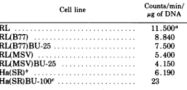
ability to incorporate labeled thymidine (Ta-ble 1). Thus RL(MSV)BU-25 cells, despite theirabilitytogrowin5-BUdR,arenotlacking
thymidine kinase. The RL(MSV)BU-25 and
RL(B77)BU-25 growing in 5-BUdR medium
werelight sensitive.
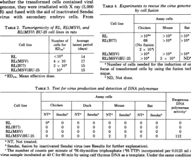
Tumorigenicity of cell lines. All four cell
lines were tested for their abilitytoinduce tu-mors in 1-wk-old rats. RL cells and RL(MSV) cells were highly oncogenic for 1-wk-old rats.
It isstriking that the injection ofaslittleas40
cells into rats gave rise to progressively
grow-ing tumors. On the other hand, the RL(B77)
cells required a thousand times more cells for
tumor induction in 50% of the inoculated
ani-mals (Table 2).
Virus production and presence of DNA
polymerases. To determine whether the in-vestigated cell lines are producing viral
par-ticles, the following experiments were done.
Tissue culture medium harvested from these
cells was 100-fold concentrated by
ultracen-TABLE 1. Incorporation ofthymidine-6-3HintoDNA
Cell line
Counts/min/
Cellline p~,gofDNA
RL.11.500a
RL(B77) ... 8.840
RL(B77)BU-25.7.500
RL(MSV). 5.400
RL(MSV)BU-25.4.150
Ha(SR). 6.190
Ha(SR)BU-100C.23
aThe values represent the averages of
determina-tions on duplicate cultures oftwo different
experi-ments.The rangewas notgreater than10%.
b Hamster cells transformed with the
Schmidt-Ruppin strain of Roussarcomavirus.
cThe Schmidt-Ruppin Rous sarcoma virus
trans-formed hamster cells resistant to 5-BUdR and
de-ficient in thymidine kinase activity (C. Altaner,
preparedforpublication).
trifugation,andthe material was platedon dif-ferentsecondary embryocells(Table3).
To testthe
possibility
that viralparticlesareproduced with different viral envelopes, the
techniqueof fusion
by
inactivated Sendaivirus wasused. Underthese conditions weobservedon November 10, 2019 by guest
http://jvi.asm.org/
[image:3.493.100.390.67.337.2] [image:3.493.251.442.400.492.2]ALTANER AND HLAVAYOVA
production of virus infectious for mouse cells fromRL(MSV)BU-25 cellsonly.
Tissue culture fluid from RL, RL(B77), RL(MSV), and RL(MSV)BU-25 cells was
con-centrated 100-fold by the procedure used for concentration of viruses. The search for
DNA polymerase activity in the sedimented
material obtained from RL, RL(B77), and RL(MSV) cells was negative. A small activity
wasfoundinthematerialfromRL(MSV)BU-25 cells.
The uridine-5-3H labeling technique with
subsequent bandingofthe viral material in su-crose gradient centrifugation gave negative
re-sultsinthecaseofRL,RL(MSV), and RL(B77) cell lines. These cell lines were also negative
in theultraviolet-XC plaque assay forthe leu-kemia viruses (18).
Attempts to recover the viral genome
from transformed cells by fusion with
diff-erent kinds of embryo cells. To determine whether the transformed cells contained viral
[image:4.493.61.449.286.604.2]genome,
they
wereirradiated with X ray(5,000 R) and fused with the aidofinactivatedSendai virus with secondary embryo cells. FromTABLE 2. Tumorigenicity ofRL,RL(MSV),and
RL(MSV)BU-25 cell lines in rats
Number of Average Cell line cellsfor latentperiod
ED50a (days)
RL <103 14
RL(MSV) 4 x 10 17
RL(B77) 2 x 10⁵ 15
RL(MSV)BU-25 102 15
aED50,
Mean effective dose.RL(B77) cells, infectious viruswasreadily
res-cuedbyfusionwith chickenembryocells.
Cells of the RL(MSV)BU-25linegave riseto
typical MSV foci after fusion with mouse
em-bryo cells, whereas the RL cells and the RL(MSV) cells did not induce focus formation after fusion of 106 cells with mouse, rat, and chicken embryo cells
(Table
4).Also,
culture fluid fromthese mixedcultures didnotcontain anyinfectiousvirusformouseembryo cells.The virus rescued from RL(B77) cells was
characterized by its ability to transform chickenand duck cells in comparison with the
virus which was used for their transformation and with the viruses rescued from the three
different clones ofratembryo cells transformed with B77 virus (Table 5). The virus rescued
from rat livercellswas more than 1,000 times
less effective in transformation of duck cells than the virus obtained from transformed rat
embryo cells.
TABLE4. Experimentsto rescuethevirus genome
by cellfusion
Assay cells Cell line
Chicken Mouse Rat
RL >lo6a >106 >106
RL(B77) 68 >10f >106
(Nofusion 2 x 10⁴)
RL(MSV) >106 >106 >106
RL(MSV)BU-25 >106 2 x 10³
NDW
aNumberofcells needed for the induction of one
focus oftransformed cells by using the fusion
tech-nique.
"ND, Not done.
TABLE 3. Test for virusproduction and detection of DNA polymerase
Assay cells
~~~~~~Exogenous
Cell line Chicken Duck Mouse Rat DNA
polymerase
NTa Sendai' NTa Sendai' NTa Sendai' NT| Sendai'
activityc
RL Od ° 0 T 0 0 0 0 0
RL(B77) 0 0 0 0 0 0 0 0 0
RL(MSV) 0 0 0 0 0 0 0 0
RL(MSV)BU-25 0 0 0 0 2 2
0
0 112aNT, Not treated.
ISendai,fusionby inactivated Sendai virus (see Results for further explanation).
cThenumberofcountsper minute of3H-thymidine triphosphate
(3H-TTP)
incorporated per 0.0125 ml ofvirussampleincubated at 40 C for 60min by using calf thymus DNA as a template. Under the same conditions
ofthe assayapreparation ofchicken sarcoma virus B77 of 106 FFU/ml gave 18.580 counts/min of 3H-TTP
incorporation.
dThe number representstheaveragenumber of foci on plates infected with 0.2 ml of 100-fold concentrated
tissueculture fluid harvested from confluent cultures.
180 J. VIROL.
on November 10, 2019 by guest
http://jvi.asm.org/
TABLE 5. Relative plating efficiency of viruses rescued from RL(B77) and R(B77) cells
Relative plating Viruses rescuedfrom: efficiency
(duck/chicken)
RL(B77).9.1 x 10-,a
R(B77) no. 1.1.1 x 10-2
R(B77) no. 2.1.4 x 10-2
R(B77) no. 3.7. 1 x 10- 2
Original B77 8.0 x 10⁻²
aViruses were rescued from rat liver-transformed
cells RL(B77) and from different clones of
trans-formed rat embryo cells [R(B77) no. 1; R(B77)no. 2;
R(B77) no. 3] by fusion with chicken embryo cells.
These viruses and virus B77 originally used for
infec-tion of rat liver cells were assayed on duck and
chicken embryo cells. Relative plating efficiency
(duck/chicken) was calculated. Titers of all viruses
used werearound 106FFU/mlonchicken cells.
DISCUSSION
The chicken sarcoma virus B77 is known as a virus with broad host range. It is able to
in-duce sarcomas in rats (8) and to transform some mammalian cells in vitro (5). For the study ofcell transformation, primarily embryo fibroblasts were used.
Under certain conditions it is possible to
cultivate mammalian liver cells in tissue
cul-ture (6, 19). Rat liver cells kept in tissue
cul-ture are of
epithelioid morphology.
It wasshownby Coon (6) that such cells are
produc-ing some serum proteins, and several
enzy-matic activities typical for liver tissue have
beenfound.
We have proved that rat liver cells are
sus-ceptible
to transformation with chickensar-coma virus B77 and murine sarcoma virus. In
both cases the transformed cells are
tumori-genic and contain the whole viral genome. The
greatdifference inthe
ability
toinducetumors in ratsbetween RL(MSV) cellsorRLcells and RL(B77) cells could beexplained by
their abil-ity to grow in vitro, theirmorphology,
andby
the presence oftumor-specific
transplantation
antigens, or
by
a combination of these prop-erties. RL(MSV) cells and RL cells aresmall,
rapidly growing cells, whereas RL(B77) cells
are epithelioid with slower growth rate. The
higher doses of RL(B77) cells needed to
in-duce tumors in rats could be in connection with the presence oftumor-specific
transplan-tation antigen on the surface of cells
trans-formedwithchickensarcomaviruses.
RL(B77) cells did not differ from other rat
embryo cells transformed with B77 virus.
They
are virogenic without production of an
infec-tious virus or viral particles. After fusion of
these cells with chicken embryo cells, infec-tious virus could be rescued. This virus
diff-ered from the original one and from viruses rescued from transformed rat embryo cells in its property to transform duck cells. Its ability totransform duck cells is much lower, showing
that host modification of virus occurred. This is in agreement with earlier observations of
B77 virus, which we interpreted as a conse-quence of virus genome integration into the
hostcell genetic material(5).
A number of tissue culture mouse cell lines after long-term cultivation appeared to be transformed in vitro. Some lines produce viral
particles
and some do not. The presence ofvirus particles does notseems tobeassociated with malignancy(7).
Our line of rat liver cells transformed
spon-taneouslyin vitro ishighly tumorigenicin rats. It did not produce any detectable infectious
virus or viral particles. Also, preliminary ex-periments to try to induce some virus by
5-BUdR by using the uridine-5-3H technique
gave negative results. Therefore, it seemsthat this neoplastic transformation is not a conse-quence of some virus action, but more
experi-mentsmustbe doneinthis direction.
There are several cell lines transformed by MSV with no detectable virus
production
(9, 2). Such nonproducer cells release MSV when super-infected with a helper leukemia virus in vitro (9, 17, 23) or in vivo (13). Infectiousvirus canbe rescuedafter cell fusion with cells sheddingamurineleukemiavirus(10).
The RL(MSV) cells behaved as
nonproduc-ers. These cells did not produce infectious
virus, virus particles, or particle-bound DNA
polymerase
activity as detected by differenttechniques. The RL(MSV) cell line did not
produce any viral particles which could have the character of MSV(O) described by Ting
(23). The RL(MSV) cell line was adapted for growth in a medium containing 5-BUdR. These cells incorporated labeled thymidine and did grow inHATG
medium,
which meansthatthey are not
deficient
inthymidine kinaseactivity. The ability to grow in medium con-taining 5-BUdR could be in connection with the presence of ahigh level ofrepair enzymes.
Recently it was found that murine leukemia viruses can be activated in vitro by treatment
of cells with 5-BUdR or 5-iododeoxyuridine
(3, 15).Focus-formingvirus canbe also induced
from
nonproducer
cells transformed by MSV(1, 11).
on November 10, 2019 by guest
http://jvi.asm.org/
ALTANER AND HLAVAYOVA
It was found that the RL(MSV)BU-25 cell line is producing infectious virus which
in-ducedtypical MSV focion mouseembryo cells. The induced sarcoma virus is able to trans-form mouse cells, but these cells did not pro-duce infectious virus showing that this virusis unable to replicate in the absence of helper leukemia virus (C. Altaner, unpublished data).
ACKNOWLEDGMENTS
Wewishtothank H. M. Temin forhelpfulcommentsand
S. Bodocka fortechnical assistance. Theinvestigation was partially supportedby PublicHealth Service research grant CA 07175 from theNational CancerInstitute. A part ofthe workreported in this paper was undertaken during the ten-ureof aTravel Fellowshipawardedto onefromus(C. A.)
by theIntemationalAgency for Research on Cancer in Mc-Ardle Laboratory for CancerResearch,University of Wiscon-sin,Madison, Wisconsin.
LITERATURECITED
1. Aaronson, S. A. 1971.Chemical induction of focus-form-ing virus from nonproducer cells transformed by murine sarcoma virus. Proc. Nat. Acad. Sci. U.S.A. 68:3069-3072.
2. Aaronson, S. A., and W. P. Rowe. 1970. Nonproducer clones of murine sarcoma virus transformed BALB/
3T3cells.Virology42:9-19.
3. Aaronson, S.A.,G.J.Todaro,and E. M.Scolnick.1971.
Inductionof murineC-type viruses from clonal lines of virus freeBALB/3T3cells. Science 174:157-159. 4. Altaner, C.,and F. Svec. 1966. Virusproductionin rat
tumors induced by chicken sarcoma virus. J. Nat. Cancer Inst. 37:745-752.
5. Altaner, C., and H. M. Temin.1970. Carcinogenesis by
RNAsarcoma viruses. XII. Aquantitative study of infection of rat cells in vitro by aviansarcoma viruses. Virology 40:118-134.
6. Coon, H. G. 1968. Clonal cultureofdifferentiated rat livercells.J.CellBiol. 39:29a.
7. Franks, L. M., andS. Henzell. 1970. Thedevelopment
of"spontaneous" malignant transformation in vitro of cells from youngand old mice. Eur. J.Cancer 6:357-364.
8. Hlavayova, E., J.Smida,C.Altaner,and F.Svec.1964.
Pathogenicityof virus B77 inrats.Folia Biol.(Prague) 10:301-306.
9. Huebner, R. J., J. W.Hartley,W.P.Rowe,W. T.Lane,
and W.I.Capps. 1966. Rescueofthe defectivegenome of aMoloney sarcoma virus fromanoninfectious ham-ster tumorand theproductionofpseudotypesarcoma viruses with various murineleukemia viruses. Proc. Nat.Acad. Sci. U.S.A. 56:1164-1169.
10. Kelloff, G. J., R. J.Huebner, C. Long, and R. V. Gilden. 1971.Rapid rescue of the defective M-MSV genomeby
the use of cell fusion.Virology46:965-968.
11. Klement,V., M. 0.Nicolson, and R. J. Huebner. 1971. Rescue of thegenome of focusforming virus fromrat non-productive lines by 5'-bromodeoxyuridine. Na-tureN. Biol. 234:12-14.
12. Kit, S., D. R. Dubbs, L. J. Piekarski, and T. C. Hsu. 1963. Deletion ofthymidine kinase activity from
L-cells resistanttobromodeoxyuridine. Exp. Cell.Res. 31:297-312.
13. Law, L. W., and R. C. Ting. 1969. Rescue in vivo in adult mice of murine sarcoma virus(MSV)from non-producer rat neoplasm. Proc. Soc. Exp. Biol. Med. 131:960-963.
14. Littlefield, J. W. 1965. Studies on thymidine kinase in cultured mouse fibroblasts. Biochim. Biophys. Acta 95:14-22.
15. Lowy, D. R., W. P. Rowe, N. Teich, and J. W. Hartley. 1971. Murineleukemia virus:high frequency activation in vitroby 5-iododeoxyuridine and 5-bromodeoxyuri-dine.Science 174:155-156.
16. Reed, L. J., and H. Muench. 1938. A simple method of estimating fifty per cent endpoints. Amer. J. Hyg. 27:493-497.
17. Rowe, W. P. 1971. The kinetics of rescue of the murine sarcoma virus genome from a nonproducer line of transformed mousecells. Virology 46:369-374. 18. Rowe, W. P., W. E. Pugh, and J. W. Hartley. 1970.
Plaqueassaytechniques for murine leukemia viruses. Virology 42:1136-1139.
19. Rutzky, P. L., W. G. Taylor and R. W. Pumper. 1971. In vitrostimulation of rat liver cells by homologous partial hepactomy serum. In Vitro 7:1-5.
20. Temin, H. M., and S. Mizutani. 1970. RNA-dependent DNA polymerase in virions of Rous sarcoma virus. Nature (London) 226:1211-1213.
21. Thurzo, V., J. Smida, V. Smidova-Kovarova and D.
Simkovic. 1963. Someproperties of fowl virus tumor B77.Acta Unio lnt. Contra Cancrum 19:304-305. 22. Ting, R. C. 1966. In vitro transformation of rat
em-bryo cells by a murine sarcoma virus. Virology 28: 783-785.
23. Ting, R. C. 1968. Biological and serological properties ofviral particles from a nonproducer rat neoplasm in-duced by murine sarcomavirus (Moloney). J. Virol. 2:865-868.
182 J. VIROL.