R E S E A R C H A R T I C L E
Open Access
PU.1 is involved in the immune response to
Aspergillus fumigatus
through upregulating
Dectin-1 expression
Min Wang
1†, Zhicheng Liu
2†, Chenyang Liu
2, Ting Wu
1, Feng Cai
1, Quan Wang
3, Xin Su
1*and Yi Shi
1*Abstract
Background:Invasive aspergillosis is a life-threatening disease, and its incidence has increased in the recent past. Dectin-1 recognizesβ-glucans and mediates innate immune responses toAspergillus fumigatus. Transcription factor PU.1 has been the focus of recent research due to its role in inflammation and infection. However, its role in Dectin-1 regulation duringA. fumigatusinfection remains to be elucidated.
Methods:THP-1 cells were stimulated withA. fumigatusconidia. We then used real-time RT-PCR, Western blot, and immunofluorescence assays to analyze the mRNA and protein levels and cellular distribution, respectively, of Dectin-1 and PU.1 in stimulated THP-1 cells. Additionally, we used the luciferase reporter assays, chromatin immunoprecipitation (ChIP) assays, electrophoretic mobility shift assays (EMSA), and RNA interference experiments to investigate the role of PU.1 in Dectin-1 regulation.
Results:Our results revealed that Dectin-1 mRNA and protein levels as well as the PU.1 protein level were increased in THP-1 cells stimulated withA. fumigatusconidia, while the mRNA expression level did not significantly change between the stimulated and control groups. We also observed that PU.1 translocated into the nucleus in stimulated THP-1 cells. The results of the luciferase reporter assay showed that PU.1 promoted human Dectin-1 (hDectin-1) gene activity. ChIP and EMSA indicated that PU.1 could bind with hDectin-1 gene promoter at three potential transcription factor-binding sites (TFBSs). In addition, knockdown of PU.1 significantly decreased Dectin-1 expression.
Conclusions:This study demonstrated the novel role of PU.1 in the immune response toA. fumigatusthrough upregulation of Dectin-1 expression and its translocation to the nucleus inA. fumigatus-stimulated THP-1 cells.
Keywords:PU.1, Dectin-1,Aspergillus fumigatus, Immunity
Background
Invasive aspergillosis (IA) is a life-threatening disease whose incidence is growing worldwide due to the increas-ing number of immunocompromised patients over the last decades [1]. Despite therapeutic advances and better diag-nostic tools and criteria, IA remains difficult to diagnose and treat, and the mortality associated with this condition remains high, ranging from 60 % to 90 % [2]. Aspergillus fumigatus is a ubiquitous opportunistic fungal pathogen that accounts for more than 90 % of all cases of IA [3].
The innate immune system serves as the first line of defense against fungal infection, as it is capable of recogniz-ing and initiatrecogniz-ing an effective response to eliminate invadrecogniz-ing A. fumigatus[4]. However, there is limited knowledge of the regulation and detailed immune mechanism involved in the defense againstA. fumigatusinfection. Therefore, further in-vestigation of the mechanism would help develop effective immunoprophylaxis and to improve patient prognosis.
Pattern recognition receptors (PRRs) recognize pathogen-associated molecular patterns (PAMPs) expressed in the cell wall during fungal infection [4, 5]. C-type lectin receptors (CLRs) are a large family of receptors that bind to car-bohydrates in a calcium-dependent manner. They are expressed by several cell types such as macrophages and dendritic cells (DCs), which phagocytose various * Correspondence:[email protected];[email protected]
†Equal contributors
1Department of Respiratory and Critical Care Medicine, Jinling Hospital,
Medical School of Nanjing University, 305 East Zhongshan Road, Nanjing, China
Full list of author information is available at the end of the article
glycoproteins and microbes for clearance and antigen presentation to T lymphocytes. CLRs are widely recog-nized to mediate innate immune responses to fungi, with Dectin-1 being the best characterized receptor in the context of fungal infections [5]. Dectin-1 initiates antifungal immunity by recognizing the β-glucan present in the cell walls ofA. fumigatus[6, 7].
PU.1, a member of the E-twenty-six (ETS) family, is a transcription factor that is essential for the development of hematopoietic stem cells and regulation of the immune response [8, 9]. In addition, PU.1 participates in regulating the expression of a variety of PRRs, including toll-like receptor (TLR)-4 [10], TLR-9 [11], mannose receptor (MR) [12], a dendritic cell-specific C-type lectin (DC-SIGN) [13], and Dectin-1 [14, 15]. In a previous study, pneumocystis-infected mice exhibited reduced Dectin-1 and PU.1 expres-sion levels decreases in alveolar macrophages (AMs) [15]. However, another study incubated human bronchial epithe-lial cells (HBE) withA. fumigatus and found that Dectin-1 was upregulated in these cells [16]. Despite these studies, the role of PU.1 in regulating the transcription and expres-sion of the hDectin-1 gene during A. fumigatusinfection remains unclear. Therefore, we investigated the role of PU.1 regulating Dectin-1 expression in THP-1 cells stimu-lated withA. fumigatus. Additionally, we characterized the transcription factor-binding sites (TFBSs) in the hDectin-1 gene promoter, indicated the regulation of hDectin-1 gene via PU.1, and further determined changes in the expression of Dectin-1 following knockdown of PU.1.
Methods
Cells, reagents, and antibodies
THP-1 (human monocytic leukemia) cells were cultured in RPMI-1640 medium (Gibco; Invitrogen, Carlsbad) and human embryonic kidney (HEK)-293 T cells were cultured in Dulbecco’s modified Eagle’s medium (DMEM; Gibco). These two cell lines were gifts from Prof. Xing (Laboratory of Immunology and Virology, Nanjing University, China) [17]. Both the culture mediums were supplemented with 10 % fetal bovine serum (FBS; Hyclone, Thermo Scientific, Logan), and 1 % antibiotic-antimycotic solution (Gibco). The cells were cultured at 37 °C in an atmosphere con-taining 5 % CO2. Polymerase chain reaction (PCR)
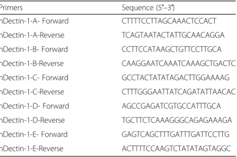
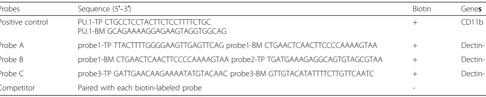
primers were synthesized by GenScript (Nanjing, China), while probes for EMSA were synthesized by Viagene Bio-tech (Ningbo, China). All the primers and probes used in this study are listed in Table 1, 2 and 3. The following antibodies were used for Western blot analysis: rabbit anti-PU.1, rabbit anti-β-tubulin (Cell Signaling Technol-ogy, Danvers); goat anti-Dectin-1 (N-16, Santa Cruz Biotechnology, Santa Cruz); mouse anti-GAPDH, mouse anti-β-actin, and rabbit anti-Histone (Abmart, Shanghai, China). Other secondary antibodies were purchased from Cell Signaling Technology. Rabbit anti-PU.1 (H-135, Santa
Cruz Biotechnology) and rabbit IgG (Upstate Biotechnol-ogy, Waltham) were used in ChIP. Antibodies for Alexa Fluor-488 donkey anti-rabbit IgG and Alexa Fluor-568 don-key anti-goat IgG were purchased from Life Technologies.
A. fumigatusstrain and stimulation with conidia
A. fumigatus A1 was kindly provided by the Microbio-logical Laboratory of Jinling Hospital. After culture on Sabouraud dextrose agar plates for 7 days at 28 °C, co-nidia were harvested by washing the plates with sterile phosphate-buffered saline (PBS)-0.1 % (vol/vol) Tween 20 (PBST). The suspension was then gently filtered through a 40-μm cell strainer to separate conidia from mycelium. Then, the conidia were thoroughly washed, centrifuged, and resuspended in sterile PBST at concentra-tions ranging from 5.0 × 10⁷to 1.0 × 10⁸ colony-forming units (CFU)/mL, and stored at 4 °C within 2 weeks for further experiments. The THP-1 cells were cultured in RPMI-1640 medium at a density of 1.0–1.5 × 10⁶/mL, and stimulated with A. fumigatus conidia at a multiplicity of infection (MOI) of 1.
Quantitative real-time RT-PCR
[image:2.595.305.540.99.195.2]The total RNA precipitated from the lysates of THP-1 cells stimulated withA. fumigatusconidia and the lysates of un-stimulated THP-1 cells were used for reverse transcription (RT) with a Primescript RT reagent kit (R036A; Takara, Tokyo). The mRNA expression levels were analyzed by real-time PCR, as described previously [18]. The specificity Table 1Primer sequences for real-time PCR
Primers Sequence (5′–3′)
hDectin-1-Forward GCAATACCAGGATAGCTGTTG
hDectin-1-Reverse CCAAGCATAGGATTCCCAA
hPU.1- Forward CGTGCACAGCGAGTTCGA
hPU.1-Reverse GCTCTGGAGCTCCGTGAAGT
GAPDH- Forward ACAGTCAGCCGCATCTTCTT
GAPDH-Reverse ACGACCAAATCCGTTGACTC
Table 2Primer sequences for PCR of ChIP
Primers Sequence (5′–3′)
hDectin-1-A- Forward CTTTTCCTTAGCAAACTCCACT
hDectin-1-A-Reverse TCAGTAATACTATTGCAACAGGA
hDectin-1-B- Forward CCTTCCATAAGCTGTTCCTTGCA
hDectin-1-B-Reverse CAAGGAATCAAATCAAAGCTGACTC
hDectin-1-C- Forward GCCTACTATATAGACTTGGAAAAG
hDectin-1-C-Reverse CTTTGGGAATTATCAGATATTAACAC
hDectin-1-D- Forward AGCCGAGATCGTGCCATTTGCA
hDectin-1-D-Reverse TGCTTCTCAAAGGGCAGAGAAAGA
hDectin-1-E- Forward GAGTCAGCTTTGATTTGATTCCTTG
[image:2.595.306.540.577.732.2]of primers was confirmed by melting curve analysis. The relative expression values were standardized by an internal glyceraldehyde-3-phosphate dehydrogenase (GAPDH) con-trol. The fold changes of mRNA were calculated according to the formula 2(ΔCTof gene–ΔCTof GAPDH), whereCTis the
threshold cycle.
Western blot analysis
THP-1 cell lysates of the mock or A. fumigatus conidia-stimulated cells were subjected to sodium dodecyl sulfate-polyacrylamide gel electrophoresis (SDS-PAGE) and trans-ferred to polyvinylidene difluoride (PVDF) membranes (Millipore, Billerica), and then incubated with primary anti-bodies overnight. After washing the membranes, they were incubated with horseradish peroxidase (HRP)-conjugated secondary antibodies for 90 min at room temperature. Im-ages were captured using Tanon Imaging System (Tanon, Shanghai, China). For the fractionation of lysates, stimu-lated or control cells were washed, and cytoplasmic and nuclear fractions were isolated using cytoplasmic/nuclear protein extraction kit (KeyGene Biotechnology, Nanjing, China). The fractions were processed as described above for immunoblot analysis.
Immunofluorescence microscopy
Stimulated and unstimulated THP-1 cells were washed twice with PBS at various time points, fixed with 4 % para-formaldehyde for 30 min, and permeabilized with 0.1 % Triton X-100 for 10 min, and then washed with PBS. After blocking with 5 % bovine serum albumin (BSA) in PBS for 60 min at 37 °C, the cells were incubated with both rabbit anti-PU.1 and goat anti-Dectin-1 antibodies (1:100 dilutions for both antibodies) at 4 °C overnight. The cells were washed with PBST, and then incubated with Alexa Fluor-488 donkey anti-rabbit IgG and Alexa Fluor-568 donkey anti-goat IgG (1:200 dilution for both antibodies) at 37 °C for 1 h. Then, the cells were washed and stained with 1μg/ mL of 4′,6-diamidino-2-phenylindole (DAPI). After wash-ing, the slides were carefully observed under a confocal microscope (Carl Zeiss Jena, Oberkochen).
Plasmid constructs and luciferase reporter assays
The 2.0-kb DNA fragment containing the Dectin-1 gene promoter was PCR amplified from human genomic DNA
using the following primers: 5′ -GCGGTACCGCAGATC-GAGACCATTCTGGTTAACAC-3′and 5′-GCAAGCTT GCGAAACTATGCTGTGGTAATTTT-3′. Plasmid pGL3 -Decin-1 was constructed by cloning the amplified frag-ment into the firefly luciferase reporter vector pGL3 (Pro-mega, Madison). The PU.1 expression plasmid pRK5-PU.1 was obtained by cloning human PU.1 cDNA into EcoRI-digested pRK5-HA (Promega) and a selection of plasmids with properly oriented inserts. HEK-293 T cells were seeded in 24-well plates in total DMEM until 50–70 % confluence was achieved. The cells were then transfected with pRK5-HA or with various amounts of pRK5-PU.1, along with pGL3-Dectin-1 or pGL3-basic, and pRL8-SV40 by using Lipofectamine 2000 (Invitrogen). The total amount of DNA was maintained at a constant level by adding empty control plasmid. At 24 h post-transfection, the cells were stimulated by 10 ng/mL of phorbol-12-myristate-13-acetate (PMA) for 12 h, and cell lysates were analyzed for the activities of firefly and Renilla luciferases (Promega) according to the manufacturer’s instructions.
Chromatin immunoprecipitation assays
ChIP assays were performed using EZ-ChIP™kit (Upstate Biotechnology) according to the manufacturer’s instruc-tions. In short, 1–1.5 × 10⁷THP-1 cells were stimulated by 10 ng/mL PMA for 2 h, cross-linked with 1 % formalde-hyde, and lysed in SDS lysis buffer. The chromatin samples were sonicated at high power for 7 min (in cycles of 5-s pulses with 10-s pause) using a sonicator (Bioruptor™; Diagenode, Belgium). After preclearing with salmon sperm DNA/Protein A-agarose, the protein extracts were sub-jected immunoprecipitated with antibodies against PU.1 (H-135), RNA polymerase, and normal rabbit IgG. The re-sultant immune complexes were extensively washed, and the DNA was recovered by phenol:chloroform extraction and resuspended in 50-μL volume. PCR amplification was performed by using the primers listed in Table 2. The re-sultant PCR products were analyzed by 2 % agarose gel electrophoresis.
Electrophoretic mobility shift assays (EMSA)
[image:3.595.59.536.97.193.2]THP-1 cells were plated on RPMI-1640 at a density of 1.0–1.5 × 10⁷in the presence of 10 ng/mL PMA, and col-lected after 2 h. Nuclear extracts from PMA-stimulated Table 3Probe sequences for EMSA
Probes Sequence (5′–3′) Biotin Genes
Positive control PU.1-TP CTGCCTCCTACTTCTCCTTTTCTGC PU.1-BM GCAGAAAAGGAGAAGTAGGTGGCAG
+ CD11b
Probe A probe1-TP TTACTTTTGGGGAAGTTGAGTTCAG probe1-BM CTGAACTCAACTTCCCCAAAAGTAA + Dectin-1
Probe B probe1-BM CTGAACTCAACTTCCCCAAAAGTAA probe2-TP TGATGAAAGAGGCAGTGTAGCGTAA + Dectin-1
Probe C probe3-TP GATTGAACAAGAAAATATGTACAAC probe3-BM GTTGTACATATTTTCTTGTTCAATC + Dectin-1
-and control cells were prepared using nuclear protein ex-traction kit (KeyGene Biotechnology). The probes used for EMSA are listed in Table 3. Briefly, 3 μg of nuclear protein samples were incubated with 10× binding buffer, 1.0μg/μL poly (dI-dC), and 0.5 μL biotin-labeled probes in an EMSA kit (Viagene Biotech). Where indicated, 0.83 μL or 3.33μL of specific, cold-competitor probe in 10× or 100× competing buffer was added before the biotin-labeled probe. The incubation mixtures were sepa-rated by 6.5 % non-denaturing PAGE, and the bands were detected by autoradiography.
RNA interference experiments
PU.1-specific small interfering RNA (siRNA) or a nonspe-cific siRNA control (siCtrl) were chemically synthesized at
GenePharma (Shanghai, China). The sequence of the PU.1 siRNA was as follows: 5′-UAUAGAUCCGUGUCAUAG GGCACCA-3′. A 5-μL of aliquot of 20μM siRNA was in-troduced into 1 × 10⁶THP-1 cells by using Lipofectamine 2000, according to the manufacturer’s instructions. The transfected cells were incubated for 24 h,and the cells were harvested and subjected to real-time PCR and Western blot analyses to detect PU.1, Dectin-1, and the control cell populations.
Statistical analysis
A two-tailed Student’s pairedttest was used to evaluate the data by SPSS software (IBM SPSS, Armonk, USA), and values ofp< 0.05 were considered to be statistically significant.
[image:4.595.58.538.282.661.2]Results
Dectin-1 and PU.1 mRNA and protein expression levels in A. fumigatusconidia-stimulated THP-1 cells
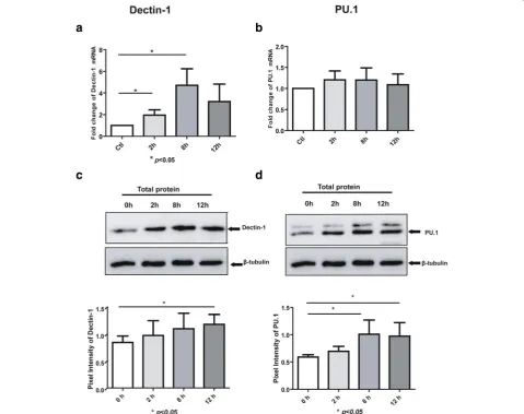
In order to study the PU.1 and Dectin-1 expression pat-terns in the antifungal immune response, THP-1 cells were exposed toA. fumigatusconidia at an MOI of 1 for 2, 8, and 12 h. Real-time PCR and Western blot analyses were then used to analyze the Dectin-1 and PU.1 mRNA and protein expression levels, respectively, in THP-1. It was observed that both Dectin-1 mRNA and protein levels were upregulated in the conidia-stimulated THP-1 cells compared to the controls (Fig. 1a and c). Interest-ingly, the results also found that PU.1 mRNA levels remained unchanged in THP-1 cells; however, the PU.1 protein expression was higher in the stimulated cells than in the control cells (Fig. 1b and d).
Cellular localization of Dectin-1 and PU.1
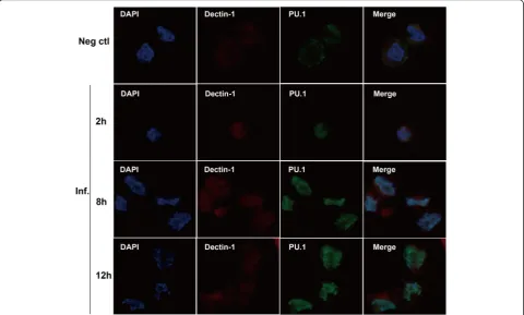
We performed immunofluorescence analysis to the cellular localization of Dectin-1 and PU.1 in THP-1 cells. The stim-ulated (MOI = 1) or unstimstim-ulated THP-1 cells, fixed at different time points after stimulation (2, 8, and 12 h), were stained with diluted goat Dectin-1 and rabbit anti-PU.1 antibodies. In the confocal study, Dectin-1 protein
(red) was dispersed in the membrane of conidia-exposed THP-1 cells (Fig. 2). Additionally, Dectin-1 protein expres-sion increased with time of exposure (Fig. 2), as explained previously (Fig. 1c). Meanwhile, translocation of PU.1 pro-tein (green) into the nucleus was markedly stimulated byA. fumigatus conidia (Fig. 2), which was not the case in the control cells.
A. fumigatusactivates PU.1 nuclear translocation
To gain further insights into the role ofA. fumigatusin the activation of PU.1, we studied nuclear translocation by Western blot analysis of the cytoplasmic and nuclear cell fractions. In the cytoplasm, the PU.1 protein level started to decrease at 8 h after stimulation (Fig. 3a). Meanwhile, in the nucleus, the PU.1 protein levels were higher compared to the corresponding levels in the control cells; moreover, the PU.1 protein levels in the nuclear lysates of the stimulated cells increased with time (Fig. 3b). The histone and β -tubulin protein levels in the cytoplasmic and nuclear frac-tions were similar in the stimulated and control cells.
PU.1 promotes hDectin-1 gene activity
To investigate whether PU.1 accounts for the hDectin-1 gene promoter activity, HEK-293 T cells were cotransfected
[image:5.595.58.539.394.683.2]with the reporter plasmid pGL3-hDectin-1 and the PU.1-expressing plasmid pRK5-PU.1. HEK-293 T cells were chosen for this experiment instead of monocytes because they normally do not express PU.1; therefore, the influence of endogenous PU.1 can be avoided. Results of the im-munoblotting analyses demonstrated the presence of PU.1 protein in transfected cell lysates, indicating that pRK5-PU.1 plasmid could successfully express pRK5-PU.1 (Fig. 4b). As shown in Fig. 4a, luciferase activity of HEK-293 T cells transfected with 0.10, 0.25, and 0.50 μg of pRK5-PU.1 in-creased by 3.2–, 8.5–, and 23.0-fold that of the control, re-spectively, indicating that PU.1 activated hDectin-1 expression in a dose-dependent manner.
PU.1 binds directly to the hDectin-1 gene promoter
To investigate the regulation of hDectin-1 gene expression, we analyzed the 2000-bp region upstream from the tran-scription initiation start site of the hDectin-1 gene pro-moter for candidate TFBSs using TRANSDAC [19] and JASPAR [20]. A total of five potential TFBSs were identified for PU.1 (Fig. 5a), which including A (GGGAAGT, –1053 to–1047), B (AGGCAGT,–719 to–713), C (AGAAAAT,
–527 to –521), D (AGAAAGA, –1411 to –1405), and E (AGAAAGA,–618 to–612).
The results of the luciferase reporter assays indicate that PU.1 activates the hDectin-1 gene promoter (Fig. 4a). We performed ChIP to determine whether PU.1 interacts with the hDectin-1 gene promoter in THP-1 cells (Fig. 5b). To verify the five putative PU.1 binding motifs, the DNA frag-ments were amplified with specific hDectin-1 gene-targeting primers (Table 2). The results showed that three regions relative to the transcription start site (+1) of the hDectin-1 gene:–1102 to–1000,–768 to–653, and–564 to –443 were co-precipitated by PU.1, confirming that PU.1 can bind with hDectin-1 gene promoter.
To further verify the ChIP results, EMSA was performed by using nuclear extracts from THP-1 cells. Three biotin-labeled double-stranded oligonucleotide probes (Table 3) carrying the hDectin-1 gene promoter regions A (–1053 to
–1047), B (–719 to –713), and C (–527 to –521) were designed and synthesized. Meanwhile, the PU.1-binding sequence from the CD11b gene promoter [21, 22] was chosen as the positive control. Each of these four probes had an unlabeled oligonucleotide probe as the competitor, and the competitor concentrations were 25-fold and 100-fold. An NF-kB probe was used to verify whether the EMSA system worked properly. PMA activates the protein kinase C (PKC) pathway by mimicking diacylglycerol, a nat-ural ligand and activator of PKCs [23], and can alter gene
[image:6.595.56.540.88.359.2]expression via the activation of PKC and modulating the activity of transcriptional factors that bind cis elements such as NF-kB [24], AP-1 [25], and AP-2 [26]. As only active transcription factors could bind to the specific probe, so we prepared two groups of nuclear extracts: one from THP-1 cells that were treated with PMA for 2 h [27] and one group from untreated cells. As shown in Fig. 5c, Probe A bound to nuclear extracts from PMA-stimulated THP-1
cells (Fig. 5c, lane 8), and the band was located at the same position with the CD11b probe (Fig. 5c, lane 4), indicating the specificity of this proteDNA complex. Adding in-creasing amounts of competitors (25- and 100-fold) into the assay resulted in a decreasing intensity of PU.1, demon-strating its presence in this complex (Fig. 5c, lanes 9 and 10). These data suggest that the PU.1 protein directly binds to the –1053 to –1047 gene promoter region. A specific
Fig. 4Effect of PU.1 on Dectin-1 activation.aHEK-293 T cells were cotransfected with the indicated amount of pRK5-PU.1, 1μg pGL3-hDectin1, and 10 ng pRL8-SV40 plasmids, and pGL3-basic and pRK5-HA were used as the negative controls. At 24 h post-transfection, the cells were harvested, lysed, and assayed for luciferase activity. The resultant ratios were normalized to the fold change values obtained from HEK-293 T cells cotransfected with pGL3-basic and pRK5-HA.bCell lysates were analyzed by immunoblotting with rabbit anti-PU.1 antibodies to detect the expression of pRK5-PU.1.
[image:7.595.58.541.87.560.2]PU.1 band could be observed in EMSA using probes B and C (for the–719 to–713 and–527 to–521 regions, respect-ively Fig. 5d). These results indicated that PU.1 recognizes and binds to the –1053 to –521 region of the hDectin-1 gene promoter through at least three different sites.
Effect of PU.1 knockdown on hDectin-1 gene expression
In order to further confirm that PU.1 regulates the expres-sion of the hDectin-1 gene, THP-1 cells were transfected
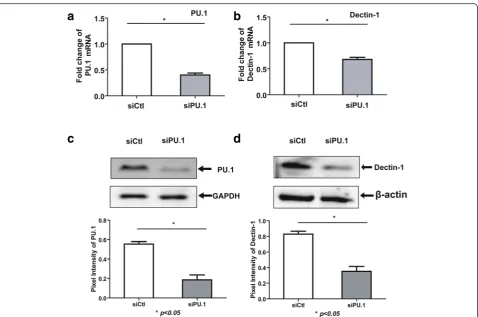
with control or PU.1 siRNA, and assayed for the expression levels of PU.1 and Dectin-1. Transfection with PU.1 siRNA resulted in approximately 60 % knockdown of PU.1 mRNA expression (Fig. 6a) relative to the control siRNA. In addition, PU.1 silencing decreased Dectin-1 mRNA expres-sion by approximately 32 % in THP-1 cells relative to the expression level in the controls (Fig. 6b). Results of the Western blot analysis also confirmed that THP-1 cells trans-fected with PU.1 siRNA showed a significant reduction in
[image:8.595.58.540.88.473.2]the Dectin-1 protein expression (Fig. 6c and d), but cells transfected with control siRNA showed no such reduction. These results indicate that PU.1 may participate in the regu-lation of hDectin-1 gene expression in THP-1 cells.
Discussion
Dectin-1 is a member of the CLR family and is expressed on the surface of many cells [28]. Its involvement in the immune defense against A. fumigatus has been widely confirmed [29]. In our previous study, A. fumigatuswas shown to induce the upregulation of Dectin-1 in HBE cells in a TLR2-dependent manner [16]. In the present study, we found that Dectin-1 mRNA and protein expression levels were increased in THP-1 cells stimulated with A. fumigatusconidia.
PU.1 belongs to the ETS family and has a great im-pact on the immunity, primarily through its control of immune cell development [30]. As PU.1 has been the focus of increasing attention for its role in inflam-mation and infection [31–33], we evaluated PU.1 ex-pression at the mRNA and protein levels in THP-1 monocyte cells exposed to A. fumigatus conidia. The results of the present study demonstrated for the first
time that the PU.1 protein expression was increased in THP-1 cells incubated with A. fumigatus conidia compared to the control cells, while the mRNA level showed no significant differences between the exposed and control cells.
In our study, the PU.1 mRNA level was not correlated with its protein level. Similar to our finding, a previous study reported that the PU.1 mRNA level in preadipocytes was high, while the PU.1 protein is not expressed in these cells [34]. Regulation of gene expression is a complicated and meticulous process, and it is controlled at multiple levels, including transcriptional, post-transcriptional, trans-lational, and post- translational levels [35]. MicroRNAs are a large class of small, noncoding RNAs in plants and ani-mals, and have been known to mediate post-transcriptional regulation [36]. PU.1 is a direct target for miR-155 and the target sequence for miR-155 is localized in the 3′ -untrans-lated region (UTR) of PU.1. Also, miR-155 overexpression in THP-1 cells has been found to decrease the PU.1 protein level [13]. Further studies are required to investigate the precise mechanisms responsible for the different patterns of PU.1 mRNA and protein expression during stimulation withA. fumigatus.
[image:9.595.60.539.85.402.2]In our study, PU.1 protein was localized in the cyto-plasm of unstimulated THP-1 cells, and translocated into the nucleus following exposure toA. fumigatus co-nidia. To the best of our knowledge, this is the first study to illustrate the translocation of PU.1 stimulated by A. fumigatus conidia. Previously, in HeLa and RAW264.7 macrophage cell models, PU.1 has been shown to enter the nucleus via passive diffusion and ac-tive transport. The latter can be facilitated by: (i) the classical pathway requiring importin α and β, (ii) the non-classical pathway requiring only importin β, or (iii) direct interaction with nucleoporins [37, 38]. However, to date, little is known regarding the mechanisms of PU.1 nuclear transport during A. fumigatusstimulation. Thus, future studies are necessary to investigate the mechanisms by whichA. fumigatus drives the transport of PU.1 from the cytoplasm to the nucleus.
The regulation of Dectin-1 expression has not been ex-tensively studied to date; however, downregulation of PU.1 is known to result in decreased Dectin-1 expression in AMs in Pneumocystis-infected mice [15]. In another study, BLT1 was found to modulate the transcription of Dectin-1 via a GM-CSF/PU.1 cascade [14]. However, the role of PU.1 in regulating the transcription and expression of hDectin-1 gene during A. fumigatus stimulation in THP-1 cells remains unknown. In our study, the results of the luciferase reporter assay showed that PU.1 promoted hDectin-1 gene activity in a dose-dependent manner. Additionally, five potential TFBSs for PU.1 were predicted by using the TFBS databases. Moreover, the ChIP assay in our study indicated that PU.1 could bind to the hDectin-1 gene promoter at the–1053 to–1047,–719 to–713, and
–527 to–521 regions. The synthetic polymer poly(dI-dC) is considered to be one of the most potent, and likely the most widely used, nonspecific competitor in EMSA. Its addition to the reaction mixture before addition of crude nuclear proteins has been proven to be an efficient way of reducing any nonspecific interactions by facilitating detec-tion of the complexes of interest [39]. Therefore, we used poly(dI-dC) in the EMSA analysis in this study. Further EMSA results confirmed that PU.1 could bind to the abovementioned three regions of the hDectin-1 gene promoter. Based on the results of our study and those of previous reports, we suggest that PU.1 can bind to the hDectin-1 gene promoter.
To confirm that PU.1 regulates the expression of the hDectin-1 gene, we knocked down the expression of PU.1 in THP-1 cells using siRNA. Although Dectin-1 expres-sion has been reported to be controlled by PU.1 [15] and leukotriene B4 via a GM-CSF/PU.1 axis [14] in mouse models, our results are the first to demonstrate that silen-cing the expression of PU.1 could significantly decrease the hDectin-1 gene mRNA and protein expression levels in THP-1 cells. Lack of PU.1 expression is known to
contribute to decreased maturation, differentiation, and surfactant metabolism in AMs of patients with pulmonary alveolar proteinosis [40]. In addition, PU.1 knockdown in macrophages has shown to decrease phagocytic activity [15]. However, further studies are warranted to examine how knockdown or overexpression of PU.1 can affect the innate immune and inflammatory responses toA. fumiga-tusinfection and the precise mechanisms underlying this process.
Conclusions
As Dectin-1 is a major recognition receptor for numer-ous fungi, including species ofCandida, Aspergillus, His-toplasma, Cryptococcus, and Coccidioides [41], our data suggest a potentially broad role for PU.1 in antifungal defense. Meanwhile, Dectin-1 may participate in recog-nizing other microbes as well, and it may therefore be important that future studies investigate the potential roles of PU.1 in defense against infections by other path-ogens. In conclusion, this study provides novel insights to modulate the immune response toA. fumigatus, and lays a foundation for the diagnosis and therapeutic strat-egy forA. fumigatus aspergillosis.
Abbreviations
BSA, Bovine serum albumin; ChIP, Chromatin immunoprecipitation; CLR, C-type lectin receptor; DAPI, 4′,6-Diamidino-2-phenylindole; DMEM, Dulbecco’s modified Eagle’s medium; EMSA, Electrophoretic mobility shift assays; ETS, E-twenty-six; FBS, Fetal bovine serum; HBE, Human bronchial epithelial; IA, Invasive aspergillosis; MOI, Multiplicity of infection; NSB, Nonspecific binding; PAGE, Polyacrylamide gel electrophoresis; PAMP, Pathogen-associated molecular pattern; PBS, Phosphate-buffered saline; PBST, Phosphate-Phosphate-buffered saline (PBS)-0.1 % (vol/vol) Tween 20; PCR, Polymerase chain reaction; PMA:Phorbol-12-myristate-13-acetate; PRR, Pattern recognition receptor; PVDF, Polyvinylidene difluoride; RT, Reverse transcription; SDS, Sodium dodecyl sulfate; TFBS, Transcription factor-binding site; UTR, Untrans-lated region
Acknowledgements
The authors would like to thank Prof. Xing for providing cell lines. This work was supported by grants from the National Natural Science Foundation of China (Grant No. 81270064 and 81330035).
Availability of data and materials
The datasets supporting the conclusions of this article are included within the article.
Authors’contributions
SY and SX conceived and designed the study, and critically revised the manuscript. WM and LZC performed the experiments, analyzed the data and drafted the manuscript. LCY, WT, CF and WQ helped in experiment implementation. All authors read and approved the final manuscript.
Competing interests
The authors have declared that they have no competing interests.
Consent for publication
Not applicable.
Ethics approval and consent to participate
Author details
1Department of Respiratory and Critical Care Medicine, Jinling Hospital,
Medical School of Nanjing University, 305 East Zhongshan Road, Nanjing, China.2Southern Medical University, Guangzhou, China.3Department of Respiratory Medicine, BenQ Medical Center, Nanjing, China.
Received: 5 February 2016 Accepted: 7 June 2016
References
1. Patterson KC, Strek ME. Diagnosis and treatment of pulmonary aspergillosis syndromes. Chest. 2014;146(5):1358–68.
2. Bassetti M, Righi E, De Pascale G, De Gaudio R, Giarratano A, Mazzei T, Morace G, Petrosillo N, Stefani S, Antonelli M. How to manage aspergillosis in non-neutropenic intensive care unit patients. Crit Care. 2014;18(4):458. 3. Sugui JA, Kwon-Chung KJ, Juvvadi PR, Latge JP, Steinbach WJ.
Aspergillus fumigatus and related species. Cold Spring Harb Perspect Med. 2015;5(2):a19786.
4. Romani L. Immunity to fungal infections. Nat Rev Immunol. 2011;11(4):275–88. 5. Hardison SE, Brown GD. C-type lectin receptors orchestrate antifungal
immunity. Nat Immunol. 2012;13(9):817–22.
6. Jia XM, Tang B, Zhu LL, Liu YH, Zhao XQ, Gorjestani S, Hsu YMS, Yang L, Guan JH, Xu GT. CARD9 mediates Dectin-1-induced ERK activation by linking Ras-GRF1 to H-Ras for antifungal immunity. J Exp Med. 2014;211(11): 2307–21.
7. Lee DH, Kim HW. Innate immunity induced by fungal beta-glucans via dectin-1 signaling pathway. Int J Med Mushrooms. 2014;16(1):1–16. 8. Kastner P, Chan S. PU.1: A crucial and versatile player in hematopoiesis and
leukemia. Int J Biochem Cell Biol. 2008;40(1):22–7.
9. Moreau-Gachelin F, Tavitian A, Tambourin P. Spi-1 is a putative oncogene in virally induced murine erythroleukaemias. Nature. 1988;331(6153):277–80. 10. Pedchenko TV. Inducible binding of PU.1 and interacting proteins to the
Toll-like receptor 4 promoter during endotoxemia. AJP. 2005;289(3):L429–37. 11. Schroder K, Lichtinger M, Irvine KM, Brion K, Trieu A, Ross IL, Ravasi T, Stacey
KJ, Rehli M, Hume DA. PU.1 and ICSBP control constitutive and IFN--regulated Tlr9 gene expression in mouse macrophages. J Leukocyte Biol. 2007;81(6):1577–90.
12. Egan BS, Lane KB, Shepherd VL. PU.1 and USF are required for macrophage-specific mannose receptor promoter activity. J Biol Chem. 1999;274(13):9098–107. 13. Martinez-Nunez RT, Louafi F, Friedmann PS, Sanchez-Elsner T. MicroRNA-155 modulates the pathogen binding ability of dendritic cells (DCs) by down-regulation of DC-specific intercellular adhesion molecule-3 grabbing non-integrin (DC-SIGN). J Biol Chem. 2009;284(24):16334–42.
14. Serezani CH, Kane S, Collins L, Morato-Marques M, Osterholzer JJ, Peters-Golden M. Macrophage dectin-1 expression is controlled by leukotriene B4 via a GM-CSF/PU.1 axis. J Immunol. 2012;189(2):906–15.
15. Zhang C, Wang SH, Liao CP, Shao S, Lasbury ME, Durant PJ, Lee CH. Downregulation of PU.1 Leads to Decreased Expression of Dectin-1 in Alveolar Macrophages during Pneumocystis Pneumonia. Infect Immun. 2010;78(3):1058–65.
16. Sun WK, Lu X, Li X, Sun QY, Su X, Song Y, Sun HM, Shi Y. Dectin-1 is inducible and plays a crucial role in Aspergillus-induced innate immune responses in human bronchial epithelial cells. Eur J Clin Microbiol. 2012; 31(10):2755–64.
17. Qu B, Qi X, Wu X, Liang M, Li C, Cardona CJ, Xu W, Tang F, Li Z, Wu B. Suppression of the Interferon and NF- B Responses by Severe Fever with Thrombocytopenia Syndrome Virus. J Virol. 2012;86(16):8388–401. 18. Wu X, Qi X, Qu B, Zhang Z, Liang M, Li C, Cardona CJ, Li D, Xing Z. Evasion
of Antiviral Immunity through Sequestering of TBK1/IKK/IRF3 into Viral Inclusion Bodies. J Virol. 2014;88(6):3067–76.
19. Wingender E. The TRANSFAC project as an example of framework technology that supports the analysis of genomic regulation. Brief Bioinform. 2008;9(4):326–32.
20. Bryne JC, Valen E, Tang MH, Marstrand T, Winther O, Da PI, Krogh A, Lenhard B, Sandelin A. JASPAR, the open access database of transcription factor-binding profiles: new content and tools in the 2008 update. Nucleic Acids Res. 2008; 36(Database issue):D102–6.
21. Dominguez-Soto A, Puig-Kroger A, Vega MA, Corbi AL. PU.1 Regulates the Tissue-specific Expression of Dendritic Cell-specific Intercellular Adhesion Molecule (ICAM)-3-grabbing Nonintegrin. J Biol Chem. 2005;280(39):33123–31.
22. Nika E, Brugnoli F, Piazzi M, Lambertini E, Grassilli S, Bavelloni A, Piva R, Capitani S, Bertagnolo V. hnRNP K in PU.1-containing complexes recruited at the CD11b promoter: a distinct role in modulating granulocytic and monocytic differentiation of AML-derived cells. Biochem J. 2014;463(1):115– 22.
23. Ghamlouch H, Ouled-Haddou H, Guyart A, Regnier A, Trudel S, Claisse JF, Fuentes V, Royer B, Marolleau JP, Gubler B. Phorbol myristate acetate, but not CD40L, induces the differentiation of CLL B cells into Ab-secreting cells. Immunol Cell Biol. 2014;92(7):591–604.
24. Ghosh S, Baltimore D. Activation in vitro of NF-kappa B by phosphorylation of its inhibitor I kappa B. Nature. 1990;344(6267):678–82.
25. Bos TJ, Bohmann D, Tsuchie H, Tjian R, Vogt PK. v-jun encodes a nuclear protein with enhancer binding properties of AP-1. Cell. 1988;52(5):705–12. 26. Williams T, Admon A, Luscher B, Tjian R. Cloning and expression of AP-2, a
cell-type-specific transcription factor that activates inducible enhancer elements. Genes Dev. 1988;2(12A):1557–69.
27. Foster N, Lea SR, Preshaw PM, Taylor JJ. Pivotal Advance: Vasoactive intestinal peptide inhibits up-regulation of human monocyte TLR2 and TLR4 by LPS and differentiation of monocytes to macrophages. J Leukocyte Biol. 2007;81(4):893–903. 28. Wevers BA, Geijtenbeek TB, Gringhuis SI. C-type lectin receptors orchestrate
antifungal immunity. Future Microbiol. 2013;8(7):839–54.
29. Saijo S, Iwakura Y. Dectin-1 and Dectin-2 in innate immunity against fungi. Int Immunol. 2011;23(8):467–72.
30. Gallant S, Gilkeson G. ETS transcription factors and regulation of immunity. Arch Immunol Ther Exp (Warsz). 2006;54(3):149–63.
31. Qian F, Deng J, Lee YG, Zhu J, Karpurapu M, Chung S, Zheng J, Xiao L, Park GY Christman JW. The transcription factor PU.1 promotes alternative macrophage polarization and asthmatic airway inflammation. J Mol Cell Biol. 2015;7(6):557–67.
32. Steinwede K, Tempelhof O, Bolte K, Maus R, Bohling J, Ueberberg B, Langer F, Christman JW, Paton JC, Ask K. Local delivery of GM-CSF protects mice from lethal pneumococcal pneumonia. J Immunol. 2011;187(10):5346–56. 33. Zhang G, Zhou B, Li S, Yue J, Yang H, Wen Y, Zhan S, Wang W, Liao M,
Zhang M. Allele-specific induction of IL-1beta expression by C/EBPbeta and PU.1 contributes to increased tuberculosis susceptibility. PLoS Pathog. 2014; 10(10):e1004426.
34. Wang F, Tong Q. Transcription factor PU.1 is expressed in white adipose and inhibits adipocyte differentiation. Am J Physiol Cell Physiol. 2008;295(1):C213–20. 35. Marcel V, Catez F, Diaz JJ. p53, a translational regulator: contribution to its
tumour-suppressor activity. Oncogene. 2015;34(44):5513–23. 36. Chen K, Rajewsky N. The evolution of gene regulation by transcription
factors and microRNAs. Nat Rev Genet. 2007;8(2):93–103.
37. Kwok JC, Perdomo J, Chong BH. Identification of a monopartite sequence in PU.1 essential for nuclear import, DNA-binding and transcription of myeloid-specific genes. J Cell Biochem. 2007;101(6):1456–74. 38. Zhong H, Takeda A, Nazari R, Shio H, Blobel G, Yaseen NR.
Carrier-independent nuclear import of the transcription factor PU.1 via RanGTP-stimulated binding to Nup153. J Biol Chem. 2005;280(11):10675–82. 39. Larouche K, Bergeron MJ, Leclerc S, Guerin SL. Optimization of competitor
poly(dI-dC).poly(dI-dC) levels is advised in DNA-protein interaction studies involving enriched nuclear proteins. Biotechniques. 1996;20(3):439–44. 40. Bonfield TL, Raychaudhuri B, Malur A, Abraham S, Trapnell BC, Kavuru MS,
Thomassen MJ. PU.1 regulation of human alveolar macrophage differentiation requires granulocyte-macrophage colony-stimulating factor. Am J Physiol Lung Cell Mol Physiol. 2003;285(5):L1132–6.