quantity of the L transcript late in infection is lower than those of the other two because of transcriptional
control. In bacteriophage ⌽8 and its close relatives, transcription of L is not controlled; instead, the L
transcript is turned over rapidly late in infection. The three messages are produced in approximately equal amounts early in infection, but the amount of L is less than 10% of the amounts of the others late in infection.
The decay of the⌽8 L message depends upon the production of protein Hb, which is encoded in segment L.
It also depends upon a target site within theHgene region. Phage mutants lacking either theHbgene or the
target region do not show the late control of L message quantity. In addition to having a role as a negative regulator, Hb functions to neutralize the activity of protein J, encoded by segment S, which causes the degradation of all viral transcripts.
Bacteriophage⌽8 is a member of theCystoviridae, a family of phages that have genomes of three double-stranded RNA (dsRNA) molecules packaged within a polyhedral capsid struc-ture (11). The first-discovered isolate of this family was bacte-riophage⌽6 (26). The life cycle of⌽6 involves attachment to a pilus, which retracts to place the virus at the outer membrane of its host,Pseudomonas syringae. The external membrane of
⌽6 fuses with the outer membrane of the host, which results in the entry of the viral nucleocapsid into the periplasmic space (21). A viral muramidase digests the cell wall, and the viral nucleocapsid enters the cell (1). The nucleocapsid loses the shell of protein 8 (P8). The core particle is composed of four proteins: P1, the major structural protein; P2, the RNA poly-merase; P4, a hexameric NTPase essential for genomic pack-aging; and P7, an auxiliary protein involved in RNA synthesis and packaging (14). The core particle, once inside the cell, transcribes the three genomic dsRNA molecules, L, M, and S, to produce messages l, m, and s, which are extruded from the particle. In the case of ⌽6, the concentrations of the early transcripts are almost equimolar, but translation of L predom-inates (5). At later times, the level of the L transcript is about 5 to 10% of those of M and S, and translation of their genes predominates. The plus strands of the three segments have an 18-base consensus sequence at the 5⬘ end. There is a 1-base difference between the sequence of L and those of M and S. The L sequence begins with GU, while those of M and S begin with GG (16). In vitro transcription by nucleocapsids results in the production of transcripts of M and S in buffers that do not contain manganese ions. In vitro studies showed that the se-quence difference is responsible for the differences between both late transcription and transcription from nucleocapsids
and early transcription in infected cells (8). The control of⌽6 transcription is dependent upon the activity of a host protein.
⌽8 transcription is independent of this protein (unpublished results).
⌽8 is somewhat similar to ⌽6 in structure and genetic makeup but has almost no sequence similarity to⌽6 (10).⌽8 plus strands have a shorter 5⬘consensus sequence, but the first 7 nucleotides are identical in all three plus strands (10).⌽8 is the only member of theCystoviridaewhose second nucleotide of L is identical to those of S and M. In vitro transcription from inner core particles produces approximately equal amounts of the three plus strands, yet there is little L transcript evident late in the infection cycle (this study). Genetic studies showed that⌽8 differs from⌽6 in several ways. A striking difference is manifested in the arrangement of genes on genomic segment L (Fig. 1). In all other members of theCystoviridae, the order of genes is14-7-2-4-1. However, in the case of⌽8, the order is 14-H-2-4-1-7, where gene7has been moved out from its usual position and a pair of genes,HaandHb, now precedes gene2 (25). Deletion ofHbprevents normal phage development, but the deletion can be partially complemented by cloned geneHb (this study). In this report, we will show that geneHb is in-volved in the temporal control of the abundance of the L transcript by regulating the L transcript’s turnover.
MATERIALS AND METHODS
Bacterial strains, phage, and plasmids.Cells and plasmids used in this study are listed in Table 1. Phages are listed in Table 2. LM2489 is a rough derivative ofP. syringaepv. phaseolicola HB10Y (26) and was used as the primary host for plating⌽8 (17). LM128 was also used, and LM2691 is LM128 carrying plasmid pLM1086, which is a derivative of pRK290 (7) and pAR1219 (6) and expresses T7 RNA polymerase in pseudomonads. Plasmids pLM2622, pLM2638, and pLM2743 are derivatives of pT7T319U with T7 polymerase promoters and cDNA copies of the⌽8 segments L, M, and S, respectively. Details of the construction of the plasmids are available from the authors. Plasmid transfor-mation intoEscherichia coliused strain JM109 (28) or Stratagene supercompe-tent XL1-Blue cells {recA1 endA1 gyrA96 thi-1 hsdR17 supE44 relA1 lac[F⬘ proAB lacIq
Z⌬M15 Tn10(Tetr
)]}. Complementation of phage mutants and deletions was accomplished by inserting the relevant genes from cDNA plasmids
* Corresponding author. Mailing address: Public Health Research Institute Center at NJMS, 225 Warren Street, Newark, NJ 07103. Phone: (973) 854-3420. Fax: (973) 854-3453. E-mail: mindicle@umdnj .edu.
䌤Published ahead of print on 29 October 2008.
633
on November 8, 2019 by guest
http://jvi.asm.org/
of⌽8 into the shuttle vector pLM254, which is a derivative of plasmids pUC8 and RSF1010 (15).
Media.The media used were LC and M8 (24). Ampicillin plates contained 200 g of ampicillin per ml in LC agar. Kanamycin was used at 40g per ml.
Reverse genetics of⌽8.Mutant forms of the phage were prepared by con-structing deletions or modifications in plasmids containing cDNA copies of the genomic segments. These were then electroporated into host cells containing plasmids expressing T7 RNA polymerase. In general, thousands of plaques were produced, samples were purified, and their RNA was analyzed by gel behavior and by the preparation of DNA copies by reverse transcription-PCR and
sub-sequently checked by sequence analysis at the DNA sequencing core facility of UMDNJ.
Labeling of RNA during phage infection.Host cells were diluted from an overnight culture in synthetic M8 medium and grown to 5⫻108
[image:2.585.93.490.67.332.2]cells per ml. One-milliliter aliquots were placed on ice, and phage was added at a multiplicity of infection of 10. The cells were left on ice for 30 minutes and then transferred to 28°C. [5,6-3H]uracil (10Ci) was added at various times. The cultures were spun at 6,000⫻g10 minutes later, and the cells were resuspended in 200l lysis buffer (20 mM Tris-HCl, pH 8, 1% sodium dodecyl sulfate, 5 mM EDTA) with 2 M sodium acetate, pH 5.4. The mixture was then frozen at⫺20°C for 20 min
FIG. 1. Restriction maps of⌽8 genomic segment cDNA copies.
TABLE 1. Bacteriophages used in this study
Phage Relevant characteristic(s)a Figure(s) Reference or source
⌽8 wild type 1, 2, 3, 4, 5 10
⌽2797 ⌬14⌬H, deletion of nucleotides 329 to 1152
3, 5, 6, 7 This study
⌽2833 ⌬14⌬HmJ 4 This study
⌽2834 ⌬14⌬H⌬J 3, 4, 5 This study
⌽2825 ⌬14⌬H⌬8 This study
⌽2835 ⌬14⌬Hm8 4 This study
⌽2806 ⌬14⌬Hm8 mJ 2, 3, 4 This study
⌽2821 ⌬14⌬Hm8⌬J 4 This study
⌽2826 ⌬8 4 This study
⌽6 wild type 5⬘GU in L 2 26
⌽2807 5⬘GG in L 2 This study
⌽2837 ⌬14⌬Hin L,Hinsertion in gene
3aof M
This study
⌽2781 Deletion of nucleotides 329 to 11526
6 This study
⌽2895 Deletion of nucleotides 329 to 781 6 This study
⌽2896 Deletion of nucleotides 329 to 841 6 This study
⌽2897 Deletion of nucleotides 329 to 901 6 This study
⌽2916 Deletion of nucleotides 838 to 1152 6 This study
amJ, mutation in gene J; m8, mutation in gene 8.
on November 8, 2019 by guest
http://jvi.asm.org/
[image:2.585.47.541.513.716.2]and then spun at 8,000⫻gfor 10 min at room temperature. The supernatant liquid was extracted twice with phenol-chloroform and precipitated with ethanol. The precipitate was resuspended in 20l DNA buffer and analyzed on 0.8% SeaKem GTG agarose (FMC BioProducts) in Tris-borate-EDTA with 2g/ml ethidium bromide. The bands were visualized with UV light and subjected to autoradiography.
Transposon mutagenesis.EZ::TN (Epicentre), which is a complex of trans-posase and transposon (9), was electroporated into cells of LM2489, which were then plated on LB agar containing kanamycin. Colonies were tested by cross-streaking for sensitivity to bacteriophages⌽8,⌽6, and⌽13. Candidates that showed resistance to⌽8 were tested further by plating dilutions of phage on lawns. The sites of insertions were determined by preparation of EcoRI restric-tion digests of chromosomal DNA, ligating the DNA to form circles, and then amplifying inserts by PCR using forward and reverse primers supplied by Epi-centre. The PCR products were then sequenced using the forward primer sup-plied by Epicentre, and the sequences were compared with those in the NIH data bank using the BLAST search facility. All insert sequences showed high identity to sequences ofP. syringaepv. phaseolicola 1448A (GenBank accession number CP000058).
CloningrnrofP. syringaepv. phaseolicola.Thernr(vacB) gene was copied from chromosomal DNA using oligonucleotides olm824 (CCCGTCGACG-GAGTTGACAAATGGCCGATTGGCA) for the 5⬘end and olm829 (TGACG-GCCGTAGATTTTTTCCAGC) for the 3⬘end. The PCR product was cut with restriction enzymes SalI and EagI and inserted into the shuttle vector pLM254. The base sequence of thisrnrgene is 99% identical to that ofPseudomonas syringaepv. phaseolicola 1448A (locus tag AAZ33054), and the amino acid sequence has 99% identity.
Nucleotide sequence accession numbers.Open reading frames (ORFs)Ha andHbin segment L identified in this study have been deposited in GenBank as accession number AF226851. ORFJin segment S is GenBank accession number AF226853.
RESULTS
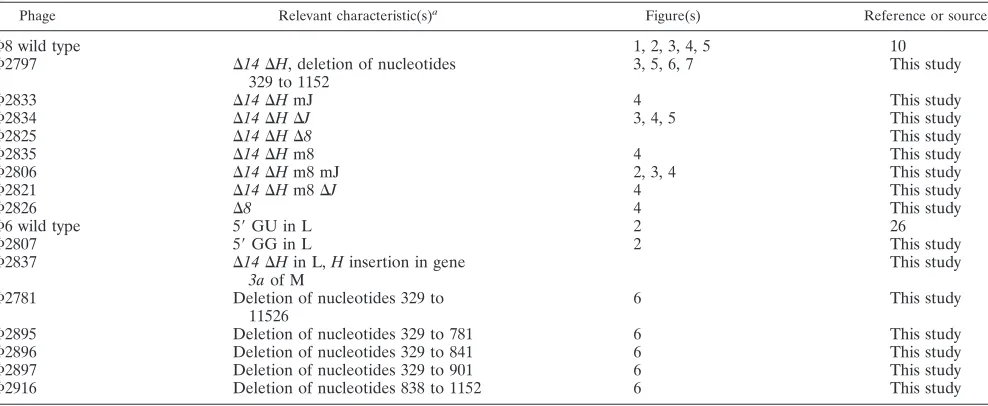
Temporal control of plus-strand abundance in⌽6 and⌽8
infections. In vitro transcription using nucleocapsids of ⌽6
results in unbalanced synthesis, in that the transcripts of seg-ments S and M are more than 10 times as abundant as those of L (19) (Fig. 2). This behavior is dependent upon the difference in the second nucleotide of the L plus strand from that of S or M (8). The sequence at the 5⬘end of L is GU, while it is GG for S and M. Early in infection, there is a higher level of the L transcript than seen in the in vitro transcription, but late in the infection period, the amount of L transcript diminishes dras-tically. We have prepared⌽6 phage with GG as the first 2 nucleotides of L and demonstrate that production of the L transcript is much higher than normal in late infection (Fig. 2).
The elucidation of the control of⌽6 transcription is the subject of a separate publication.
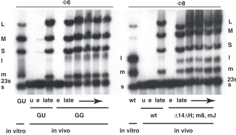
In vitro transcription using nucleocapsids of ⌽8 results in approximately equimolar synthesis of the three transcripts (Fig. 2). Yet, late in infection there is a dramatic reduction in the relative amount of the L transcript. This reduction is not the result of differential transcription but is due to turnover of the L transcript (Fig. 3). The amount of L transcript is seen to be low even after very short pulses of labeling. However, transcription in⌽8 involves displacement of the plus strand from the double-stranded template by the newly synthesized transcript. Therefore, the plus strand of the dsRNA is a mea-sure of the amount of transcription taking place. We observe that labeling of the dsRNA for L is commensurate with that for S and M. Denaturation of the genomic dsRNA shows that plus strands of L are being synthesized at rates comparable to those of S and M (Fig. 3).
LM3671 pLM2934 14,Ha, andHbin pLM254 (SmaI/HindIII) This study
LM3722 pLM3102 Hbin pLM254 (XbaI/HindIII from pLM3099 in SmaI/HindIII) This study
a
[image:3.585.45.543.81.238.2]LPS, lipopolysaccharide.
FIG. 2. Autoradiogram of an agarose gel with [3H]uracil-labeled
infected cells of LM2489. Infection was with⌽6 or⌽8. In vitro col-umns show products of transcription reactions of inner cores of puri-fied virions. Note that, in vitro,⌽6 shows very little L transcript but that⌽8 has abundant L transcript. Also note that late transcripts in ⌽6-infected cells show little L transcript but that the GG mutant has a significant amount. In the case of⌽8-infected cells, there is also little L transcript unless gene His missing (deletion of positions 329 to 1152). wt, wild type; u, uninfected cells; e, early labeling (15 min); late, labeling at 60, 90, 120, 165, and 210 min.⌬H, m8, and mJ refer to phage⌽2806. m8, mutation in gene 8; mJ, mutation in gene J.
on November 8, 2019 by guest
http://jvi.asm.org/
[image:3.585.300.540.488.625.2]Deletion of theHregion. TheH region of segment L con-tains two ORFs, Ha and Hb. A protein product has been observed only forHb. We prepared virus with a deletion of the Hregion in order to determine its role in the life cycle of⌽8. This was accomplished by creating an NsiI site at position 1152 of the cDNA copy of segment L and then deleting from NsiI positions 329 to 1152, which eliminates part of gene14, all of Ha, and most ofHband is designated the⌬14⌬Hconstruct. Gene14has been found to be dispensable in this study. The resulting plasmid, pLM3007, along with plasmids pLM2743 and pLM3056, which carry the cDNA copies of normal seg-ment S and segseg-ment M, respectively, in whichHaandHbare embedded in gene3a, was electroporated into host cells con-taining plasmids expressing T7 RNA polymerase and gene3a. Plaques were obtained, and the phage was crossed with a phage with normal segment M but with a deletion in gene4of segment L. The resulting phage, ⌽2797, contained normal segments S and M and the deletion of theHregion in segment L. Phage from these plaques did not propagate well on normal host strains. The efficiency of plating was orders of magnitude lower than that found on the strain carrying a plasmid contain-ing theHregion. Only a few plaques were obtained, and these were very small. Propagation of these phages led to the pro-duction of larger plaques,⌽2806, that were found to be capa-ble of reproduction on normal host cells. Analysis of these phages showed that they indeed carried the deletion ofHand that they carried suppressor mutations in segment S. The sup-pressor mutations were located in gene8, which encodes a membrane protein in⌽8 but the determinant of a middle shell in⌽6, and in gene J, which is a unique gene, found only in segment S of⌽8 (Fig. 1). GeneJis found between nucleotides 2850 and 3086. The mutation in gene8is A738G (K3R), and
[image:4.585.85.244.69.231.2]the mutation in Jis T2872C (F3S). P8 contains 366 amino acids, while protein J contains 78. Although the mutant phages with the deletion ofHand suppressor mutations in genes8and Jwere able to propagate well, they showed a new pattern of RNA labeling late in infection (Fig. 2, 3, and 4). Whereas the concentration of the transcript of segment L was usually low at late times in normal virus infection, it was close to equimolar with those of segments M and S in the mutant viruses. The phage with the deletion of H but lacking the suppressor mu-tations showed low levels of transcripts for all three segments (Fig. 3, 4, and 5). It appears thatHis necessary for the down-regulation of the L transcript late in infection, while it also seems to be necessary for the normal levels of the S and M transcripts. In the absence of the normal geneJproduct, either because of mutation or deletion, H is not necessary for the normal levels of S and M transcripts. GeneJis not necessary for normal phage reproduction (data not shown), but whenJis present,His required to counter its effects. The mutation in gene8is necessary for plaque formation in the absence ofH (data not shown), but it does not have a role in the regulation FIG. 3. Autoradiogram showing the synthesis of plus strands of L
despite their absence in single-stranded RNA. Note that in wild-type (wt) infection, there is visible M transcript (m⫹) but not L transcript (l⫹) in the nondenatured lanes (lanes n); however, in the lanes with denatured RNA (lanes d) (18), the L transcript is apparent. Phage with
Hdeleted (deletion of positions 329 to 1152) but with genes8andJ
mutated (⌽2806) orJdeleted (⌽2834) show L transcript in the non-denatured lanes. Phage withHdeleted (⌽2797) shows very low levels of RNA synthesis in wild-type cells but increased levels in cells pro-ducing protein H intrans. Note that the L transcript is visible because
transcomplementation of H does not restore degradation of l⫹ be-causeHbis missing from genomic segment L.
FIG. 4. Autoradiogram showing that the dsRNA and transcripts of the genomic segments are missing when geneHis deleted (deletion of positions 329 to 1152) but that deletion of geneJ(⌽2834) or mutation inJ(⌽2833) suffices to restore synthesis and to prevent the decay of the L transcript. Deletion (⌽2825) or mutation (⌽2835) of gene 8
shows little effect despite their requirement for plaque formation when
[image:4.585.348.493.562.687.2]His deleted. wt, wild type.
FIG. 5. Autoradiogram showing that geneHintrans(LM3671) is able to complement a deletion of H (⌬329 to 1152) in the phage genome with respect to the effects ofJ. However,Hintransdoes not restore the downregulation of the L transcript. wt, wild type.
on November 8, 2019 by guest
http://jvi.asm.org/
of RNA decay (Fig. 4). The nature of the role of the geneJ product will be the subject of another publication.
The loss ofHcan be complemented intransby geneHbin a plasmid (pLM3102) so as to restore the levels of the S and M transcripts, but in this case, the transcript of L is not com-pletely downregulated (Fig. 5 and 6). It appears that the tar-geting of the L transcript requires aciselement. IfHis deleted from segment L and moved to segment M, the transcript level of segment M is found to be diminished and that of L is elevated (not shown).
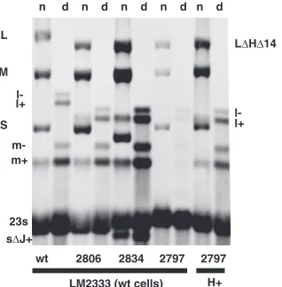
Fine structure of region H. There is an ORF starting at
position 507 in segment L that continues to nucleotide 1211. We designate this ORFH. It contains 234 amino acids. There is a smaller ORF that starts at position 514 and continues to position 744 that we designate ORFHa (Fig. 1) and whose product is 76 amino acids, and there is an ORF in the same reading frame as ORFHthat starts at position 741 and con-tinues to position 1211 that we designate ORFHband whose product is 156 amino acids. Although both ORFHand ORF Ha have reasonable Shine-Dalgarno sequences preceding an initiating methionine codon, we have not seen gene products for ORF H or ORF Ha in infected cells, but we do see a product of the size of ORFHbthat correlates with the pres-ence of that gene. Phage mutants that are missing ORFHb behave as if they are missing the entireH region; however, mutants missing gene14 and part of ORFHa can plate on normal cells and show downregulation of the L transcript. An amber mutation in ORFHadoes not show any abnormalities in terms of RNA levels or reproduction. A fusion of the N terminus of P14 with Hb that is missing the first five amino acids (⌽2781) behaves as if the Hb protein is intact but that the target of H is partially missing. The resulting fusion protein can be seen on labeled protein gels. Fusions that lack the first 34 or 54 amino acids behave as if both the protein and the target are missing (Fig. 6). This suggests that the target(s) for the action of Hb on the turnover of the L transcript is between gene14 and part of geneHb. We believe that ORFHbmakes an active protein and that several parts of the region involving genesHa andHbare the target for the action of protein Hb and other cellular components. Deletions of regions within segment L result in partial loss of downregulation of L transcript abun-dance whenHbis intrans.
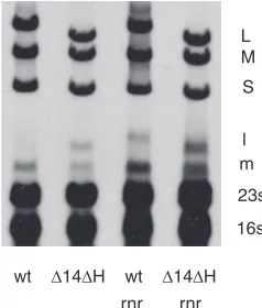
Transposon knockouts of rnr.In parallel experiments, we
[image:5.585.111.476.65.200.2]used transposon mutagenesis in a search for host genes involved in the propagation of bacteriophage ⌽8. The EZ::TN system of Epicentre (23) was used to create inser-tions in the genome ofP. syringaestrain LM2489. Kanamy-cin-resistant colonies were produced after electroporation of the transposon-transposase complex into cells. The colo-nies were cross-streaked against ⌽8, ⌽13, and⌽6. Strains that were resistant to ⌽8 were further tested by plating phage dilutions on lawns. Several promising cultures that produced very small plaques were isolated. Chromosomal DNA was prepared from these, restricted with EcoRI, li-gated, and PCR amplified with primers supplied by Epicen-tre. The PCR products were then sequenced with the for-ward primer supplied by Epicentre. Several of the inserts were in genes involved with surface proteins and were not pursued further. Among the isolates were insertions into mdoHandmgoG, which are glycosyltransferase genes inP. syringae pv. phaseolicola listed under accession number AAZ34542.1 in GenBank. Another set of inserts was in a DedA family protein which is probably an integral mem-brane protein, and another was in a glucosyl UTP trans-ferase. However, several of the inserts were in a gene with high identity toP. syringaepv. phaseolicolarnr, which codes for RNase R (locus tag AAZ33054.1). This enzyme plays a role in the modification of the structure and regulation of RNA production inE. coli(2, 3). The gene was cloned from the chromosomal DNA of LM2489 and was found to com-plement the knockout strain with respect to plaque size when it was expressed on an expression plasmid, pLM254. We found that the rnr knockouts showed a less dramatic downregulation of the L transcript late in⌽8 infection than the wild type, indicating that RNase R acts with the geneH product to downregulate the L transcript (Fig. 7). The loss of RNase R does not entail as strong an effect as the loss of geneH, suggesting that other elements may also participate or substitute in the decay of the L transcript. It was of interest that thernrknockouts did not suppress the down-regulation shown by the geneJproduct, suggesting that the J product acts either alone or with an element apart from RNase R.
FIG. 6. Map showing deletion constructs involving the fusion of the N terminus of the ORF14product with constructs with deletions ofHb. The designations of viruses with corresponding deletions are shown. Pink lines indicate constructs that do not downregulate the L transcript even whenHbis supplied intrans. Green lines indicate deletions that are able to partially downregulate the L transcript whenHbis supplied intrans.
on November 8, 2019 by guest
http://jvi.asm.org/
DISCUSSION
Temporal control of gene expression is a common feature of virus infection. In the case of theCystoviridae, there is a dra-matic difference in the relative quantities of segment L tran-scripts at early versus late times of infection. The genes of segment L code for the proteins of the procapsid or internal core of the virions, and these proteins are expressed early in infection. In⌽6, transcription of segment L is dependent upon a host protein.⌽6 nucleocapsids do not transcribe segment L in vitro unless host protein or manganese ions are present; however, changing the second 5⬘base in segment L from U to G to make GG facilitates the transcription of segment L (Fig. 2). In⌽8, nucleocapsids transcribe all three genomic segments in vitro without special assistance (Fig. 2). Yet there is little L transcript present in the late period of⌽8 infection. Whereas most members of theCystoviridae regulate transcription, ⌽8 regulates the amount of the L transcript by its degradation during the late period of infection.
TheHregion apparently plays two roles in⌽8 infection. (i) TheHbproduct acts with RNase R, and probably with other host elements, to degrade the transcript of L late in infection. This works effectively, since the amount of protein Hb must reach a particular level before it has a significant effect, and therefore it does not cut off the translation of L at early times in infection. (ii) At late times in infection, it appears to be necessary to bolster the amounts of the M and S transcripts, since these are under attack from the product of gene J (Fig. 4).
The involvement of RNase R in the regulation of the L transcript is of special interest because the role of RNase R in bacteria is not very clear. There is evidence that it is involved in the degradation of defective rRNA and that it enhances the expression of genes on pathogenicity plasmids inE. coliand Shigellaspp. (2, 4). The enzyme appears to act in both negative and positive manners. In the latter case, it seems that it must inactivate or interfere with a repressor system of some sort. Since RNase R is involved in large complexes, it is possible that the regulation of L is also mediated by a large complex. RNase R has also been found to be associated with the SsrA RNA complex, which is for peptide degradation (13). We do not
know how the combination of Hb and RNase R affects the degradation of the L transcript, but our working model is that Hb guides the RNase, probably in a complex to a site within theHregion.
It is not clear why⌽8 has adopted this unique set of controls of RNA turnover. In the other members of theCystoviridae, it appears that temporal control is mediated by the difference in sequence at positions 2 of the plus strands, which affects the relative rates of transcription (initiation) and a difference in half-life of the plus strands where the L transcript is longer lived than the other two. The⌽8 arrangement seems so much more complex. The role ofJis even more obscure in that the phage does quite well without geneJ. The effect ofJseems to be specific for⌽8. A plasmid expressing protein J inhibits the plaque formation of ⌽8 but not that of ⌽6 or ⌽13 (20). It appears that J and H might be involved in a control circuit that modulates the temporal control of phage protein synthesis. H seems to be responsible for the high turnover of the L tran-script late in infection and the prevention of the high turnover of M and S transcripts at the same time.
The temporal control of gene expression during bacterio-phage infection is usually determined by differential transcrip-tion, with early genes being transcribed by normal host RNA polymerase and late genes being transcribed by host polymer-ase with new or modified sigma factors or by virally encoded RNA polymerase (27). In the case of T4, two proteins that play a role in the temporal control of message degradation have been identified. One is dmd, a small protein involved in middle and late message stability but that also seems to facilitate the degradation of some middle messages (12). The other is regB, an endoribonuclease that selectively cleaves early messages (22). regB is of particular interest because it attacks message in a subset of Shine-Dalgarno sequences with the collaboration of ribosomal protein S1. Both proteins have affinity for Shine-Dalgarno sequences, and it is thought that S1 modifies the structure of the stem-loops in a manner that converts them to substrates for regB endoribonuclease activity. We have found no RNase activity in Hb so far, and we have not found any interaction in vitro with RNase E, RNase R, or protein S1 (data not shown). However, there is a strong expectation that the nuclease activity might involve a large complex that is difficult to demonstrate in vitro. The role of thernr product would be in the further degradation of molecules cut by Hb and its cofactor.
One possible explanation for the unique transcriptional con-trol of⌽8 is that it facilitates the ability of the phage to infect unrelated bacteria.⌽8 is the only member of theCystoviridae that can form plaques on strains ofSalmonella entericaserovar Typhimurium (17). The other members of the Cystoviridae require a host protein for transcriptional control. Although mutations to independence have been isolated, they do not confer completely normal production.
ACKNOWLEDGMENT
This work was supported by grant GM34352 from the National Institutes of Health.
REFERENCES
[image:6.585.102.221.70.210.2]1.Caldentey, J., and D. H. Bamford.1992. The lytic enzyme of the Pseudomo-nasphage⌽6. Purification and biochemical characterization. Biochim. Bio-phys. Acta1159:44–50.
FIG. 7. Autoradiogram showing that wild-type⌽8 downregulates the L transcript (l) but that the⌬14⌬Hdeletion mutant (⌽2797) does not downregulate and that thernrknockout strain shows poor down-regulation even in infections with wild-type (wt) virus.
on November 8, 2019 by guest
http://jvi.asm.org/
of Rhizobium meliloti. Proc. Natl. Acad. Sci. USA77:7347–7351. 8.Frilander, M., M. Poranen, and D. H. Bamford.1995. The large genome
segment of dsRNA bacteriophage⌽6 is the key regulator in the in vitro minus and plus strand synthesis. RNA1:510–518.
9.Goryshin, I. Y., J. Jendrisak, L. M. Hoffman, R. Meis, and W. S. Reznikoff.
2000. Insertional transposon mutagenesis by electroporation of released Tn5 transposition complexes. Nat. Biotechnol.18:97–100.
10.Hoogstraten, D., X. Qioa, Y. Sun, A. Hu, S. Onodera, and L. Mindich.2000. Characterization of⌽8, a bacteriophage containing three double-stranded RNA genomic segments and distantly related to⌽6. Virology272:218–224. 11.Jaalinoja, H. T., J. T. Huiskonen, and S. J. Butcher.2007. Electron cryomi-croscopy comparison of the architectures of the enveloped bacteriophages phi6 and phi8. Structure15:157–167.
12.Kai, T., and T. Yonesaki.2002. Multiple mechanisms for degradation of bacteriophage T4 soc mRNA. Genetics160:5–12.
13.Karzai, A. W., M. M. Susskind, and R. T. Sauer.1999. SmpB, a unique RNA-binding protein essential for the peptide-tagging activity of SsrA (tmRNA). EMBO J.18:3793–3799.
14.Mindich, L.2004. Packaging, replication and recombination of the seg-mented genome of bacteriophage Phi6 and its relatives. Virus Res.101:
83–92.
15.Mindich, L., G. MacKenzie, J. Strassman, T. McGraw, S. Metzger, M. Romantschuk, and D. Bamford.1985. cDNA cloning of portions of the bacteriophage⌽6 genome. J. Bacteriol.162:992–999.
21.Romantschuk, M., V. M. Olkkonen, and D. H. Bamford.1988. The nucleo-capsid of bacteriophage⌽6 penetrates the host cytoplasmic membrane. EMBO J.7:1821–1829.
22.Sanson, B., R. M. Hu, E. Troitskayadagger, N. Mathy, and M. Uzan.2000. Endoribonuclease RegB from bacteriophage T4 is necessary for the degra-dation of early but not middle or late mRNAs. J. Mol. Biol.297:1063–1074. 23.Shevchenko, Y., G. G. Bouffard, Y. S. Butterfield, R. W. Blakesley, J. L. Hartley, A. C. Young, M. A. Marra, S. J. Jones, J. W. Touchman, and E. D. Green.2002. Systematic sequencing of cDNA clones using the transposon Tn5. Nucleic Acids Res.30:2469–2477.
24.Sinclair, J. F., J. Cohen, and L. Mindich.1976. The isolation of suppressible nonsense mutants of bacteriophage⌽6. Virology75:198–208.
25.Sun, Y., X. Qiao, J. Qiao, S. Onodera, and L. Mindich. 2003. Unique properties of the inner core of bacteriophage phi8, a virus with a segmented dsRNA genome. Virology308:354–361.
26.Vidaver, A. K., R. K. Koski, and J. L. Van Etten.1973. Bacteriophage⌽6: a lipid-containing virus ofPseudomonas phaseolicola. J. Virol.11:799–805. 27.Wong, K., G. A. Kassavetis, J. P. Leonetti, and E. P. Geiduschek.2003.
Mutational and functional analysis of a segment of the sigma family bacte-riophage T4 late promoter recognition protein gp55. J. Biol. Chem.278:
7073–7080.
28.Yanisch-Perron, C., J. Vieira, and J. Messing.1985. Improved M13 phage cloning vectors and host strains: nucleotide sequences of the M13mp18 and pUC19 vectors. Gene33:103–119.