J
OURNAL OFV
IROLOGY, Sept. 2009, p. 8353–8363
Vol. 83, No. 17
0022-538X/09/$08.00
⫹
0
doi:10.1128/JVI.00780-09
Copyright © 2009, American Society for Microbiology. All Rights Reserved.
Virus Entry via the Alternative Coreceptors CCR3 and FPRL1 Differs
by Human Immunodeficiency Virus Type 1 Subtype
䌤
†
R. Nedellec,
1‡ M. Coetzer,
1‡ N. Shimizu,
2H. Hoshino,
2V. R. Polonis,
3L. Morris,
4U. E. A. Mårtensson,
5J. Binley,
6J. Overbaugh,
7and D. E. Mosier
1*
Department of Immunology, The Scripps Research Institute, La Jolla, California 92037
1; Department of Virology and
Preventive Medicine, Gunma University Graduate School of Medicine, Showa-machi, Maebashi, Gunma 371-8511,
Japan
2; Division of Retrovirology, Walter Reed Army Institute of Research, Washington, DC 20307
3;
National Institute for Communicable Diseases, Johannesburg, Private Bag X4, Sandringham 2131,
Johannesburg, South Africa
4; Division of Molecular Neurobiology, Wallenberg Neuroscience Center,
Lund University, SE-223 62 Lund, Sweden
5; Torrey Pines Institute for Molecular Studies,
San Diego, California 92121
6; and Division of Human Biology,
Fred Hutchinson Cancer Research Center, Seattle, Washington 98109
7Received 16 April 2009/Accepted 13 June 2009
Human immunodeficiency virus type 1 (HIV-1) infects target cells by binding to CD4 and a chemokine
receptor, most commonly CCR5. CXCR4 is a frequent alternative coreceptor (CoR) in subtype B and D HIV-1
infection, but the importance of many other alternative CoRs remains elusive. We have analyzed HIV-1
envelope (Env) proteins from 66 individuals infected with the major subtypes of HIV-1 to determine if virus
entry into highly permissive NP-2 cell lines expressing most known alternative CoRs differed by HIV-1 subtype.
We also performed linear regression analysis to determine if virus entry via the major CoR CCR5 correlated
with use of any alternative CoR and if this correlation differed by subtype. Virus pseudotyped with subtype B
Env showed robust entry via CCR3 that was highly correlated with CCR5 entry efficiency. By contrast, viruses
pseudotyped with subtype A and C Env proteins were able to use the recently described alternative CoR FPRL1
more efficiently than CCR3, and use of FPRL1 was correlated with CCR5 entry. Subtype D Env was unable to
use either CCR3 or FPRL1 efficiently, a unique pattern of alternative CoR use. These results suggest that each
subtype of circulating HIV-1 may be subject to somewhat different selective pressures for Env-mediated entry
into target cells and suggest that CCR3 may be used as a surrogate CoR by subtype B while FPRL1 may be used
as a surrogate CoR by subtypes A and C. These data may provide insight into development of resistance to
CCR5-targeted entry inhibitors and alternative entry pathways for each HIV-1 subtype.
Human immunodeficiency virus type 1 (HIV-1) infects
tar-get cells by binding first to CD4 and then to a coreceptor
(CoR), of which C-C chemokine receptor 5 (CCR5) is the
most common (6, 53). CXCR4 is an additional CoR for up to
50% of subtype B and D HIV-1 isolates at very late stages of
disease (4, 7, 28, 35). Many other seven-membrane-spanning
G-protein-coupled receptors (GPCRs) have been identified as
alternative CoRs when expressed on various target cell lines in
vitro, including CCR1 (76, 79), CCR2b (24), CCR3 (3, 5,
17, 32, 60), CCR8 (18, 34, 38), GPR1 (27, 65), GPR15/BOB
(22), CXCR5 (39), CXCR6/Bonzo/STRL33/TYMSTR (9, 22,
25, 45, 46), APJ (26), CMKLR1/ChemR23 (49, 62), FPLR1
(67, 68), RDC1 (66), and D6 (55). HIV-2 and simian
immu-nodeficiency virus SIVmac isolates more frequently show
ex-panded use of these alternative CoRs than HIV-1 isolates (12,
30, 51, 74), and evidence that alternative CoRs other than
CXCR4 mediate infection of primary target cells by HIV-1
isolates is sparse (18, 30, 53, 81). Genetic deficiency in CCR5
expression is highly protective against HIV-1 transmission (21,
36), establishing CCR5 as the primary CoR. The importance of
alternative CoRs other than CXCR4 has remained elusive
despite many studies (1, 30, 70, 81). Expansion of CoR use
from CCR5 to include CXCR4 is frequently associated with
the ability to use additional alternative CoRs for viral entry (8,
16, 20, 63, 79) in most but not all studies (29, 33, 40, 77, 78).
This finding suggests that the sequence changes in HIV-1
env
required for use of CXCR4 as an additional or alternative CoR
(14, 15, 31, 37, 41, 57) are likely to increase the potential to use
other alternative CoRs.
We have used the highly permissive NP-2/CD4 human
gli-oma cell line developed by Soda et al. (69) to classify virus
entry via the alternative CoRs CCR1, CCR3, CCR8, GPR1,
CXCR6, APJ, CMKLR1/ChemR23, FPRL1, and CXCR4.
Full-length molecular clones of 66
env
genes from most
prev-alent HIV-1 subtypes were used to generate infectious virus
pseudotypes expressing a luciferase reporter construct (19, 57).
Two types of analysis were performed: the level of virus
entry mediated by each alternative CoR and linear
regres-sion of entry mediated by CCR5 versus all other alternative
CoRs. We thus were able to identify patterns of alternative
CoR use that were subtype specific and to determine if use
of any alternative CoR was correlated or independent of
CCR5-mediated entry. The results obtained have
implica-tions for the evolution of
env
function, and the analyses
* Corresponding author. Mailing address: Department of
Immunol-ogy, IMM-7, The Scripps Research Institute, 10550 North Torrey
Pines Road, La Jolla, CA 92037. Phone: (858) 784-9121. Fax: (858)
784-9190. E-mail: [email protected].
‡ R.N. and M.C. contributed equally.
† Supplemental material for this article may be found at http://jvi
.asm.org/.
䌤
Published ahead of print on 24 June 2009.
8353
on November 8, 2019 by guest
http://jvi.asm.org/
revealed important differences between subtype B Env
func-tion and all other HIV-1 subtypes.
MATERIALS AND METHODS
Typing of alternative coreceptor use.NP-2/CD4 cells engineered to express the GPCR proteins CCR5, CCR3, CMKLR1/ChemR23, APJ, CCR1, CCR6, CCR8, CXCR6/Strl33/BONZO, GPR1, RDC1, FPRL1, or CXCR4 (49, 65–69) were used as target cells for infection by luciferase reporter viruses (19)
pseudotyped with Env proteins expressed from full-lengthenvclones, as
de-scribed previously (56, 57). The NP-2/CD4/CoR cell lines were kindly provided by the Hoshino (all but CMKLR1/ChemR23) and Mårtensson (CMKLR1/ ChemR23) laboratories. NP-2 glioma cells were chosen for target cells because the more commonly used U87/CD4 cells could be infected by virus pseudotyped by several different Env proteins in the absence of any exogenous CoR expres-sion (see Fig. 1A), confirming a previous report of GPR1 (66) and an uniden-tified endogenous CoR present in this cell line (74). Each NP-2/CD4/CoR line
was permissive for high-level entry (⬎106relative light units [RLU]) mediated by
at least two independent Env proteins, and most are documented to mediate
entry by at least a subset of HIV-1 Env proteins (49, 58, 65, 74). To confirm expression of functional CoRs, we performed entry assays with viruses
pseudotyped with two SIVmac239envclones (one gp160 and a second gp140)
and one SIVmac251 gp140envclone (see Fig. 1B). NP-2/CD4 cells expressing
CCR1 or CCR6 gave relatively low signals with SIV Env-mediated entry and did
not support entry above background levels for any of the HIV-1envclones
studied here. All other alternative CoRs supported robust entry function medi-ated by SIV Env (except CCR3) or by many HIV-1 Env proteins. Entry was
scored as positive if the RLU signal was⬎log 3.5, the highest background value
observed on NP-2/CD4 cells in the absence of an identified CoR (Fig. 1A).
envmolecular clones.We collected a total of 66 full-lengthenvclones, each derived from an individual patient infected with subtype A, B, C, or D HIV-1.
Standard panels of 12 subtype Benvclones from the United States, Italy, and
Trinidad; 12 subtype Cenvclones from South Africa and Zambia (43, 44, 50);
and 10 subtype Cenvclones from India were obtained from the NIH AIDS
Research and Reference Reagent Program, Division of AIDS, National Institute of Allergy and Infectious Diseases, NIH (Indian subtype C clones were from R. Paranjape, S. Kulkarni, and D. Montefiori). All of these clones were isolated directly from the blood of recently infected individuals and thus represented
FIG. 1. (A) Entry mediated by eight, late-stage subtype C
env
clones determined on U87-CD4 or NP2/CD4 cell lines lacking any exogenous
CoR. Two independent
env
clones from the same patient (4-1, triangle; 4-3, inverted triangle), and one
env
clone from a separate patient (11-1,
filled diamond) were able to mediate virus entry into U87-CD4 cells well above the background of the assay, whereas no
env
clone could use
endogenous coreceptors expressed on NP2-CD4 cells at levels that were scored positive (3.5 log RLU). (B) Entry mediated by two SIVmac239
env
clones (one gp160 and a second gp140) and one SIVmac251 gp140
env
clone on NP-2.CD4 cells expressing the indicated alternative CoR. The
SIVmac239 results are shown in black or open symbols, while the SIVmac251 results are shown as gray-filled symbols.
on November 8, 2019 by guest
http://jvi.asm.org/
[image:2.585.128.458.65.467.2]early/transmitted strains. Severalenvclones from subtype B laboratory strains (BaL, ADA, SF162, and JR-CSF) were included in the analysis, bringing the total
to 18. Subtype Aenvclones were from Kenya and included envelope variants
from seven chronically infected mothers and two infants as well as six variants from recently infected women (10, 47, 59, 75), for a total of 15. Most subtype D
envclones were from Uganda and have been previously described (13), but two
were added from one transmission case (QB857 [11]) and an early infection sample from the Rakai cohort (42) that was cloned in our laboratory (GenBank
accession number GQ245681), for a total of 11 clones. Two SIVmac239env
clones and one SIVmac251envclone were also tested for comparison (see above
and Fig. 1B). Allenv-pseudotyped viruses were freshly prepared for each assay
since we observed that freeze-thawing cycles reduced infectivity.
Statistical analysis.Linear regression analysis of log-transformed RLU for CCR5-mediated entry versus entry mediated by individual alternative CoRs was performed using Prism 5 statistical programs (GraphPad Software, San Diego,
CA). Pairwise comparison of the level of entry mediated byenvclones from
different subtypes via the same CoR was performed with the Mann-Whitney U test (two-tailed) implemented with Prism 5. Analysis of the differences between
the fraction of positive and negativeenvclones for one alternative CoR was
performed using two-by-two contingency tables and a Fisher’s exact test, also implemented with Prism 5.
RESULTS
Use of alternative coreceptors by SIVmac Env proteins.
As
noted above, SIVmac239 and SIVmac251 Env proteins could
mediate entry via all the alternative CoRs expressed including
CCR1, CCR6, and CXCR4 (Fig. 1B) although entry via CCR3
was at a very low level. These results confirm the previously
reported promiscuous use of alternative CoRs by SIV isolates
(26, 27) and show a surprisingly high level of entry via CXCR4.
Use of alternative CoRs by subtype A HIV-1 Env proteins.
Fifteen
env
clones from 15 different individuals infected with
FIG. 2. (A) Entry mediated by 15 full-length
env
clones from different HIV-1 subtype A-infected patients via CCR5 or the alternative CoRs
CCR3, CCR8, APJ, CMKLR1 (ChemR23), FPRL1, GPR1, or CXCR4. Data are expressed as log RLU, with the mean
⫾
standard error shown
by horizontal lines. Only data for positive use of CoR are shown, defined as RLU of
⬎
3.2
⫻
10
3(log 3.5), with background of
⬍
500.
env
clones
from chronic infection are shown in black or open symbols while
env
clones from recent transmission are shown in gray. (B) Linear regression
analysis of entry via CCR5 (
x
axis) versus entry via alternative coreceptors. The bold line indicates significant correlation between the regression
lines fitted by Prism, version 5.0, for CCR5 and FPRL1. Use of CCR5 and other CoRs (including CCR3; shown here for comparison with Fig. 3)
did not show statistically significant correlation.
V
OL. 83, 2009
VIRUS ENTRY VIA CCR3 AND FPRL1 VARIES BY HIV-1 SUBTYPE
8355
on November 8, 2019 by guest
http://jvi.asm.org/
[image:3.585.135.452.65.470.2]subtype A HIV-1 were used to generate pseudotyped virus for
alternative CoR determination. The results are depicted in Fig.
2A for all
env
clones that mediated entry above background
(
⬎
log 3.5 RLU). Three CoRs were permissive for virus entry
by all 15 subtype A
env
clones: CCR5, CCR3, and FPRL1
(Table 1). CCR5 was the best CoR, FPRL1 was the next most
efficient CoR (1.47 log lower than CCR5), and CCR3 was less
efficient (2.22 log lower than CCR5) (Fig. 2A and Table 1).
Other alternative CoRs were used by fewer
env
clones and with
less efficiency. Eleven of 15
env
clones mediated entry via
CCR8, 6/15 clones mediated entry via APJ, and 5/15 mediated
entry via CMKLR1. Only three
env
clones could utilize GPR1
for entry, and only one
env
could mediate entry via CXCR4 at
low levels.
Eight of the subtype A
env
clones were from acute/early
infection, and seven were from chronically infected mothers
who transmitted infection to their infants. The data in Fig. 2A
suggest that
env
clones from acute/early infection showed
trends toward a higher level of entry via CCR5, CCR8, APJ,
CMKLR1, and FPRL1 than for
env
clones from chronic
infec-tion. More samples will be needed to verify these trends.
Linear regression analysis was performed to determine if
entry mediated via CCR5 correlated with entry via other CoRs.
The results are shown in Fig. 2B and demonstrate that entry
mediated by FPRL1 was highly significantly correlated with
entry via CCR5 (
P
value of
⬍
0.0005), whereas entry via CCR3
was not significantly correlated with entry via CCR5. The best
alternative CoR for subtype A samples was thus FPRL1, and
CCR3 was a less efficient and less predictable alternative CoR.
Alternative CoR use by subtype B HIV-1 Env proteins.
A
similar analysis was performed with viruses pseudotyped with
18 subtype B Env proteins expressed from the panel of subtype
B
env
molecular clones (mostly from early infection) described
in Materials and Methods. All 18 subtype B
env
clones could
mediate entry via both CCR5 and CCR3. Although CCR5 was
highly efficient for mediating virus entry (Fig. 3A and Table 1),
CCR3 was statistically equivalent to CCR5 (Table 1). APJ and
CCR8 could be used by a majority of
env
clones with lesser
efficiency: 15/18 could use APJ, and 14/18 could use CCR8.
Fewer
env
clones mediated entry via CMKLR1 or FPRL1:
11/18 could use CMKLR1, and 11/18 could use FPRL1. Only
5/18
env
clones could use CXCR4 and all with poor efficiency
(Table 1), as might be expected given the early stage of
infec-tion from which the isolates were derived.
Figure 3B shows the results of the linear regression analysis
of entry mediated by CCR5 versus other CoRs. Entry via
CCR5 and CCR3 was equivalent and highly significantly
cor-related, and entry via CCR5 and APJ or CCR8 showed a
correlation that was less significant. By contrast to subtype A
Env function, there was no correlation between entry via
CCR5 and FPRL1.
Alternative CoR use by subtype C HIV-1 Env proteins.
We
proceeded to analyze virus entry mediated by 22 subtype C Env
proteins incorporated in pseudotyped viruses using the 22
env
molecular clones described in Materials and Methods. The
results of the entry assays are shown in Fig. 4A and Table 1. All
22
env
clones mediated entry via CCR5 more efficiently than
any alternative CoR. The second most efficient CoR was
FPRL1, which could also be used by all 22
env
clones. CCR3
and CCR8 were able to mediate virus entry for the majority of
subtype C
env
clones: 18/22 clones could use CCR3, and 17/22
clones could use CCR8. APJ could be used for entry by 9/22
clones, whereas only 5 clones could use CXCR4 and only 3
could use CMKLR1 at levels barely above background.
The regression analysis displayed in Fig. 4B shows that entry
via CCR5 was highly correlated with entry via FPRL1 and also
that entry via CCR5 and CCR3 or CCR8 was significantly
correlated although less impressively so than for FPRL1.
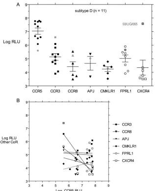
[image:4.585.43.548.81.225.2]Alternative CoR use by subtype D HIV-1 Env proteins.
A
similar analysis of CoR use was performed with 11 Env
pro-teins from subtype D isolates from nine different patients
de-scribed in Brown et al. (13) and two additional
env
clones
described in Materials and Methods. One of these isolates,
93/UG/065, was known to use CXCR4 and not CCR5 for
TABLE 1. Summary of alternative coreceptor use by HIV-1 subtype
Receptor
CoR use by the indicated HIV-1 subtype
Efficiency of CoR use for all
subtypesa
A B C D
Efficiency
of CoR usea
Level of entry
(log10RLU)
with CoR of CoR useEfficiencya
Level of entry
(log10RLU)
with CoR of CoR useEfficiencya
Level of entry
(log10RLU)
with CoR of CoR useEfficiencya
Level of entry
(log10RLU)
with CoR
Mean 95% CI Mean 95% CI Mean 95% CI Mean 95% CI
CCR5
15/15 (100)
7.66
7.46–7.85
18/18 (100)
6.99
6.63–7.35
22/22 (100)
7.00
6.71–7.29
11/11 (100)
7.04
6.52–7.56
100
CCR3
15/15 (100)
5.55
5.08–6.02
18/18 (100)
6.90
6.52–7.27 18/22 (82)
4.99
4.53–5.45
11/11 (100)
5.14
4.59–5.69
95
CCR8
11/15 (73)
4.69
4.14–5.25 14/18 (78)
4.69
4.35–5.03 17/22 (77)
5.23
4.71–5.75
6/11 (55)
4.42
3.52–5.32
71
APJ
6/15 (40)
4.43
3.30–5.56 15/18 (84)
b4.64
4.25–5.02
9/22 (41)
4.55
4.09–5.01
3/11 (27)
4.66
2.49–6.84
48
CMKLR1
5/15 (33)
3.91
3.55–4.27 11/18 (61)
4.12
3.68–4.56
3/22 (14)
c3.64
3.28–4.00
6/11 (55)
4.24
3.77–4.71
41
FPRL1
15/15 (100)
6.19
5.82–6.57 11/18 (61)
d4.68
4.16–5.19
22/22 (100)
5.83
5.36–6.30
8/11 (73)
5.02
4.40–5.65
84
CXCR4
1/15 (7)
3.94
NA
f5/18 (28)
3.91
3.69–4.12
5/22 (23)
4.18
3.94–4.42
7/11 (64)
e4.32
3.03–5.61
31
aCalculated as the number of positiveenvclones/number of clones tested (% positive). Boldface indicates 100% positive.
bPercentage of subtype Benvclones using APJ is significantly higher than the percentage of subtype A (P⫽0.0120, Fisher’s exact test), subtype C (P⫽0.0092),
or subtype D (P⫽0.0045).
cPercentage of subtype Cenvclones using CMKLR1 is significantly lower than the percentage of subtype B (P⫽0.0014) but not significantly lower than the
percentage of subtype A or D.
dPercentage of subtype Benvclones using FPRL1 is significantly lower than the percentage of subtypes A or C (P⫽0.0008).
ePercentage of subtype Denvclones using CXCR4 is significantly higher than the percentage of subtype A (P⫽0.0005) but not significantly higher than the
percentage of subtype B or C.
fNA, not available.
on November 8, 2019 by guest
http://jvi.asm.org/
infection (13), while the remaining 10
env
clones used CCR5
much better than CXCR4 or other alternative CoRs (Fig. 5A
and Table 1). Only CCR5 and CCR3 were able to mediate
entry by all 11 subtype D
env
clones. A majority of subtype D
env
clones could mediate virus entry via other alternative
CoRs. FPRL1 was used by 8/11 clones, and CCR8 and
CMKLR1 were used by 6/11 clones. Only 3/11 subtype D
env
clones could mediate entry via APJ, but 7/11 clones could
use CXCR4, with clone 93/UG/065 capable of robust entry via
CXCR4 (Fig. 5A and Table 1). The previously characterized
X4 93/UG/065
env
clone was the poorest at mediating entry via
CCR5 and the best at mediating entry via CCR3 and CXCR4
(Fig. 5A) but probably should be characterized as “dual/X4”
based on our results. Excluding that one high value (7.41 log
RLU), the mean entry level via CXCR4 for the remaining six
clones was 3.87 log RLU (95% confidence interval [CI], 3.44 to
4.18).
Linear regression analysis of virus entry via CCR5 versus
any other CoR did not show any significant correlation (Fig.
5B). The smaller
env
clone sample size would make
signif-icant correlation more difficult to observe, but the plotted
regression lines shown in Fig. 5B show no hint of positive
correlation.
Comparison of entry via alternative CoRs between different
HIV-1 subtypes.
Table 1 and Fig. 6 show summary data for
all experiments, and Table S1 in the supplemental material
gives the
env
clone name, accession number, and entry data
for each CoR assayed. The major differences between entry
mediated by
env
clones from different subtypes was in the
use of CCR3 and FPRL1, so only data for CCR5, CCR3,
and FPRL1 are shown in Fig. 6. Use of CCR5 was not
significantly different between subtypes although subtype A
env
clones showed a trend toward a higher level of entry.
Entry function via CCR3 was much higher for subtype B
env
clones than for other subtypes, and this difference was very
highly significant (
P
⬍
0.0001). Subtype A and C
env
clones
FIG. 3. (A) Entry mediated by 18 subtype B
env
clones from different patients plotted as described in the legend of Fig. 2. (B) Linear regression
analysis of entry via CCR5 versus via alternative CoR, analyzed as described in the legend of Fig. 2. The bold regression lines for CCR3 and APJ
indicate significant correlation with CCR5-mediated entry, but entry via FPRL1 did not show significant correlation with CCR5.
V
OL. 83, 2009
VIRUS ENTRY VIA CCR3 AND FPRL1 VARIES BY HIV-1 SUBTYPE
8357
on November 8, 2019 by guest
http://jvi.asm.org/
[image:5.585.136.451.67.462.2]mediated significantly higher levels of entry via FPRL1 than
subtype B or D
env
clones. Subtype D had a unique
pheno-type, with a low level of entry via CCR3, like subtypes A and
C, and a low level of entry via FPRL1, like subtype B and
unlike subtypes A and C.
Additional significant differences between HIV-1 subtypes
were a larger fraction of subtype B
env
clones than all other
subtypes that could use APJ for virus entry, a smaller fraction
of subtype C
env
clones than subtype B that could use
CMKLR1, and a larger fraction of subtype D
env
clones than
subtype A that could use CXCR4 (Table 1).
DISCUSSION
The most striking finding of these studies is that we were
able to define an alternative CoR entry phenotype that
distin-guished each HIV-1 subtype examined (Fig. 6). Robust use of
the primary CCR5 CoR was common to HIV-1 Env from all
subtypes, but subtypes A and C shared low levels of infection
via CCR3 and high levels via FPRL1, in contrast to subtype B,
which displayed high levels of infection via CCR3 and low
levels via FPRL1. Subtype D Env proteins were poor at
me-diating infection via either CCR3 or FPRL1. All of these
differences in entry phenotypes were highly significant (Fig. 6,
legend). These subtype-specific patterns of CoR use go beyond
differences previously observed (8, 61, 71), primarily because
of the use of a highly permissive indicator cell line, stringent
criteria for positive entry, and the addition of FPRL1 (67) to
the list of alternative CoRs assayed. The implications of these
results are that HIV-1
env
has evolved differently in subtypes A
and C compared to either subtype B or D infection, perhaps in
response to subtle selection pressure influenced by the host
genetics of the infected population or founder effects from
FIG. 4. (A) Entry mediated by 22 subtype C
env
clones from different patients at early stages of infection plotted as described in the legend
of Fig. 2. (B) Linear regression analysis of entry via CCR5 versus three alternative CoRs showing significant correlation with CCR5: CCR3, CCR8,
and FPRL1.
on November 8, 2019 by guest
http://jvi.asm.org/
[image:6.585.133.452.67.482.2]early in the epidemic. CCR3 and FPRL1, the two favored
alternative CoRs, show little primary sequence homology in
their extracellular domains (49), and neither has a particularly
close homology to CCR5. Subtle differences in CCR5 binding
by CD4-triggered Env protein may contribute to the
subtype-specific patterns of alternative CoR use we have identified and
may possibly relate to the development of resistance to
CCR5-targeted entry inhibitors (72).
These experiments were predicated on the hypothesis that
the ability of HIV-1 isolates to utilize CoRs other than CCR5
(clearly the primary CoR) might fall into two or more
catego-ries that would have implications for how Env evolution
im-pacts CoR use. The first category would be defined by use of
alternative CoRs that was correlated with entry activity
medi-ated by CCR5, a finding that would suggest that alternative
CoRs are surrogates for CCR5 and would generally be less
efficient than CCR5 at mediating virus entry. The second
cat-egory would be defined by independent use of alternative
CoRs that is not predicted by entry efficiency via CCR5, and
entry efficiency via the alternative CoRs might be greater or
less than via CCR5. In the first category, the driving force for
Env evolution is improved ability to use CCR5 for entry, and
expanded use of alternative CoRs is a direct consequence of
this evolutionary pathway. In the second category, Env
evolu-tion is more complicated, and pathways leading to CCR5 or
alternative CoR use must diverge at some point prior to
sam-pling of the HIV-1 quasispecies.
The results obtained favor the first hypothesis, namely, that
the use of the preferred alternative CoR is correlated with (or
predicted by) virus entry efficiency via CCR5. Linear
regres-sion analysis of the correlation between entry via CCR5 and
other CoRs revealed a subtype-specific phenotype. For subtype
A Env proteins, entry via CCR5 and FPRL1 was highly
corre-lated (Fig. 2B). For subtype B, entry via CCR5 and CCR3 was
highly correlated (Fig. 3B). For subtype C, entry via CCR5 was
highly correlated with entry via FPRL1, CCR3, and CCR8
(Fig. 4B). For subtype D, entry via CCR5 did not correlate with
entry mediated by any alternative CoR (Fig. 5B). We also
FIG. 5. (A) Entry mediated by 11 subtype D
env
clones from different patients plotted as described in the legend of Fig. 2. One subtype D
env
clone (93/UG/065) is depicted in lighter fill because it was the only one capable of robust entry via CXCR4. (B) Linear regression analysis of entry
via CCR5 versus via alternative CoRs. No significant correlations were observed.
V
OL. 83, 2009
VIRUS ENTRY VIA CCR3 AND FPRL1 VARIES BY HIV-1 SUBTYPE
8359
on November 8, 2019 by guest
http://jvi.asm.org/
[image:7.585.135.451.65.456.2]noted some subtype-specific differences in the frequency with
which alternative CoRs could be used for entry (Table 1).
Significant differences were a more frequent use of APJ in
subtype B, a less frequent use of CMKLR1 in subtype C, a less
frequent use of FPRL1 in subtype B, and a more frequent use
of CXCR4 in subtype D.
Several prior reports have noted the high frequency of R5R3
viruses that show efficient entry via both CCR5 and CCR3
(1–3, 5, 17, 61), and some (77, 78) suggest possible target cell
adaptation by this prevalent subset of HIV-1 phenotypes.
Dif-ferences in cell lines used for typing CCR3 use could have
contributed to distinct outcomes between our results and prior
reports; specifically, U87/CD4/CCR3 cells obtained from the
AIDS Research and Reference Reagent Program and used in
the study by Morris et al. (54) express 100-fold lower levels of
CCR3 than the NP-2/CD4/CCR3 cells we have employed (data
not shown). Other studies employing transiently transfected
target cell lines have also found a lower incidence of R5R3
viruses (see, e.g., reference 81), so it is likely that typing of
CCR3 use is highly dependent upon the level of CCR3
ex-pressed on a given target cell. The demonstration of HIV-1
entry via alterative CoRs other than CCR5 or CXCR4 is often
dismissed as an artifact of using target cell lines that
overex-press a given GPCR, a view that is bolstered by blocking
infection of primary target cells by either CCR5 or CXCR4
inhibitors (30, 53, 80, 81). The phenotypes that we have
ob-served are entirely based on overexpression of alternative
CoRs on one cell line and do not address the important issue
of whether or not these CoRs can be used for productive
infection of natural target cells in HIV-1-infected individuals.
However, we speculate that high entry efficiency via a CoR
expressed at high levels may translate into low entry efficiency
via the same CoR expressed at physiological levels.
FPRL1 emerged as a potentially important CoR in our
stud-ies. There have been prior studies suggesting that a peptide
derived from either the C4-V4 region of subtype B Env (23) or
from the V3 region (64) could react with FPRL1, but its use as
an alternative CoR has only recently been documented (67).
FPRL1 is expressed on a variety of human cell types, including
monocytes, dendritic cells, and T cells (52), that potentially
could serve as targets for HIV-1 infection. The importance of
FPRL1 for infection of natural target cells remains to be
es-tablished, but our results suggest that this may be a fruitful
area for further investigation.
Use of CMKLR1/ChemR23 was less frequent in subtype C,
in agreement with prior reports (49, 62). However, entry
func-tion via CMKLR1/ChemR23 did not correlate with CCR3 use
despite the homology in the extracellular domains noted in a
prior report (49). CMKLR1 was efficiently used for entry by
SIVmac
env
-pseudotyped viruses (Fig. 1B), and we used the
same cell line employed in prior studies, so these differences in
results appear to reflect the selection of distinct
env
clones. We
also noted a higher than expected frequency (5/22) of subtype
C
env
clones that could mediate low-level entry via CXCR4.
This may reflect our unique assay conditions since SIVmac
env
clones could also infect CXCR4-expressing target cells (Fig.
1B). No unique features of the five subtype C
env
clones
capable of CXCR4 use were found, either in time of isolation
or use of other CoRs, when they were compared to the other
17 clones incapable of mediating entry via CXCR4.
The results in Fig. 2 to 6 show only a subset of alternative
CoRs. The primary reason for this is that few HIV-1
env
clones
were able to generate pseudotyped virus that could enter target
cells via GPR1, CCR1, CCR6, CXCR6, or RDC1 although
SIVmac
env
clones could mediate entry via these CoRs (Fig.
1B). We also did not have access to NP-2/CD4 target cells
expressing GPR15 or D6, two alternative CoRs previously
reported to mediate infection of other target cell lines (22, 55).
It is possible that examination of these two additional
alterna-tive CoRs could add to the differences between HIV-1
sub-types that we observed. We also had access to only limited
numbers of
env
clones from subtypes E, A/D, A/C, CRF01_AE,
or CRF02_AG HIV-1 infections, and thus we cannot comment
on the properties of these variants. Future analysis of a larger
collection of natural recombinant viruses may be of use in
dissecting the determinants for the subtype differences
ob-served here.
[image:8.585.43.284.68.220.2]Expansion of alternative CoR use to receptors other than
CXCR4 and the relationship between CCR5-mediated entry
and alternative CoR use distinguish different HIV-1 subtypes.
Much of the data on alternative CoR use has been derived
from the study of subtype B isolates, and larger sample sets
from other subtypes need to be carefully examined for
alter-native CoR use. Even in subtype B-infected patients, reduction
in levels of CCR5 or treatment with CCR5 inhibitors may
increase the selective pressure to expand CoR use beyond
CXCR4, as in the well-studied patient of Gorry et al. (30).
Although there are many pathways to CCR5 inhibitor
resis-tance (48, 73), these data suggest that we should remain alert
for selection of rare variants with expanded CoR use beyond
CXCR4, particularly in patients with non-subtype B HIV-1
infection.
FIG. 6. Comparison of virus entry mediated by
env
clones from
different subtypes (A, B, C, or D), with log RLU values depicted in
box-and-whisker plots such that the vertical bars represent the range of
observed values and the box represents the 25th to 75th percentile.
Results for each subtype are color-coded with subtype A in blue,
subtype B in magenta, subtype C in green, and subtype D in salmon.
The difference in virus entry mediated by CCR3 for subtype B versus
subtypes A, C, and D is highly significant (
***
,
P
⬍
0.0001;
Mann-Whitney U test). The difference in virus entry mediated by FPRL1 for
subtype B versus subtype A (
**
,
P
⫽
0.005) or versus subtype C (
**
,
P
⫽
0.0094) is significant, but there is no significant difference between
subtype B and subtype D entry via FPRL1.
on November 8, 2019 by guest
http://jvi.asm.org/
ACKNOWLEDGMENTS
We thank Paul Clapham for supplying NP-2/CD4 cell lines
express-ing selected alternative coreceptors (with the permission of H.
Hoshino) and Oliver Laeyendecker for the Rakai Health Sciences
Program for supplying samples from the Rakai, Uganda cohort from
which subtype D
env
clone H23706_A6_9-4 was isolated.
This research was supported by NIH grants AI052778, AI071935
(D.E.M.), and AI058763 (J.B.) and by the James B. Pendleton
Char-itable Trust.
This is publication 20108 from The Scripps Research Institute.
REFERENCES
1.Aasa-Chapman, M. M., K. Aubin, I. Williams, and A. McKnight.2006. Primary CCR5 only using HIV-1 isolates does not accurately represent the
in vivo replicating quasi-species. Virology351:489–496.
2.Aasa-Chapman, M. M., C. R. Seymour, I. Williams, and A. McKnight.2006. Novel envelope determinants for CCR3 use by human immunodeficiency
virus. J. Virol.80:10884–10889.
3.Alkhatib, G., E. A. Berger, P. M. Murphy, and J. E. Pease.1997. Determi-nants of HIV-1 coreceptor function on CC chemokine receptor 3. Impor-tance of both extracellular and transmembrane/cytoplasmic regions. J. Biol.
Chem.272:20420–20426.
4.Alkhatib, G., C. C. Broder, and E. A. Berger.1996. Cell type-specific fusion cofactors determine human immunodeficiency virus type 1 tropism for T-cell
lines versus primary macrophages. J. Virol.70:5487–5494.
5.Bazan, H. A., G. Alkhatib, C. C. Broder, and E. A. Berger.1998. Patterns of CCR5, CXCR4, and CCR3 usage by envelope glycoproteins from human
immunodeficiency virus type 1 primary isolates. J. Virol.72:4485–4491.
6.Berger, E. A.1997. HIV entry and tropism: the chemokine receptor
connec-tion. AIDS11:S3–S16.
7.Berson, J. F., D. Long, B. J. Doranz, J. Rucker, F. R. Jirik, and R. W. Doms.
1996. A seven-transmembrane domain receptor involved in fusion and entry
of T-cell-tropic human immunodeficiency virus type 1 strains. J. Virol.70:
6288–6295.
8.Bjorndal, A., H. Deng, M. Jansson, J. R. Fiore, C. Colognesi, A. Karlsson, J. Albert, G. Scarlatti, D. R. Littman, and E. M. Fenyo.1997. Coreceptor usage of primary human immunodeficiency virus type 1 isolates varies according to
biological phenotype. J. Virol.71:7478–7487.
9.Blaak, H., P. H. Boers, R. A. Gruters, H. Schuitemaker, M. E. van der Ende, and A. D. Osterhaus.2005. CCR5, GPR15, and CXCR6 are major corecep-tors of human immunodeficiency virus type 2 variants isolated from
individ-uals with and without plasma viremia. J. Virol.79:1686–1700.
10.Blish, C. A., R. Nedellec, K. Mandaliya, D. E. Mosier, and J. Overbaugh.
2007. HIV-1 subtype A envelope variants from early in infection have
vari-able sensitivity to neutralization and to inhibitors of viral entry. AIDS21:
693–702.
11.Blish, C. A., Z. Jalalian-Lechak, S. Rainwater, M.-A. Nguyen, O. C. Dogan, and J. Overbaugh.27 May 2009. Cross-subtype neutralization sensitivity despite monoclonal antibody resistance among early subtype A, C, and D envelope variants of human immunodeficiency virus type 1. J. Virol. doi.10.1128/JVI.00673-09.
12.Bron, R., P. J. Klasse, D. Wilkinson, P. R. Clapham, A. Pelchen-Matthews, C. Power, T. N. Wells, J. Kim, S. C. Peiper, J. A. Hoxie, and M. Marsh.1997. Promiscuous use of CC and CXC chemokine receptors in cell-to-cell fusion mediated by a human immunodeficiency virus type 2 envelope protein.
J. Virol.71:8405–8415.
13.Brown, B. K., J. M. Darden, S. Tovanabutra, T. Oblander, J. Frost, E. Sanders-Buell, M. S. de Souza, D. L. Birx, F. E. McCutchan, and V. R. Polonis.2005. Biologic and genetic characterization of a panel of 60 human immunodeficiency virus type 1 isolates, representing clades A, B, C, D, CRF01_AE, and CRF02_AG, for the development and assessment of
can-didate vaccines. J. Virol.79:6089–6101.
14.Cann, A. J., M. J. Churcher, M. Boyd, W. O’Brien, J. Q. Zhao, J. Zack, and I. S. Chen.1992. The region of the envelope gene of human immunodefi-ciency virus type 1 responsible for determination of cell tropism. J. Virol.
66:305–309.
15.Chesebro, B., K. Wehrly, J. Nishio, and S. Perryman.1996. Mapping of independent V3 envelope determinants of human immunodeficiency virus type 1 macrophage tropism and syncytium formation in lymphocytes. J.
Vi-rol.70:9055–9059.
16.Choe, H., M. Farzan, M. Konkel, K. Martin, Y. Sun, L. Marcon, M. Cay-abyab, M. Berman, M. E. Dorf, N. Gerard, C. Gerard, and J. Sodroski.1998. The orphan seven-transmembrane receptor Apj supports the entry of pri-mary T-cell-line-tropic and dualtropic human immunodeficiency virus type 1.
J. Virol.72:6113–6118.
17.Choe, H., M. Farzan, Y. Sun, N. Sullivan, B. Rollins, P. D. Ponath, L. Wu, C. R. Mackay, G. LaRosa, W. Newman, N. Gerard, C. Gerard, and J. Sodroski.1996. The beta-chemokine receptors CCR3 and CCR5 facilitate
infection by primary HIV-1 isolates. Cell85:1135–1148.
18.Cilliers, T., S. Willey, W. M. Sullivan, T. Patience, P. Pugach, M. Coetzer, M.
Papathanasopoulos, J. P. Moore, A. Trkola, P. Clapham, and L. Morris.
2005. Use of alternate coreceptors on primary cells by two HIV-1 isolates.
Virology339:136–144.
19.Connor, R. I., B. K. Chen, S. Choe, and N. R. Landau.1995. Vpr is required for efficient replication of human immunodeficiency virus type-1 in
mono-nuclear phagocytes. Virology206:935–944.
20.Connor, R. I., K. E. Sheridan, D. Ceradini, S. Choe, and N. R. Landau.1997. Change in coreceptor use correlates with disease progression in HIV-1–
infected individuals. J. Exp. Med.185:621–628.
21.Dean, M., M. Carrington, C. Winkler, G. A. Huttley, M. W. Smith, R. Allikmets, J. J. Goedert, S. P. Buchbinder, E. Vittinghoff, E. Gomperts, S. Donfield, D. Vlahov, R. Kaslow, A. Saah, C. Rinaldo, R. Detels, Hemophilia Growth and Development Study, Multicenter AIDS Cohort Study, Multi-center Hemophilia Cohort Study, San Francisco City Cohort, ALIVE Study, and S. J. O’Brien.1996. Genetic restriction of HIV-1 infection and
progres-sion to AIDS by a deletion allele of theCKR5structural gene. Science
273:1856–1862.
22.Deng, H. K., D. Unutmaz, V. N. KewalRamani, and D. R. Littman.1997. Expression cloning of new receptors used by simian and human
immunode-ficiency viruses. Nature388:296–300.
23.Deng, X., H. Ueda, S. B. Su, W. Gong, N. M. Dunlop, J. L. Gao, P. M. Murphy, and J. M. Wang.1999. A synthetic peptide derived from human immunodeficiency virus type 1 gp120 downregulates the expression and function of chemokine receptors CCR5 and CXCR4 in monocytes by acti-vating the 7-transmembrane G-protein-coupled receptor FPRL1/LXA4R.
Blood94:1165–1173.
24.Doranz, B. J., J. Rucker, Y. Yi, R. J. Smyth, M. Samson, S. C. Peiper, M. Parmentier, R. G. Collman, and R. W. Doms.1996. A dual-tropic primary HIV-1 isolate that uses fusin and the beta-chemokine receptors CKR-5,
CKR-3, and CKR-2b as fusion cofactors. Cell85:1149–1158.
25.Edinger, A. L., T. L. Hoffman, M. Sharron, B. Lee, B. O’Dowd, and R. W. Doms.1998. Use of GPR1, GPR15, and STRL33 as coreceptors by diverse human immunodeficiency virus type 1 and simian immunodeficiency virus
envelope proteins. Virology249:367–378.
26.Edinger, A. L., T. L. Hoffman, M. Sharron, B. Lee, Y. Yi, W. Choe, D. L. Kolson, B. Mitrovic, Y. Zhou, D. Faulds, R. G. Collman, J. Hesselgesser, R. Horuk, and R. W. Doms.1998. An orphan seven-transmembrane domain receptor expressed widely in the brain functions as a coreceptor for human immunodeficiency virus type 1 and simian immunodeficiency virus. J. Virol.
72:7934–7940.
27.Farzan, M., H. Choe, K. Martin, L. Marcon, W. Hofmann, G. Karlsson, Y. Sun, P. Barrett, N. Marchand, N. Sullivan, N. Gerard, C. Gerard, and J. Sodroski.1997. Two orphan seven-transmembrane segment receptors which are expressed in CD4-positive cells support simian immunodeficiency virus
infection. J. Exp. Med.186:405–411.
28.Feng, Y., C. C. Broder, P. E. Kennedy, and E. A. Berger.1996. HIV-1 entry cofactor: functional cDNA cloning of a seven-transmembrane, G
protein-coupled receptor. Science272:872–877.
29.Ghorpade, A., M. Q. Xia, B. T. Hyman, Y. Persidsky, A. Nukuna, P. Bock, M. Che, J. Limoges, H. E. Gendelman, and C. R. Mackay.1998. Role of the beta-chemokine receptors CCR3 and CCR5 in human immunodeficiency
virus type 1 infection of monocytes and microglia. J. Virol.72:3351–3361.
30.Gorry, P. R., R. L. Dunfee, M. E. Mefford, K. Kunstman, T. Morgan, J. P. Moore, J. R. Mascola, K. Agopian, G. H. Holm, A. Mehle, J. Taylor, M. Farzan, H. Wang, P. Ellery, S. J. Willey, P. R. Clapham, S. M. Wolinsky, S. M. Crowe, and D. Gabuzda.2007. Changes in the V3 region of gp120 contribute to unusually broad coreceptor usage of an HIV-1 isolate from a
CCR5⌬32 heterozygote. Virology362:163–178.
31.Harrowe, G., and C. Cheng-Mayer.1995. Amino acid substitutions in the V3 loop are responsible for adaptation to growth in transformed T-cell lines of
a primary human immunodeficiency virus type 1. Virology210:490–494.
32.He, J., Y. Chen, M. Farzan, H. Choe, A. Ohagen, S. Gartner, J. Busciglio, X. Yang, W. Hofmann, W. Newman, C. R. Mackay, J. Sodroski, and D. Gabuzda.1997. CCR3 and CCR5 are co-receptors for HIV-1 infection of
microglia. Nature385:645–649.
33.Hoffman, T. L., E. B. Stephens, O. Narayan, and R. W. Doms.1998. HIV type I envelope determinants for use of the CCR2b, CCR3, STRL33, and
APJ coreceptors. Proc. Natl. Acad. Sci. USA95:11360–11365.
34.Horuk, R., J. Hesselgesser, Y. Zhou, D. Faulds, M. Halks-Miller, S. Harvey, D. Taub, M. Samson, M. Parmentier, J. Rucker, B. J. Doranz, and R. W. Doms.1998. The CC chemokine I-309 inhibits CCR8-dependent infection by
diverse HIV-1 strains. J. Biol. Chem.273:386–391.
35.Huang, W., S. H. Eshleman, J. Toma, S. Fransen, E. Stawiski, E. E. Paxinos, J. M. Whitcomb, A. M. Young, D. Donnell, F. Mmiro, P. Musoke, L. A. Guay, J. B. Jackson, N. T. Parkin, and C. J. Petropoulos.2007. Coreceptor tropism in human immunodeficiency virus type 1 subtype D: high prevalence of CXCR4 tropism and heterogeneous composition of viral populations. J.
Vi-rol.81:7885–7893.
36.Huang, Y., W. A. Paxton, S. M. Wolinsky, A. U. Neumann, L. Zhang, T. He, S. Kang, D. Ceradini, Z. Jin, K. Yazdanbakhsh, K. Kunstman, D. Erickson, E. Dragon, N. R. Landau, J. Phair, D. D. Ho, and R. A. Koup.1996. The role
V
OL. 83, 2009
VIRUS ENTRY VIA CCR3 AND FPRL1 VARIES BY HIV-1 SUBTYPE
8361
on November 8, 2019 by guest
http://jvi.asm.org/
of a mutant CCR5 allele in HIV-1 transmission and disease progression. Nat.
Med.2:1240–1243.
37.Hwang, S. S., T. J. Boyle, H. K. Lyerly, and B. R. Cullen.1991. Identification of the envelope V3 loop as the primary determinant of cell tropism in HIV-1.
Science253:71–74.
38.Jinno, A., N. Shimizu, Y. Soda, Y. Haraguchi, T. Kitamura, and H. Hoshino.
1998. Identification of the chemokine receptor TER1/CCR8 expressed in brain-derived cells and T cells as a new coreceptor for HIV-1 infection.
Biochem. Biophys. Res. Commun.243:497–502.
39.Kanbe, K., N. Shimizu, Y. Soda, K. Takagishi, and H. Hoshino.1999. A CXC chemokine receptor, CXCR5/BLR1, is a novel and specific coreceptor for
human immunodeficiency virus type 2. Virology265:264–273.
40.Karlsson, I., L. Antonsson, Y. Shi, A. Karlsson, J. Albert, T. Leitner, B. Olde, C. Owman, and E. M. Fenyo.2003. HIV biological variability unveiled: frequent isolations and chimeric receptors reveal unprecedented variation of
coreceptor use. AIDS17:2561–2569.
41.Kiselyeva, Y., R. Nedellec, A. Ramos, C. Pastore, L. B. Margolis, and D. E. Mosier.2007. Evolution of CXCR4-using human immunodeficiency virus type 1 SF162 is associated with two unique envelope mutations. J. Virol.
81:3657–3661.
42.Kiwanuka, N., O. Laeyendecker, M. Robb, G. Kigozi, M. Arroyo, F. Mc-Cutchan, L. A. Eller, M. Eller, F. Makumbi, D. Birx, F. Wabwire-Mangen, D. Serwadda, N. K. Sewankambo, T. C. Quinn, M. Wawer, and R. Gray.2008. Effect of human immunodeficiency virus Type 1 (HIV-1) subtype on disease progression in persons from Rakai, Uganda, with incident HIV-1 infection.
J. Infect. Dis.197:707–713.
43.Li, M., F. Gao, J. R. Mascola, L. Stamatatos, V. R. Polonis, M. Koutsoukos, G. Voss, P. Goepfert, P. Gilbert, K. M. Greene, M. Bilska, D. L. Kothe, J. F. Salazar-Gonzalez, X. Wei, J. M. Decker, B. H. Hahn, and D. C. Montefiori.
2005. Human immunodeficiency virus type 1envclones from acute and early
subtype B infections for standardized assessments of vaccine-elicited
neu-tralizing antibodies. J. Virol.79:10108–10125.
44.Li, M., J. F. Salazar-Gonzalez, C. A. Derdeyn, L. Morris, C. Williamson, J. E. Robinson, J. M. Decker, Y. Li, M. G. Salazar, V. R. Polonis, K. Mlisana, S. A. Karim, K. Hong, K. M. Greene, M. Bilska, J. Zhou, S. Allen, E. Chomba, J. Mulenga, C. Vwalika, F. Gao, M. Zhang, B. T. Korber, E. Hunter, B. H. Hahn, and D. C. Montefiori.2006. Genetic and neutralization properties of
subtype C human immunodeficiency virus type 1 molecularenvclones from
acute and early heterosexually acquired infections in Southern Africa. J.
Vi-rol.80:11776–11790.
45.Liao, F., G. Alkhatib, K. W. Peden, G. Sharma, E. A. Berger, and J. M. Farber.1997. STRL33, A novel chemokine receptor-like protein, functions as a fusion cofactor for both macrophage-tropic and T cell line-tropic HIV-1.
J. Exp. Med.185:2015–2023.
46.Loetscher, M., A. Amara, E. Oberlin, N. Brass, D. Legler, P. Loetscher, M. D’Apuzzo, E. Meese, D. Rousset, J. L. Virelizier, M. Baggiolini, F. Arenzana-Seisdedos, and B. Moser.1997. TYMSTR, a putative chemokine receptor selectively expressed in activated T cells, exhibits HIV-1 coreceptor function.
Curr. Biol.7:652–660.
47.Long, E. M., S. M. Rainwater, L. Lavreys, K. Mandaliya, and J. Overbaugh.
2002. HIV type 1 variants transmitted to women in Kenya require the CCR5 coreceptor for entry, regardless of the genetic complexity of the infecting
virus. AIDS Res. Hum. Retrovir.18:567–576.
48.Marozsan, A. J., S. E. Kuhmann, T. Morgan, C. Herrera, E. Rivera-Troche, S. Xu, B. M. Baroudy, J. Strizki, and J. P. Moore.2005. Generation and properties of a human immunodeficiency virus type 1 isolate resistant to the
small molecule CCR5 inhibitor, SCH-417690 (SCH-D). Virology338:182–199.
49.Mårtensson, U. E., E. M. Fenyo, B. Olde, and C. Owman.2006. Character-ization of the human chemerin receptor–ChemR23/CMKLR1–as co-recep-tor for human and simian immunodeficiency virus infection, and
identifica-tion of virus-binding receptor domains. Virology355:6–17.
50.Mascola, J. R., P. D’Souza, P. Gilbert, B. H. Hahn, N. L. Haigwood, L. Morris, C. J. Petropoulos, V. R. Polonis, M. Sarzotti, and D. C. Montefiori.
2005. Recommendations for the design and use of standard virus panels to assess neutralizing antibody responses elicited by candidate human
immu-nodeficiency virus type 1 vaccines. J. Virol.79:10103–10107.
51.McKnight, A., M. T. Dittmar, J. Moniz-Periera, K. Ariyoshi, J. D. Reeves, S. Hibbitts, D. Whitby, E. Aarons, A. E. Proudfoot, H. Whittle, and P. R. Clapham.1998. A broad range of chemokine receptors are used by primary isolates of human immunodeficiency virus type 2 as coreceptors with CD4.
J. Virol.72:4065–4071.
52.Migeotte, I., D. Communi, and M. Parmentier.2006. Formyl peptide recep-tors: a promiscuous subfamily of G protein-coupled receptors controlling
immune responses. Cytokine Growth Factor Rev.17:501–519.
53.Moore, J. P., S. G. Kitchen, P. Pugach, and J. A. Zack.2004. The CCR5 and CXCR4 coreceptors—central to understanding the transmission and patho-genesis of human immunodeficiency virus type 1 infection. AIDS Res. Hum.
Retrovir.20:111–126.
54.Morris, L., T. Cilliers, H. Bredell, M. Phoswa, and D. J. Martin.2001. CCR5 is the major coreceptor used by HIV-1 subtype C isolates from patients with
active tuberculosis. AIDS Res. Hum. Retrovir.17:697–701.
55.Neil, S. J., M. M. Aasa-Chapman, P. R. Clapham, R. J. Nibbs, A. McKnight,
and R. A. Weiss.2005. The promiscuous CC chemokine receptor D6 is a functional coreceptor for primary isolates of human immunodeficiency virus
type 1 (HIV-1) and HIV-2 on astrocytes. J. Virol.79:9618–9624.
56.Pastore, C., R. Nedellec, A. Ramos, O. Hartley, J. L. Miamidian, J. D. Reeves, and D. E. Mosier.2007. Conserved changes in envelope function during human immunodeficiency virus type 1 coreceptor switching. J. Virol.
81:8165–8179.
57.Pastore, C., R. Nedellec, A. Ramos, S. Pontow, L. Ratner, and D. E. Mosier.
2006. Human immunodeficiency virus type 1 coreceptor switching: V1/V2 gain-of-fitness mutations compensate for V3 loss-of-fitness mutations. J.
Vi-rol.80:750–758.
58.Peters, P. J., J. Bhattacharya, S. Hibbitts, M. T. Dittmar, G. Simmons, J. Bell, P. Simmonds, and P. R. Clapham.2004. Biological analysis of human immunodeficiency virus type 1 R5 envelopes amplified from brain and lymph node tissues of AIDS patients with neuropathology reveals two distinct tropism phenotypes and identifies envelopes in the brain that confer an
enhanced tropism and fusigenicity for macrophages. J. Virol.78:6915–6926.
59.Rainwater, S. M., X. Wu, R. Nduati, R. Nedellec, D. Mosier, G. John-Stewart, D. Mbori-Ngacha, and J. Overbaugh.2007. Cloning and character-ization of functional subtype A HIV-1 envelope variants transmitted through
breastfeeding. Curr. HIV Res.5:189–197.
60.Ross, T. M., and B. R. Cullen.1998. The ability of HIV type 1 to use CCR-3 as a coreceptor is controlled by envelope V1/V2 sequences acting in
con-junction with a CCR-5 tropic V3 loop. Proc. Natl. Acad. Sci. USA95:7682–
7686.
61.Rucker, J., A. L. Edinger, M. Sharron, M. Samson, B. Lee, J. F. Berson, Y. Yi, B. Margulies, R. G. Collman, B. J. Doranz, M. Parmentier, and R. W. Doms.1997. Utilization of chemokine receptors, orphan receptors, and her-pesvirus-encoded receptors by diverse human and simian immunodeficiency
viruses. J. Virol.71:8999–9007.
62.Samson, M., A. L. Edinger, P. Stordeur, J. Rucker, V. Verhasselt, M. Shar-ron, C. Govaerts, C. Mollereau, G. Vassart, R. W. Doms, and M. Parmentier.
1998. ChemR23, a putative chemoattractant receptor, is expressed in mono-cyte-derived dendritic cells and macrophages and is a coreceptor for SIV and
some primary HIV-1 strains. Eur. J. Immunol.28:1689–1700.
63.Scarlatti, G., E. Tresoldi, A. Bjorndal, R. Fredriksson, C. Colognesi, H. K. Deng, M. S. Malnati, A. Plebani, A. G. Siccardi, D. R. Littman, E. M. Fenyo, and P. Lusso.1997. In vivo evolution of HIV-1 co-receptor usage and
sensitivity to chemokine-mediated suppression. Nat. Med.3:1259–1265.
64.Shen, W., P. Proost, B. Li, W. Gong, Y. Le, R. Sargeant, P. M. Murphy, J. Van Damme, and J. M. Wang.2000. Activation of the chemotactic peptide receptor FPRL1 in monocytes phosphorylates the chemokine receptor CCR5 and attenuates cell responses to selected chemokines. Biochem.
Bio-phys. Res. Commun.272:276–283.
65.Shimizu, N., Y. Soda, K. Kanbe, H. Y. Liu, A. Jinno, T. Kitamura, and H. Hoshino.1999. An orphan G protein-coupled receptor, GPR1, acts as a coreceptor to allow replication of human immunodeficiency virus types 1 and
2 in brain-derived cells. J. Virol.73:5231–5239.
66.Shimizu, N., Y. Soda, K. Kanbe, H. Y. Liu, R. Mukai, T. Kitamura, and H. Hoshino.2000. A putative G protein-coupled receptor, RDC1, is a novel
coreceptor for human and simian immunodeficiency viruses. J. Virol.74:
619–626.
67.Shimizu, N., A. Tanaka, T. Mori, T. Ohtsuki, A. Hoque, A. Jinno-Oue, C. Apichartpiyakul, S. Kusagawa, Y. Takebe, and H. Hoshino. 2008. A formylpeptide receptor, FPRL1, acts as an efficient coreceptor for primary
isolates of human immunodeficiency virus. Retrovirology5:52.
68.Shimizu, N., A. Tanaka, A. Oue, T. Mori, C. Apichartpiyakul, and H. Hoshino.2008. A short amino acid sequence containing tyrosine in the N-terminal region of G protein-coupled receptors is critical for their poten-tial use as co-receptors for human and simian immunodeficiency viruses.
J. Gen. Virol.89:3126–3136.
69.Soda, Y., N. Shimizu, A. Jinno, H. Y. Liu, K. Kanbe, T. Kitamura, and H. Hoshino.1999. Establishment of a new system for determination of core-ceptor usages of HIV based on the human glioma NP-2 cell line. Biochem.
Biophys. Res. Commun.258:313–321.
70.Sullivan, W. M., P. Dorr, M. Perros, R. Hudson, J. Leif, K. Luzuriaga, and P. R. Clapham.2008. Lack of alternative coreceptor use by pediatric HIV-1 R5 isolates for infection of primary cord or adult peripheral blood
mono-nuclear cells. Arch. Virol.153:363–366.
71.Tscherning, C., A. Alaeus, R. Fredriksson, A. Bjorndal, H. Deng, D. R. Littman, E. M. Fenyo, and J. Albert.1998. Differences in chemokine
core-ceptor usage between genetic subtypes of HIV-1. Virology241:181–188.
72.Tsibris, A. M., M. Sagar, R. M. Gulick, Z. Su, M. Hughes, W. Greaves, M. Subramanian, C. Flexner, F. Giguel, K. E. Leopold, E. Coakley, and D. R. Kuritzkes. 2008. In vivo emergence of vicriviroc resistance in a human
immunodeficiency virus type 1 subtype C-infected subject. J. Virol.82:8210–
8214.
73.Westby, M., C. Smith-Burchnell, J. Mori, M. Lewis, M. Mosley, M. Stock-dale, P. Dorr, G. Ciaramella, and M. Perros.2007. Reduced maximal inhi-bition in phenotypic susceptibility assays indicates that viral strains resistant to the CCR5 antagonist maraviroc utilize inhibitor-bound receptor for entry.
J. Virol.81:2359–2371.
on November 8, 2019 by guest
http://jvi.asm.org/
74.Willey, S. J., J. D. Reeves, R. Hudson, K. Miyake, N. Dejucq, D. Schols, E. De Clercq, J. Bell, A. McKnight, and P. R. Clapham.2003. Identification of a subset of human immunodeficiency virus type 1 (HIV-1), HIV-2, and simian immunodeficiency virus strains able to exploit an alternative
corecep-tor on untransformed human brain and lymphoid cells. J. Virol.77:6138–
6152.
75.Wu, X., A. B. Parast, B. A. Richardson, R. Nduati, G. John-Stewart, D. Mbori-Ngacha, S. M. Rainwater, and J. Overbaugh.2006. Neutralization escape variants of human immunodeficiency virus type 1 are transmitted
from mother to infant. J. Virol.80:835–844.
76.Xiao, L., D. L. Rudolph, S. M. Owen, T. J. Spira, and R. B. Lal.1998. Adaptation to promiscuous usage of CC and CXC-chemokine coreceptors in
vivo correlates with HIV-1 disease progression. AIDS12:F137–F143.
77.Xu, Y., H. Zhu, C. K. Wilcox, A. van’t Wout, T. Andrus, N. Llewellyn, L. Stamatatos, J. I. Mullins, L. Corey, and T. Zhu.2008. Blood monocytes harbor HIV type 1 strains with diversified phenotypes including
macro-phage-specific CCR5 virus. J. Infect. Dis.197:309–318.
78.Zerhouni-Layachi, B., M. Husain, M. J. Ross, D. Marras, M. Sunamoto, X. Liu, P. E. Klotman, and M. E. Klotman.2006. Dual tropism of HIV-1 envelopes derived from renal tubular epithelial cells of patients with
HIV-associated nephropathy. AIDS20:621–624.
79.Zhang, L., T. He, Y. Huang, Z. Chen, Y. Guo, S. Wu, K. J. Kunstman, R. C. Brown, J. P. Phair, A. U. Neumann, D. D. Ho, and S. M. Wolinsky.1998. Chemokine coreceptor usage by diverse primary isolates of human
immu-nodeficiency virus type 1. J. Virol.72:9307–9312.
80.Zhang, Y., B. Lou, R. B. Lal, A. Gettie, P. A. Marx, and J. P. Moore.2000. Use of inhibitors to evaluate coreceptor usage by simian and simian/human immunodeficiency viruses and human immunodeficiency virus type 2 in
pri-mary cells. J. Virol.74:6893–6910.
81.Zhang, Y. J., T. Dragic, Y. Cao, L. Kostrikis, D. S. Kwon, D. R. Littman, V. N. KewalRamani, and J. P. Moore.1998. Use of coreceptors other than CCR5 by non-syncytium-inducing adult and pediatric isolates of human
immunodeficiency virus type 1 is rare in vitro. J. Virol.72:9337–9344.
V
OL. 83, 2009
VIRUS ENTRY VIA CCR3 AND FPRL1 VARIES BY HIV-1 SUBTYPE
8363
on November 8, 2019 by guest
http://jvi.asm.org/