with Entry Factors and Lipoproteins
Dorothea Bankwitz,aGabrielle Vieyres,aKathrin Hueging,aJulia Bitzegeio,a*Mandy Doepke,aPatrick Chhatwal,aSibylle Haid,a Maria Teresa Catanese,b,cMirjam B. Zeisel,d,eAlfredo Nicosia,f,gThomas F. Baumert,d,eLars Kaderali,hThomas Pietschmanna
Division of Experimental Virology, TWINCORE, Centre for Experimental and Clinical Infection Research, Medical School Hannover (MHH) and Helmholtz Centre for Infection Research (HZI), Hannover, Germanya
; King’s College London School of Medicine, Division of Immunology, Infection and Inflammatory Disease, Department of Infectious Diseases, London, United Kingdomb
; Center for the Study of Hepatitis C, Laboratory of Virology and Infectious Disease, The Rockefeller University, New York, New York, USAc
; Inserm, U1110, Strasbourg, Franced
; Université de Strasbourg, Strasbourg, Francee
; CEINGE, Naples, Italyf
; Department of Molecular Medicine and Medical Biotechnology, University of Naples Federico II, Naples, Italyg
; Institute for Medical Informatics and Biometry, Medical Faculty, Technische Universität Dresden, Dresden, Germanyh
ABSTRACT
Hepatitis C virus (HCV) particles associate with lipoproteins and infect cells by using at least four cell entry factors. These fac-tors include scavenger receptor class B type I (SR-BI), CD81, claudin 1 (CLDN1), and occludin (OCLN). Little is known about specific functions of individual host factors during HCV cell entry and viral domains that mediate interactions with these fac-tors. Hypervariable region 1 (HVR1) within viral envelope protein 2 (E2) is involved in the usage of SR-BI and conceals the viral CD81 binding site. Moreover, deletion of this domain alters the density of virions. We compared lipoprotein interaction, surface attachment, receptor usage, and cell entry between wild-type HCV and a viral mutant lacking this domain. Deletion of HVR1 did not affect CD81, CLDN1, and OCLN usage. However, unlike wild-type HCV, HVR1-deleted viruses were not neutralized by anti-bodies and small molecules targeting SR-BI. Nevertheless, modulation of SR-BI cell surface expression altered the infection effi-ciencies of both viruses to similar levels. Analysis of affinity-purified virions revealed comparable levels of apolipoprotein E (ApoE) incorporation into viruses with or without HVR1. However, ApoE incorporated into these viruses was differentially rec-ognized by ApoE-specific antibodies. Thus, SR-BI has at least two functions during cell entry. One of them can be neutralized by SR-BI-targeting molecules, and it is critical only for wild-type HCV. The other one is important for both viruses but apparently is not inactivated by the SR-BI binding antibodies and small molecules evaluated here. In addition, HVR1 modulates the confor-mation and/or epitope exposure of virus particle-associated ApoE.
IMPORTANCE
HCV cell entry is SR-BI dependent irrespective of the presence or absence of HVR1. Moreover, this domain modulates the prop-erties of ApoE on the surface of virus particles. These findings have implications for the development of SR-BI-targeting antivi-rals. Furthermore, these findings highlight separable functions of SR-BI during HCV cell entry and reveal a novel role of HVR1 for the properties of virus-associated lipoproteins.
H
epatitis C virus (HCV) is an enveloped, hepatotropic virus with a single-stranded RNA genome of positive polarity that belongs to the familyFlaviviridae(1). Chronic HCV infection is associated with severe liver disease, including hepatitis, liver cir-rhosis, and hepatocellular carcinoma, and it is one of the most frequent indications for liver transplantation (2). A hallmark of HCV is its high degree of sequence variability that likely contrib-utes to its ability to establish chronic infections. Patient isolates are grouped into seven genotypes, which differ from each other by ca. 31 to 33% at the nucleotide level (3). The highest degree of se-quence variability within the HCV genome can be found in hyper-variable region 1 (HVR1), a 27-amino-acid (aa) domain at the N terminus of the viral glycoprotein E2 (3). Notably, HVR1 contains epitopes that are recognized by patients’ antibodies (4–7). How-ever, since this domain tolerates substantial variability, it permits continuous evolution of viral escape variants. As a consequence, antibodies targeting this viral domain are rather strain specific and not broadly cross-neutralizing. It has been shown that the HVR1 sequence does not evolve in a gammaglobulin-deficient patient, supporting the notion that sequence diversity within this region is driven primarily by humoral immune pressure (8). Of note, se-quence variability of HVR1 is not random, and several basicresi-dues conserved across viral genotypes have been identified (9), suggesting that functional constraints limit the evolution of HVR1. Furthermore, recent studies from others and us suggest that HVR1 is an essential viral domain that shields highly con-served virus-neutralizing epitopes and thus facilitates immune es-cape (10,11). Besides the involvement in immune escape, HVR1 has been reported to be important for infectivity of low-density particles and to be involved in viral entry (10,11).
The formation of virus particles and their release from infected cells require essential components of the cellular very-low-density
Received22 April 2014Accepted13 August 2014
Published ahead of print20 August 2014
Editor:M. S. Diamond
Address correspondence to Thomas Pietschmann, [email protected].
* Present address: Julia Bitzegeio, Aaron Diamond AIDS Research Center, The Rockefeller University, New York, New York, USA.
Copyright © 2014, American Society for Microbiology. All Rights Reserved.
doi:10.1128/JVI.01145-14
on November 7, 2019 by guest
http://jvi.asm.org/
lipoprotein (VLDL) machinery (12–14). As a consequence, HCV particles circulating in serum are highly enriched with triglycer-ides and cholesterol and are tightly associated with apolipoprotein E (ApoE) and ApoB (summarized in reference15). HCV particles released from hepatocytes vary in their degree of lipid and apoli-poprotein association as well as in their buoyant densities (16).
Not only virus assembly but also virus entry is linked to lipid metabolism of hepatocytes, since three lipid transfer molecules on the cellular surface have been implicated in viral entry. First, the low-density lipoprotein receptor (LDL-R) mediates cellular up-take of HCV RNA but may be nonessential for productive infec-tion (17). Second, the cholesterol transporter Niemann-Pick-C1-like 1 (NPC1L1) has been shown to support HCV entry, but its exact role remains to be determined (18). Finally, scavenger recep-tor class B type I (SR-BI), which is best known for cholesteryl ester uptake from high-density lipoproteins (HDLs), is essential for HCV infectionin vitroandin vivo(19–22). SR-BI acts at different steps during the HCV entry process. SR-BI might first interact with the lipoprotein component of the lipoviral particle in an E2 binding-independent manner (23,24). However, later during en-try, the interaction between SR-BI and the lipoviroparticle be-comes E2 dependent, with HVR1 playing a major role (23). Be-sides the lipid transport molecules, the tetraspanin molecule CD81 and the tight junction proteins claudin 1 (CLDN1) and occludin (OCLN) are essential for HCV entry (25–27).
To further shed light into the role of HVR1 during the complex HCV entry process, we analyzed the influence of this domain on HCV interactions with SR-BI, CD81, CLDN1, and OCLN. Inter-estingly, we and others recently showed that HVR1 is important for production and infectivity of low-density HCV particles (10,
11). Since this may indicate that HVR1 modulates HCV interac-tion with lipoproteins, in this work, we also explored the role of HVR1 for lipoprotein association.
MATERIALS AND METHODS
Plasmids.Plasmids pFK-Jc1 (28), pFK-Luc-Jc1 (29), pFK-Jc1/⌬HVR1, and pFK-Luc-Jc1/⌬HVR1 (10), encoding the Jc1 chimera with or with-out HVR1 and with or withwith-out a firefly luciferase reporter gene, were described previously. The Jc1 derivative Jc1/FlagE2, encoding E2 N-ter-minally tagged with a Flag epitope, was described previously (16). Jc1/ FlagE2/⌬HVR1, JcR-2a/FlagE2, and JcR-2a/FlagE2/⌬HVR1 were con-structed by standard PCR-based techniques and verified by sequencing. Detailed sequence information is available upon request.
Cell culture.The Huh7 cell-derived cell clone Huh-7.5 is highly per-missive for HCV replication (30) and was used for virus production, virus titrations with a limiting-dilution assay, and a luciferase reporter virus infection assay if not stated otherwise.
Huh-7.5/shSRBIkd cells expressing a doxycycline-inducible lentiviral vector with a short hairpin RNA (shRNA) targeting the 3=untranslated region of SR-BI mRNA were described previously (31). To achieve SR-BI downregulation, Huh-7.5/shSRBIkd cells were incubated with 100 ng/ml doxycycline (Sigma) for 96 h. Huh-7.5-SRBIhighcells were created by stable transduction with pWPI hSRBI BLR and selected by 5g/ml blas-ticidin.
Huh-7.5/OCLNlowcells were described recently (32). Huh-7.5 and
Huh-7.5-derived cell lines were grown in Dulbecco’s modified Eagle’s medium (DMEM) (Invitrogen, Karlsruhe, Germany) supplemented with 2 mML-glutamine (Invitrogen), nonessential amino acids (Invitrogen),
100 U/ml penicillin (Invitrogen), 100g/ml streptomycin (Invitrogen), and 10% fetal calf serum (PAA, Coelbe, Germany). Chinese hamster ovary (CHO) cells were maintained in RPMI supplemented with HEPES, 2 mM
L-glutamine, nonessential amino acids, 100 U/ml penicillin, 100g/ml
streptomycin, and 10% fetal calf serum.
Compounds and antibodies.The monoclonal anti-CLDN1 antibody (OM-7D3-B3) was described recently (33). The CD81-specific monoclo-nal antibody (MAb) (JS81) was purchased from BD Biosciences. Mono-clonal anti-SR-BI antibodies NK-8HE-E3 and C16-71 were described pre-viously (20,34), and C8 and C15 were a kind gift from Alfredo Nicosia. The SR-BI-specific small molecules ITX5061 and ITX7650 as well as the negative-control small molecule ITX7874 were kind gifts from Flossi Wong-Staal and were described previously (35). The goat anti-ApoE polyclonal antibody was obtained from Calbiochem (Cb). The mouse monoclonal anti-ApoE antibody was obtained from Progen (Pg).
In vitrotranscription and electroporation.Methods forin vitro tran-scription and electroporation were described previously (29,36).
Quantification of HCV infection.Authentic viruses were titrated by using a limiting-dilution assay (37). The 50% tissue culture infectious dose (TCID50) was calculated based on methods described previously by
Spearman and Kärber (55,56). Luciferase reporter virus-associated infec-tivity was determined as described previously (38).
Enzyme-linked immunosorbent assay. The amount of human ApoE in purified virus particles was determined by an ApoE-specific enzyme-linked immunosorbent assay (ELISA) (Mabtech, Nacka Strand, Sweden) as recommended by the manufacturer. HCV core protein was quantified by using a diagnostic kit (Architect HCV Ag assay; Abbott, Abbott Park, IL).
Immunoblot analysis.For Western blotting, cells were scraped into sample buffer containing 2% sodium dodecyl sulfate (SDS), boiled for 5 min at 98°C, and loaded onto a 12% SDS gel. After electrophoresis, pro-teins were transferred onto a polyvinylidene difluoride membrane with a semidry blotter. The membrane was blocked with 5% milk in phosphate-buffered saline (PBS) containing 0.5% Tween. Proteins were detected with SR-BI-specific (NB104; Novus Biology) (1:1,000 dilution), OCLN-specific (clone OC-3F10; Zymed/Invitrogen, Darmstadt, Germany) (1: 1,000), or beta-actin-specific (Sigma-Aldrich, Munich, Germany) (1: 1,000) antibody and a secondary antibody coupled to horseradish peroxidase (Sigma-Aldrich) (1:20,000) or coupled to fluorescent mole-cules (IRDye-800RD; Li-Cor) (1:15,000). The antibody signal was de-tected with either the ECL Prime detection reagent (GE Healthcare Eu-rope, Freiburg, Germany) or an Odyssey imager (Li-Cor).
Neutralization of HCV infection.For inhibition of HCV infection, 200l of an Huh-7.5 cell suspension (3⫻105cells per ml) was seeded into
each well of a 96-well plate 24 h prior to inoculation. Luciferase reporter viruses were mixed with serial dilutions of antibodies indicated in the text and were used to inoculate cells for 72 h.
Binding of HCVcc to CHO cells.CHO cells, mock transduced or transduced with SR-BI, were incubated with cell culture-derived HCV (HCVcc) particles for 2 h at 37°C. After binding, the cells were washed extensively with PBS, and total RNA was isolated.
Quantitative measurement of viral RNA.RNA was isolated from cells by using a Nucleo Spin RNAII kit (Macherey-Nagel) as recommended by the manufacturer. Two microliters of the RNA sample was used for quan-titative reverse transcription-PCR (qRT-PCR) analysis using a Light-Cycler 480 device (Roche, Mannheim, Germany). HCV-specific RT-PCRs were conducted in duplicate by utilizing a one-step RT-PCR LightCycler 480 RNA master hydrolysis probes kit (Roche, Mannheim, Germany) and the following HCV-specific probe (TIB Molbiol, Berlin, Germany) and primers (MWG-Biotech, Martinsried, Germany): A-195 (5=-6-FAM [6-carboxyfluorescein]-AAA GGA CCC AGT CTT CCC GGC AAT T-TAMRA (tetra-chloro-6-carboxyfluorescein]-3=), S-146 (5=-TCT GCG GAA CCG GTG AGT A-3=), and A-219 (5=-GGG CAT AGA GTG GGT TTA TCC A-3=). Reactions were performed in three stages under the following conditions: stage 1 for 3 min at 63°C (reverse transcription), stage 2 for 30 s at 95°C (initial denaturation), and stage 3 for 35 cycles of 15 s at 95°C and 30 s at 60°C (amplification). The amount of HCV RNA was calculated in comparison to serially dilutedin vitrotranscripts.
Role of HVR1 for Receptor and Lipoprotein Interaction
on November 7, 2019 by guest
http://jvi.asm.org/
Production of soluble E2 glycoproteins.Expression plasmids encod-ing HCV E2 (aa 384 to 661) protein genotype 1a (H77) or 2a (J6) and containing a His6tag were transfected into 293T cells by using polyethyl-eneimine (PEI). At 48 h posttransfection, cell supernatants were harvested and passed through 0.45-m-pore-size filters.
Measurement of E2 binding to SR-BI.Soluble recombinant E2 glyco-protein was added to CHO cells expressing SR-BI (CHO-SR-BI cells) or an empty vector (CHO-BLR cells) for 2 h at room temperature, and bind-ing was quantified by flow cytometry after incubation with anti-His MAb (Qiagen).
Statistical analysis. Statistical analyses were performed by using Welch’s 2-samplettest, the Kolmogorov-Smirnoff test, and, alternatively, the Wilcoxon test (for⬎5 biological replicates).Pvalues of⬍0.1 were considered marginally significant (indicated by single asterisks in the fig-ure legends),Pvalues of⬍0.05 were considered statistically significant (indicated by double asterisks), andPvalues of⬍0.01 were considered highly significant (indicated by triple asterisks).
RESULTS
Deletion of HVR1 does not affect usage of CD81, CLDN1, and
OCLN during HCV cell entry.To analyze the role of HVR1 for
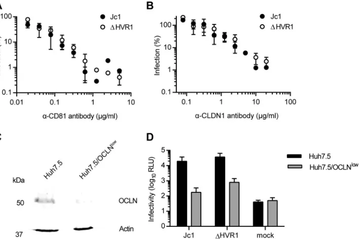
receptor usage and lipoprotein association, we used the Jc1 chimera, which grows to high virus titers in tissue culture (32), and compared Jc1 and a Jc1 mutant lacking HVR1, i.e., the 27 amino-terminal resi-dues of E2 (10,11). Removal of HVR1 did not affect utilization of CD81 or CLDN1 since antibodies specific for these receptors com-peted with HCV infection to a similar extent irrespective of whether cells were challenged with wild-type (wt) Jc1 or Jc1/⌬HVR1 (Fig. 1A
andB). Usage of OCLN was also not altered by deletion of HVR1, since a knockdown of OCLN impaired the infectivity of Jc1 with and without HVR1⬃10-fold (Fig. 1CandD).
Deletion of HVR1 confers viral resistance to SR-BI-targeting
antibodies and compounds.Recently, we reported that HCV
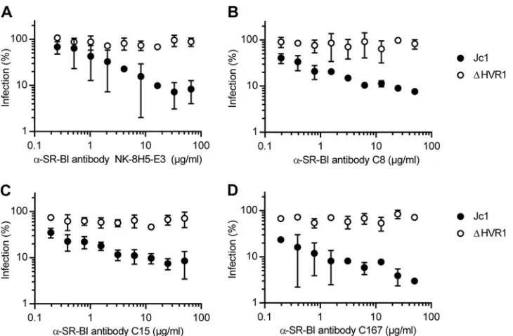
par-ticles lacking HVR1 were no longer neutralized by an SR-BI-spe-cific antiserum that efficiently competed with infection of the pa-rental virus (10). To explore if ⌬HVR1 particles are generally resistant to SR-BI-targeting antibodies and/or compounds, we took advantage of a set of monoclonal antibodies (C8, C15, C167, and NK-8H5-E3) that potently repress infection by HCVin vitro as well as in humanized mice (20,34,39,40). Strikingly, all these antibodies inhibited infection only with wild-type HCV but not with the mutant lacking HVR1 (Fig. 2). In parallel, we investigated the effect of the SR-BI-specific small-molecule inhibitors ITX5061 and ITX1650 on Jc1 and Jc1/⌬HVR1 infectivity (Fig. 3). Admin-istration of ITX5061 to mice or humans increased plasma levels of HDL, probably through inhibiting SR-BI-mediated HDL lipid transfer (41). Moreover, it was shown previously that both ITX compounds inhibit HCVcc infection of human hepatoma cell lines and HCV pseudoparticle (HCVpp) infection of primary hu-man hepatocytes (35). Similar to what we found for all SR-BI-specific antibodies tested, only Jc1 was inhibited by ITX5061 and ITX1650 in a dose-dependent manner, whereas removal of HVR1 rendered the virus resistant to both inhibitors (Fig. 3AandB). Together, these results indicate that deletion of HVR1 ablated or strongly reduced HCV neutralization by molecules targeting SR-BI.
Modulation of the SR-BI expression level affects infectivity
of HCV with and without HVR1.The findings described above in
turn suggested that viruses lacking HVR1 may be independent of SR-BI usage for cell entry. To test this hypothesis and to evaluate possible direct and indirect roles of SR-BI for infection by HCV, we modulated SR-BI expression on the surface of Huh-7.5 cells by overexpression or RNA interference-mediated silencing. SR-BI overexpression was shown to enhance HCV infectivity 4-fold FIG 1HCV particles lacking HVR1 are not affected in their usage of CD81, CLDN1, or OCLN. (A and B) Huh-7.5 cells were inoculated with Jc1 or Jc1/⌬HVR1 (⌬HVR1) luciferase reporter viruses mixed with increasing doses of CD81-specific antibodies (A) or CLDN1-specific antibodies (B) for 72 h at 37°C. Luciferase activity was determined and is expressed relative to infections performed in the absence of antibodies. (C) Expression of OCLN and actin in cell extracts of Huh-7.5 and Huh-7.5/OCLNlowcells determined by Western blotting using specific antibodies. (D) Luciferase activity determined in given cell lines at 72 h
postinoculation with given viruses. Mean values of triplicate measurements, including standard deviations, are given. RLU, relative light units.
on November 7, 2019 by guest
http://jvi.asm.org/
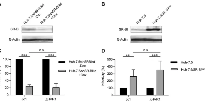
[image:3.585.118.473.67.303.2](21). In agreement with this, Jc1 infection of cells overexpressing SR-BI was increased ca. 2- to 3-fold compared to infection of parental cells expressing endogenous levels of SR-BI (Pvalue of 0.023) (Fig. 4BandD). Unexpectedly, infection of⌬HVR viruses was increased to a similar extent by overexpression of SR-BI (P value of 0.007). This finding suggested that SR-BI surface density influences HCV infectivity in both the presence and absence of HVR1 (Fig. 4BandD). In agreement with this notion, downregu-lation of SR-BI expression using a doxycycline-regulated SR-BI-targeting shRNA (31) reduced permissiveness of these cells to in-fection by both parental HCV Jc1 as well as the Jc1/⌬HVR1 mutant (Pvalues of 0.001 and 0.005 for Jc1 and⌬HVR1, respec-tively) (Fig. 4AandC). Thus, modulation of SR-BI expression clearly modifies the permissiveness of cells to HCV lacking HVR1. Importantly, silencing or overexpression of SR-BI did not influ-ence replication of HCV replicons, indicating that SR-BI abun-dance does not affect RNA replication of HCV (data not shown).
Therefore, we concluded that these viruses still rely on SR-BI tions during their cell entry. Of note, however, these critical func-tions do not seem to be ablated by SR-BI-targeting antibodies or small molecules. Therefore, these results suggest that SR-BI has at least two functions during cell entry. One of these functions can be neutralized by SR-BI-targeting molecules, and it is critical for wild-type HCV only. The other one is important for both viruses but is not inactivated by the SR-BI-specific antibodies and small molecules evaluated here.
Expression of human SR-BI enhances binding of HCVcc
with and without HVR1 to CHO cells.Previous studies indicated
that recombinant soluble E2 devoid of HVR1 exhibits increased CD81 binding yet decreased interactions with SR-BI compared to wild-type E2 comprising this domain (42). Combined with the finding that HCVpp as well as HCVcc lacking HVR1 were not neutralized by SR-BI-targeting antibodies (10,43), we and others speculated that HCV particles lacking HVR1 may no longer bind FIG 2HCV particles devoid of HVR1 are more resistant to neutralization with SR-BI-specific antibodies. Huh-7.5 cells were inoculated with the indicated luciferase reporter viruses in the presence of increasing doses of SR-BI-specific monoclonal antibodies. The efficiency of infection was determined 72 h later by a luciferase reporter assay and is expressed relative to infections performed in the absence of antibodies. Means⫾standard deviations of data from three independent experiments are shown.
FIG 3HCV particles devoid of HVR1 are more resistant to inhibition with SR-BI-specific inhibitors. Huh-7.5 cells were inoculated with the indicated luciferase reporter viruses in the presence of increasing doses of SR-BI-specific inhibitors (A and B) or a control (C). The efficiency of infection was determined 72 h later by a luciferase reporter assay and is expressed relative to efficiencies of infections performed in the absence of antibodies. Means⫾standard deviations of data from three independent experiments are shown.
Role of HVR1 for Receptor and Lipoprotein Interaction
on November 7, 2019 by guest
http://jvi.asm.org/
[image:4.585.112.477.64.303.2] [image:4.585.76.511.581.683.2]and use SR-BI. The results described above, however, indicate that viruses lacking HVR1 are still dependent on SR-BI.
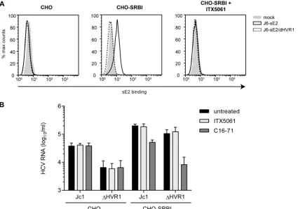
Therefore, we wished to determine if this function of SR-BI was independent of an interaction of the⌬HVR1 virus with SR-BI. To test this, we employed a cell binding assay similar to the one de-scribed previously by Evans et al. (25). Specifically, binding of soluble E2 or HCVcc particles with or without HVR1 to parental Chinese hamster ovary (CHO) cells or to CHO cells ectopically overexpressing SR-BI was determined by a fluorescence-activated cell sorter (FACS) assay or quantitative RT-PCR, respectively. In order to reduce the background from E2 or virus adherence to glycosaminoglycans (GAGs), we used a CHO cell line with defec-tive xylosyltransferase activity for glycan processing, resulting in strongly reduced cell surface-expressed GAGs (44). Ectopic ex-pression of SR-BI at the cell surface of these cells was confirmed by FACS analysis (data not shown). As expected, soluble J6CF-de-rived E2 (note that in Jc1, the structure genes, including E1-E2, are encoded by genotype 2a isolate J6CF [28,45]) bound to CHO cells in an SR-BI-dependent fashion, and this binding was inhibited by ITX5061 (Fig. 5A). In contrast, E2-⌬HVR1 did not bind these cells irrespective of whether SR-BI was expressed (Fig. 5A). Notably, both recombinant proteins bound to CHO cells overexpressing CD81, and this binding was neutralized by the addition of CD81-specific antibodies (data not shown). Thus, we can rule out that E2-⌬HVR1 was globally misfolded. In fact, as reported previously, E2-⌬HVR1 bound to CHO-CD81 cells much more efficiently than did parental soluble E2, confirming the previously reported observations that deletion of HVR1 exposes the CD81 binding site and increases the binding of soluble E2 to cell surface-expressed CD81 (data not shown) (10,42).
When Jc1 and Jc1/⌬HVR1 HCVcc preparations normalized for equal HCV RNA titers were incubated with parental CHO cells, the parental virus bound more efficiently than Jc1/⌬HVR1, as evidenced by ca. 5-fold-higher levels of viral RNA associated
with CHO cells (Fig. 5B). Interestingly, when CHO-SR-BI cells were incubated with these particle types, in both cases, substan-tially higher levels of viral RNA were associated with the cells, indicating that binding of both viruses was facilitated by expres-sion of SR-BI. Notably, under these circumstances, binding of Jc1/⌬HVR1 particles was essentially as effective as the binding of parental Jc1 particles. Strikingly, the addition of SR-BI-specific monoclonal antibody C16-71 repressed both Jc1 as well as Jc1/
⌬HVR1 virus binding to the basal level observed in the absence of SR-BI (Fig. 5B). This finding indicates that the gain of virus bind-ing observed upon ectopic expression of SR-BI in these CHO cells was due to binding of these particles to SR-BI. Notably, addition of ITX5061 at a dose that neutralizes ca. 80 to 90% of Jc1 infection (1
M) (Fig. 3B) reduced neither Jc1 nor Jc1/⌬HVR1 binding to
CHO-SR-BI cells (Fig. 5B). Collectively, these observations con-firm that HVR1 is critical for binding of soluble E2 to cell surface-expressed SR-BI. However, this domain is dispensable for binding of HCVcc particles to CHO-SR-BI cells. Therefore, features other than HVR1 likely mediate SR-BI-dependent cell surface attach-ment of HCV particles in this cellular model. Moreover, virus particle binding, unlike binding of soluble E2, is not inhibited by ITX5061, further confirming a different mode of SR-BI interac-tion between soluble E2 and virus particles. Finally, since viruses lacking HVR1 displayed lower levels of cell surface binding to parental CHO cells, this viral domain modulates interactions with the cell surface in the absence of SR-BI.
HCV particles lacking HVR1 display reduced ApoE and LDL-R dependence in cell surface attachment to CHO cells.
Since HCV particles associate with lipoproteins, and as⌬HVR1 particles have an altered buoyant density (10,11), we speculated that different particle compositions with regard to lipoproteins and/or lipids may be responsible for the lower level of binding of
⌬HVR1 particles to parental CHO cells. Moreover, recent evi-dence indicates that virus-associated ApoE facilitates cell surface FIG 4HCV infection of hepatoma cells with down- or upregulated SR-BI. (A and B) SR-BI expression levels in Huh-7.5/shSRBIkd cells pretreated with or without doxycycline (Dox) for 96 h (A) and Huh-7.5 and Huh-7.5/SR-BIhighcells (B) were determined by Western blotting. (C) Jc1 or Jc1/⌬HVR1 (⌬HVR1) was
added to Huh-7.5/shSRBIkd cells pretreated or not with doxycycline, and HCV infection was determined at 72 h postinfection (hpi) by luciferase reporter activity. Infectivity in the absence of doxycycline is set to 100%. (D) Jc1 or Jc1/⌬HVR1 (⌬HVR1) was added to Huh-7.5 or Huh-7.5/SR-BIhighcells, and HCV
infection was determined at 72 h postinfection by luciferase reporter activity. Infection of Huh-7.5 cells is set to 100%. Means⫾standard deviations of data from three independent experiments are shown. n.s., not significant.
on November 7, 2019 by guest
http://jvi.asm.org/
[image:5.585.93.496.64.263.2]attachment through binding to the low-density lipoprotein recep-tor (LDL-R) (46) and cell surface-resident GAGs (47). Thus, we repeated the binding assay in the presence of two different ApoE-specific antibodies and an anti-LDL-R antibody to assess the role of ApoE and LDL-R in cell surface binding of parental Jc1 and Jc1/⌬HVR1 particles (Fig. 6). Jc1 binding to these cells was indeed reduced with comparable efficiencies by polyclonal anti-ApoE an-tibody Cb (ca. 3-fold;Pvalue of 0.015) and the LDL-R anti-body (ca. 3-fold; Pvalue of 0.072), suggesting that ApoE and LDL-R were involved in the binding of these viruses to naive CHO cells, possibly through a direct interaction between virus-associ-ated ApoE and cell surface-resident LDL-R. Of note, monoclonal anti-ApoE antibody Pg did not significantly block binding of the virus variants to CHO or CHO-SR-BI cells (Pvalues of 0.591 and 0.863, respectively), probably because the antibody recognizes a different epitope than the Cb antibody. Interestingly, binding of Jc1/⌬HVR1 particles to parental CHO cells was not significantly repressed by the ApoE- or the LDL-R-specific antibodies (Pvalues of 0.439 and 0.547, respectively), suggesting that these viruses lacking HVR1 display reduced levels of ApoE- and LDL-R-depen-dent cell surface binding. As described above, both Jc1 and Jc1/
⌬HVR1 particles showed increased binding to SR-BI-expressing CHO cells (Fig. 5and6). Interestingly, cell surface binding of HCV particles with or without HVR1 to CHO-SR-BI cells was poorly neutralized by ApoE- and LDL-R-specific antibodies, and
there was no statistically significant difference in binding in the presence or absence of ApoE- or LDL-R-specific antibodies. Therefore, we conclude that SR-BI binding is dominant over at-tachment via ApoE and LDL-R in this cell system.
FIG 5Attachment of HCVcc particles and soluble E2 to CHO cells expressing SR-BI or not. (A) CHO cells expressing SR-BI (CHO-SRBI) or not (CHO) were incubated with soluble E2 (sE2)-J6 or soluble E2-J6/⌬HVR1 for 2 h at room temperature. Binding was performed in the presence or absence of SR-BI-inhibiting compound ITX5061 (1M). Bound soluble E2 was detected by using an anti-His antibody and FACS measurements. (B) Equal amounts of Jc1 and Jc1/⌬HVR1 (⌬HVR1) were incubated with CHO cells or CHO-SRBI cells for 2 h at 37°C. Incubation was performed in either the presence or absence of SR-BI-inhibiting compound ITX5061 (1M) or monoclonal anti-SRBI antibody C16-71 (5g/ml). The number of bound HCVcc particles was measured by quantifying HCV RNA genomes in lysates of virus-incubated cells. Means⫾standard deviations of data from three independent experiments are shown.
FIG 6ApoE- and LDL-R-mediated attachment of HCVcc particles to CHO cells in the presence or absence of SR-BI. Equal amounts of Jc1 and⌬HVR1 were incubated with CHO cells expressing SR-BI (CHO-SR-BI) or a vector control (CHO) for 2 h at 37°C. Infection was performed in either the presence or absence of anti-ApoE antibodies from Calbiochem (Cb) (1/40 dilution) or Progen (Pg) (2g/ml) or an anti-LDL-R antibody (5g/ml). After washing, the number of bound HCVcc particles was assessed by determining the num-ber copies of viral RNA in lysates of virus-incubated cells. Means⫾standard deviations of data from three independent experiments are shown. Statistical significance was tested relative to untreated cells.
Role of HVR1 for Receptor and Lipoprotein Interaction
on November 7, 2019 by guest
http://jvi.asm.org/
[image:6.585.83.508.67.366.2] [image:6.585.308.532.508.630.2]Combined with the previously reported observation that
⌬HVR1 particles display a different buoyant density than wild-type HCV particles (10, 11), these results suggest that⌬HVR1 particles may incorporate different levels (or types) of lipids or lipoproteins. This in turn may alter usage of cellular lipoprotein receptors (LDL-R and SR-BI) during cell entry.
Deletion of HVR1 does not change the abundance of ApoE in
HCV lipoviroparticles.To test the two hypotheses mentioned
above and to analyze incorporation of ApoE into Jc1 and Jc1/
⌬HVR1 particles, we took advantage of a recently described virus variant carrying a Flag tag at the N terminus of E2 (Jc1/FlagE2) (16,48). In parallel, we created a novel Jc1/⌬HVR1 variant com-prising a Flag epitope at the N terminus of the truncated E2 pro-tein (Jc1/FlagE2/⌬HVR1). Importantly, insertion of the tag into both viruses was well tolerated (16,48) and did not decrease in-fectivity of the particles (data not shown). Moreover, similar to the untagged viruses, Flag-tagged⌬HVR1 particles also displayed an altered distribution in density gradients (data not shown).
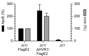
Using Flag-specific antibodies, we affinity purified Jc1/FlagE2 and Jc1/FlagE2/⌬HVR1 particles and determined the amounts of ApoE and core in the purified viruses by ELISA. Although Jc1/FlagE2 and Jc1/⌬HVR1/FlagE2 virus preparations were normalized to equal core protein levels before affinity purification, Jc1/⌬HVR1/FlagE2 parti-cles were consistently more efficiently precipitated, probably because they bind more efficiently to anti-Flag antibodies and therefore are captured more readily (Fig. 7). Consequently, about two times more core and two times more ApoE were detected in the precipitation of Jc1/⌬HVR1/FlagE2 particles than in the precipitation of parental Jc1/ FlagE2. Importantly, the amount of coprecipitated ApoE was com-mensurately increased for the Jc1/FlagE2/⌬HVR1 particles com-pared to the parental viruses. Thus, in conclusion, these experiments indicate that deletion of HVR1 does not grossly affect the amount of ApoE incorporated into HCV particles.
HCV particles with or without HVR1 are differentially
rec-ognized and neutralized by ApoE-specific antibodies.Next, we
assessed whether the differences between Jc1 and Jc1/⌬HVR1 bind-ing to CHO cells correlated with divergent properties of virus-associ-ated ApoE. To this end, we utilized the anti-ApoE-specific antibodies described above to neutralize infection of parental Jc1 and Jc1/
⌬HVR1 particles. Interestingly, although polyclonal ApoE anti-body Cb prevented attachment of Jc1 particles to parental CHO cells more efficiently than binding of Jc1/⌬HVR1 viruses, in the infection assay, Jc1/⌬HVR1 was more efficiently neutralized. Indeed, smaller amounts of the antibodies were required for a greater reduction of
infection (Fig. 8A). Also, monoclonal anti-ApoE antibody Pg, which had not efficiently inhibited cell surface binding of these viruses to parental CHO cells (Fig. 6), interfered with infection of both viruses in a dose-dependent fashion (Fig. 8B). Therefore, these data indicate that the inhibition of cell surface binding of HCV particles to CHO cells by these antibodies does not directly correlate with their ability to neutralize infection of Huh-7.5 cells by HCV particles. Moreover, these results provide additional evidence that Jc1 and Jc1/⌬HVR1 particles may be differentially targeted by ApoE-specific antibodies
(Fig. 6and8A).
To further corroborate this notion, we investigated the binding of these ApoE-specific antibodies to Jc1 and Jc1/⌬HVR1 particles. To this end, equal amounts of these particles (normalized for equal viral RNA levels) were incubated with different ApoE-spe-cific antibodies or isotype-matched control antibodies. Subse-quently, antibody complexes were precipitated by using protein A beads, and the amount of coprecipitating viral RNA was deter-mined by quantitative RT-PCR (Fig. 8CandD). Moreover, the amount of residual HCV infectivity remaining in the supernatant was determined (Fig. 8EandF). Using this approach, we observed comparable coprecipitations of viral RNA between Jc1 and Jc1/
⌬HVR1 in the case of polyclonal anti-ApoE antibody Cb particles
(Fig. 8C). Likewise, these polyclonal antibodies depleted viral
in-fectivity to similar extents when incubated with virus-containing supernatants (Fig. 8E). These results therefore suggest that despite the differential neutralization of Jc1 and Jc1/⌬HVR1 particles, there is no evidence for differential binding of virus particles by these antibodies. In contrast, the monoclonal ApoE Pg anti-body neutralized these virus particle types with a similar efficacy but precipitated slightly larger quantities of viral RNA and infec-tious virus in the case of Jc1/⌬HVR1 than in the case of Jc1 (Fig. 8DandF). Collectively, these results further support the notion that ApoE associated with Jc1/⌬HVR1 particles is differentially targeted by ApoE-specific antibodies compared to ApoE on Jc1 viruses. Thus, it is conceivable that ApoE, although incorporated into both particles types to similar levels, may display different epitopes and/or adopt a slightly different conformation between Jc1 and Jc1/⌬HVR1 particles, thus resulting in differential recog-nition and neutralization by distinct ApoE-targeting antibodies.
DISCUSSION
In this study, we explored the role of HVR1 during HCV cell entry, thereby focusing on the interaction of HCV with entry receptors and lipoproteins. When analyzing inhibition of infection by HCV with or without HVR1 by antibodies specific for CD81 or CLDN1 or by si-lencing of OCLN expression, we observed similar reductions of virus infection between these virus types (Fig. 1). Since these treatments decrease the cell surface availability of targeted entry factors, these results indicate that HCV particles with or without HVR1 similarly depend on the availability of accessible CD81, CLDN1, and OCLN molecules for productive infection of Huh-7.5 cells. Therefore, dele-tion of HVR1 apparently does not modulate the requirement for utilization of these three critical HCV entry factors. In contrast, our results indicate that HVR1 is important for certain but not all func-tions that SR-BI plays during the entry process. Moreover, we provide evidence suggesting that deletion of HVR1 changes the properties of ApoE on the virus particle without affecting the amount of ApoE incorporated into the virion (Fig. 6to8).
We previously reported that HCV particles lacking HVR1, un-like wild-type particles, were resistant to neutralization by a poly-FIG 7ApoE incorporation into HCV particles is not altered by deletion of
HVR1. The amounts of ApoE and core in affinity-purified Jc1/FlagE2 or Jc1/ ⌬HVR1/FlagE2 particles were assessed by ELISA. The amounts of ApoE and core in affinity purified Jc1/FlagE2 particles are set to 100%. Means⫾standard deviations of data from three independent experiments are shown.
on November 7, 2019 by guest
http://jvi.asm.org/
[image:7.585.77.252.63.174.2]clonal SR-B1-specific antiserum, suggesting that this viral surface domain modulated SR-B1 dependency (10). In this study, we compared the neutralization of these viruses by a panel of different SR-BI-specific monoclonal antibodies as well as two small mole-cules (ITX5061 and ITX7650) targeting this HCV entry factor. Of note, the monoclonal anti-SR-BI antibodies have different modes of action. While the mechanism by which the C8 and C15 anti-bodies preclude infection by wild-type HCV has to our knowledge not been explored, the NK-8H5-E3 antibody was reported to in-terfere with a postbinding step during HCV entry without inter-fering with soluble envelope glycoprotein E2 or HCVcc particle binding to the target cell surface (34). In contrast, the C167 anti-body, recognizing a conformational epitope on the receptor, was shown to inhibit binding of E2 and HDL to SR-BI and to interfere with SR-BI-mediated lipid transfer (20). In addition, the two
SR-BI specific inhibitors ITX7650 and ITX5061 both interfere with HDL-mediated lipid transfer of SR-BI (35) and block the interaction of soluble E2 with SR-BI. Despite their different modes of action, these antibodies and small molecules targeting SR-BI consistently inhibited Jc1 infection in a dose-dependent fashion, whereas they did not decrease infection by the Jc1/⌬HVR1 variant
(Fig. 2and3). These results therefore extend and refine
observa-tions previously reported by us and others (10,49) and indicate that HVR1 modulates HCV utilization of SR-BI and that Jc1 par-ticles lacking this domain display modified SR-BI dependence during cell entry, as they are no longer neutralized by these SR-BI-targeting molecules.
These experiments involving SR-BI binding compounds sug-gested that Jc1/⌬HVR1 particles may be completely independent of SR-BI usage for infection. To test this, we next modulated SR-BI FIG 8ApoE exposure on HCV particles with or without HVR1. (A and B) Huh-7.5 cells were inoculated with the indicated luciferase reporter viruses in the presence of increasing doses of a polyclonal anti-ApoE antibody (Cb) (A) and a monoclonal ApoE-specific antibody (Pg) (B). The efficiency of infection was determined 72 h later by a luciferase reporter assay and is expressed relative to infections performed in the absence of antibodies. Results of one representative experiment out of five independent repetitions are shown. Statistical analysis was performed on the five independent repetitions, and the statistical significances of the differences between Jc1 and⌬HVR1 were tested with a pairedttest. (C and D) Equal amounts of Jc1 and Jc1/⌬HVR1 particles were subjected to an immunocapture assay by using Cb (C) or Pg (D) anti-ApoE antibodies. HCV RNA contained in each captured sample was quantified by qRT-PCR. RNA copy numbers captured by control IgGs are set to 100%. (E and F) The infectivity of the postcapture supernatant of the samples shown in panels C and D was determined by a TCID50assay. Data points and means of data from seven to nine independent experiments are shown.
Role of HVR1 for Receptor and Lipoprotein Interaction
on November 7, 2019 by guest
http://jvi.asm.org/
[image:8.585.136.452.66.465.2]cell surface levels by overexpression or shRNA-mediated silenc-ing. Using these approaches, we noted that the infection efficien-cies of both wild-type Jc1 and the variant lacking HVR1 were significantly increased by SR-BI overexpression and decreased by SR-BI downregulation (Fig. 4). Therefore, we concluded that in-fection by both viruses was similarly dependent on SR-BI surface expression. This indicates that deletion of HVR1 does not ablate SR-BI dependence of HCV cell entry. Nevertheless, these results support a model where wild-type Jc1 particles,⌬HVR1 virions, require an additional SR-BI function(s) that is inhibited by the SR-BI binding molecules tested by us.
Previous reports have established that HVR1 is critical for binding of soluble E2 to SR-BI (42), suggesting that viruses lacking HVR1 may no longer interact with this entry factor. It was con-ceivable that viruses lacking HVR1 could indirectly rely on SR-BI for infection without direct binding of E2 being involved. There-fore, we explored whether deletion of HVR1 abrogated binding of HCVcc particles to this entry factor. First, we confirmed that de-letion of HVR1 in the context of the J6CF (GT2a) isolate, as for H77 (GT1a)-derived E2 (42), prevented the interaction of soluble E2 with SR-BI (Fig. 5A). Next, we utilized an established HCVcc cell binding assay that involves CHO cells that lack endogenous expression of human HCV entry factors. Using these cells, Evans et al. recently reported that HCVcc particles attach to the cell surface in an SR-BI-dependent fashion (25). Accordingly, we ob-served ca. 5-fold-increased binding of Jc1 particles to CHO-SR-BI cells compared to parental cells lacking SR-BI expression (Fig. 5B). Moreover, this additional binding was inhibited by SR-BI-specific antibody C16-71 (Fig. 5B). In contrast, ITX5061 was not able to inhibit this SR-BI-dependent binding, suggesting that the mechanism by which this molecule interferes with HCV infection is not a block of virus attachment to SR-BI. Strikingly, cell surface binding of Jc1/⌬HVR1 particles to these CHO cells was increased
⬎10-fold by overexpression of SR-BI, and this additional viral cell binding in the presence of SR-BI was fully ablated by the addition of the SR-BI-specific monoclonal antibody (Fig. 5B). These data indicate that HCVcc particles lacking HVR1, unlike soluble E2 lacking this domain, attach to the cell surface in an SR-BI-depen-dent manner. Therefore, viral domains other than HVR1 and/or host constituents of HCV particles are important for the interac-tion with SR-BI. Finally, we observed that wild-type Jc1 particles bound ca. 5-fold more efficiently to parental CHO cells than did Jc1/⌬HVR1 particles (Fig. 5B). Since these cells lack human HCV cell entry factors and are unable to produce GAGs, we speculate that this may be due to increased interactions of wild-type parti-cles with hamster cell-derived cell surface proteins. At present, we do not know which cellular factor is responsible for the differential binding of Jc1 and Jc1/⌬HVR1 particles to the surface of CHO cells. However, recent reports indicate that HCV particles lacking HVR1 have a higher buoyant density than wild-type particles (10,
11) and that virus-associated ApoE facilitates attachment by way of interaction with LDL-R (46). Moreover, these CHO cells do express LDL-R (50). Therefore, we tested the influence of different ApoE-specific antibodies as well as of an antibody targeting LDL-R on the binding of HCVs to CHO cells. One of the two ApoE-specific antibodies as well as the LDL-R-targeting antibody significantly decreased cell surface attachment of wild-type Jc1 particles to parental CHO cells, suggesting that an interplay be-tween virus-resident ApoE and cell surface-expressed LDL-R measurably contributes to attachment of these viruses to parental
CHO cells. We do not know why the monoclonal anti-ApoE Pg antibody did not interfere with HCV binding, but this may be because the epitope targeted by this antibody is not involved in ApoE binding to LDL-R. Interestingly, binding of Jc1 wild-type particles to parental CHO cells was repressed by the polyclonal anti-ApoE Cb antibodies and the LDL-R-specific antibody ap-proximately to the level of binding achieved by Jc1/⌬HVR1 par-ticles in the absence of antibodies. Moreover, addition of these antibodies to Jc1/⌬HVR1 particle binding assay mixtures only weakly reduced attachment of these viruses (⬍2-fold). Finally, once SR-BI was overexpressed, inhibition of virus binding by the ApoE- and LDL-R-specific antibodies was less pronounced. Taken together, these observations are consistent with a model where virus binding to SR-BI occurs for viruses with and without HVR1 and may be the dominant mode of cell surface attachment. Moreover, the ApoE-specific antibodies used here do not effi-ciently block this interaction. Finally, wild-type Jc1 particles at-tach to parental CHO cells in an ApoE- and LDL-R-dependent fashion. Of note, our binding assays were conducted with CHO cells that are unable to produce GAGs. Therefore, this system does not reflect the contribution of protein interactions with GAGs, which have been shown to additionally contribute to virus attach-ment to human hepatocytes (29,47,51,52).
It is currently unclear why Jc1/⌬HVR1 particles show reduced ApoE–LDL-R-dependent binding to parental CHO cells com-pared to parental Jc1 particles. However, it is possible that either HVR1 may contribute to LDL-R binding and/or the abundances or the properties of ApoE between wild-type and HVR1-deleted viruses may differ, thus causing differential attachment through LDL-R. Clearly, more work will be necessary to prove, or disprove, a role of HVR1 in binding to LDL-R. However, given that HVR1-deleted particles display an aberrant distribution in density gradi-ents that is characterized by reduced numbers of particles with low density (11,53), we speculated that aberrant loading of these par-ticles with lipids and/or lipoproteins may be responsible.
To obtain evidence supporting this hypothesis, we compared the abundances and the properties of virus particle-associated ApoE of parental Jc1 and Jc1/⌬HVR1 particles. Using Flag-E2-tagged versions of both viral variants, we affinity purified equal numbers of these particles using Flag-specific antibodies. Impor-tantly, insertion of the Flag epitope at the N terminus of E2 is well tolerated (16,48) and did not alter the differential distribution of these particle types in density gradients (data not shown). While we consistently observed greater affinity purification of the Jc1/ FlagE2/⌬HVR1 virus than of the parental virus with HVR1, we coprecipitated almost identical relative amounts of ApoE protein
(Fig. 7). Therefore, these results exclude gross differences in the
abundances of virus-incorporated ApoE between these particle types. Finally, we compared binding and neutralization of Jc1 vi-ruses in the presence and in the absence of HVR1 by the above-mentioned mono- or polyclonal ApoE-specific antibodies. Using this approach, we observed striking differences between these par-ticle types with regard to their interactions with these ApoE-spe-cific antibodies. First, the polyclonal anti-ApoE Cb antibody neu-tralized Jc1/⌬HVR1 particles more efficiently than parental Jc1 particles, although it bound both particle types with comparable efficiencies. Second, the monoclonal anti-ApoE Pg antibody neu-tralized both particle types to similar levels; however, it bound viruses lacking HVR1 slightly more effectively (Fig. 8). Based on these findings, we conclude that the ability of these ApoE-specific
on November 7, 2019 by guest
http://jvi.asm.org/
antibodies to bind HCV particles does not directly correlate with their HCV neutralization properties. Moreover, the ability of these antibodies to prevent HCV particle binding to CHO cells does not predict their capability to neutralize infection. The latter finding may suggest that, in the CHO cell binding assay, HCV associates primarily via ApoE-independent routes and that ApoE may exert postattachment functions during cell entry. Finally, these results provide evidence supporting the conclusion that Jc1/
⌬HVR1 particles incorporate ApoE with differential conforma-tion and/or epitope exposure compared to parental Jc1 particles. Moreover, different amounts of lipids present in wild-type com-pared with HVR1-deleted viruses may result in differential epitope exposures of ApoE, thus explaining the different inter-plays of these particle types with these antibodies. Interestingly, the conformation of ApoE is known to be influenced by the lipo-protein and lipid composition on a given lipolipo-protein particle, and a large body of literature supports the assumption that conforma-tional opening of the N-terminal domain of ApoE modulates its receptor binding activity (summarized in reference54). Finally, it is possible that an altered conformation of virus-associated ApoE in HVR1-deleted particles is in part responsible for the reduced infectiousness of these viruses (10). Careful lipid and protein pro-filing of Jc1/⌬HVR1 particles may help to validate this assump-tion.
Collectively, our data indicate that HVR1 contributes to effi-cient cell surface attachment of HCV Jc1 particles. This function of HVR1 may be linked to its involvement in the incorporation of ApoE, a natural ligand of LDL-R. Our data support a model where HVR1 modulates the conformation/epitope exposure of virus-resident ApoE but not its total abundance on the virus particle. These altered properties of ApoE, which we documented by the differential interplays between these viruses and ApoE-specific antibodies, could influence virus attachment through LDL-R and possibly postbinding entry steps facilitated by ApoE. As a conse-quence, they may be responsible for the observed lower level of infectiousness of Jc1/⌬HVR1 particles. Finally, we show that vi-ruses lacking HVR1 remain SR-BI dependent in infection, high-lighting the important role of SR-BI for HCV cell entry. Notably, SR-BI-targeting antibodies and small molecules inhibit wild-type but not HVR1-deleted Jc1 in vitro. This finding confirms that SR-BI fulfills additional “druggable” functions that are specifically required by wild-type HCV and that these functions are targeted by the antibodies and small molecules described above. Whether viral variants independent of these SR-BI functions and, thus, pos-sibly resistant to these SR-BI-targeting modalities can arise re-mains to be shown.
ACKNOWLEDGMENTS
We are grateful to Takaji Wakita and Jens Bukh for JFH1 and J6CF iso-lates, respectively; Charles Rice for Huh-7.5 cells and the E9E10 antibod-ies; Sandra Ciesek and Thomas von Hahn for Huh-7.5/OCLNlow cells; and Flossie Wong-Staal for providing the ITX compounds. We also thank all members of the Institute of Experimental Virology for helpful sugges-tions and discussions.
This work was supported by a grant from the Deutsche Forschungs-gemeinschaft (SFB 900, project A6) and by a grant from the Helmholtz Association, SO-024, to T.P. T.F.B. acknowledges funding through the ANRS and Laboratoire d’Excellence LabEx HEPSYS (Investissement d’Avenir; ANR-10-LAB-28).
REFERENCES
1.Lindenbach BD, Thiel HJ, Rice CM.2007. Flaviviridae: the viruses and their replication, p 1101–1152.InKnipe DM, Howley PM, Griffin DE, Lamb RA, Martin MA, Roizman B, Straus SE (ed), Fields virology, 5th ed. Lippincott Williams & Wilkins, Philadelphia, PA.
2.Brown RS.2005. Hepatitis C and liver transplantation. Nature436:973– 978.http://dx.doi.org/10.1038/nature04083.
3.Simmonds P, Bukh J, Combet C, Deleage G, Enomoto N, Feinstone S, Halfon P, Inchauspe G, Kuiken C, Maertens G, Mizokami M, Murphy DG, Okamoto H, Pawlotsky JM, Penin F, Sablon E, Shin IT, Stuyver LJ, Thiel HJ, Viazov S, Weiner AJ, Widell A.2005. Consensus proposals for a unified system of nomenclature of hepatitis C virus genotypes. Hepatol-ogy42:962–973.http://dx.doi.org/10.1002/hep.20819.
4.Kato N, Sekiya H, Ootsuyama Y, Nakazawa T, Hijikata M, Ohkoshi S, Shimotohno K.1993. Humoral immune response to hypervariable region 1 of the putative envelope glycoprotein (gp70) of hepatitis C virus. J. Virol. 67:3923–3930.
5.Kato N, Ootsuyama Y, Sekiya H, Ohkoshi S, Nakazawa T, Hijikata M, Shimotohno K.1994. Genetic drift in hypervariable region 1 of the viral genome in persistent hepatitis C virus infection. J. Virol.68:4776 – 4784. 6.Taniguchi S, Okamoto H, Sakamoto M, Kojima M, Tsuda F, Tanaka T,
Munekata E, Muchmore EE, Peterson DA, Mishiro S.1993. A structur-ally flexible and antigenicstructur-ally variable N-terminal domain of the hepatitis C virus E2/NS1 protein: implication for an escape from antibody. Virol-ogy195:297–301.http://dx.doi.org/10.1006/viro.1993.1378.
7.Weiner AJ, Geysen HM, Christopherson C, Hall JE, Mason TJ, Saracco G, Bonino F, Crawford K, Marion CD, Crawford KA.1992. Evidence for immune selection of hepatitis C virus (HCV) putative envelope glycopro-tein variants: potential role in chronic HCV infections. Proc. Natl. Acad. Sci. U. S. A.89:3468 –3472.http://dx.doi.org/10.1073/pnas.89.8.3468. 8.Kumar U, Monjardino J, Thomas HC.1994. Hypervariable region of
hepatitis C virus envelope glycoprotein (E2/NS1) in an agammaglobuline-mic patient. Gastroenterology106:1072–1075.
9.Penin F, Combet C, Germanidis G, Frainais PO, Deleage G, Pawlotsky JM.2001. Conservation of the conformation and positive charges of hep-atitis C virus E2 envelope glycoprotein hypervariable region 1 points to a role in cell attachment. J. Virol.75:5703–5710.http://dx.doi.org/10.1128 /JVI.75.12.5703-5710.2001.
10. Bankwitz D, Steinmann E, Bitzegeio J, Ciesek S, Friesland M, Herrmann E, Zeisel MB, Baumert TF, Keck ZY, Foung SK, Pecheur EI, Pietschmann T.2010. Hepatitis C virus hypervariable region 1 modulates receptor interactions, conceals the CD81 binding site, and protects con-served neutralizing epitopes. J. Virol.84:5751–5763.http://dx.doi.org/10 .1128/JVI.02200-09.
11. Prentoe J, Jensen TB, Meuleman P, Serre SB, Scheel TK, Leroux-Roels G, Gottwein JM, Bukh J.2011. Hypervariable region 1 differentially impacts viability of hepatitis C virus strains of genotypes 1 to 6 and impairs virus neutralization. J. Virol. 85:2224 –2234. http://dx.doi.org/10.1128 /JVI.01594-10.
12. Chang KS, Jiang J, Cai Z, Luo G.2007. Human apolipoprotein E is required for infectivity and production of hepatitis C virus in cell culture. J. Virol.81:13783–13793.http://dx.doi.org/10.1128/JVI.01091-07. 13. Huang H, Sun F, Owen DM, Li W, Chen Y, Gale M, Jr, Ye J.2007.
Hepatitis C virus production by human hepatocytes dependent on assem-bly and secretion of very low-density lipoproteins. Proc. Natl. Acad. Sci. U. S. A.104:5848 –5853.http://dx.doi.org/10.1073/pnas.0700760104. 14. Gastaminza P, Cheng G, Wieland S, Zhong J, Liao W, Chisari FV.2008.
Cellular determinants of hepatitis C virus assembly, maturation, degrada-tion, and secretion. J. Virol.82:2120 –2129.http://dx.doi.org/10.1128/JVI .02053-07.
15. Felmlee DJ, Hafirassou ML, Lefevre M, Baumert TF, Schuster C.2013. Hepatitis C virus, cholesterol and lipoproteins—impact for the viral life cycle and pathogenesis of liver disease. Viruses5:1292–1324.http://dx.doi .org/10.3390/v5051292.
16. Merz A, Long G, Hiet MS, Brugger B, Chlanda P, Andre P, Wieland F, Krijnse-Locker J, Bartenschlager R.2011. Biochemical and morpholog-ical properties of hepatitis C virus particles and determination of their lipidome. J. Biol. Chem.286:3018 –3032.http://dx.doi.org/10.1074/jbc .M110.175018.
17. Albecka A, Belouzard S, Op de Beeck A, Descamps V, Goueslain L, Bertrand-Michel J, Terce F, Duverlie G, Rouille Y, Dubuisson J.2012. Role of HVR1 for Receptor and Lipoprotein Interaction
on November 7, 2019 by guest
http://jvi.asm.org/
Role of low-density lipoprotein receptor in the hepatitis C virus life cycle. Hepatology55:998 –1007.http://dx.doi.org/10.1002/hep.25501. 18. Sainz B, Jr, Barretto N, Martin DN, Hiraga N, Imamura M, Hussain S,
Marsh KA, Yu X, Chayama K, Alrefai WA, Uprichard SL.2012. Iden-tification of the Niemann-Pick C1-like 1 cholesterol absorption receptor as a new hepatitis C virus entry factor. Nat. Med.18:281–285.http://dx .doi.org/10.1038/nm.2581.
19. Zeisel MB, Koutsoudakis G, Schnober EK, Haberstroh A, Blum HE, Cosset FL, Wakita T, Jaeck D, Doffoel M, Royer C, Soulier E, Schvoerer E, Schuster C, Stoll-Keller F, Bartenschlager R, Pietschmann T, Barth H, Baumert TF.2007. Scavenger receptor class B type I is a key host factor for hepatitis C virus infection required for an entry step closely linked to CD81. Hepatology46:1722–1731.http://dx.doi.org/10.1002/hep.21994. 20. Catanese MT, Graziani R, von Hahn T, Moreau M, Huby T, Paonessa
G, Santini C, Luzzago A, Rice CM, Cortese R, Vitelli A, Nicosia A.2007. High-avidity monoclonal antibodies against the human scavenger class B type I receptor efficiently block hepatitis C virus infection in the presence of high-density lipoprotein. J. Virol.81:8063– 8071.http://dx.doi.org/10 .1128/JVI.00193-07.
21. Grove J, Huby T, Stamataki Z, Vanwolleghem T, Meuleman P, Farqu-har M, Schwarz A, Moreau M, Owen JS, Leroux-Roels G, Balfe P, McKeating JA. 2007. Scavenger receptor BI and BII expression levels modulate hepatitis C virus infectivity. J. Virol.81:3162–3169.http://dx .doi.org/10.1128/JVI.02356-06.
22. Dorner M, Rice CM, Ploss A.2013. Study of hepatitis C virus entry in genetically humanized mice. Methods59:249 –257.http://dx.doi.org/10 .1016/j.ymeth.2012.05.010.
23. Dao Thi VL, Granier C, Zeisel MB, Guerin M, Mancip J, Granio O, Penin F, Lavillette D, Bartenschlager R, Baumert TF, Cosset FL, Dreux M.2012. Characterization of hepatitis C virus particle subpopulations reveals multiple usage of the scavenger receptor BI for entry steps. J. Biol. Chem.287:31242–31257.http://dx.doi.org/10.1074/jbc.M112.365924. 24. Maillard P, Huby T, Andreo U, Moreau M, Chapman J, Budkowska A.
2006. The interaction of natural hepatitis C virus with human scavenger receptor SR-BI/ClaI is mediated by ApoB-containing lipoproteins. FASEB J.20:735–737.http://dx.doi.org/10.1096/fj.05-4728fje.
25. Evans MJ, von Hahn T, Tscherne DM, Syder AJ, Panis M, Wolk B, Hatziioannou T, McKeating JA, Bieniasz PD, Rice CM.2007. Claudin-1 is a hepatitis C virus co-receptor required for a late step in entry. Nature 446:801– 805.http://dx.doi.org/10.1038/nature05654.
26. Ploss A, Evans MJ, Gaysinskaya VA, Panis M, You H, de Jong YP, Rice CM.2009. Human occludin is a hepatitis C virus entry factor required for infection of mouse cells. Nature457:882– 886.http://dx.doi.org/10.1038 /nature07684.
27. Pileri P, Uematsu Y, Campagnoli S, Galli G, Falugi F, Petracca R, Weiner AJ, Houghton M, Rosa D, Grandi G, Abrignani S.1998. Binding of hepatitis C virus to CD81. Science282:938 –941.http://dx.doi.org/10 .1126/science.282.5390.938.
28. Pietschmann T, Kaul A, Koutsoudakis G, Shavinskaya A, Kallis S, Steinmann E, Abid K, Negro F, Dreux M, Cosset FL, Bartenschlager R. 2006. Construction and characterization of infectious intragenotypic and intergenotypic hepatitis C virus chimeras. Proc. Natl. Acad. Sci. U. S. A. 103:7408 –7413.http://dx.doi.org/10.1073/pnas.0504877103.
29. Koutsoudakis G, Kaul A, Steinmann E, Kallis S, Lohmann V, Piet-schmann T, Bartenschlager R.2006. Characterization of the early steps of hepatitis C virus infection by using luciferase reporter viruses. J. Virol. 80:5308 –5320.http://dx.doi.org/10.1128/JVI.02460-05.
30. Blight KJ, McKeating JA, Rice CM.2002. Highly permissive cell lines for subgenomic and genomic hepatitis C virus RNA replication. J. Virol.76: 13001–13014.http://dx.doi.org/10.1128/JVI.76.24.13001-13014.2002. 31. Catanese MT, Ansuini H, Graziani R, Huby T, Moreau M, Ball JK,
Paonessa G, Rice CM, Cortese R, Vitelli A, Nicosia A.2010. Role of scavenger receptor class B type I in hepatitis C virus entry: kinetics and molecular determinants. J. Virol.84:34 – 43.http://dx.doi.org/10.1128 /JVI.02199-08.
32. Ciesek S, Westhaus S, Wicht M, Wappler I, Henschen S, Sarrazin C, Hamdi N, Abdelaziz AI, Strassburg CP, Wedemeyer H, Manns MP, Pietschmann T, von Hahn T.2011. Impact of intra- and interspecies variation of occludin on its function as coreceptor for authentic hepatitis C virus particles. J. Virol.85:7613–7621.http://dx.doi.org/10.1128/JVI .00212-11.
33. Fofana I, Krieger SE, Grunert F, Glauben S, Xiao F, Fafi-Kremer S, Soulier E, Royer C, Thumann C, Mee CJ, McKeating JA, Dragic T,
Pessaux P, Stoll-Keller F, Schuster C, Thompson J, Baumert TF.2010. Monoclonal anti-claudin 1 antibodies prevent hepatitis C virus infection of primary human hepatocytes. Gastroenterology139:953–964.http://dx .doi.org/10.1053/j.gastro.2010.05.073.
34. Zahid MN, Turek M, Xiao F, Thi VL, Guerin M, Fofana I, Bachellier P, Thompson J, Delang L, Neyts J, Bankwitz D, Pietschmann T, Dreux M, Cosset FL, Grunert F, Baumert TF, Zeisel MB.2013. The postbinding activity of scavenger receptor class B type I mediates initiation of hepatitis C virus infection and viral dissemination. Hepatology57:492–504.http: //dx.doi.org/10.1002/hep.26097.
35. Syder AJ, Lee H, Zeisel MB, Grove J, Soulier E, Macdonald J, Chow S, Chang J, Baumert TF, McKeating JA, McKelvy J, Wong-Staal F.2011. Small molecule scavenger receptor BI antagonists are potent HCV entry inhibitors. J. Hepatol.54:48 –55.http://dx.doi.org/10.1016/j.jhep.2010.06 .024.
36. Wakita T, Pietschmann T, Kato T, Date T, Miyamoto M, Zhao Z, Murthy K, Habermann A, Krausslich HG, Mizokami M, Bartenschlager R, Liang TJ.2005. Production of infectious hepatitis C virus in tissue culture from a cloned viral genome. Nat. Med.11:791–796.http://dx.doi .org/10.1038/nm1268.
37. Vieyres G, Pietschmann T.2013. Entry and replication of recombinant hepatitis C viruses in cell culture. Methods59:233–248.http://dx.doi.org /10.1016/j.ymeth.2012.09.005.
38. Owsianka A, Clayton RF, Loomis-Price LD, McKeating JA, Patel AH. 2001. Functional analysis of hepatitis C virus E2 glycoproteins and virus-like particles reveals structural dissimilarities between different forms of E2. J. Gen. Virol.82:1877–1883.
39. Meuleman P, Catanese MT, Verhoye L, Desombere I, Farhoudi A, Jones CT, Sheahan T, Grzyb K, Cortese R, Rice CM, Leroux-Roels G, Nicosia A.2012. A human monoclonal antibody targeting scavenger re-ceptor class B type I precludes hepatitis C virus infection and viral spread in vitro and in vivo. Hepatology55:364 –372.http://dx.doi.org/10.1002 /hep.24692.
40. Lacek K, Vercauteren K, Grzyb K, Naddeo M, Verhoye L, Slowikowski MP, Fafi-Kremer S, Patel AH, Baumert TF, Folgori A, Leroux-Roels G, Cortese R, Meuleman P, Nicosia A.2012. Novel human SR-BI antibodies prevent infection and dissemination of HCV in vitro and in humanized mice. J. Hepatol.57:17–23.http://dx.doi.org/10.1016/j.jhep.2012.02.018. 41. Masson D, Koseki M, Ishibashi M, Larson CJ, Miller SG, King BD, Tall AR.2009. Increased HDL cholesterol and apoA-I in humans and mice treated with a novel SR-BI inhibitor. Arterioscler. Thromb. Vasc. Biol. 29:2054 –2060.http://dx.doi.org/10.1161/ATVBAHA.109.191320. 42. Roccasecca R, Ansuini H, Vitelli A, Meola A, Scarselli E, Acali S,
Pezzanera M, Ercole BB, McKeating J, Yagnik A, Lahm A, Tramontano A, Cortese R, Nicosia A.2003. Binding of the hepatitis C virus E2 glyco-protein to CD81 is strain specific and is modulated by a complex interplay between hypervariable regions 1 and 2. J. Virol.77:1856 –1867.http://dx .doi.org/10.1128/JVI.77.3.1856-1867.2003.
43. Bartosch B, Vitelli A, Granier C, Goujon C, Dubuisson J, Pascale S, Scarselli E, Cortese R, Nicosia A, Cosset FL.2003. Cell entry of hepatitis C virus requires a set of co-receptors that include the CD81 tetraspanin and the SR-B1 scavenger receptor. J. Biol. Chem.278:41624 – 41630.http: //dx.doi.org/10.1074/jbc.M305289200.
44. Esko JD, Stewart TE, Taylor WH.1985. Animal cell mutants defective in glycosaminoglycan biosynthesis. Proc. Natl. Acad. Sci. U. S. A.82:3197– 3201.http://dx.doi.org/10.1073/pnas.82.10.3197.
45. Yanagi M, Purcell RH, Emerson SU, Bukh J.1999. Hepatitis C virus: an infectious molecular clone of a second major genotype (2a) and lack of viability of intertypic 1a and 2a chimeras. Virology262:250 –263.http://dx .doi.org/10.1006/viro.1999.9889.
46. Owen DM, Huang H, Ye J, Gale M, Jr. 2009. Apolipoprotein E on hepatitis C virion facilitates infection through interaction with low-density lipoprotein receptor. Virology394:99 –108.http://dx.doi.org/10 .1016/j.virol.2009.08.037.
47. Jiang J, Cun W, Wu X, Shi Q, Tang H, Luo G.2012. Hepatitis C virus attachment mediated by apolipoprotein E binding to cell surface heparan sulfate. J. Virol.86:7256 –7267.http://dx.doi.org/10.1128/JVI.07222-11. 48. Menzel N, Fischl W, Hueging K, Bankwitz D, Frentzen A, Haid S,
Gentzsch J, Kaderali L, Bartenschlager R, Pietschmann T.2012. MAP-kinase regulated cytosolic phospholipase A2 activity is essential for pro-duction of infectious hepatitis C virus particles. PLoS Pathog.8:e1002829.
http://dx.doi.org/10.1371/journal.ppat.1002829.
49. Prentoe J, Serre SB, Ramirez S, Nicosia A, Gottwein JM, Bukh J.2014.
on November 7, 2019 by guest
http://jvi.asm.org/
Hypervariable region 1 deletion and required adaptive envelope muta-tions confer decreased dependency on scavenger receptor class B type I and low-density lipoprotein receptor for hepatitis C virus. J. Virol.88: 1725–1739.http://dx.doi.org/10.1128/JVI.02017-13.
50. Ji ZS, Brecht WJ, Miranda RD, Hussain MM, Innerarity TL, Mahley RW.1993. Role of heparan sulfate proteoglycans in the binding and up-take of apolipoprotein E-enriched remnant lipoproteins by cultured cells. J. Biol. Chem.268:10160 –10167.
51. Barth H, Schafer C, Adah MI, Zhang F, Linhardt RJ, Toyoda H, Kinoshita-Toyoda A, Toida T, Van Kuppevelt TH, Depla E, Von Weizsacker F, Blum HE, Baumert TF.2003. Cellular binding of hepatitis C virus envelope glyco-protein E2 requires cell surface heparan sulfate. J. Biol. Chem.278:41003– 41012.http://dx.doi.org/10.1074/jbc.M302267200.
52. Jiang J, Wu X, Tang H, Luo G.2013. Apolipoprotein E mediates attach-ment of clinical hepatitis C virus to hepatocytes by binding to cell surface
heparan sulfate proteoglycan receptors. PLoS One8:e67982.http://dx.doi .org/10.1371/journal.pone.0067982.
53. Anggakusuma, Colpitts CC, Schang LM, Rachmawati H, Frentzen A, Pfaender S, Behrendt P, Brown RJ, Bankwitz D, Steinmann J, Ott M, Meuleman P, Rice CM, Ploss A, Pietschmann T, Steinmann E.2014. Turmeric curcumin inhibits entry of all hepatitis C virus genotypes into human liver cells. Gut 63:1137–1149.http://dx.doi.org/10.1136/gutjnl -2012-304299.
54. Narayanaswami V, Ryan RO. 2000. Molecular basis of exchangeable apolipoprotein function. Biochim. Biophys. Acta1483:15–36.http://dx .doi.org/10.1016/S1388-1981(99)00176-6.
55. Spearman C.1908. The method of “right and wrong cases” (“constant stimuli”) without Gauss’s formulae. Br. J. Psychol.2:227–242.
56. Kärber G.1931. Beitrag zur kollektiven Behandlung pharmakologischer Reihenversuche. Arch. Exp. Pathol. Pharmakol.162:480 – 487.
Role of HVR1 for Receptor and Lipoprotein Interaction