STUDY OF BRANCHING PATTERN OF LEFT
CORONARY ARTERY IN 50 SPECIMENS
Dissertation submitted in partial fulfillment of the requirement for
M.S. DEGREE EXAMINATION
ANATOMY
BRANCH V
INSTITUTE OF ANATOMY MADURAI MEDICAL COLLEGE
MADURAI
MARCH 2009
THE TAMILNADU DR.M.G.R. MEDICAL UNIVERSITY
CHENNAI – 600 032
CERTIFICATE
This is to certify that the dissertation entitled “STUDY OF BRANCHING PATTERN OF LEFT CORONARY ARTERY IN 50
SPECIMENS” submitted by Dr. M. Sobana, Post Graduate in Anatomy, to the
Faculty of Anatomy, The Tamilnadu Dr. M.G.R. Medical university, Chennai in partial fulfillment of the requirement for the award of M.S. Degree in Anatomy is a bonafide work carried out by her during the period 2006 –2008 under my direct supervision and guidance.
Dr. V.Rajaram DLO., M.S.,
Director & Professor I/C,
Department of Anatomy,
ACKNOWLEDGEMENT
I sincerely thank the DEAN, Madurai Medical College, Madurai for
permitting me to use the college and Department facilities for my study.
I profoundly thank Dr.V.Rajaram, DLO., M.S., Director & Professor I/C,
Institute of Anatomy, Madurai Medical College, Madurai for his constant guidance, encouragement and untiring technical help rendered throughout the period of this study.
I am grateful to Dr.T.Hariharan M.S., (Rtd.,) Director, Institute of
Anatomy, Madurai Medical College, Madurai for his advice and guidance in choosing the topic.
I am thankful to Dr.K.Meiyazhagan M.D., Professor and HOD,
Department of Forensic Medicine, Madurai Medical College, Madurai, for permitting me to collect the required specimens for my study.
I thank Dr.T.Jeeva M.S., and Dr.S.Sundari M.S., other faculty and non
DECLARATION
I Dr. M.Sobana, solemnly declare that the dissertation entitled “STUDY OF BRANCHING PATTERN OF LEFT CORONARY ARTERY IN 50
SPECIMENS” has been prepared by me under the able guidance and
supervision of my guide Director & Prof. I/C Dr.V.RAJARAM DLO., M.S.,
Institute of Anatomy, Madurai Medical College, Madurai, in partial fulfillment of the requirement for the award of M.S. (ANATOMY) degree examination of The
Tamilnadu Dr. M.G.R. Medical University, Chennai to be held in March 2009. This work has not formed the basis for the award of any other degree to me previously from any other university.
Place : Madurai
Date : (Dr.M.SOBANA)
CONTENTS
Chapter Title Page No.
1 INTRODUCTION 1
2 AIM OF THE STUDY 4
3 REVIEW OF LITERATURE 5
4 MATERIAL AND METHODS 14
5 OBSERVATIONS 22
6 DISCUSSION 33
7 CONCLUSION 52
BIBLIOGRAPHY
INTRODUCTION
The existence of heart was well known to ancient Greeks who gave it
the name Kardia.
Cardiovascular disease (CVD) is one of the leading cause of mortality
worldwide. According to World Health Organization, 17.5 million people
died of cardiovascular disease in 2005, developing countries contributing to
80%. In 2010, Cardiovascular disease is projected to be the leading cause of
death in developing countries with coronary heart disease (CHD)
predominating. Between 1990 and 2020, coronary heart disease mortality is
expected to increase by 120% in women and by 137% in men in developing
countries. It is estimated that the annual number of deaths caused by
cardiovascular disease in developing countries will rise to 11.1 million in
2020 (Hurst, 2008). The stress on need for detailed study of coronary disease
is further emphasized by the fact of early age of cardiovascular disease
deaths (Park, 2007).
The branching structure of vascular system has been the subject of
much discussion and debate since it was first suggested that these systems
heart and their branching characteristics have been the subject of particular
attention among researchers.
Coronary artery anomalies are a diverse group of congenital disorders
whose manifestations and pathophysiological mechanisms are highly
variable (Angelini et. al., 2002). The subject of coronary artery anomalies is
undergoing profound evolutionary changes related to the definition,
morphogenesis, clinical presentation, diagnostic workup, prognosis and
treatment of these anomalies.
According to Engel (1975), 1-2% of patients studied by selective
coronary arteriography, one or more major elements of the coronary arterial
system originated from the sinuses of valsalva in an ectopic manner. The
majority of variations involved the left coronary artery. The failure to
recognise variations in coronary arterial origin can prolong arteriographic
procedures and lead to errors in interpretation of coronary artery anatomy
and pathology.
Though a number of recent techniques namely Electron Beam
Computerized Tomography (EBCT), 64 slice CT Angio (2005) and
intravascular ultrasound have been introduced, selective coronary
anatomy (Grossman, 2000). The performance of high quality coronary
arteriography safely defining each and every coronary stenosis in an optimal
view is an important measure of an operator’s skill in cardiac catheterisation
which emphasizes the importance of complete knowledge of coronary artery
AIM OF THE STUDY
• Knowledge of the morphological characteristics of the main trunk
of left coronary artery and its variations is essential for
haemodynamic and surgical manipulations as well as for correctly
interpreting angiographic data.
• Coronary artery anomalies occur in 0.64% to 1.3% of patients
undergoing coronary angiography (Yamanaka et. al., 1990).
Hence, this study of Branching pattern of left coronary artery has
been undertaken to highlight the variations of branches of left
REVIEW OF LITERATURE
Study of Anatomy began atleast as early as 1600 B.C., the date of the
ancient Egyptian, Edward Smith Papyrus. The ebers papyrus (1550 B.C)
features a treatise on the heart. It notes that the heart is the centre of the
blood supply with vessels attached for every member of the body.
In the third century B.C., in Alexander the Great’s city of Alexandria,
there was the first anatomical revolution. Some of the first human
dissections were carried out by Greek anatomist Herophilus (Late 4th century
B.C.) and his younger follower Erasistratus. In his studies of the heart and
blood vessels, Erasistratus came very close to working out the circulatory
system of heart (Pioneers of Heart Anatomy).
Arteries contained blood and not air was discovered by Greek
physician, Claudius Galen (131-200 A.D), the Father of Experimental
Physiology who knew that the heart set the blood in motion (Lionel, 1997).
After human dissections resumed in the sixteenth century, the long
held teachings of Galen were overturned by the work of Flemish anatomist
The correct description of circulation of blood was provided by the
English Physician, William Harvey (1578-1657). He was the first to
discover that blood flows in a continuous circle from the heart to the arteries
to the veins and back to the heart.
The discovery of capillaries by Italian anatomist, Marcello Malphighi
(1628-1694), in 1663 provided the fractual evidence to confirm Harvey’s
theory of blood circulation (Pioneers of Heart Anatomy).
Since William Heberden wrote his classic account of angina in 1768
and Edward Jenner and Calab Parry were the first to suspect a coronary
etiology for Angina which parry published in 1799 (Ryle and Russel, 1949).
According to Counard, Claude Bernard in 1844 was the first to insert
a catheter into the heart of animals to measure temperature and pressure
(Grossman, 2000).
In 1901, Osler called the anterior branch Artery of sudden death. In
1903, Banchi first described single coronary artery. The concept that
coronary thrombosis was always fatal was finally dispelled by James
Cardiac catheterisation in humans was inconceivable risk until Werner
Frossman, a 29 yr old surgical resident in Germany, performed a self
catheterisation in 1929. Morriz in Libson (1931) and Castellanos in Cuba
(1937) were the first to image the interior of heart with intravenous
angiograms (Grossman, 2000).
Schlesinger (1938) used a radiopaque injection mass to study the
distribution of the vessels and stated that judged by the method he employed,
the coronary artery in normal human hearts, are functionally end ones
without anastamoses. The term dominant coronary artery was introduced by
Schlesinger (1940) who used it to indicate the areas of heart supplied by
each artery.
Prinzmetal (1947) by using a finer injection medium of radioactive
red cells and microscopic glass beads showed that within the heart, the artery
and arterioles anastamose with each other and also anastamose directly with
veins.
Both coronary arteries have also been reported arising by a single
According to Smith (1950), an example of a single coronary artery
was reported by Thebesius in 1716. Essenberg (1950) showed a case with
three separate arteries arising from the left posterior aortic sinus, one
representing the anterior interventricular artery, another a twig to the aorta
and pulmonary artery and the third the left marginal and presumably the
circumflex branch of left coronary artery.(Hollinshead, 1961)
The high susceptibility of single coronary artery to atherosclerosis due
to absence of intercoronary collaterals was shown by Alexander and Griffith
(1956). Angiogram became the essence of cardiovascular imaging for
several decades after mid twentieth century, vital to diagnosis and
management of coronary disease during 1960’s and continue to play a
central role.
Polacek and Zechmeister (1968) tried to contribute to the selection of
an ideal experimental model by giving a classification of the coronary
vascular pattern in different species.
According to Ogden (1970), although anomalies of coronary artery
are relatively rare causes of cardiac pathology, atypical position or branching
may be found in upto about 2% of human beings. Ogden (1970) showed the
with this anomaly are at risk from sudden death. According to Ogden and
Goodyer (1973), single coronary artery incidence in general population is
0.01-0.04%.
Liberthson et. al., (1974) also showed the origin of left coronary
artery from anterior aortic sinus. Engel (1975) showed that one of the vessel
was very tiny so that essentially a single coronary artery was present.
In man, as in all mammals, birds and reptiles, the arterial supply to the
heart is achieved by two arteries which are the only branches of the
ascending aorta. In each case these artery branch in such a manner that
occupy the atrioventricular and interventricular sulci in the shape of a
“Crown”. Hence, they are called the “Coronary Artery” (Allwork, 1976).
Morettin (1976) reported complete duplication of a coronary artery or
one of its branches in approximately 1% of his cases. A very rare variant is
the origin of left circumflex artery, branch of left coronary artery from
pulmonary artery (Ott, Cooley, 1978).
Single coronary artery is rare in normal hearts but occurs with some
frequency in congenitally malformed hearts. Lipton (1979) described a
In most individuals with left dominance, the right coronary artery is
usually small and often fails to reach the acute right margin of the heart
(Raphael, Hawtin, Allwork, 1980) so that an acute proximal occlusion could
have disastrous consequence.
According to Spindola – Franco (1983) and colleagues, the incidence
of dual anterior interventricular artery in otherwise normal heart is about 1%
and found frequently with congenital malformations of heart.
Although the major branches of coronary artery are subepicardial,
they are frequently contained in places by strands of myocardium which are
mostly small and of no significance. Boucek and Judkine (1984), observed
myocardial bridges occur in upto 60% of normal hearts. They occur most
often over the anterior interventricular artery of left coronary artery and its
diagonal branch (Sally Allwork, 2008).
Ferguson (1985) reported a case with single coronary artery,
According to Berth (1986) anomalous origin of the left coronary artery from
the right sinus of valsalva (RSOV) with its course between the aorta and
pulmonary trunk is rare, causing sudden exercise related cardiac death in
The overall incidence of atherosclerotic disease in coronary artery was
68% in those who had undergone angiography (Charles et.al., 1988).
Electron Beam Computerised Tomography (EBCT) introduced in 1990 has
become a popular way to detect early coronary disease. Patients with
anomalies with left coronary artery from right Sinus of valsalva (RSOV) are
prone for myocardial infarction and sudden death was studied by Taylor et.
al., (1992) by Transesophageal two dimensional Echocardiogram .
Fineschi et. al., (1998) showed that all three major coronary arteries
arose separately from right sinus with separate ostia.
Harikrishnan et. al., (2001) concluded that the most common anomaly
was separate origin of anterior interventricular artery and left circumflex
artery (35.3%). The next most common anomalies were the origin of left
circumflex artery from the right sinus (20%). Incidence of primary
congenital coronary anomalies varies from 0.95%-2% in the adult
population undergoing coronary angiography. Many of the anomalies are
silent and discovered as incidental findings during coronary arteriography.
Coronary artery fistulas are rare congenital anomalies with incidence
tomography angio (2005) provides detailed anatomy of coronary artery and
its wall motion.
The term “anomaly” is used for variations that occur in less than 1%
of the general population (Angelini et. al., 2007).
“Median artery” is the artery deriving from the main trunk in addition
to two terminal branches originating from the left coronary artery and ramus
lateralis (ramus diagonalis) as branches originating from arteria
interventricularis anterior (Cenk Kilic et. al., 2007).
Recent studies show that the coronary artery disease is the leading
cause of death not only in men but also in women and an important cause of
disability though the risk is underestimated accounting for one third of all
MATERIAL AND METHODS
Material
Fifty specimens taken up for this present study of Branching pattern
of left coronary artery was obtained from the cadavers of Institute of
Anatomy and Department of Forensic Medicine with age, sex, socio
economic status etc no par.
In the dissection hall, the heart specimens were obtained by the
following method described below with the help of following materials.
1. Forceps (Non-toothed)
2. Scalpel
3. Scissors
4. Cotton
5. 0.4mm Thread
6. Measuring Scale
7. Knife and bone cutter
8. Gloves and Apron
Method (as in Romanes’ Cunningham’s manual of practical anatomy)
A transverse cut was made through the manubrium of the sternum
immediately inferior to its junction with the first costal cartilage. The cut
was more superficial taking care not going deeper. The parietal pleura was
cut through in the first intercostal space (ICS) on both sides. This cut
extended as far back as possible. The next cut was made inferiorly through
the second and subsequent ribs and intercostal spaces from the posterior end
of the pleural incision to the level of the xiphisternal joint. A cut was made
through the internal thoracic vessels in the first intercostal space. The
inferior part of the sternum was gently elevated with the costal cartilages and
anterior parts of the ribs. A cut through the parietal pleura was made where it
leaves the sternum. The anterior part of the sternum is lifted away and
hinged on the superior part of the abdominal wall. The cut through the
parietal pleura was extended along its line of reflection from the sternum on
to the mediastinum to the level of the lower border of the heart.
After defining the heart with its pericardium, a vertical cut was made
through each side of the pericardium immediately anterior to the line of the
phrenic nerve. The lower ends of these two incisions were joined by a
transverse cut approximately one cm above the diaphragm. The flap of the
attachment of the flap of the pericardium to the superior venacava, aorta and
pulmonary trunk was determined and a cut through these attachments were
made separating the heart which was taken out of the cadaver. This same
procedure was also followed in autopsy by the personnel of forensic
department for collection of heart specimens.
The specimens thus obtained from both these departments were
preserved in 10% formalin solution before undertaking the study.
During the study, the visceral pericardium was stripped off the left
coronary artery issuing from the ascending aorta from its left posterior aortic
sinus (LPAS) identified and its course traced as per Gray’s Text Book of
Anatomy (2005) who describes as follows:
It’s initial stem, between the ostium in the left posterior aortic sinus
and its first branches varies in length from a few millimeters to a few
centimeters. It lies between the pulmonary trunk and the left auricular
appendage emerging into the atrioventricular sulcus, in which it turns left;
this part is loosely embedded in the subepicardial fat. Reaching the coronary
sulcus the left coronary artery divides into two or three main rami. The two
main rami are
b. Circumflex artery
A) Anterior Interventricular Artery (AIVA)
This branch being commonly described as its continuation descends
obliquely forwards and left in the interventricular sulcus, sometimes deeply
embeded or crossed by bridges of myocardial tissue and by the Great cardiac
vein. It reaches apex almost always, terminating there in one third of
specimens. But more often turning around the apex into the posterior
interventricular sulcus (PIVS) in which it traverses a third to half of its
length, to meet the terminal twigs of corresponding right coronary ramus.
The anterior interventricular artery produces right and left anterior
ventricular, anterior septal and variable, corresponding posterior rami.
The major branches of anterior interventricular artery are
i) Diagonal Artery
ii) Left Conus Artery
i) Diagonal Artery
From two to nine large left anterior ventricular arteries, one is often
large and may arise separately from the left coronary trunk (which then ends
by trifurcation).
Small left conus artery frequently leaves the anterior interventricular
artery near its commencement anastamosing on the conus with that of right
coronary artery (RCA).
B) Circumflex Artery
The circumflex artery curves left in the atrioventricular sulcus
continuing round the left cardiac border into the posterior part of the sulcus
and ending left of the crux in most hearts. The major branches are
i) Left marginal artery
ii) Posterior interventricular artery (occasionally)
i) Left marginal artery
In 90%, a large ventricular branch, the left marginal artery arises
perpendicularly from it to ramify over the rounded obtuse margin.
ii) Posterior interventricular artery (PIVA)
Rarely, posterior interventricular artery (PIVA) is seen as a
continuation of circumflex artery. Such a left posterior interventricular artery
is frequently double or triple.
Atrial rami from the circumflex artery supply the left atrium.
a) The artery to the sinoatrial (SA) node which is a branch in 35%
usually from anterior circumflex segment.
b) The artery to the atrioventricular (AV) node, the terminal ramus in
20% arises near the crux and then the circumflex usually supplies a
posterior interventricular ramus.
c) Kugel’s anastamotic artery, usually from its anterior part traversing
the interatrial septum to establish anastamosis with right coronary
artery.
After having a complete anatomical knowledge about left coronary
artery (LCA) its origin, course, branches and termination, this study of
Branching pattern of left coronary artery has been undertaken under the
following headings.
1. Location of ostium
2. Level of ostium with relation to sinotubular junction (STJ)
3. Length of main trunk
4. Division of main trunk of left coronary artery
5. Median artery
6. Anterior interventricular artery (AIVA)
a. Left conus artery
b. Diagonal artery
c. Atrial rami
d. Ventricular rami
8. Myocardial bridges over anterior interventricular artery
9. Termination of anterior interventricular artery
10.Left Circumflex Artery
11.Branches of left circumflex artery (LCX)
a. Sinoatrial nodal artery
b. Left marginal artery
c. Posterior interventricular artery
d. Atrioventricular nodal artery
12.Termination of left circumflex artery
13.Coronary dominance
OBSERVATIONS
This current study of
branching pattern of left coronary artery
(LCA)
in fifty specimens reveals the following results :-
I. Location of ostium
The ostium of left coronary artery in the study was seen in left
posterior aortic sinus in all fifty specimens.
Table – 1 :
Location of ostium
Name of Sinus Frequency Percentage
Left Posterior aortic sinus 50 100%
Others Nil -
II. Level of Ostium with relation to Sinotubular junction (STJ)
In this study, ostium of left coronary artery was at the level of
sinotubular junction in five specimens (Figure No 1) and below sinotubular
junction in forty five (Fig No. 2).
Table – 2 :
Level of Ostium with relation to Sinotubular junction
Position of ostium Frequency PercentageAbove Nil -
At 5 10%
Below 45 90%
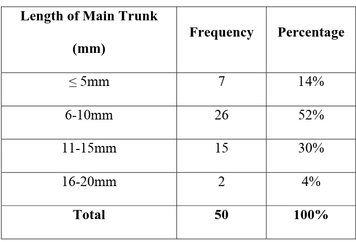
[image:25.612.131.485.307.391.2]III. Length of Main Trunk
In this present study, trunk of seven specimens were very short
ranging ≤ 5mm, twenty six specimens ranged between 6-10mm, fifteen
specimens ranged between 11-15 mm and two specimens ranged between
16-20mm. The mean length was about 9.32mm.
The minimum length was 5mm.
[image:26.612.130.481.360.598.2]The maximum length was 19mm.
Table 3: Length of Main Trunk
Length of Main Trunk
(mm)
Frequency Percentage
≤ 5mm 7 14%
6-10mm 26 52%
11-15mm 15 30%
16-20mm 2 4%
IV. Division of Main Trunk of left coronary artery
In this current study, left coronary artery was seen bifurcating into its
two main branches in thirty one specimens (Figure 3), trifurcation in sixteen
specimens (Figure 4) and quadrification in three specimens (Figure 5).
Among the sixteen trifurcating specimens, in five specimens the
diagonal artery which was not seen arising from the anterior interventricular
artery was seen at the junction between its two branches and in eleven
specimens, diagonal artery was seen arising from anterior interventricular
artery and an additional branch was seen at the junction called the “Median
Artery”.
Table – 4 : Division of Main Trunk of left coronary artery
Number of Branches Frequency Percentage
Bifurcation 31 62%
Trifurcation 16 32%
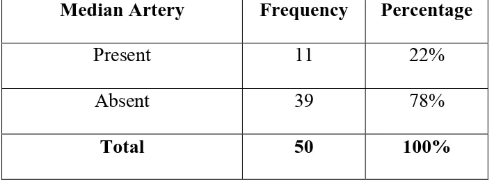
V. Median Artery
In this present study, eleven trifurcating specimens showed a branch
at the junction of its two main branches along with the diagonal artery
[image:28.612.130.485.305.436.2]arising separately from anterior interventricular artery (Figure 6).
Table - 5 : Median Artery
Median Artery Frequency Percentage
Present 11 22%
Absent 39 78%
Total 50 100%
VI. Anterior Interventricular Artery (AIVA)
This major branch of left coronary artery was seen in all fifty
specimens coursing in the anterior interventricular sulcus. This branch is
also called the left anterior descending artery (LAD)
VII. Branches of anterior interventricular artery
Left conus artery was present in all fifty specimens (Figure 7).
Diagonal artery was seen in all fifty specimens. It was seen as
trifurcation in five specimens (sp.13, 20, 30, 41, 42) (Figure 8), parallel
branch to diagonal artery was seen in specimens 31 and 37 (Figure 9) .
Atrial rami and anterior ventricular rami were seen in all fifty
specimens.
Table - 6 : Branches of anterior interventricular artery
Name of Branches Frequency Percentage
Left conus artery 50 100%
Diagonal artery 50 100%
Atrial Rami 50 100%
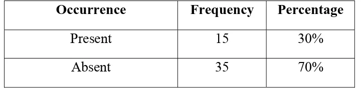
VIII. Myocardial bridges over anterior interventricular artery
In this present study, myocardial bridges was seen over anterior
[image:30.612.127.488.252.341.2]interventricular artery in fifteen specimens (Figure 9).
Table – 7 : Myocardial bridges over anterior interventricular artery
Occurrence Frequency Percentage
Present 15 30%
Absent 35 70%
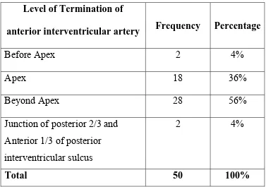
IX. Termination of anterior interventricular artery
In this study, anterior interventricular artery terminated at various
levels. In two specimens, it terminated before apex in the sternocostal
surface ( Figure 10) , in eighteen specimens it terminated over the apex, in
twenty eight specimens it terminated crossing the apex and going to the
diaphragmatic surface (Figure 11) and in two specimens it terminated at the
junction of posterior two third and anterior one third of posterior
Table – 8 :
Termination of anterior interventricular artery
Level of Termination of
anterior interventricular artery Frequency Percentage
Before Apex 2 4%
Apex 18 36%
Beyond Apex 28 56%
Junction of posterior 2/3 and Anterior 1/3 of posterior interventricular sulcus
2 4%
Total 50 100%
X. Left Circumflex Artery (LCX)
The next major branch of left coronary artery namely the left
[image:31.612.118.495.119.384.2]circumflex artery was seen in all fifty specimens.
Table – 9 : Left Circumflex Artery
Occurrence Frequency Percentage
Present 50 100%
XI. Branches of left circumflex artery
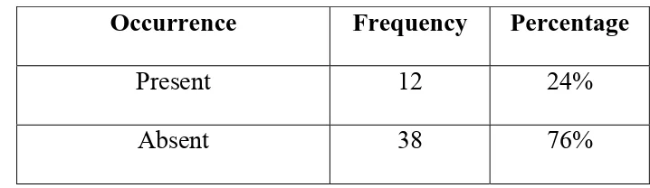
a) Sinoatrial Nodal Artery
This artery which is usually a branch from right coronary artery was
seen arising as first branch of left circumflex artery in twelve specimens
[image:32.612.122.481.256.360.2](Figure No 12).
Table – 10 : Sinoatrial Nodal Artery
Occurrence Frequency Percentage
Present 12 24%
Absent 38 76%
b) Left Marginal Artery
This branch which was seen in all fifty specimens had parallel
branches in ten specimens (Fig -13,14) and seen as termination of left
circumflex artery in ten specimens (Fig - 15).
Table – 11: Left Marginal Artery
Occurrence Frequency Percentage
Parallel branch to LMA 10 20%
Termination of LCX as Left marginal
10 20%
[image:32.612.119.485.536.651.2]c) Posterior interventricular artery (PIVA)
This branch which determines coronary dominance was seen arising
from left circumflex artery in five specimens (Sp. 20, 28, 38, 39, 45). (Figure
[image:33.612.126.483.234.334.2]- 16) and in one specimen (Sp.No.20) it had a parallel branch (Figure - 17).
Table – 12: Posterior interventricular artery
Occurrence of PIVA Frequency Percentage
Present 5 10%
Absent 45 90%
d) Atrioventricular Nodal Artery
In this study, atrioventricular nodal artery was seen arising as a septal
branch of posterior interventricular artery in the Posterior interventricular
sulcus since, in five specimens left circumflex artery continued down as
posterior interventricular artery (Sp 20, 28, 38, 39, 45).
Table – 13: Atrioventricular Nodal artery
Occurrence Frequency Percentage
Present 5 10%
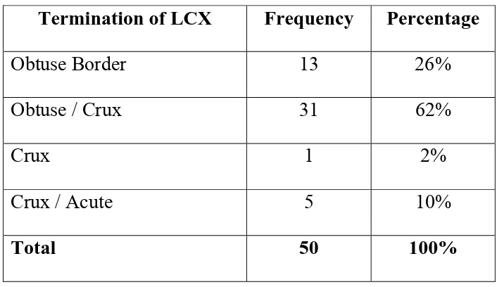
XII. Termination of left circumflex artery
In this study, left circumflex artery terminated at the obtuse border in
thirteen specimens, between obtuse border and crux in thirty one specimens,
at the level of crux in one specimen (Figure – 18). Five specimens showed
the left circumflex artery crossing the crux and running towards the acute or
inferior border. Left circumflex artery terminated as left marginal artery in
nearly ten specimens.
Table – 14:
Termination of left circumflex artery
Termination of LCX Frequency Percentage
Obtuse Border 13 26%
Obtuse / Crux 31 62%
Crux 1 2%
Crux / Acute 5 10%
Total 50 100%
[image:34.612.129.482.365.569.2]XIII. Coronary Dominance
In this present study, posterior interventricular artery was seen arising
from left circumflex artery showing left coronary dominance in five
specimens (Sp.20, 28, 38, 39, 45) and the rest of the specimens showed right
[image:35.612.132.484.299.401.2]dominance.
Table – 15: Coronary Dominance
Dominance Frequency Percentage
Right 45 90%
Left 5 10%
XIV. Anomalous Left Coronary Artery
In this present study, no anomalous origin of left coronary artery was
DISCUSSION
I. Location of Ostium
Aortic sinus is one of the anatomic dilatations of the ascending aorta
which occurs just above the aortic valve. The ostium of left coronary artery
is seen commonly in left posterior aortic sinus. The left coronary opening
may be double, leading into major initial branches, usually the circumflex
and anterior interventricular; one may lead into a stem common to one such
branch and a diagonal ventricular ramus (Gray’s Text Book of Anatomy,
2005).
Duplication of the ostia within the left aortic sinus is occasionally the
consequence of a very short or even absent left main trunk and separated
origin of the branches. This phenomenon is also designated as ‘early’
division of the left coronary artery. It occurs in 1-8% of otherwise normal
hearts (Baroldi and scomazzoni , 1965, Vlodaver et.al., 1976 : Angelini
1989)
In this study, the ostium of left coronary artery was seen in left
In the left aortic sinus there are reports of the coexistence of an
independent origin for anterior interventricular artery and circumflex artery
or duplication of either (Waller, 1983).
The situation of coronary orifices in the aortic sinus varies both cross
sectionally and in frontal plane. Left coronary artery may originate in the
mid third of sinus (87%), from posterior one third (10%) and anterior one
third (3%) (Reigvilallonga, 2003).
According to Angelini et.al., (2007) 0.15% incidence of anomalous
origination of the left coronary artery from the right aortic sinus was
reported.
II. Level of Ostium with Sinotubular junction
Position of coronary orifices is described in terms of their relation to
the sinotubular junction. A high left coronary orifice is usually associated
with a long left coronary artery and is therefore at a greater risk of injury
during surgery (Neufeld and Schneeweiss, 1983). Most haemodynamics
agree that high or low coronary orifices represent an added difficulty in
The most frequent position of the coronary orifice is at the level of
sinotubular junction or below it (56%) followed by a high left orifice and a
low right orifice or at sinotubular junction (30%) (Vlodaver et. al., 1976).
In this present study, ostium of left coronary artery was at the level of
sinotubular junction in 10% specimens and below sinotubular junction in
90% specimens. None of the ostium was seen above sinotubular junction
level.
III. Length of Main Trunk
In 92-95.5% of autopsy cases examined by Angelini (1989) the left
coronary artery had a single initial stem or trunk of variable length (2-40mm,
mean 13.5mm).
In this study seven specimens were ≤5mm, twenty six specimens
ranged between 6-10mm and fifteen specimens ranged between
11-15mm and two specimens ranged between 16-20mm. The maximum
length was found to be 19mm.
The mean length was 9.32 mm.
Anatomically, the length of main trunk has been found to range
between 1-26mm before bifurcation. Short common trunk presents the same
When the common trunk is above 15mm, it is considered long which
is seen in 11.5%-18% (Petit and Reig, 1993). It is considered ‘short’ when it
is less than or equal to 5mm (Vlodaver et.al., 1976) which is frequently
between 7-12% (Petit and Reig, 1993).
In patients with the short main left coronary artery, the atherosclerotic
lesions in the anterior descending and circumflex branch appear earlier ,
progress faster at higher level of severity and lead more frequently to
myocardial infarction , than in cases with the long left coronary trunk (
Gazetopoulos et.al., 1976)
Going with the above study, 4% specimens in this study were
considered ‘long’ with their main trunk exceeding above 15mm, 14%
specimens were considered ‘short’ with the length of main trunk less than or
equal to five millimeters.
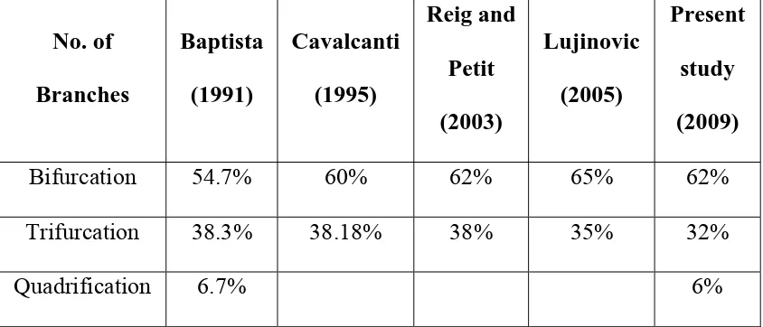
IV. Division of Main trunk of left coronary artery
According to Davidson’s Text Book (2006), the single main stem of
left coronary artery within 2.5cm of its origin divides into left anterior
The division of the common trunk into anterior interventricular,
circumflex and median or intermediate artery is a variation found in between
25-40% of cases and number of studies had been undertaken by different
authors regarding branching of left coronary artery with varying percentages
[image:40.612.96.518.282.463.2]which are shown below.
Table – 16: Division of main trunk of left coronary artery
No. of Branches Baptista (1991) Cavalcanti (1995) Reig and Petit (2003) Lujinovic (2005) Present study (2009)
Bifurcation 54.7% 60% 62% 65% 62%
Trifurcation 38.3% 38.18% 38% 35% 32%
Quadrification 6.7% 6%
V. Median artery
According to James 1961, Median artery is one which
1) Originates in the vertex of the angle formed by the main terminal
arteries of the left coronary artery or in the first millimeters.
3) Has an area of distribution extending half-way down the free wall
of left ventricle.
Levin (1983) states that angiographic examination of left coronary
artery should not be focused on the two main branches alone, since the
involvement of median artery may, depending on is distribution be as
dangerous as the involvement of the two arteries (Roberts et. al. 1986)
According to Helen Genevier (2004), in 30-37% of patients the left
main coronary artery terminates in a bifurcation in which case it also gives
rise to the ramus intermedius artery that is directed laterally.
‘Median Artery’ is the artery deriving from the main trunk in addition
to two terminal branches originating from the left coronary artery and
‘ramus lateralis’ (ramus diagonalis) as branches originating from arteria
interventricularis anterior. The most frequent type of division of the main
trunk of left coronary artery was bifurcation and arteria mediana was
detected in seven hearts (Cenk kilic et.al., 2007).
VI. Anterior interventricular artery
Left coronary artery was replaced by anterior interventricular artery
which commenced from left posterior aortic sinus and travelled in anterior
interventricular sulcus where it gave rise to left circumflex branch (Keshaw
Kumar, 2006).
It originates in the retropulmonary portion of left coronary artery,
passes above the interventricular groove, adopting an S shape, and in most
cases reaches the apex. It occasionally continues through the posterior
interventricular groove known as Mouchet’s posterior interventricular artery.
In this present study, anterior interventricular artery was seen in all
fifty specimens.
VII. Branches of anterior interventricular artery
- Left conus artery which is the first ventricular branch of anterior
interventricular artery usually forms an anastamosis with the likewise branch
of the right coronary artery. This anastamosis lies on the distal part of the
arterial conus and pulmonary trunk and is known as the vieussens arterial
ring. The functional significance of this anastamosis is still under question.
collateral path between the right coronary artery and left coronary artery.
(Reig Vilallonga, 2003)
In this present study, left conus artery was present in all fifty
specimens.
- The diagonal artery which runs parallel to the anterior
interventricular artery never reaches the anterior interventricular groove
(Spindola – Franco et. al., 1983). The importance of it is above all surgical,
since if it is ignored, during coronary bypass there is a risk that only a part of
the vessel affected will be revascularised.
Anterior interventricular artery ramifies into two to nine large
branches and one is often large and may arise separately from the left
coronary artery (which then ends by trifurcation). This diagonal artery
reported in 33-50% or more cases is occasionally duplicated (20%) (Gray’s
Anatomy, 2005).
Diagonals are branches of anterior interventricular artery that run
diagonally away from the anterior interventricular artery and towards the left
edge in front of heart (Kaimkhani et.al, 2005)
‘Intermediate’ and ‘Median’ refer to the origin but ‘diagonal’ refers to
the course of artery (Cenk kilic et.al, 2007). In this study, diagonal artery
was seen at the junction of branching appearing as trifurcation in 10%
specimens (Sp. 13, 20, 30, 41, 42) and in 4% specimens (Sp. 31 and 37)
there were parallel branches to diagonal artery.
- Small atrial and ventricular rami were seen in all 100% specimens.
VIII. Termination of anterior interventricular artery
Situated in the incisura apicis cordis, some 1-3 cm to the right of the
apex cordis (Bosco, 1935) this artery may end before reaching the apex, in
the apex itself or more frequently pass around the apex and reach the
posterior interventricular sulcus.
According to James (1961), termination of anterior interventricular
[image:44.612.140.470.488.667.2]artery was as follows
Table – 17 : Termination of anterior interventricular artery
Termination of anterior
interventricular artery Number Percentage Anterior apex
Posterior apex 2.5cm up PIVS >5cms up PIVS
18 24 44 20 17 23 42 18
Total 106 100
According to Gray’s Anatomy (2005), it reaches the apex almost
always terminating there in one third of specimens, but more often turning
round the apex into the posterior interventricular sulcus in which it traverses
one third to half of its length.
In this study, it terminates before apex in 4% specimens, at the level
of apex in 36% specimens, beyond the apex in 56% specimens and at the
junction of posterior two third and anterior one third of posterior
interventricular sulcus in 4% specimens.
IX. Myocardial Bridges over anterior interventricular artery
It was first described by Reyman in 1737 and the artery coursing in
the myocardium is called the Tunnelled Artery or coronary mural
(Geiringer, 1951).
Polacek and Kralove (1961) found that relative frequency of
myocardial bridges exclusively involving the anterior interventricular artery
was 70%.
Ozlem soran et. al., (2000) showed the incidence of myocardial
bridges between 15 and 85% in pathologic series but angiographic evidence
Vanildo Junior (2002) found that myocardial bridges are more
frequently found in the middle one third of anterior interventricular artery.
The diameter of anterior interventricular branch of left coronary artery under
the myocardial bridges may be smaller than after the bridge.
Ji – Shen chen (2003) found myocardial bridges in 5-86% in anatomic
studies but only observed in 0.5 – 12% of patients undergoing coronary
angiography.
Andrew N Pelech (2006) showed myocardial bridging in 5-25% of
patients as normal variant.
According to Angelini (2007), the fact that such bridges are surely
present in >1% of the general population suggest that they may be a normal
variant.
Angiographic study by Huxin – Ying (2007) suggest the incidence of
myocardial bridges is <2% in general population.
Myocardial Bridges is only 0.5-1.6% in the general population. It is
reported 28% in children and 30-50% in adults with hypertrophic
cardiomyopathy (Hurst, 2008).
Various studies carried out regarding myocardial bridges by dissection
Table – 18 : Myocardial bridges over anterior interventricular artery (AIVA)
Study of myocardial bridges over AIVA by Dissection method Angio study
By % By %
Geiringer (1951) 23% Kramer (1982) 12% Penther et al (1977) 17.6% Irvin (1982) 7.5%
Angelini (1983) 5.5%
This study showed 30% occurrence of myocardial bridges
(15 specimens) over anterior interventricular artery.
X. Left Circumflex Artery (LCX)
The next major branch of left coronary artery which presents the
greatest variability in terms of length and distribution was present in all fifty
specimens.
XI. Branches of left circumflex artery
a) Sinoatrial (SA) Nodal Artery
Sinoatrial nodal artery arises from the right coronary artery (54%)
from circumflex artery in (42%) from both arteries in (2%) and in 2% origin
The origin of the artery of sinoatrial node from the proximal portion
of trunk of left coronary artery was 12% and from left circumflex artery
(30%) (Didio et. al., 1995).
Among its branches, sinoatrial nodal artery which is responsible for
irrigating the structure which is in charge of initiating each heart beat is one
of the most important branch (Sanudo et. al., 1998).
In this study, sinoatrial nodal artery was seen in 24% specimens.
b) Left Marginal Artery (LMA)
The left circumflex artery commonly extends to the left ventricular
margin where it often terminates as left marginal branch. Occasionally, the
left circumflex artery passes round the margo sinister to reach the
diaphragmatic surface of the left ventricle (Angelini et. al., 1989).
In this study, left marginal artery was seen in 100% specimens but
with some variations. In specimen 22, the left marginal artery was much
bigger than the left circumflex artery. It appeared very small in specimens 23
and 30. Parallel branches to left marginal artery was seen in 20% specimens.
Left circumflex artery terminated as left marginal artery in nearly 20%
c) Posterior Interventricular Artery (PIVA)
The origin of posterior interventricular artery is one of the parameters
on which Schlesinger (1940) system of arterial dominance is based. In rare
cases, the posterior interventricular artery is entirely replaced by a dominant
long anterior interventricular artery (Levin and Baltaxe, 1972). In most
cases, the posterior interventricular artery terminates halfaway between the
crux and the apex (Angelini et. al., 1989) reporting single posterior
interventricular artery in 70% cases. Double posterior interventricular artery
accompanied by right or left branch in 6% and in 10% cases replaced by a
left coronary artery.
In this study, posterior interventricular artery was seen as continuation
of left circumflex artery in 10% specimens (Sp.20, 28, 38, 39 45). In 8%
specimens (Sp.20, 28, 38, 39) posterior interventricular artery was found
single whereas double posterior interventricular artery was seen in 2%
(Sp.45).
With regard to termination of posterior interventricular artery, in
specimens 38, 39 the posterior interventricular artery terminated in the
middle of posterior interventricular groove whereas in specimens 20, 28 and
45 it terminated at the junction of anterior one third and posterior two third
d. Atrioventricular (AV) Nodal Artery
Habitually, the atrioventricular node is irrigated by the artery that
reaches the crux cordis and supplies posterior interventricular artery
although as noted by McAlpine (1975) coronary dominance does not reflect
the origin of the node artery.
This artery which is the branch of right coronary artery (86%) was
seen arising from the left coronary artery (12%) or in both arteries (2%)
(Petit and Reig, 1993).
In this study, atrioventricular Nodal artery was seen as a branch of
posterior interventricular artery in 10% specimens.
XII. Termination of left circumflex artery
Many authors suggest that the termination of left circumflex artery as
highly variable. Table – 19 : Termination of left circumflex artery
Authors Year Cases Obtuse Border Obtuse /Crux Crux Crux/Acute
Banchi 1904 100 19% 70%
Crainicianu 1922 200 15% 75% 10%
Mouchet 1933 100 10% 82% 81%
Bosco 1935 135 25% 45% 12% 8%
James 1961 106 22% 60% 9% 9%
Baroldi and
Scamazoni 1965 522 25% 63% 5% 7%
This study showed the termination of left circumflex artery at obtuse
border in 26%, between obtuse border and crux in 62% and at crux in 2%,
beyond crux in posterior interventricular sulcus in 10%.
XIII. Coronary Dominance
The origin of posterior interventricular artery is one of the parameters
on which Schlesinger (1940), reported left dominance in 18%. Cavalcanti
(1995) showed left dominance in 11% of his study. In left dominance, the
posterior interventricular artery originated in the circumflex artery in
10-15% of cases (Ludinhausen, 2002).
In this study, left coronary dominance was found in 10% of
specimens.
Marios Loukas (2006) showed that the presence of bridges appeared
to be related to coronary dominance especially in left coronary circulation.
66.6% of the hearts with bridges were left dominant.
In this present study, 20% of the hearts with bridges were left
XIV. Anomalous Left Coronary Artery
Separate origins from the aorta of the anterior descending and
circumflex branches of the left coronary artery have been reported; the
former arose in the proper location of the left coronary but the latter arose
from the right or anterior aortic sinus close to the origin of the right coronary
artery and circled the aorta posteriorly to assume the position of a normal
circumflex branch (Hepburn, 1895).
The incidence of left coronary artery arising from the pulmonary trunk
and then assuming the course and distribution pattern of a coronary artery of
normal origin is designated as Bland – Garland syndrome (Bankl, 1977).
According to Topaz et. al., (1992), 27% patients had an anomalous
circumflex artery and 11% patients presented an anomalous anterior
interventricular artery. In 6% patients only, the anomalous coronary artery
was solely responsible for a clinical event. Coronary anomalies are found
in 0.2-1.2% of the population (Lipsett et. al., 1994) with left coronary artery
arising from the right aortic sinus or pulmonary artery.
A very rare angiographic study of Harikrishnan et. al., (2002)
concluded that the most common anomaly was separate origins of the
anomalous coronaries are more prone to atherosclerosis which was found in
[image:53.612.80.537.168.380.2]32% of patients.
Table – 20 : Anomalous left coronary artery
Left coronary anomalies Yamanaka et. al., (1990)
Harikrishnan et. al., (2002)
Anomalous arteries 1.6% 0.46%
Separate origin of anterior interventricular artery and LCX
30.4% 35.3%
LCX from RAS/RCA 17.6% 17.6%
Origin of anterior interventricular artery from RCS
38/1461 pt 1 pt
LCX – Left circumflex artery
RAS – Right aortic sinus
RCA – Right coronary artery
RCS – Right coronary sinus
CONCLUSION
This current study of Branching pattern of left coronary artery was
carried out in fifty specimens.
• The left coronary artery was seen to arise from the left posterior aortic
sinus in all fifty specimens (100%).
• With relation to Sinotubular Junction (STJ), the ostium was found at the
level of sinotubular junction in five specimens (10%), below the level
of sinotubular junction in forty five specimens (90%), none was seen
above the level of sinotubular junction.
• The mean length of the main trunk of left coronary artery was found to
be 9.32 mm.
• Regarding branching pattern, there was bifurcation of main trunk of left
coronary artery in thirty one specimens (62%), trifurcation in sixteen
specimens (32%), quadrification in three specimens (6%).
• Apart from diagonal artery arising at the junction of two major branches
showing trifurcation in five specimens (10%) one another branch called
median artery was found in eleven specimens (22%).
• Anterior interventricular artery with all its branches was seen in all fifty
specimens and it terminated before apex in two specimens (4%), at
specimens (56%) and in posterior interventricular sulcus in two
specimens (4%).
• Myocardial bridges over anterior interventricular artery was seen in
fifteen specimens (30%).
• Left circumflex artery was seen in all fifty specimens (100%).
Regarding its branches, sinoatrial nodal artery from left circumflex was
seen in twelve specimens (24%). Left marginal artery was seen in all
fifty specimens with parallel branch (1, 2 or 3) seen in ten specimens
(20%) and the left circumflex artery terminating as left marginal artery
in ten (20%) specimens.
• Posterior interventricular artery determining the coronary dominance
was seen to arise in five specimens (10%) and in one specimen (2%)
there was a parallel branch to posterior interventricular artery.
Atrioventricular nodal artery from posterior interventricular artery was
found in five specimens (10%).
• Termination of left circumflex artery was found at the obtuse border in
thirteen specimens (26%), between obtuse border and crux in thirty one
specimens (62%), at the crux in one specimen (2%) and between crux
• Left coronary dominance was seen in five specimens (10%) with
posterior interventricular artery arising from left circumflex artery.
• There was not a single left coronary anomaly found in this study.
Recent technical advances in the study of coronary arteries make it
necessary for all angiographers and cardiac surgeons to be familiar with
variants of left coronary artery, because accurate identification and
delineation of coronary arteries in the presence of coronary artery disease is
integral to proper surgical revascularization of myocardium as surgical
Level of Ostium with relation to
Sinotubular Junction
0 10
90
0 20 40 60 80 100
Above At Below
P
er
cen
ta
g
14
52
30
4
0
10
20
30
40
50
60
Pe
rc
e
n
ta
ge
≤
5mm
6-10mm
11-15mm 16-20mm
Myocardial Bridges Over AIVA
30%
70%
Present Absent
Termination of AIVA
4 36 56 4 0 20 40 60
Before Apex Apex Beyond Apex Junction of
posterior 2/3 & Anterior 1/3 of
PIVS Pe rc e n ta ge
PIVA
10%
90%
Present Absent
PIVA – Posterior Interventricular Artery
AV Nodal Artery
10%
90%
Present Absent
Coronary Dominance
10%
90%
MASTER CHART ABBREVIATIONS
LCA - Left coronary artery
LPAS - Left Posterior Aortic Sinus
STJ - Sinotubular junction
SA - Sinoatrial
AV - Atrioventricular
LCX - Left circumflex artery
PIVA - Posterior interventricular artery
O - Obtuse Border
O/C - Obtuse / Crux
C/A - Crux / Acute
C - Crux
M - Median Artery D - Diagonal Artery LMA - Left Marginal Artery R - Right
L - Left
Jn - Junction of posterior two third and
Fig No : 1 – Left Coronary Ostium – At STJ
Fig No : 2 – Left Coronary Ostium – Below STJ
Fig No : 3 – Bifurcation of left Coronary Artery
[image:65.612.119.498.417.674.2]62
32
6
0 20 40 60 80
Pe
rc
e
n
ta
ge
Bifurcation Trifurcation Quadrification
Division of Main Trunk of LCA
[image:67.612.118.495.77.321.2]LCA – Left coronary artery
→ - Diagonal Artery from anterior interventricular artery → - Median Artery
Median Artery
22%
78%
Fig No : 8 – Trifurcation of left coronary artery with diagonal artery at the junction
[image:70.612.146.427.370.701.2]→ - Intramural course of anterior interventricular artery
Fig No : 10 – Termination of AIVA over sternocostal surface before apex
[image:71.612.175.436.403.657.2]AIVA – Anterior Interventricular Artery
SA Nodal Artery
76%
24%
Present Absent
[image:73.612.168.444.347.637.2]SA – Sinoatrial
Fig No: 14 – Two Parallel Branches to Left Marginal Artery
[image:74.612.161.454.391.659.2]Left Marginal Artery
20 20
0 5 10 15 20 25
Parallel branch Termination of LCX as Left marginal
P
e
rcen
ta
g
e
LCX – Left Circumflex
Interventricular Artery
Fig No: 18 – Termination of Left Circumflex Artery at Crux
Termination of LCX
26 62 2 10 0 10 20 30 40 50 60 70 80 90 100
Obtuse Border Obtuse / Crux Crux Crux / Acute
P er cen ta g e
BIBLIOGRAPHY
1. Alexander R.W. & Griffith G.C. Anomalies of the coronary artery and their clinical significance 1956; 14: 800-805.
2. Allwork S.P. The Anatomy of the coronary arteries; 1976, 15-25.
3. Andrew N Pelech. Coronary artery anomalies, 2006, www.emedicine.com. 4. Angelini P, Trivellato M, Donis J, Leachman RD. Myocardial bridges; a
review, prog cardiovascular dis 1983; 26 : 75-78.
5. Angelini P. Normal and anomalous coronary arteries definitions and classifications. Am Heart J 1989, 117: 418 – 434.
6. Angelini P. Velasco JA, Flamm S, Coronary anomalies – incidence, pathophysiology and clinical relevance, circulation, 2002, 105, 20, 2449-60.
7. Angelini, Coronary artery anomalies, circulation 2007: 115; 1296-1305. 8. Banchi A. Mortfologia della arteriae aoronariae cordis: Arch Ital. Anat.
Embroil 1904 3:87.
9. Bankl H, congenital malformations of the heart and Great Vessels. Urban and Schwarzenberg, 1977.
11. Baroldi G and Scomazzoni G. Coronary circulation in the normal and pathologic heart. Armed forces institute of pathology: Washington, 1965; pp 1 – 37.
12.Berth CW, Roberts WC, Left main coronary artery originating from the right sinus of valsalva and coursing between Aorta and pulmonary trunk Jam Coll Cardiol 1986; 7: 366-73.
13.Bosco GA. Diagnostico anatomo – topografico de la obstruccion arterial coronaria. Artes graficas modernas. Buenos Aires, 1935, pp 17 - 179
14.Cavalcanti J.S. Anatomic variations of the coronary arteries Arq Bras Cardiol 1995; Dec;65(6): 489-92.
15.Cenk Kilic, Yakin Kirici; Third branch derived from left coronary artery: The Median Artery; Gulhane Tip Dergisi 2007; 49: 232-235.
16.Charles E Wilkins, coronary artery anomalies, Texas Heart Institute Journal 1988; 15: 166-173.
17.Crainicianu AC, Anatomische studien ber die coronararaterien and experimentelle untersuchunger uber ihre Durchgngigkeit, virchow’s Arch path anat 1922; 238 : 1 – 75.
19.Donald S.Baim, William Grossman, Grossman’s Cardiac Catheterization, Angiography and intervention, Lippincott Williams and Wilkins, 2000, 6th edition pg. 3, 1211,.
20.Engel HJ, Torres C, Page HL Jr. Major variations in anatomical origin of the coronary arteries: Angiographic observation in 4250 patients without associated congenital heart disease. Cathet Cardiovasc Diagn 1975; 1:157. 21.Essenberg J.M. An anomalous left coronary artery in a human fetus. Its
passage through the left atrium and possible discharge into the right atrium anat Rec 1950; 108: 709.
22.Ferguson DW, Henkle JQ, Haws CW. Absence of left anterior descending coronary artery associated with anomalous origin of left circumflex coronary from the right coronary artery Cathet Cardiovasc Diagn 1985: 11(1); 55-61.
23.Fineschi M, Del Sordo, M Leosco D, A rare anatomic variation of anomalous origin of all three major coronary arteries from right sinus of valsalva; 1998: 28; 564-566.
24.Gazetopoulos N, PJ Loannidis, C Karydis, C Lola. Short left coronary trunk as a risk factor in the development of coronary atherosclerosis, pathological study, British Heart Journal, 1976; 38 : 1160-1165.
26.Greenberg, MA Fish BG, Spindola-Franco H, Congenital anamolies of the coronary arteries, classification and significance 1989; 27: 1127-1146.
27.Harikrishnan S, Coronary anomalies in adults, Indian Heart J 2002; 54: 271-275.
28.Harikrishnan S, P Jacob SP. Coronary anomalies in adults, Sree Chitra Tirunal Institute for Medical Sciences and Tech; 2001, 53; 619.
29.Helen Genevier, ramus intermedius, 2004, www.proz.com
30.Henry Hollinshead W, Textbook of Anatomy for surgeons Harper & Row, 1961, 3rd Edition, 115-116.
31.Hepburn D. A rare abnormal arrangement of the cardiac coronary arteries. J. Anat and Physiol 29: 459, 1895.
32.Hurst, Hurst’s The Heart Foster O’ Kourke, Walsh Poole-Wilson, 2008, 12th edition, pg. 19-20.
33.Hu-Xin Ying, Zhooda-Xin, Multiple myocardial bridges affecting left anterior descending artery, Chinese Medical Journal, 2007, Vol.120, No.8, 734-746.
34.Irvin RG. The angiographic prevalence of myocardial bridging in man. Chest 1982, 81: 196-202.
36.Ji – Shen chen, Chin-Lon Lin. Myocardial bridging, Tzu chi Med J 2003; 15 : 357-362.
37.Kaimkhani ZA, Ali MM, Faruqi AM, Pattern of coronary arterial distribution and its relation to coronary artery DM. Journal of Ayub Medical college (2005). JAMC 17(1): 40-3.
38.Keshaw Kumar, Anterior interventricular artery replacing left coronary artery with absence of arterial anastamosis in human heart, J Anat. Soc. India, 2006, 55 (1) : 42-44.
39.Kramer JR, Kitazume H, Proudfit WL, Sones, FM Jr. Clinical significance of isolated coronary bridges benign and frequent condition involving the left anterior descending artery. Am Heart J, 1982; 103: 282-288.
40.Kugel, M.A. Anatomical studies on the coronary arteries and their branches I arteria anastomotica auricularis magna. Amer Heart J. 1928; 3: 260-270. 41.Levin DC, Baltaxe HA, Angiographic demonstration of important anatomic
variations of the posterior descending coronary artery 1972, A JR 116 : 41-49.
42. Levin DC. Anomalies and variations of the coronary arteries, In: Abrams HL (ed) : coronary arteriography. A practical approach. Little, Brown co, Boston, 1983; pg 283-299.
44.Lionel H Opie The Heart physiology from cell to circulation Lippincott. Raven, 1997 3rd edition, pg -3.
45.Lipsett J, Cohle S.D., Russel G, Berry PJ and Byard K.W. Anomalous coronary arteries : a multicentre paediatric autopsy study. Pediatric pathology ; 1994 : 14 : 287-300.
46.Lipton MJ, Barry WH, Obrez I, Silverman JF, Wexler L. Isolated single coronary artery: diagnosis angiographic classification, and clinical significance. Radiology 1979; 130 : 39-47.
47.Ludinghausen M.V. Advances in anatomy embryology and cell biology 2002: 167; 15-32.
48.Lujinovic A, Ovcina F, Voljevica A, Hasanovia A. Branching of main trunk of left coronary artery and importance of her diagonal branch in cases of coronary insufficiency. Bosn J Basic Med Sci 2005; 5: 69-73.
49.Maleszka. Giant coronary arteriovenous fistula. A case report and review of literature Z Kardiol, 2005; 94 (1): 38-43.
50.Mandelbrott ‘The fractal geometry of nature, WH freeman and company, 1977:41.
52.MC Alpine WA, Heart and coronary arteries; an anatomical atlas for clinical diagnosis, radiological investigation and surgical treatment – New York; springer – Verlag 1975, pp.133-209.
53.Morettin LB: Coronary Arteriorgraphy; in common observations Radiol Clin North Am 1976: 14; 189-209.
54.Mouchet A. Les arteres coronaires du cocur chez L homme. Paris, Norbert Maloine 1933.
55.Neufeld HN and Schneeweiss A. Coronary artery disease in infants and children. Lea and febiger; Philadelphia, 1983 : pp 65-78.
56.Nicholas A, Brian R, Nicki R, John .A.A. Hunter, Davidson’s principles and practice of Medicine, Churchill living stone, 2006, 20th Edition pg. 523. 57.Ogden JA. Congenital anomalies of the coronary artery. American Journal
of cardiology (1970); 25: 474-479.
58.Ogden JA; Goodyer AV: Patterns of distribution of the single coronary artery. 1973; 43: 11-21.
59.Ott D.A Cooley D.A. Pinskeil, WW & Mollins, CE anomalous origin of circumflex artery from right pulmonary artery. Journal of Thoracic and cardiovascular surgery. 1978; 76: 190-194.