J
OURNAL OFV
IROLOGY, Nov. 2005, p. 14465–14469
Vol. 79, No. 22
0022-538X/05/$08.00
⫹
0
doi:10.1128/JVI.79.22.14465–14469.2005
Copyright © 2005, American Society for Microbiology. All Rights Reserved.
The Herpes Simplex Virus Type 1 Locus That Encodes the
Latency-Associated Transcript Enhances the
Frequency of Encephalitis in Male
BALB/c Mice
Clinton Jones,
1* Melissa Inman,
1Weiping Peng,
1Gail Henderson,
1Alan Doster,
1Guey-Chuen Perng,
2and Anisa Kaenjak Angeletti
3Department of Veterinary and Biomedical Sciences, Nebraska Center for Virology, University of Nebraska, Fair Street at
East Campus Loop, Lincoln, Nebraska 68583-0905
1; Department of Ophthalmology, College of Medicine,
University of California at Irvine, UCIMC, Bldg. 55, Room 204, 101 The City Drive, Orange,
California 92868-4380-02
2; and School of Biological Sciences, Nebraska Center
for Virology, University of Nebraska, Lincoln, Nebraska
3Received 26 May 2005/Accepted 26 August 2005
Herpes simplex virus type 1 (HSV-1) is the leading cause of virus-induced encephalitis; however, the viral
genes that regulate encephalitis have not been well characterized. In this study, we tested whether the LAT
(latency-associated transcript) locus regulates the frequency of encephalitis in male or female mice. Male
BALB/c mice are more susceptible to HSV-1-induced encephalitis than age-matched female BALB/c mice.
Deletion of LAT coding sequences reduced the frequency of encephalitis. A recombinant virus containing the
first 1.5 kb of the LAT coding sequence induces levels of encephalitis in male BALB/c mice similar to those
induced by wild-type HSV-1.
Approximately 90% of the population is infected with
her-pes simplex virus type 1 (HSV-1) (29, 55). Following acute
infection, latency is established in sensory neurons in
trigemi-nal ganglia or sacral dorsal root ganglia (19, 50). Reactivation
from latency periodically occurs, resulting in virus transmission
and recurrent disease (19, 20).
The latency-associated transcript (LAT) is abundantly
tran-scribed in latently infected neurons (8, 10, 11, 22, 27, 44, 48, 51,
52). An 8.3-kb LAT transcript is spliced, yielding the stable
2-kb LAT intron that localizes to the nucleus (2, 31, 49) and an
unstable 6.3-kb transcript (11, 44, 57). Proper splicing of LAT
is necessary for establishment and maintenance of latency (21).
The HSV-1 McKrae strain is frequently shed in tears of
la-tently infected rabbits because of spontaneous reactivation
(39–43), and LAT gene expression is crucial for spontaneous
reactivation. LAT inhibits apoptosis in cultured cells and
tri-geminal ganglia of infected rabbits or mice (1, 14, 17, 18, 21,
37). The ability of LAT to interfere with apoptosis correlates
with spontaneous reactivation (17, 18).
HSV-1-induced encephalitis is a severe form of focal
necro-tizing encephalitis that affects approximately 2,000 individuals
each year in the United States (12, 24, 54, 55). Without
anti-viral therapy, the mortality rate can be 70% (45, 46).
Approx-imately 2/3 of HSV-1-induced encephalitis cases are due to
reactivation from latency (56); consequently, high morbidity
and long-term complications occur despite antiviral treatment
(23, 26, 45).
In general, females induce a stronger immune response
be-cause they express higher levels of lymphocytes and cytokines
(3, 5, 16). Susceptibilities to encephalomyocarditis virus (9),
vesicular stomatitis virus (4), coxsackievirus B3 (15), and
HSV-1 infections in mice (13) differ based on gender.
Further-more, a HSV-2 vaccine trial in humans succeeded in females
but not in males (47). In female mice, a HSV resistance locus
(sex modifier locus) has been identified (25). HSV-1
hematog-enous infection is more pathogenic to females (6), and
estro-gen influences pseudorabies virus infections in the central
ner-vous system (53), suggesting that gender-specific factors, male
as well as female, influence host-pathogen interactions.
Based on the observations that gender can influence the
pathogenic potential of certain viruses, we tested whether LAT
influences the frequencies of encephalitis in male and female
mice. Adult female or male BALB/c mice were infected with
the wild-type (wt) McKrae strain or the LAT locus mutant,
dLAT2903 (Fig. 1). As previously described for 129 Sv//Ev or
RGKO male mice (13), male BALB/c mice infected with wt
HSV-1 or dLAT2903R (rescued dLAT2903) (Fig. 1A) had
frequencies of survival significantly lower than those of female
BALB/c mice (Fig. 2A and B). A Tukey-Kramer
multiple-comparison post-analysis of variance test revealed that survival
rates for male and female mice infected with wt HSV-1 or
dLAT2903R were significantly different (P
⫽
0.004). Male
mice infected with dLAT2903 had significantly higher (P
⫽
0.003) levels of survival than those infected with wt HSV-1 or
dLAT2903R. As previously reported (38), female mice
in-fected with dLAT2903, wt HSV-1, or dLAT2903R exhibited
similar survival rates.
The deletion within dLAT2903 prevents expression of at
least four genetic elements (Fig. 1C): (i) the stable 2-kb LAT;
(ii) the first 1.5 kb of LAT, which contains only part of the
* Corresponding author. Mailing address: Department of
Veteri-nary and Biomedical Sciences, Nebraska Center for Virology,
Univer-sity of Nebraska, Fair Street at East Campus Loop, Rm. 104, Lincoln,
NE 68583-0905. Phone: (402) 472-1890. Fax: (402) 472-9690. E-mail:
14465
on November 8, 2019 by guest
http://jvi.asm.org/
FIG. 1. Schematic representation of wild-type and mutant viruses. (A) The prototypic HSV-1 genomic structure of the wt McKrae strain is
shown at the top. The viral repeat regions are shown as open rectangles. TR
Lis the terminal long repeat. IR
Lis the internal (or inverted) long
repeat. TR
Sis the terminal short repeat. IR
Sis the internal (or inverted) short repeat. The unique long (U
L) and unique short (U
S) regions are
[image:2.585.112.473.45.281.2]each represented by a solid line. Below the genomic structure, the LAT region (one in each long repeat) is shown in expanded form. The primary
8.3-kb LAT is shown as a long arrow. The stable 2-kb LAT is shown as a solid rectangle.
⫹
1 and the small arrows indicate the starts of LAT
transcription. The relative locations of mRNAs encoding ICP0 and ICP34.5 are shown for reference. (B) dLAT2903 is a LAT null mutant that
contains deletions (XXXX) in both copies of LAT from LAT nucleotides
⫺
161 to
⫹
1667. dLAT2903 is missing the core LAT promoter, a putative
secondary LAT promoter just upstream of the 2-kb LAT, and the first 1,667 nucleotides of the primary LAT RNA. (C) Schematic of genes within
the LAT locus. The primary LAT transcript is indicated by the large arrow. The solid rectangle represents the very stable 2-kb LAT intron. The
LAT TATA box is indicated, and the striped box denotes the LAT promoter. The start of LAT transcription is indicated by the arrow at
⫹
1
(genomic nucleotide 118801). Several restriction enzyme sites and the relative locations of the ICP0, ICP34.5, UOL (upstream of LAT), and AL
(antisense to LAT) transcripts are also shown for reference. The nucleotide positions of the 5
⬘
and 3
⬘
ends of UOL (30) and AL (36) are depicted
and are relative to the start of LAT transcription. (D) LAT3.3A is a recombinant virus that was constructed using dLAT2903, and it contains a
fragment spanning from
⫺
1800 to
⫹
1499. LAT sequences were inserted between UL37 and UL38. The construction of LAT3.3A was described
previously (41). (E) LAT1.8A is a recombinant virus that was constructed using dLAT2903, and it contains a fragment spanning from
⫺
1800 to
⫹
76. LAT sequences were inserted between UL37 and UL38. The construction of LAT1.8A was described previously (38).
FIG. 2. Survival of BALB/c mice after infection with HSV-1. Adult male and female BALB/c mice (54 to 82 days old) were obtained from
Charles River Laboratory. Female and male mice were housed in separate rooms during the course of all experiments. Mice were ocularly infected
with doses of 1
⫻
10
5PFU/eye. Mice were bilaterally infected without scarification by placing the virus (2
l of inoculum) into the conjunctival
cul-de-sac, closing the eye, and rubbing the lid gently against the eye for 30 seconds. Prior to infection, mice were lightly anesthetized using
isoflurane (Halocarbon Laboratories, River Edge, NJ). Mice were observed daily during the studies and were euthanized by CO
2inhalation if they
exhibited severe neurological symptoms. (A) The percentages of surviving mice are presented. (B) The number of mice used for each virus and
the percentages of mice that survived are shown. (C) Survival data were plotted based on the times that mice succumbed to fatal encephalitis after
infection. There were no dramatic differences between wt McKrae, LAT3.3A, and dLAT2903R; consequently, these data were combined and
designated as LAT
⫹
virus data. Since there was no difference between dLAT2903 and LAT1.8A, these data were combined and designated as
LAT
⫺
data. The mice that were euthanized because they were lethargic and unresponsive were scored as having undergone fatal encephalitis on
the day they were euthanized.
14466
on November 8, 2019 by guest
stable 2-kb LAT and is sufficient for high levels of spontaneous
reactivation in the rabbit eye model (41) or for trigeminal
ganglia explant-induced reactivation in mice (38); (iii) the AL
transcript (36); and (iv) the UOL transcript (30). To localize
regions of LAT necessary for enhancing encephalitis in male
BALB/c mice, we tested LAT3.3A (Fig. 1D) and LAT1.8A
(Fig. 1E). LAT3.3A expresses UOL, AL, and one copy of the
first 1.5 kb of LAT but does not express the intact stable 2-kb
LAT. LAT1.8A contains one copy of the minimal UOL coding
sequences but not AL or LAT. Male, but not female, BALB/c
mice infected with LAT3.3A had a frequency of encephalitis
significantly higher than that for age-matched male mice
in-fected with LAT1.8A (P
⫽
0.003) (Fig. 2A and B).
Further-more, LAT3.3A, wt HSV-1, and dLAT2903R induced similar
levels of encephalitis in male BALB/c mice, indicating that
expression of the intact stable 2-kb LAT does not regulate
gender-specific encephalitis. Finally, LAT1.8A induced levels
of encephalitis in male BALB/c mice similar to those induced
by dLAT2903.
Following infection of male or female mice with the
respec-tive viruses, the mice that died typically succumbed to fatal
encephalitis between 7 and 10 days after infection (Fig. 2C).
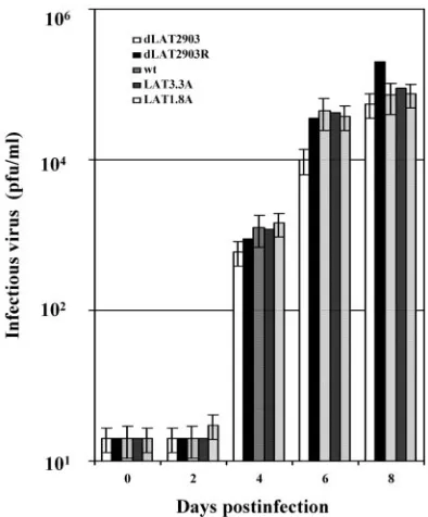
Similar virus titers were also detected in brain homogenates at
2, 4, or 8 days after infection, regardless of the virus used to
infect male (Fig. 3) or female BALB/c mice (data not shown).
Consistent with previous studies (28, 35, 38), we also found
similar levels of infectious virus in ocular swabs from male and
female mice following infection with the virus strains. These
studies suggested that the reduced frequency of encephalitis in
male BALB/c mice infected with dLAT2903 was not due to
reduced viral titers in the brain.
Since the LAT locus inhibits interferon (IFN) RNA
expres-sion in neuronal cell types (34), we tested whether IFN was
differentially expressed in brains of mice during acute infection
(2 to 8 days after infection) or after latency was established (14
days after infection). Using primers that specifically amplify
mouse gamma IFN (IFN-
␥
) (Fig. 4C), male mice infected with
dLAT2903 contained readily detectable levels of IFN-
␥
RNA
[image:3.585.314.528.70.254.2]at 6, 8, and 14 days after infection, but mock-infected mice did
[image:3.585.64.261.374.612.2]FIG. 3. Analysis of virus titers in brains of male BALB/c mice
following infection with HSV-1. Male BALB/c mice were infected with
the designated virus as described in the legend for Fig. 2. Brains from
individual mice were minced into small pieces and suspended in 2 ml
of phosphate-buffered saline, and tissue was disrupted using a Polytron
tissue homogenizer. Samples were then subjected to three cycles of
freeze-thawing (
⫺
86°C to 37°C), and debris was removed by
centrifu-gation (18,000
⫻
g
for 30 min at 4°C). One-tenth of the supernatant
from a single brain was used for a measurement of the amount of
infectious virus, which was performed as previously described (35, 38).
The data represent values obtained from brains of eight mice for each
virus and time point. The average of these results are shown.
FIG. 4. Gene expression in male mice infected with HSV-1. As
described in the legend for Fig. 2, adult male (A) and female
(B) BALB/c mice were infected with the designated strains of HSV-1.
Brains from individual mice were collected following euthanasia and
snap-frozen on dry ice. One ml of TRIZOL reagent was added
(In-vitrogen, Carlsbad, CA), and RNA was prepared according to the
manufacturer’s instructions. For the designated times after infection
(numbers of days shown at the tops of the gels), RNA was prepared
from brains of three mice. RNA samples were subjected to DNase I
digestion to remove trace amounts of contaminating DNA. Samples
from mock-infected mice are designated C. Semiquantitative reverse
transcriptase PCR was performed as described previously (34) using
RNA from the brain of a single mouse. RNA concentrations were
determined initially by measuring the optical density at 260 nm.
Run-ning a 1.2% formaldehyde agarose gel using 0.5 to 1
g of RNA
monitored the integrity of total RNA and confirmed the optical density
reading. First-strand cDNA synthesis was performed with the
Super-Script first-strand synthesis system for reverse transcriptase PCR
(In-vitrogen) using equal amounts of total RNA (1 to 2
g) and
oli-go(dT)
12-16(for cellular genes) or random hexamers (for HSV-1
genes). PCRs were carried out in 20-
l reaction mixtures that
con-tained 0.5
M of the designated gene-specific primers shown in panel
A, 1/10 of the cDNA reaction mixture, 2.5 to 5 mM MgCl
2, 25 to 100
M dNTP, and 1 U
Taq
DNA polymerase. LAT cDNA was amplified
using the GC-rich PCR system (Roche). Without reverse transcriptase
in the reaction mixture, there were no specific amplified products (data
not shown). These results are representative of three separate
exper-iments using RNA prepared from different mice. Upper gel in panel B,
IFN-
␥
; lower gel in panel B,

-actin. (C) All primers were described
previously (32, 34) and are presented in a 5
⬘
-to-3
⬘
direction. For each
gene, the first primer is the forward primer and the second is the
reverse primer.
V
OL. 79, 2005
NOTES
14467
on November 8, 2019 by guest
http://jvi.asm.org/
not (Fig. 4A). In contrast, male mice infected with the McKrae
strain or dLAT2903R expressed IFN-
␥
RNA at 6 and 8 days,
but not at 14 days, after infection. Viral glycoprotein (gC) was
detected on days 6 and 8 regardless of the virus used for
infection. At 4 days after infection, low levels of gC RNA were
detected in male mice infected with the McKrae strain or with
dLAT2903R but not in those infected with dLAT2903. gC
RNA expression levels were similar in brains of male mice at 6
and 8 days after infection, regardless of which virus was used.
As expected, LAT was not detected in mice infected with
dLAT2903 (Fig. 4A). Female mice infected with dLAT2903,
dLAT2903R, or wt HSV-1 expressed IFN-
␥
RNA 6 and 8 days
after infection (Fig. 4B). These findings suggested that a
cor-relation exists between survival of male mice infected with
dLAT2903 and prolonged IFN-
␥
RNA expression in brains.
Our results are also consistent with studies demonstrating that
IFN-
␥
protects against HSV infection (7, 13).
In summary, these studies indicated that the ability of
HSV-1 to induce high levels of fatal encephalitis in male
BALB/c mice was dependent on expression of the first 1.5 kb
of LAT, expression of UOL, and/or expression of AL. We
further suggest that the male mouse model may be useful for
identifying sequences in the LAT locus that regulate the
laten-cy-reactivation cycle and neuropathogenesis. The first 1.5 kb of
LAT promotes spontaneous reactivation (40, 41) and inhibits
caspase 8-induced apoptosis efficiently (33). The functions of
AL and UOL are not known. Additional studies are required
to prove whether AL, UOL, or the 1.5-kb LAT sequence
reg-ulates neurovirulence in male BALB/c mice. Since expressions
of the 1.5-kb LAT, of UOL, and of AL are regulated by the
same promoter and their coding sequences overlap one
an-other, preparing recombinant viruses that express just one of
the respective gene products is a challenge.
This work was supported by Public Health Service grant
1P20RR15635 and by two USDA grants (2002-35204 and 2003-02213).
REFERENCES
1.Ahmed, M., M. Lock, C. G. Miller, and N. W. Fraser.2002. Regions of the herpes simplex virus type 1 latency-associated transcript that protect cells from apoptosis in vitro and protect neuronal cells in vivo. J. Virol.76:717– 729.
2.Ahmed, M., and N. Fraser.2001. Herpes simplex virus type 1 2-kilobase latency-associated transcript intron associates with ribosomal proteins and splicing factors. J. Virol.75:12070–12080.
3.Aida, T., N. Ishikawa, and K. Shinkai.1998. Sex differences in immune responses to cephalothin in guinea pigs. J. Toxicol. Sci.23:87–91. 4.Barna, M., T. Komatsu, Z. Bi, and C. S. Reiss.1996. Sex differences in
susceptibility to viral infection of the central nervous system. J. Neuroim-munol.67:31–39.
5.Barrat, F., B. Lesourd, H. J. Boulouis, D. Thibault, N. S. Vincent, B. Gjata, A. Louise, T. Neway, and C. Pilet.1997. Sex and parity modulate cytokine production during murine aging. Clin. Exp. Immunol.109:562–568. 6.Burgos, J. S., C. Ramirez, I. Sastre, J. M. Alfaro, and F. Valdivieso.2005.
Herpes simplex virus type 1 infection via the bloodstream with apolipopro-tein E dependence in the gonads is influenced by gender. J. Virol.79:1605– 1612.
7.Cantin, E., B. Tanamachi, H. Openshaw, J. Mann, and K. Clarke.1999. Gamma interferon (IFN-␥) receptor null-mutant mice are more susceptible to herpes simplex virus type 1 infection than IFN-␥ligand null-mutant mice. J. Virol.73:5196–5200.
8.Croen, K. D., J. M. Ostrove, L. J. Dragovic, J. E. Smialek, and S. E. Straus.
1987. Latent herpes simplex virus in human trigeminal ganglia. Detection of an immediate early gene “anti-sense” transcript by in situ hybridization. N. Engl. J. Med.317:1427–1432.
9.Curiel, R. E., M. H. Miller, R. Ishikawa, D. C. Thomas, and N. J. Bigley.
1993. Does the gender difference in interferon production seen in picorna-virus-infected spleen cell cultures from ICR Swiss mice have any in vivo significance? J. Interferon Res.13:387–395.
10.Deatly, A. M., J. G. Spivack, E. Lavi, and N. W. Fraser.1987. RNA from an immediate early region of the type 1 herpes simplex virus genome is present in the trigeminal ganglia of latently infected mice. Proc. Natl. Acad. Sci. USA
84:3204–3208.
11.Deatly, A. M., J. G. Spivack, E. Lavi, D. R. D. O’Boyle, and N. W. Fraser.
1988. Latent herpes simplex virus type 1 transcripts in peripheral and central nervous system tissues of mice map to similar regions of the viral genome. J. Virol.62:749–756.
12.Gesser, R. M., and S. C. Koo.1997. Latent herpes simplex virus type 1 gene expression in ganglia innervating the human gastrointestinal tract. J. Virol.
71:4103–4106.
13.Han, X., P. Lundeberg, B. Tanamachi, H. Openshaw, J. Longmate, and E. Cantin.2001. Gender influences herpes simplex virus type 1 infection in normal and gamma interferon-mutant mice. J. Virol.75:3048–3052. 14.Henderson, G., W. Peng, L. Jin, G.-C. Perng, A. B. Nesburn, S. L. Wechsler,
and C. Jones.2002. Regulation of caspase 8- and caspase 9-induced apo-ptosis by the herpes simplex virus latency-associated transcript. J. Neurovi-rol.8:103–111.
15.Huber, S., and B. Pfaeffle.1994. Differential Th1 and Th2 cell responses in male and female BALB/c mice infected with coxsackievirus group B type 3. J. Virol.68:5126–5132.
16.Huppert, F. A., W. Solomou, S. O’Connor, K. Morgan, P. Sussams, and C. Brayne.1998. Aging and lymphocyte subpopulations: whole-blood analysis of immune markers in a large population sample of healthy elderly individ-uals. Exp. Gerontol.33:593–600.
17.Inman, M., G. C. Perng, G. Henderson, H. Ghiasi, A. B. Nesburn, S. L. Wechsler, and C. Jones.2001. Region of herpes simplex virus type 1 latency-associated transcript sufficient for wild-type spontaneous reactivation pro-motes cell survival in tissue culture. J. Virol.75:3636–3646.
18.Jin, L., W. Peng, G.-C. Perng, A. B. Nesburn, C. Jones, and S. L. Wechsler.
2003. Identification of herpes simplex virus type 1 latency associated tran-script sequences that both inhibit apoptosis and enhance the spontaneous reactivation phenotype. J. Virol.77:6556–6561.
19.Jones, C.1998. Alphaherpesvirus latency: its role in disease and survival of the virus in nature. Adv. Virus Res.51:81–133.
20.Jones, C.2003. Herpes simplex virus type 1 and bovine herpesvirus 1 latency. Clin. Microbiol. Rev.16:79–95.
21.Kang, W., R. Mukerjee, and N. F. Fraser.2003. Establishment and mainte-nance of HSV latent infection is mediated through correct splicing of the LAT primary transcript. Virology312:233–244.
22.Krause, P. R., K. D. Croen, S. E. Straus, and J. M. Ostrove.1988. Detection and preliminary characterization of herpes simplex virus type 1 transcripts in latently infected human trigeminal ganglia. J. Virol.62:4819–4823. 23.Lahat, E., J. Barr, G. Barkai, G. Paret, N. Brand, and A. Barzilai.1999. Long
term neurological outcome of herpes encephalitis. Arch. Dis. Child.80:69– 71.
24.Lohr, J. M., J. A. Nelson, and M. B. Oldstone.1990. Is herpes simplex virus associated with peptic ulcer disease? J. Virol.64:2168–2174.
25.Lundberg, P., P. Welander, H. Openshaw, C. Nalibandian, C. Edwards, L. Moldawer, and E. Cantin.2003. A locus on mouse chromosome 6 that determines resistance to herpes simplex virus also influences reactivation, while an unlinked locus augments resistance of female mice. J. Virol.77:
11661–11673.
26.McGrath, N., N. E. Anderson, M. C. Croxson, and K. F. Powell.1997. Herpes simplex encephalitis treated with acyclovir: diagnosis and long term out-come. J. Neurol. Neurosurg. Psychiatry63:321–326.
27.Mitchell, W. J., R. P. Lirette, and N. W. Fraser.1990. Mapping of low abundance latency-associated RNA in the trigeminal ganglia of mice latently infected with herpes simplex virus type 1. J. Gen. Virol.71:125–132. 28.Mott, K., N. Osorio, L. Jin, D. Brick, J. Naito, J. Cooper, G. Henderson, M.
Inman, C. Jones, S. L. Wechsler, and G.-C Perng.2003. The bovine herpes-virus 1 LR ORF2 is crucial for this gene’s ability to restore the high reacti-vation phenotype to a herpes simplex virus-1 LAT null mutant. J. Gen. Virol.
84:2975–2985.
29.Nahmias, A. J., and B. Roizman.1973. Infection with herpes-simplex viruses 1 and 2. 3. N. Engl. J. Med.289:781–789.
30.Naito, J., R. Mukerjee, K. R. Mott, W. Kang, N. Osorio, N. W. Fraser, and G.-C. Perng.2005. Identification of a protein encoded in the herpes simplex virus type 1 latency associated transcript promoter region. Virus Res.108:
101–110.
31.Nicosia, M., J. M. Zabolotny, R. P. Lirette, and N. W. Fraser.1994. The HSV-1 2-kb latency-associated transcript is found in the cytoplasm comi-grating with ribosomal subunits during productive infection. Virology204:
717–728.
32.Peng, W., G. Henderson, G.-C. Perng, A. B. Nesburn, S. L. Wechsler, and C. Jones.2003. The gene that encodes the herpes simplex virus type 1 latency-associated transcript influences the accumulation of transcripts (Bcl-xLand
Bcl-xS) that encode apoptotic regulatory proteins. J. Virol.77:10714–10718.
33.Peng, W., L. Jin, G. Henderson, G.-C. Perng, D. J. Brick, A. B. Nesburn, S. L. Wechsler, and C. Jones.2004. Mapping herpes simplex virus type 1 (HSV-1) LAT sequences that protect from apoptosis mediated by a plasmid express-ing caspase-8. J. Neurovirol.10:260–265.
on November 8, 2019 by guest
http://jvi.asm.org/
34.Peng, W., G. Henderson, M. Inman, L. BenMohamed, G.-C. Perng, S. L. Wechsler, and C. Jones.2005. The locus encompassing the latency-associ-ated transcript of herpes simplex virus type 1 interferes with and delays interferon expression in productively infected neuroblastoma cells and tri-geminal ganglia of acutely infected mice. J. Virol.79:6162–6171. 35.Perng, G.-C., B. Maguen, L. Jin, K. R. Mott, N. Osorio, S. M. Slanina, A.
Yukht, H. Ghiasi, A. B. Nesburn, M. Inman, G. Henderson, C. Jones, and S. L. Wechsler.2002. A gene capable of blocking apoptosis can substitute for the herpes simplex virus type 1 latency-associated transcript gene and restore wild-type reactivation levels. J. Virol.76:1224–1235.
36.Perng, G.-C., B. Maguen, L. Jin, K. R. Mott, J. Kurylo, L. BenMohamed, A. Yukht, N. Osorio, A. B. Nesburn, G. Henderson, M. Inman, C. Jones, and S. L. Wechsler.2002. A novel herpes simplex virus type 1 transcript (AL-RNA) antisense to the 5⬘end of latency-associated transcript produces a protein in infected rabbits. J. Virol.76:8003–8010.
37.Perng, G.-C., C. Jones, J. Ciacci-Zanella, M. Stone, G. Henderson, A. Yukht, S. M. Slanina, F. M. Hoffman, H. Ghiasi, A. B. Nesburn, and S. Wechsler.
2000. Virus-induced neuronal apoptosis blocked by the herpes simplex virus latency-associated transcript (LAT). Science287:1500–1503.
38.Perng, G.-C., D. Esmail, S. Slanina, A. Yukht, H. Ghiasi, N. Osorio, K. R. Mott, B. Maguen, L. Jin, A. B. Nesburn, and S. L. Wechsler.2001. Three herpes simplex virus type 1 latency-associated transcript mutants with dis-tinct and asymmetric effects on virulence in mice compared with rabbits. J. Virol.75:9018–9028.
39.Perng, G. C., K. Chokephaibulkit, R. L. Thompson, N. M. Sawtell, S. M. Slanina, H. Ghiasi, A. B. Nesburn, and S. L. Wechsler.1996. The region of the herpes simplex virus type 1 LAT gene that is colinear with the ICP34.5 gene is not involved in spontaneous reactivation. J. Virol.70:282–291. 40.Perng, G. C., E. C. Dunkel, P. A. Geary, S. M. Slanina, H. Ghiasi, R. Kaiwar,
A. B. Nesburn, and S. L. Wechsler.1994. The latency-associated transcript gene of herpes simplex virus type 1 (HSV-1) is required for efficient in vivo spontaneous reactivation of HSV-1 from latency. J. Virol.68:8045–8055. 41.Perng, G. C., H. Ghiasi, S. M. Slanina, A. B. Nesburn, and S. L. Wechsler.
1996. The spontaneous reactivation function of the herpes simplex virus type 1 LAT gene resides completely within the first 1.5 kilobases of the 8.3-kilobase primary transcript. J. Virol.70:976–984.
42.Perng, G. C., S. M. Slanina, H. Ghiasi, A. B. Nesburn, and S. L. Wechsler.
1996. A 371-nucleotide region between the herpes simplex virus type 1 (HSV-1) LAT promoter and the 2-kilobase LAT is not essential for efficient spontaneous reactivation of latent HSV-1. J. Virol.70:2014–2018. 43.Perng, G. C., S. M. Slanina, A. Yukht, B. S. Drolet, W. Keleher, Jr., H.
Ghiasi, A. B. Nesburn, and S. L. Wechsler.1999. A herpes simplex virus type 1 latency-associated transcript mutant with increased virulence and reduced spontaneous reactivation. J. Virol.73:920–929.
44.Rock, D. L., A. B. Nesburn, H. Ghiasi, J. Ong, T. L. Lewis, J. R. Lokensgard, and S. L. Wechsler.1987. Detection of latency-related viral RNAs in
tri-geminal ganglia of rabbits latently infected with herpes simplex virus type 1. J. Virol.61:3820–3826.
45.Skoldenberg, B.1991. Herpes simplex encephalitis. Scand. J. Infect. Dis. Suppl.80:40–46.
46.Skoldenberg, B., M. Forsgren, K. Alestig, T. Bergstrom, L. Burman, E. Dahlqvist, A. Forkman, A. Fryden, K. Lovgren, and K. Norlin.1984. Acy-clovir versus vidarabine in herpes simplex encephalitis. Randomised multi-centre study in consecutive Swedish patients. Lancetii:707–711.
47.Stanberry, L. R., S. L. Spruance, A. L. Cunningham, D. I. Bernstein, A. Mindel, S. Sacks, S. Tyring, F. Y. Aoki, M. Slaoui, M. Denis, P. Vandepa-peliere, and G. Dubin, for the GlaxoSmithKline Herpes Vaccine Efficacy Study Group.2002. Glycoprotein-D-adjuvant vaccine to prevent genital her-pes. N. Engl. J. Med.347:1652–1661.
48.Stevens, J. G., E. K. Wagner, G. B. Devi-Rao, M. L. Cook, and L. T. Feldman.
1987. RNA complementary to a herpesvirus alpha gene mRNA is prominent in latently infected neurons. Science235:1056–1059.
49.Thomas, D. L., M. Lock, J. M. Zabolotny, B. R. Mohan, and N. W. Fraser.
2002. The 2-kilobase intron of the herpes simplex virus type 1 latency-associated transcript has a half-life of approximately 24 hours in SY5Y and COS-1 cells. J. Virol.76:532–540.
50.Wagner, E. K., and D. C. Bloom.1997. Experimental investigation of herpes simplex virus latency. Clin. Microbiol. Rev.10:419–443.
51.Wagner, E. K., G. Devi-Rao, L. T. Feldman, A. T. Dobson, Y. F. Zhang, W. M. Flanagan, and J. G. Stevens.1988. Physical characterization of the herpes simplex virus latency-associated transcript in neurons. J. Virol.62:
1194–1202.
52.Wagner, E. K., W. M. Flanagan, G. Devi-Rao, Y. F. Zhang, J. M. Hill, K. P. Anderson, and J. G. Stevens.1988. The herpes simplex virus latency-associ-ated transcript is spliced during the latent phase of infection. J. Virol.
62:4577–4585.
53.Weiss, M. L., M. E. Dobbs, P. S. MohanKumar, S. I. Chowdhury, K. Sawrey, R. Guevara-Guzman, and J. Huang.2001. The estrous cycle affects pseudo-rabies virus (PRV) infection of the CNS. Brain Res.893:215–226. 54.Whitley, R.1991. Herpes simplex virus infections of the central nervous
system. Encephalitis and neonatal herpes. Drugs42:406–427.
55.Whitley, R.1997. Herpes simplex virus. Lippincott-Raven Publishers, Phil-adelphia, Pa.
56.Yamada, S., T. Kameyama, S. Nagaya, Y. Hashizume, and M. Yoshida.2002. Relapsing herpes simplex encephalitis: pathological confirmation of viral reactivation. J. Neurol. Neurosurg. Psychiatry74:262–264.
57.Zwaagstra, J. C., H. Ghiasi, S. M. Slanina, A. B. Nesburn, S. C. Wheatley, K. Lillycrop, J. Wood, D. S. Latchman, K. Patel, and S. L. Wechsler.1990. Activity of herpes simplex virus type 1 latency-associated transcript (LAT) promoter in neuron-derived cells: evidence for neuron specificity and for a large LAT transcript. J. Virol.64:5019–5028.
V
OL. 79, 2005
NOTES
14469
on November 8, 2019 by guest
http://jvi.asm.org/