THE EFFECTIVENESS OF REPEATED BILATERAL LEG TRAINING
TO IMPROVE LOWER EXTREMITY MOTOR PERFORMANCE IN MCA
STROKE PATIENTS
Dissertation Submitted To
The Tamilnadu Dr.MGR Medical University
In partial fulfillment for the degree of
MASTER OF PHYSIOTHERAPY
271520165
CHERRAAN’S COLLEGE OF PHYSIOTHERAY CHERAN INSTITUTE OF HEALTH SCIENCES
CERTIFICATE
The work embodied in the thesis entitled
“
THE EFFECTIVENESS OF
REPEATED BILATERAL LEG TRAINING TO IMPROVE LOWER
EXTREMITY MOTOR PERFORMANCE IN MCA STROKE PATIENTS
”
submitted to the
Tamilnadu Dr.MGR Medical University, Chennai
in the partial fulfilment of the degree of Bachelor of physiotherapy, was carried out by candidate bearing register number of 271520165 at Cherraan’s Collage of physiotherapy, Coimbatore under my supervision. This is original work done by her and has not been submitted in part or full for any degree/diploma at this or any or any other university/institute. The thesis is fit to be considered for evaluation for award of the degree of Bachelor of physiotherapy.……….
…...………
Signature of Guide Signature of Principal
Mr. GOBINATH M.P.T, Mrs.SELVARANI MPT(NEURO)
(Professor) (Professor & Principal)
Date :………… Date:……….
Internal Examiner……… External Examiner……….
DECLARATION BY THE STUDENT
I hereby declare and present my project work entitled
“
THE EFFECTIVENESS
OF REPEATED BILATERAL LEG TRAINING TO IMPROVE LOWER
EXTREMITY MOTOR PERFORMANCE IN MCA STROKE PATIENTS”.
The outcome of the original research work undertaken and carried out by me, under the guidance of professor,
MR.V.GOPBNATH M.P.T
Cherraan’s Collage of Physiotherapy, Coimbatore.I also declare that the material of the project work has not formed in any way the basis for the award of any other degree previously form the Tamil Nadu Dr. M.G.R Medical University.
………
………
signature of the supervisor
signature of the student
Date:
ACKNOWLEDGEMENT
I take this opportunity to express my heartfelt gratitude to all those who made this project possible.
My most sincere appreciation to those who the most to me. I am indebted to my parents and family members for their great support, inspiration, love and encouragement throughout this study.
I would like to express my hearty thanks and gratitude to my project guide Mr. Gobinath, MPT, Associate Professor, Cherraan’s College of Physiotherapy, who gave me a great helping hand from the beginning to the end of the project also for their immense support and constant encouragement.
I consider myself fortunate for the constant encouragement given by Mrs. Selvarani, MPT, Principal, Cherraan’s College of Physiotherapy through the course of study.
I wish to express thanks to all respectable staff members of Cherraan’s College of Physiotherapy without to co-operation this study would not have been successful.
I extend my special thanks to the participants who participated in the study that gave me an opportunity to complete this study.
Last but not the least the thesis become possible with the support love and tolerance of my classmates who provided me with timely support guidance and motivation throughout my research.
TABLE OF CONTENTS
S.NO
CHAPTER
PAGE
NO
I
INTRODUCTION
1.1 Significance of study
1.2 Aims
1.3 Objectives
1.4 Hypothesis
1
II
REVIEW OF LITERATURE
5
III
METHODOLOGY
3.1 Study design
3.2 Study settings
3.3 Study duration
3.4 Sample design
3.5 sample setting
3.6 selection of criteria
3.7 study method
3.8 parameters
3.9 variables
3.10 study procedure
13
IV
DATA PRESENTATION AND ANALYSIS
21
V
RESULT AND DISCUSSION
28
VI
CONCLUSION
33
VII
BIBLOGRAPHY
36
VIII
APPENDICES
LIST OF TABLE
S.NO
NAME OF TABLES
PAGE NO
1
Statistical analysis of fugl meyer scale using paired t
–
test
22
2
Statistical analysis of step test using paired t –test
23
3
Statistical analysis of fugl meyer scale using paired t
–
test
24
4
Statistical analysis of step test using paired t
–
test
25
5
Statistical analysis of fugl meyer scale mean difference
26
LIST OF FIGURES
S.NO
CONTENT
PAGE NO
1
Statistical analysis of fugl meyer scale using paired t
–
test
22
2
Statistical analysis of step test using paired t –test
23
3
Statistical analysis of fugl meyer scale using paired t
–
test
24
4
Statistical analysis of step test using paired t –test
25
5
Statistical analysis of fugl meyer scale mean difference
26
LIST OF ANNEXURE
ANNEXURE
NO
CONTENT
PAGE
NO
1
Concent form
41
2
Evalution form
42
3
Data analysis
56
ABSTRACT
Objective:
To find out the effectiveness of repetitive bilateral leg training along with conventional therapy in improving lower extremity motor function in MCA stroke patients.Method:
The study conducted was an experimental comparative approach. Sample of 30 subjects satisfying the criteria were divided into two groups, control (group A) and experimental (group B). Control group received range of motion exercises, functional mobility exercises, strengthening exercises, balance training, gait training. For experimental group, in addition to conventional physiotherapy repetitive bilateral leg training was given. Treatment was given for 3 weeks.Outcome Measures
: The outcome measures taken were Fugl Meyer lower extremity score and step test.Result:
The tests used for statistical analysis were paired and unpaired t test. The statistical analysis showed significant improvement in experimental group than control group.1
2
I - INTRODUCTION
Stroke is defined as the rapidly developing clinical signs of focal or gobal disturbance of cerebral blood function with symptoms lasting for 24 hours or longer or leading to death, with no apparent cause other than vascular origin.
Stroke is one of the most common causes of death and disability worldwide. It affects approximately 600000 individuals each year in United States, with an estimated number of 400000 stroke survivors. It leaves the stroke survivors with significant physical and mental disability, thus creating a major social and economical burden.
The incidence of stroke increases dramatically with age, doubling every decade after 55. For the American white men aged 65-74, the incidence is about 14.84, above75 it is 24.6 and for 85 and older it is 27%. In 2002 the greatest number of people above 65 lived in China followed by US and India. By 2025 the world population is expected to include over 830 million people above 65 will live in developing countries like India and China.
The middle cerebral artery is the single most common site of stroke. MCA stroke is characterized by common features such as contralateral spastic hemiparesis or hemiplegia and sensory deficit of face, arm and leg with the face and arm more involved than the leg. Homonymous hemianopsia (visual field defect) and loss of conjugate gaze to the opposite side also result. Lesions of the parieto-occipital cortex of the dominant hemisphere can produce aphasia. Lesions of the parietial lobe of the non dominant hemisphere can produce perceptual dysfunctions.
Stroke can be of either ischemic or hemorrhagic variety. Clinically a variety of deficits can occur as a result of stroke including changes in the level of consciousness, impairment of sensory, motor, cognitive, perceptual and language functions. Motor deficits are characterized by paralysis (hemiplegia) or weakness (hemiparesis) typically on the side of the body opposite to site of lesion. The term cerebrovascular accident (CVA) is used interchangbily with stroke very often.
3
1.1 NEED FOR STUDY:
Stroke is the major cause of disability in elderly patients. Traditional methods of compensatory treatments are shown to improve motor function.
Stroke is the largest consumer of rehabilitation services. Physiotherapist have major role in hospital based rehabilitation setting and in the community based rehabilitation setting. Re-education of motor and functional abilities are the main targets of treatment of physiotherapist.
Recently bilateral training has emerged from the motor control literature as a promising training strategy for stroke patients. Studies conducted in the upper limb indicated it as favorable method. The very limited number of preliminary studies conducted in lower limb points towards the need of a detailed study. So there is a great need to find the effectiveness simple home or clinic based bilateral training program on improving lower limb function.
1.2 AIMS:
•
To find the effectiveness of repetitive bilateral leg training in improving lower extremity motor function of stroke patients.1.3 OBJECTIVES OF STUDY:
• To find the effectiveness of conventional exercises in improving the lower extremity motor function of stroke patients.
• To find the effectiveness of repetitive bilateral leg along with conventional exercises in improving the lower extremity motor function of stroke patients.
4
1.4 HYPOTHESES:
Hypotheses to test objective 1
•
There is a statistically significant improvement in lower extremity motor function in stroke patients following the use of Conventional Physiotherapy.•
There is no statistically significant improvement in lower extremity motor function in stroke patients following the use of Conventional Physiotherapy.Hypotheses to test objective 2
•
There is a statistically significant improvement in lower extremity motor function in stroke patients following the use of repetitive bilateral leg training exercises along with Conventional Physiotherapy.•
There is no statistically significant improvement in lower extremity motor function in stroke patients following the use of repetitive bilateral leg training exercises along with Conventional Physiotherapy.Hypotheses to test objective 3
•
There is a statistically significant difference in the mean improvement in lower extremity motor function of Experimental group and control group.5
6
II REVIEW OF LITERATURES
Stroke:
World Health Organization, 1988
Stroke is defined as rapidly developing clinical signs of focal or global disturbances of cerebral blood function with symptoms lasting for 24 hours or longer or leading to death, with no apparent cause other than vascular origin.
Rown Harward, 2004
Stroke is a clinical syndrome a focal or global neurological impairment of sudden onset lasting longer than 24 hours or leading to death.
Adams et al, 2003
A sudden non conclusive loss of neurologic function due to an ischemic or hemorrhagic intracranical vascular event.
E.S.Sapna et al 2009
They suggested that apart from acute stage mortality of >20%, stroke survivors frequently exhibit persistent functional impairments that limit quality of life.
Sarah F Tyson et al 2006
7
subject’s demographics or stroke pathology, but was associated with neglect and sensation. Although group analysis showed that most participants had a similar degree of weakness in both limbs. When there was a difference, the lower limb was more frequently the stronger. Proximal joints were not more severely affected than distal joints. Patient demographics and stroke pathology factors were not associated with weakness, but stroke related impairments were.
CONVENTIONAL PHYSIOTHERAPY:
Alex Pollock et al, 2008
They found out that mixed physiotherapy approach is significantly favorable to no treatment or placebo intervention in the recovery of functional independence after stroke. This significant effect arguable demonstrates that any physiotherapy is better than none.
Yannan Fang et al, 2003
8
Ernest E, 1990
He concluded that the majority of the hard evidence implies that stroke patients benefit from rehabilitation with physiotherapy. This benefit may be statistically small, but for a given individual, it could mean the difference between living at home or in an institution.
FUGL MEYER SCALE:
Susan B O Sullivan 2001
Fugl Meyer assessment of physical performance scale has good validity and higher reliability for assessing motor function.
Duncan P et al 1983
This study establishes intratester reliability for all components of physical performance and intertester reliability for the total scores of upper and lower extremity motor performance. In a cumulative numerical scoring system devised by Fugl Meyer et al. Intertester reliability was found to be high for the total scores of upper and lower extremity motor performance. All intratester and intertester reliability coefficient were high and statistically significant. Establishing the reliability of the Fugl-Meyer method of assessing recovery of function following cerebrovascular accident has increased the usefulness of this method for clinical assessment and as a tool for the comparative analysis of the effectiveness of various therapeutic interventions.
Julie Sanford, July 1993
The purpose of this study was to establish the interrater reliability of assessments made with the Fugl Meyer evaluation of physical performance in a rehabilitation setting. Twelve patients (7 male, 5 female), aged 49 to 86 years (66), who had sustained a cerebrovascular accident participated in the study.
9
experience, assessed the patients in a randomized and balanced order using this assessment. The therapist standardized the assessment approach prior to the study but did not discuss the procedure once the study began. The overall reliability was high (overall intraclass correlation coefficient=.96), and the intraclass correlation coefficients for the subsections of the assessment varied from .61 for pain to .97 for the upper extremity. The relative merits of using the Fugl Meyer assessment as a research tool versus a clinical assessment for stroke are discussed.
David J. Gladstone, September 2002
The measurement of recovery after stroke is becoming increasingly important with the advent of new treatment options under investigation in stroke rehabilitation research. The fugl- Meyer scale was developed as the first quantitative evaluative instrument for measuring sensorimotor stroke recovery, based on Twitchell and Brunnstrom’s concept of sequential stages of motor return in the hemiplegic stroke patient. The Fugl-Meyer is a well designed, feasible and efficient clinical examination method that has been tested widely in the stroke population. Its primary value is the 100- point motor domain, which has received the most extensive evaluation.
Excellent interater and intrarater reliability and construct validity have been demonstrated, and preliminary evidence suggests that the Fugl-Meyer assessment is responsive to change. Limitations of the motor domain include a ceiling effects, omission of some potentially relevant items, and weighting of the arm more than the leg, further study should test performance of this scale in specific subgroups of stroke patients and better define its criterion validity , sensitivity to change, and minimal clinically important difference.
10
STEP TEST:
Vicki Stemmons Mercer, October 2009
The Step Test (ST) is a measure of dynamic standing balance and paretic-lower-extremity motor control in patients with stroke. The purpose of this study was to determine relationships between ST scores and measures of activity and participation during the first 6 months after stroke. This was a prospective cohort study. Thirty- three individuals (18 men, 15 women) with a diagnosis of a single, unilateral stroke participated in the study. Participants were tested one time per month from 1 to 6 months post stroke. The ST was considered an impairment-level measure. Self- selected gait sped and the Medical Outcomes Study 36-Item Short-Form Health Survey (SF-36) Physical Function Index (PFI) were used to assess physical function. Three domains (mobility, basic and instrumental activities of daily living, participation) of the Stroke Impact Scale were used to assess self-reported disability. Regression analyses were conducted to examine the bivariate association between ST scores and each physical function and disability measure at each time point (1-6months).The ST scores were positively associated with both physical function measures. The associations were stronger for self-selected gait speeds(R=.60-.79) than for the PFI scores (R2=.32-.60). During the first 6 months after stroke, each additional step with the paretic lower extremity on the ST corresponded to a 0.07-m/s to 0.09-m/s increase in gait speed, and each additional step with the nonparetic lower extremity was associated with a 0.07-m/s to 0.08-m/s gait speed increase The impairment – disability associations were weaker than the impairment –physical function associations.
Keith D. Hill Juile, October 1996
11
rehabilitation (mean age 72.5,54% males) were assessed, on average 54 days post-stroke. Retest reliability was high in a sub group of 14 healthy subjects( Intraclass Correlation Coefficents, ICC> 0.88).performance on the step test by the stroke group was significantly lower than that of the healthy elderly group (p<0.001), with only 4.8% scoring within one standard deviation of the healthy elderly mean score. Performance on the Step Test correlated significantly with functional reach and gait velocity and stride length (p<0.001). Based on the result of the study, it is recommended that the Step Test form part of abalance test battery for stroke patients.
REPETITIVE BILATERAL LEG TRAINING:
Johannsen L et al
The author sought to test whether the BATRAC strategy would transfer to the legs by improving LE motor function following ten 30 minutes sessions of bilateral leg training with rhythmic auditory cueing (BLETRAC). Twenty-four chronic stroke participants, recruited from the community, were randomized to either the BLETRAC or the BATRAC intervention. Assessments were performed before (week 0) and after (week 6) training as well as 3 months later (week 18). Change in the Fugl Meyer LE and UE subscales served as primary outcomes. Timed 10-m walk, movement parameters during treadmill walking, and a repetitive aiming task for both feet and hands were the secondary outcomes. Following an intention to treat approach, data from 21 subjects were analyzed. After training, improvements in the Fugl-Meyer LE and UE subscales tended to be better for the corresponding intervention group. The BLETRAC group also showed increases in step length during treadmill walking and performance in the repetitive foot and hand aiming tasks. No differences between the intervention groups were found at follow-up.
12
Stephen J Page et al, 2005
Bilateral training aids rehabilitation progression. Bilateral training has been shown to produce increased strength, range of motion and performance of discrete unilateral and bilateral movement in the affected limb of stroke patients. This study is to determine efficacy of a bilateral reciprocal training regimen on affected leg impairment and dynamic balance. The authors used randomized, controlled, single-blinded crossover study in an outpatient rehabilitation hospital. Seven patients who experienced stroke>1 year prior to study entry exhibiting affected leg weakness were selected. Subjects were randomly assigned to receive both of the following in a randomized, sequential order: (a) a resistance-based, reciprocal, affected leg locomotor training protocol using the Nustep apparatus (n=4) and (b) a home exercise programme (HEP) consisting of self-supervised practice with fractionated joint movements of the lower limb. Each phase of the intervention was performed for 30 minutes each session, three days a week, and conducted over an eight-week period. Outcomes were evaluated by a blinded rater using the lower extremity scale of the Fugl-Meyer and the Berg Balance Scale.
13
14
III METHODOLOGY
3.1 STUDY DESIGN:
Experimental study
3.2STUDY SETTING:
This study was conducted in ALMAS Hospital kottakkal, Malappuram.
3.3 STUDY DURATION:
Total duration of study was 4 months
3.4SAMPLE DESIGN:
Non probability purposive sampling.
3.5 SAMPLE SIZE:
30 patients satisfying the criteria were selected. They were divided into 2 groups
3.6 SELECTION CRITERIA:
3.6.1 Inclusion criteria
• Sub acute stroke patients
• Unilateral stroke
• MCA stroke
• Ischaemic stroke
• First time stroke
• Both males and females
15
3.6.2 Exclusion criteria
• Chronic stroke
• ACA and PCA territory stroke
• Bilateral lesion
• Visually impaired patients
• Recurrent stroke
• Severe joint deformities
• Rheumatoid arthritis
• Recent fractures
• Active cancer
• Neurological problems such as myopathy, leprosy, demyelinating diseases of central nervous system, degenerative disease of central nervous system like Parkinsonism and other movement disorders.
• Cognitive and mental impairment
• Non co-operative patients
• Hemorrhagic stroke
3.7 STUDY METHOD:
Thirty patients who came under the inclusion criteria were selected and were divided into two groups by non probability sampling method.
Control group- Fifteen patients received conventional physiotherapy alone.
16
3.8 PARAMETERS:
• Fugl Meyer motor assessment scale for lower limb
• Step test
3.9 VARIABLES:
Independent variables
Conventional physiotherapy, Repeated bilateral leg training.
Dependent variables
Lower limb motor function.
STATISTICAL TOOLS:
Paired t test and unpaired t test
Paired t test to assess changes within the group
Unpaired t test to assess changes between the groups
3.10 STUDY PROCEDURE:
A total of 30 stroke patients were selected who met the inclusion criteria, and were divided into two groups, Group A (Control group) and Group B (Experimental group) using randomized method. 15 patients were taken in each group. Conventional physiotherapy was given to Group A patients. Group B patients received repetitive bilateral leg training along with conventional physiotherapy 6 days per week for 3 weeks. Assessment was taken on the first day and on completion of the treatment after 3 weeks. The outcomes measures used were FMA and Step test.
17
Methodology:
Pretest
Prior to treatment the individuals were assessed using Fugl Meyer scale and step test.
Group A (Control group)
• Range of motion exercises
• Strengthening exercises
• Functional mobility exercises
• Balance training
• Gait training
group a received the following set of treatment
•
Active assisted range of motion exercise of both upper and lower
extremity.
Upper limb:
Shoulder girdle -Elevation-Depression, Protraction-retraction 5
repetitions each
Glenohumeral joint -Abduction- Adduction, Flexion- Extension 5 repetitions
each
Elbow joint -Flexion- extension 5 repetitions each
Radio Ulnar joint - Supination- Pronation 5 repetitions each
18
Metacarpo phalangeal Joint - Flexion- extension 5 repetitions each
Interphalangeal joints - Flexion- extension 5 repetitions each
Lower limb:
Hip joint -Flexion- Extension in side lying 5 repetitions each
-Abduction exercises 5 repetitions
- Internal- external rotation 5 repetitions each
Knee joint -Flexion- extension 5 repetitions each
Ankle joint -Dorsiflexion- plantarflexion 5 repetitions each
Subtalar joint -Inversion-eversion exercises 5 repetitions each
•
Functional mobility exercises:
• Bed mobility exercises
Rolling to affected side -5 Repetitions
Rolling to unaffected side -5 Repetitions
Bridging of pelvis -5 Repetitions
Prone on elbow -5 Repetitions
Prone on hands -5 Repetitions
Supine lying to sitting -5 Repetitions
19
• Walking in parallel bar -5 Repetitions
• Walking without supportive devices -5 Repetitions
Stair climbing exercises -5 repetitions
•
Balance training
Wobble board with support - 10 Repetitions
Forward and backward stepping - 10 Repetitions
Manual perturbations
Side ways - 10 Repetitions
Forward and backward - 10 Repetitions
Each subject in the control group was given the above treatment once daily 6 days a week and each session lasted for 1 hour and total treatment duration was 3 weeks.
GROUP B (EXPERIMENTAL GROUP):
In addition to the treatment given to control group, the experimental group received repeated bilateral leg training for 40 minutes. Prior to the treatment individuals were assessed using Fugl Meyer scale lower extremity score and step test.
REPETITIVE BILATERAL LEG TRAINING:
20
RANGE OF MOTION EXERCISE:
•
In supine lying position:
JOINT MOVEMENT REPETITIONS
Hip Flexion- extension 20 Repetitions Abduction-adduction 20 Repetitions Medial- lateral rotation 20 Repetitions
•
Sitting position:
JOINT MOVEMENTS REPETITIONS
Knee Flexion-extension 20 Repetitions
Ankle Plantar-dorsiflexion 20 Repetitions
Inversion – Eversion 20 Repetitions
Cycling:
Experimental group receives bilateral training using a static cycle for a period of 15- 20 minutes.
Post test:
21
DATA PRESENTATION AND
22
IV DATA PRESENTATION AND ANALYSIS
[image:32.612.76.545.175.385.2]CONTROL GROUP:
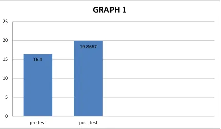
TABLE 1:STATISTICAL ANALYSIS OF FUGL MEYER SCALE USING
PAIRED T TEST
FUGL MEYER SCALE
MEAN STANDARD DEVIATION
STANDARD ERROR MEAN
T SIGNIFICANCE
Pre test 16.4 1.54919 0.40000
18.06 0.05
Post test 19.8667 1.55226 0.40079
FIGURE 1: STATISTICAL ANALYSIS OF FUGL MEYER SCALE USING
PAIRED T TEST:
16.4
19.8667
0 5 10 15 20 25
pre test post test
[image:32.612.75.507.458.710.2]23
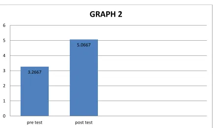
TABLE 2:STATISTICAL ANALYSIS OF STEP TEST USING PAIRED T
TEST
STEP TEST
MEAN STANDARD DEVIATION
STANDARD ERROR MEAN
T SIGNIFICANCE
Pre test 3.2667 0.79880 0.20624
12.43
0.05
Post test 5.0667 0.88371 0.22817
FIGURE 2: STATISTICAL ANALYSIS OF STEP TEST USING PAIRED T
TEST:
3.2667
5.0667
0 1 2 3 4 5 6
pre test post test
[image:33.612.74.507.446.707.2]24
EXPERIMENTAL GROUP:
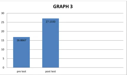
TABLE 3:STATISTICAL ANALYSIS OF FUGL MEYER SCALE USING
PAIRED T TEST:
FUGL MEYER SCALE
MEAN STANDARD DEVIATION
STANDARD ERROR MEAN
T SIGNIFICANCE
Pre test 16.8667 1.45733 0.37628
56.50 0.05
Post test 27.1333 1.76743 0.45634
FIGURE 3: STATISTICAL ANALYSIS OF FUGL MEYER SCALE USING
PAIRED T TEST:
16.8667
27.1333
0 5 10 15 20 25 30
pre test post test
[image:34.612.76.507.443.698.2]25
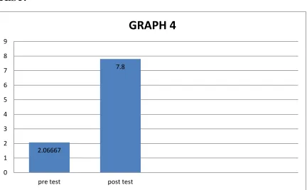
TABLE 4:STATISTICAL ANALYSIS OF AND STEP TEST USING
PAIRED T TEST:
STEP TEST
MEAN STANDARD DEVIATION
STANDARD ERROR MEAN
T SIGNIFICANCE
Pre test 2.06667 0.70372 0.18169
20.19 0.05 Post test 7.8 1.08233 0.27944
FIGURE 4: STATISTICAL ANALYSIS OF STEP TEST USING PAIRED T
TEST:
2.06667
7.8
0 1 2 3 4 5 6 7 8 9
pre test post test
[image:35.612.75.507.437.705.2]26
USING INDEPENDENT T TEST:
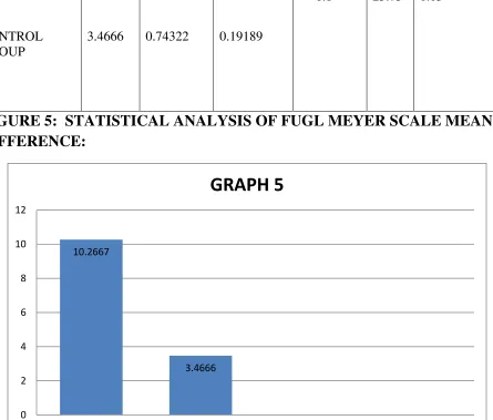
TABLE 5:STATISTICAL ANALYSIS OF FUGL MEYER SCALE MEAN
DIFFERENCE:
GROUP MEAN
STANDA RD DEVIATI ON STANDAR D ERROR MEAN MEAN DIFFERENC E
T SIGNIFICA NCE
EXPERIMENTA L GROUP
10.2667 0.703729 0.18170
6.8 25.73 0.05
CONTROL GROUP
3.4666 0.74322 0.19189
FIGURE 5: STATISTICAL ANALYSIS OF FUGL MEYER SCALE MEAN
DIFFERENCE:
10.2667 3.4666 0 2 4 6 8 10 12
EXPERIMENTAL GROUP CONTROL GROUP
[image:36.612.90.535.297.676.2]27
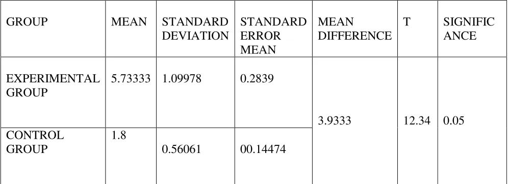
TABLE6: STATISTICAL ANALYSIS STEP TEST MEAN DIFFERENCE:
GROUP MEAN STANDARD DEVIATION STANDARD ERROR MEAN MEAN DIFFERENCE
T SIGNIFIC ANCE
EXPERIMENTAL GROUP
5.73333 1.09978 0.2839
3.9333 12.34 0.05 CONTROL
GROUP
1.8
0.56061 00.14474
[image:37.612.107.536.433.684.2]28
29
RESULTS
Fugl Meyer Assessment:
• Effectiveness of conventional physiotherapy (control group )
While comparing the pre-test and post test values of control group using paired‘t’ test, the calculated t value is 18.06. When comparing the mean values of both, the post-test mean value is 19.8667 which is greater than the pre-test mean 16.4. Hence it confirms that there is a significant difference in post-test control group than pre-test control group.
• Effectiveness of Bilateral leg training and conventional physiotherapy (experimental group )
While comparing the pre-test and post-test values of experimental group using paired
‘t’ test, the value is 56.50. When comparing the mean values of both, the post-test mean value 27.1333 which is greater than the pre-test mean 16.8667. Hence it confirms that there is a significant difference in post0test experimental group than pre-test experimental group.
STEP TEST:
• Effectiveness of conventional physiotherapy (control group )
While comparing the pre-test and post-test values of control group using paired ‘t’ test, the calculated t value is 12.43.When comparing the mean values of both, the post test mean value is 5.0667 which is greater than the pre-test mean 3.2667. Hence it confirms that there is a significant difference between the pre test and post test values in control group.
• Effectiveness of Bilateral leg training and conventional physiotherapy (experimental group)
While comparing the pre-test and post-test values of experimental group using paired
‘t’ test, the calculated t value is 20.19. There is significant difference between the pre-test and test values of experimental group. When comparing the mean values of both, the post-test mean value 7.8 is greater than the pre-post-test mean value 2.06667. Hence
30
DISCUSSION
This study was an experimental approach to find the effectiveness of repeated bilateral leg training to improve the lower extremity performance in MCA stroke patients.
The age of the subject were almost identical in both experimental and control group. The duration of the condition was three to six month after onset. Eight males and seven females were in control in experimental group.
Both groups were assessed on the first day and last day of the treatment. The tool taken to measure the outcome was Fugl Meyer lower extremity motor performance scale and the total score is thirty four. It assesses the impairment in motor performance. It has been shown to be valid and reliable tool for assessing motor function (Duncan P et al 1983). Another outcome measure used is Step test. It also has been shown to be valid and reliable (Vicki Stemmons Mercer 2009).
The control group was given conventional physiotherapy which includes range of motion exercises, functional mobility exercises, strengthening exercises, balance training and gait training. Repeated bilateral leg training was additionally given to experimental group while rests of the treatments were same.
A three week treatment program administered to the experimental group in adjunct to conventional physical therapy to improve the motor function. This support the hypothesis that repeated training program bilaterally improves the lower extremity motor function of post stroke. While comparing the gain obtained by both groups it evident that experimental group performed better than the control group.
On statistical analysis of Fugl Meyer lower extremity score, paired t test showed significant difference in pre test and post test scores of both control group and experimental group.
On statistical analysis of Fugl Meyer lower extremity examination, independent t test showed significant difference in experimental group over control group.
31
On statistical analysis of Step test score, independent t test showed significant difference in experimental group over control group.
Recently bilateral training has emerged from the motor control literature as promising strategies for stroke patients (S J Page et al). Studies conducted in upper limb indicate it as a favorable method (Jill Whittal et al).
Practicing bilateral movements in synchrony and in alteration may result in facilitation effects from the non paretic limb to the paretic limb. When bimanual movements are initiated simultaneously, the limb act as a unit that supersedes individual limb action, indicating that both limb are strongly linked as co-ordinated unit in the brain (Kelso Jas et al 1983).
Bilateral training induces functionally relevant recruitment of contralesional motor cortex in chronic stroke survives (A R Luft et al 2003).
Another proposed mechanism is plasticity. Researches demonstrated that even a simple novel thumb movement sequence repeated over a time induces cortical representation changes and these representations enlarge as learning occurs. These cortical changes following motor task learning have traditionally been called plasticity (Classen et al).
Cycling leg exercise while sitting incorporates bilateral assisted active training, the paretic limb cycles with the help of non paretic limb. Thus while strengthening the lower limb muscle; cycling exercises also encourage muscle control of lower limb which may enable patient to take more weight through affected leg while standing (Kautz S A, Brown D A 1998).
After bilateral arm training with auditory cueing, MRI showed novel or enlarged activation on the primary cortex of non injured contra-lesional hemisphere (J Whitall et al 2004).
32
Another proposed mechanism is that bilateral training is optimal for stroke patients because they receives proprioceptive and visual feedback from the unaffected limb that they do not receive during unilateral practice in which only the affected limb is used. Indeed when practicing bilateral a patient can use the unaffected extremity’s neurologically intact afferent and efferent signals, and look and feel the movement within that limb, to promote similar movement in affected limb. The visual input of seeing the unaffected limb performing an action may also provide a model with which the patient can better move the affected limb and become more successful (Stephen J Page et al 2005).
This study suggests that a bilateral training program for the lower extremites of post strokre hemiplegia patients leads to significant functional gains. The method appears simple and so it will be a very useful training method for stroke patients. All the recent datas suggests that the impropvement by bilateral training results from the mechanism of neuroplasticity. We can conclude that a bilateral training procedure for the lower extremity in hemiplegic patients is better compared to the unilateral training procedure.
33
34
SUMMARY AND CONCLUSION
Summary:
The purpose of the study determines effectiveness of repeated bilateral leg training to improve lower extremity motor performance in subacute stroke patients. For the study an experimental approach to pre-test post test control group design was used. Population included unilateral MCA stroke patients. Sample size was 30 and by random sampling method, they were divided into two groups, a control group (Group A) and an experimental group (Group B) of 15 subject each. The tools selected for measuring outcome was Fugl Meyer lower extremity motor performance score to obtain lower extremity motor performance (with a maximum score of 34) and step test.
The data was collected before and after administration of treatment program. Duration of the treatment program was three weeks. Control group was given conventional physiotherapy and experimental group was given 40 minutes of repetitive bilateral leg training in addition to conventional physiotherapy. The data obtained were analysed using‘t’ test.
The result of statistical analyses showed significant improvement in the experimental group over the control group. Thus it can be concluded that repetitive bilateral leg training can be used to improve the lower extremity motor performance in post stroke hemiplegic patients.
Conclusion
Repetitive bilateral leg training is an effective method for improving; lower extremity motor function in hemiplegic middle cerebral artery stroke patients.
35
LIMITATIONS AND SUGGESTIONS
LIMITATIONS:
• Sample size was small. Therefore study with much larger population is recommended.
• All measurements were taken manually and this may introduce human error which could threat the study reliability.
• Study was conducted for a short period of time.
• The study assessed only short term progress of the patient.
• No follow-ups could be done.
SUGGESTIONS:
• To establish efficacy of the treatment a large sample size study is required.
• To make the results more valid a long term study may be carried out.
36
37
BIBLIOGRAPHY
1. Susan b o Sulliwan, Thomas J Schmitz, physical Rehabilitation Assessment and Treatment, 1995.
2. Dettmann MA, Linder MT, Sepioc SB. Relationships amoung walking performance, postural stability, and functional assessments of the hemiplegic patients, 2002.
3. Kusoffsky A, Apel I, Hirschfeld H. Reaching lifting-placing- task during standing after stroke: coordination amoung ground forces, ankle muscle activity; and hand movement, 2010.
4. Wall JC, Turnbull GI. Gait asymmetries in residual hemiplegia,2005.
5. Bohannon RW, Larkin PA. Lower extremity weight bearing under various standing conditions in independently ambulatory patients with hemiparesis, 2007.
6. Turnbull GI, Charteris J, Wall JC. Deficiencies in standing weight shifts by ambulant hemipegic subjects, 2011.
7. Page S.J, Peter Levine “Bilateral training aids rehabilitation progression”,Biomechanics,2012.
8. Jill Whitall et al, “Repetitive bilateral training with rhythmic auditory cueing improves motor function in chronic hemiparetic stroke”. American Heart Association, 2006.
9. Raymond D Adams, Maurice Victor, Allan H.Ropper principles of neurology sixth edition,2005.
38
11.Terent, A. Marke, L. A.,, Asplund, K. Norrving, B.,Jonsson, E., and Wester, P.OCosts of stroke in Sweden: A national perspective stroke ,1994.
12.Donaldson C, Tallis R, Miller S, Sunderland A, Lemon R, Pomeroy V, “Effects of conventional physical therapy and functional strength training on upper limb motor recovery after stroke: a randomized phase two study”, Neuro rehabilitation and neural repair, 2009 .
13.Pollock Alex, Baer D. Gillian, Langhorne Peter, Pomeroy M Valerie.” Physiotherapy treatment approaches for stroke.” Stroke. 2008.
14.Yannan Fang, Xiaohua Chen, Hua Li, Jianwen Lin, Ruxun Huang and Jinsheng Zeng, “A study on additional early physiotherapy after stroke and factors affecting functional recovery” Clinical Rehabilitation 2003.
15.E Ernst. “A review of stroke rehabilitation and physiotherapy”. Stroke 1990.
16.Klunding P and Billinger SA, “Exercise induced changes of upper extremity in chronic stroke survivors” Top stroke rehabilitation, Kanasas City, 2005 .
17.Duncan PW, Propst M Nelson SG. Reliability of the Fugl Meyer assessment of the sensorimotor recovery following cerebrovascular accident, 2002.
18.Vicki Stemmons Mercer, Jancet Kues Freburger, Shuo-Hsiu Chang and Jama L. Purser.” Step test scores are related to measure of activity and participation in the first 6 months after stroke. Phys ther jnl 2009.
19.Johannsen L, Wing AM, Pelton T, Kitaka K, Zietz D, Brittle N, Van VlietbP, Riddoch J, Sackley C, McManus R. “Seated bilateral leg exercise effects on hemiparetic lower extremity function in chronic stroke”. Neurorehabilitation neural repair. 2010 .
20.Stephen J Page, Peter Levine, Jennah Teepen, Eric C Hartman clinical rehabilitation, 2008.
21.Marklund I, Klassbo M Clinical rahabilation, 2006 .
39
23.Whitall J, McCombe Waller S, Silver KH, Macko RF Stroke , 2000 .
24.Stinear JW, Byblow WD. Clin neurophysio,2004.
25.Keith D. Hill Juilie BernhardtAnee M. Mc GannDoris Maltese Dana Berkovits. ”A new test of dynamic standing balance for stroke patients. Vol.48. pp257-262.oct 1996.
26.Wood-dauphinee S,BergK,Bravo G,Williams JI;The Balance scale;Responding to c clinically meaningful changed. Canadian journal of rehabilitation1997.10;35-50
26. L.Blum and N.K.Bitensky . Usefulness of berg Balance scale in stroke rehabilitation. A Systematic review. Physical therapy 2008.88.559-566.
27.Fujiwara T,Liu M,Tsuji T,Sonoda S,Mizuno k,Akaboshi k,et al. Development of a new measure to assess trunk impairment after stroke(trunk impairment scale);its psychomentric properties. American jorunal of physical medicine rehabilitation2004.83.681-688.
28..Bonan IV .Yelnik AP,Colle FM,Michaud C,Normand E,Panigot B,ET AL.Relaince on visual information after stroke. PartII ;Effectiveness of a balance rehabilitation program with visual cue deprivation after stroke . A randomized controlled trail . Archive physical medicine rehabilitation.2004.85;274-278
29.Miranda Chantal Boonstra. The sit-to stand movement; A clinical evaluation tool for knee and hip arthroplasty patients. NCEBP.2010
30.Janssen WGM,Bussmann JBJ,Stam HJ. Determinants of the sit-to –stand movement;a review.physical therapy.2002.82;866-79
31.Fu-Ling Tung Yea-Ru Yang Chao-chung Lee Ray-yau ang. Balance outcomes after additional sit –to –stand training iinsubjects with stroke;a randomized controlled trial. Clinical rehabilitation.2010.24(6)533-542.
32.Monger C,Carr JH,Fowler V. Evallluation of a home –based exercise and rtaining program toimprove sit –to stand in patients with chronic stroke. Clinical rehabilitation 2002.16.361-367
40
41
APPENDIX I
CONSENT FORM
I………..aged………….yrs, voluntarily consent to participate
the research named “
THE EFFECTIVENESS OF REPEATED BILATERAL
LEG TRAINING TO IMPROVE LOWER EXTREMITY MOTOR
PERFORMANCE IN MCA STROKE PATIENTS
”
.
The researcher has explained me the treatment approach in brief, risk of participation and has answered all the questions pertaining to the study to my satisfaction.42
APPENDIX II
EVALUTION FORM
Subjective Assessment
Name :
Age :
Sex :
Occupation :
Address :
Chief Complaint :
History of present illness :
Past medical history :
Drug history :
Family history :
Social history :
Personal history :
General Examination :
Vital signs
43
Pulse rate
Respiratory rate
Blood pressure
Cardiovascular system
Respiratory system
Objective Assessment
Neurological examination
• Higher functions:
• Level of consciousness
Glasgow coma scale (E4 M6 V5)
• Eye opening
Spontaneous - 4
To speech - 3
To pain - 2
No response - 1
• Best motor response Follows motor commands - 6
Localizes pain - 5
Withdrawal - 4
44
Abnormal extension - 2
No response - 1
• Verbal response Oriented - 5
Confused conversation - 4
Inappropriate words - 3
Incomprehensible sounds - 2
No response - 1
• Orientation
Time
Place
Person
• Attention
• Cognition
• Fund of knowledge
• Calculation ability
• Proverb Interpretation
Mini mental state examination test is used to assess cognition
45
• Memory
Declarative
Non declarative
Long term
Short term
• Cranial nerve examination
Nerves Right Left
Olfactory
Optic
Oculomotor
Trochlear
Trigeminal
Abducent
Facial
Vestibulocochlear
Glossopharyngeal
Vagus
Spinal accessory
Hypoglossal
• Sensory Examination
46
Intact : normal, accurate
Decreased : Delayed response
Exaggerated : Increased sensitivity
Inaccurate : Inappropriate perception of a given stimulus
Absent : no response
Inconsistent : unable to assess
ASIA Sensory Scoring
• - absent
• - impaired
• - normal
NT - Not testable
Sensation includes:
Superficial (pain, touch, temperature, pressure)
Deep (movement sense, position sense, vibration sense)
Combined cortical (two point discrimination, graphasthesia, stereognosis, tactile localisation, double simultaneous stimulation, barognosis, recognition of texture)
• Motor examination
47
• - No palpable or observable muscle contraction
• – palpable muscle contraction, no observable motion
• – full available ROM against gravity minimizes plane, no resistance
• – full available ROM against gravity, no resistane
• – Full available ROM against gravity nearly moderate manual resistance
• - Full available ROM against gravity, strong manual resistance
b) Tone
Assess hypertonicity and hypotonicity
• Girth measurement
• Deep tendon reflexes
• - no response
1+ - present but depressed
2+ - average, normal
3+ - increased, brisker than average
4+ -very brisk hyperactive with clonus
Deep tendon reflexes are (biceps, bracheoradialis, triceps,fingerflexors, hamstring, quadriceps,tendo Achilles, jaw jerk
• Superficial reflex ( plantar reflex, abdominal, corneal, cremestric)
• Primitive reflexes (ATNR, STNR, tonic neck reflex, tonic labrynthine reflex, flexor withdrawal, grasp reflex, moro, startle,sucking, rooting)
48
• Co-ordination (non-equilibrium, equilibrium test)
• Balance assessment scales (berg balance scale)
• Gait assessment (observational gait analysis, step length, stride length, cadence)
• Functional assessment (Barthel index)
• Investigation
MRI and CT scan report
Other interventions (blood, EEG, ECG)
M) Problem list
Primary
Secondary
N) Aims
O) Recommendation
P) Follow up.
49
FUGL-MEYER ASSESSMENT FOR LOWER EXTREMITY
• Reflex activity
• Subject is supine or sitting.
• Attempt to elicit the Achilles and patellar reflexes.
• Assess the unaffected side first.
• Test affected side.
• Scoring (maximum possible score= 4)
• 0- No reflex activity can be elicited.
• 2- Reflex activity can be elicited. Items to be scored are Achilles and patellar reflexes.
• Flexor synergy
• Subject is supine.
• Have patient perform movement with unaffected side first.
• On the affected side, check subject’s available PROM at each joint to be tested.
• Start with leg fully extended at hip, knee, and ankle. Instruct the subject to “bring your knee to your chest” (therapist is observing for evidence of hip, knee, ankle flexion in order to assess the presence of all components of flexor synergy). Therapist can cue the patient to move any missing component.
• Test 3x on the affected sided and score best movement at each joint.
• Scoring (maximum possible score= 6)
• 0- cannot performed at all
50
• 2- full motion
Items to be scored are: hip flexion, knee flexion, and ankle dorsiflexion.
• Extensor synergy
• Subject is sidelying.
• Have patient perform movement with unaffected side first.
• On the affected side, check subject’s available PROM at each joint to be tested.
• Start in 90 degrees knee flexion and ankle dorsiflexion.
• Instruct the subject to “push your foot down and kick down and back”.(ankle plantarflexion, knee extension, hip adduction and hip extension.)
• Slight resistance should be applied in adduction which is gravity-assissted in the position to ensure subject is actively doing it.
• Test 3x on the affected side and score best movement at each joint.
• Scoring (maximum possible score=8)
• 0- No motion
• 1- partial motion
• 2- full motion
Items to be scored are: hip extension, hip adduction, knee extension, ankle plantarflexion.
• Movement combining synergies (in sitting)
• Knee flexion beyond 90 degree
51
stretch. To drecrease friction, subject’s shoes can be removed, but socks should remain on.
• Have patient perform movement with unaffected side first.
• Subject is instructed to “pull your heel back and under the chair.”
• Test 3x on the affected side and score best movement.
• Scoring (maximum possible score=2):
• 0- No active motion
• 1- From slightly extended position, knee can be flexed but not beyond 90 degree.
• 2- Knee flexion beyond 90 degree.
• Ankle dorsiflexion
• Subject is sitting, feet on floor, with knees free of chair. Calf muscles should not be on stretch.
• Have patient perform movement with unaffected side first.
• On the affected side, check subject’s available PROM at the ankle join.
• Subject is instructed to “keeping your heel on the floor, lift your foot.”
• Test 3x on the affected side and score best movement.
• Scoring (maximum possible score= 2):
• 0- No active motion
• 1- Incomplete active flexion
52
• Movement out of synergy(standing, hip at 0 degrees)
• Knee flexion
• Subject is standing, hip at 0 degrees (or full available ROM up to 0 degrees). On leg that is being tested, hip is at 0 degree (or full available ROM up to 0 degrees), but the knee is flexed, and the subject’s toes are touching the floor slightly behind. Evalutor can provide assistance to maintain balance and subject can rest hands on table.
• Have patient perform movement with unaffected side first.
• Subject is instructed to “keeping your hip back, kick your bottom with your heel”
• Test 3x on the affected sided and score best movement.
• Scoring (maximum possible score=2):
• 0- Knee cannot flex without hip flexion
• 1- knee flexion begins without hip flexion but does not reach to 90 degree or hip begins to flex in later phase of motion
2- knee flexion beyond 90 degree ( knee flexion beyond 90 degree with hip maintained in extension)
• Ankle dorsiflexion
53
can provide assistance to maintain balance and subject can rest hands on a table.
• Have patient perform movement with unaffected side first.
• On the affected side, check subject’s available dorsiflexion PROM.
• Subject is instructed to “keeping your knee extended and your heel on the floor, lift your foot”.
• Test 3x on the affected side and score best movement.
• Scoring (maximum possible score=2):
• 0- No active motion
• 1- Partial motion (less than full available range with knee extended; heel must remain on floor with medial and lateral borders of the forefoot clearing the floor during dorsiflexion.
• 2- Full motion ( within available dorsiflexion range with knee extended and heel on the floor)
• Normal Reflexes (sitting)
• Only done if the subject attains a score of 4 on section v( ie, if the subject does not score a 2 on each of the pervioys items, then score this item 0)
• The examiner shall elicit patellar and Achilles phasic reflexes with a reflex hammer and knee flexors with quick stretch of the affected leg and note if the reflexes are hyperactive or not.
• Scoring (maximum possible score= 2):
• 0- At least 2 of the 3 phasic reflexes are markedly hyperactive.
• 1- One reflex is markedly hyperactive or at least 2 reflexes are lively
54
• Coordination/speed- sitting: heel to opposite knee repetitions in rapid succession
• Subject positioned in sitting with eyes open.
• Have patient perform movement with unaffected side first.
• Subject is instructed to “Bring your heel from your opposite ankle to your opposite knee, keeping your heel on your shin bone, move as fast as possible”.
• Use a stopwatch to time how long it takes the subject to do 5 full (ankle to knee to ankle) repetitions.
• Use the full achieved active ROM in the unaffected limb as the comparison for the affected limb. If active ROM of affected limb is significantly less than that of affected limb, patient should be scored “0” for speed.
• Repeat the same movement with the affected leg. Record the time for both the unaffected and affected sides. Observe for evidence of tremor or dysmetria during movement.
• Scoring tremor (maximum possible score= 2)
• 0- marked tremor , 1- Slight tremor, 2- No tremor
• Scoring Dysmetria (maximum possible score=2)
• 0- pronounced or unsystematic dysmetria
• 1- Slight or systematic dysmetria
• 2- No dysmetria
• Scoring speed (maximum possible score=2):
• 0- Activity is more than 6 seconds longer than unaffected leg
• 1- 2-5.9 seconds longer than unaffected leg
55
STEP TEST
• The step test assesses an individual’s ability to place one foot onto a 7.5cm high step and then back down to the floor repeatedly as fast as possible for 15 seconds.
• The score is the number of steps completed in the 15 second period for each lower extremity.
• Participants were permitted to were any customary orthoses but, in accordance with published procedures for standardized administration, were not permitted to use an assistive device during testing.
• Both sides were tested, with participants completing the test first with the nonparetic foot and then with the paretic foot. Scores for lower extremity were recorded separately, as well as the sum of these 2 scores.
• Participants who were unable to stand unsupported were given a score of 0 for both lower extremities. Test-retest reliability of the ST is high, with intraclass correlation coefficients (ICCs) greater than .88 in people undergoing inpatient rehabilitation after stroke.
56
APPENDIX III
DATA ANALYSIS
FORMULAS USED FOR CALCULATIONS
1. MEAN
d =
2. STANDARD DEVIATION
S.D =
3. STANDARED MEAN ERROR
SME =
4. PAIRED ‘
t
’ TESTt =
where,
= Calculated mean difference pre-test and post-test
n = Sample size
S.D = Standard deviation
57
5. UNPAIRED ‘
t
’ TESTs
=t
=where,
n1 = Total number of subject in Group A
n2 = Total number of subject in Group B
x1 = Difference between pre test & post test values of Group A
x2 = Difference between pre test & post test values of Group B
x1 = Mean difference between pre test & post test values of Group A
x2 = Mean difference between pre test & post test values of Group B
58
APPENDIX IV
MASTER CHART
Fugl meyer assessment
Pre test and post test values of control and experimental groups
NO CONTROL GROUP EXPERIMENTAL GROUP
PRE TEST POST TEST PRE TEST POST TEST
1 14 18 18 29
2 17 19 17 28
3 15 19 15 26
4 18 22 17 27
5 15 18 19 29
6 17 19 17 26
7 16 20 16 25
8 18 22 17 27
9 17 20 18 29
10 16 20 18 28
11 17 20 16 26
12 19 23 15 25
13 18 21 17 28
14 15 19 19 30
59
STEP TEST
Pre test and post test values of control and experimental groups
NO CONTROL GROUP EXPERIMENTAL GROUP
PRE TEST POST TEST PRE TEST POST TEST
1 3 5 1 6
2 3 5 2 7
3 3 4 3 8
4 4 6 3 8
5 5 6 2 7
6 3 5 2 6
7 4 6 1 8
8 4 5 2 9
9 3 6 2 9
10 2 4 3 10
11 2 3 2 8
12 3 5 3 7
13 4 6 2 8
14 3 5 1 8