NATIONAL INSTITUTE OF SIDDHA
Chennai - 47AFFILIATED TO
THE TAMIL NADU DR. M.G.R. MEDICAL UNIVERSITY, CHENNAI - 600 032
A STUDY ON
KALANCHAGA PADAI
(DISSERTATION SUBJECT)
For the partial fulfillment of the
requirements to the Degree of
DOCTOR OF MEDICINE (SIDDHA)
BRANCH III – SIRAPPU MARUTHUVAM
INTRODUCTION
Man has eternally endeavoured to keep himself free from three types of therapeutic namely, physical, mental and spiritual. Therefore the history of medicine is as old as the history of mankind. According to Indian tradition, the primary objectives of human life are to perform religious sites of acquire wealth to satisfy the worldly desires and to attain salvation. A popular saint saying that “A sound mind in a sound body”
In additional system of medicine developed in various parts of the world during different ages.
The word siddha was derived from the word Siddhi (trans consciousness). Siddhi is interpreted in two ways. “Attaining perfection in Life” and “Heavenly bliss”. The siddha system viewed in terms of the etymological meaning can be defined as a system which can lead one to attain perfection in life and heavenly bliss.
The Siddha system, a branch of Indian systems of medicine, is holistic in its approach. The siddha medicine has its base on the principles of nature and its elements.
The siddha system is based on 96 thathuvas (philosophies). These thathuvas include fine fundamentally as well as basically known as,
1. Five basic elements (Pancha boothams) namely Vin, Kaal, Anal, Punal, Mann, 2. Five senses (Pulangal) namely Saptha, Sparisa, Ruba, Rasa, Kantha.
3. Five sensory organs (Porigal) namely Mei, Vaai, Kan, Mookku, Sevi. 4. Three humours – Vaadham, Piththam, Kabham
"mz;lj;jpy; cs;sNj gpz;lk; gpz;lj;jpy; cs;sNj mz;lk; mz;lKk; gpz;lKk; xd;Nw mwpe;J jhd; ghHf;Fk; NghNj"
- rl;lKdp Qhdk;
First of all Siddhars employed the practice of “Aasanas’ and ‘Praanayaamam’ for all the human beings to lead to longlife as ‘karpa’ medicinal therapy.
Siddhars classified in primarily the diseases based on signs & Symptoms.
Their centuries old researchful and enlightment of Siddha initially deals with etiology, pathogeneis, imbalance of humor, diagnosis and investigations (Envagai Thervugal). Secondly it is not a new one to Siddha everybody easily to get knowledge of this points of view as per the siddha text book refferences. Siddhars had a thorough knowledge of therapeutic values of herbs through their intuitions. They had tried and became very well – versed in preparations by using their scientific knowledge of a alchemy.
The whole body is governed by their physilogical factors called ‘three thathoos’ namely Vaadham, Piththam and Kabham held in the ratio of 1:1/2 :1/4 respectively. If these thathus are provoked by any external and internal factors will result as diseases. At this condition these thathoos are called ‘Three Thodam’
Air + Space = Vaatham Fire = Piththam Earth + Water = Kabham
According to the great Saint ‘Yugi muni’ the skin disorders had been classified into 18 types were dealt with in the chapter kuttam. Yugi had not explained Kalanchaga Padai as a separate disease in a detailed manner. Instead he had just made a mention in the classification of 80 vaadha diseases. Such as Kalanchaga vaadham which resembls virpodaga kuttam. The clinical features of Kalanchaga padai more or less correlated with ‘Psoriasis’ as described in modern dermatology.
“thjkyhJ Nkdp nflhJ" - NjiuaH
Morever the diseases can be formed due to changes in the mind also. Mind is influenced by various stresses in our day to day life on today world. But Siddhars also quoted very anciently that many of the diseases were caused by psychosomatic problems. So that they had advised to control one mind to get ride of stress. This was quoted by Agasthiyar as follows.
“kdkJ nrk;ikahdhy; ke;jpuQ; nrgpf;f Ntz;lhk; kdkJ nrk;ikahdhy; thAit caHj;j Ntz;lhk; kdkJ nrk;ikahdhy; thrpia epWj;j Ntz;lhk; kdkJ nrk;ikahdhy; ke;jpuQ; nrk;ikahNk"
- mfj;jpaH Fzthflk;
From the authors of this diseases study itself the very much thinking of this diseases and collected from the siddha literature which are following.
1. Internaldrug : Shenkottai Nei
-Siddha Veidhiya thirattu 2. External drug : Sivappu Ennai
- Siddha Maruthuvam Sirappu
The following pages outline the research work done on kalanchaga padai and the suggestion and recommendation offered for over coming it.
Kalanchaga Padai is by and large an incapacitating disease, bogging down the normal life of a person affected by it. Patient is very much agitated and subjected to a great matter of mentally disturbed and physically suffered. More over as per the expert experiences in medicinal therapy in all walks of medical systems, text books reveals that there would be a permanent cure as well on remedy so for reported. Available therapies are came to study with only remission and reexacerbations of this skin disorder. So for such an interactable disease the world craves for a cure which is the reason why the author has chosen this disease for her dissertation work.
Reason for selecting a shenkottai Nei is that it has equal efficacy to mercury. The main objective of the present study is to create an awarness about the siddha sciences and to high light the efficacy of sidha drugs among the public.
With this basic intention in mind fllowing specific objectives have been drawn
To thorough references in various siddha literature and modern dermatological books as literature evidences regarding the diseases Kalanchaga Padai.
To know the extent of correlation of aetiology, classification, symptomatology, diagnostic methods and the line of treatment on part with modern concepts of dermatology.
To conduct a thorough clinical study on Kalanchga padai with internal drug, that is ‘Shenkottai Nei’ in which Shenkottai is made as a main ingredient. And external drug “Sivappu ennai’ in which pungam milk is a main ingredient which are widely indicated in siddha text books are effectively valubale towards skin disorders. There by the author had anxious to go on this trial drug for Kalanchaga Padai.
To estimate the efficacy of Shenkottai Nei and Sivappu Ennai.
To make a clinical trial with necessory investigations and record all those things with follow – up study of the Patients of Kalanchaga Padai.
To have complete study of the disease Kalanchaga Padai, under the headings of
a. Pori pulangal b. Udal Kattugal c. Enn vagai thervugal d. Mukkutram etc. In order to evaluate pathology of this dreadful disease.
¾ To evaluate the pharmacological study on the trial drug.
¾ To evaluate the toxicological study on the trial drug.
¾ To study the bio – chemical analysis on the trial drug.
¾ To highlight the factors like diet, land, climatic condition and personal hygienic measures in the incidence of Kalanchaga Padai
¾ For this advise to the patient or by given counselling to the patients about their pshycological factors.
¾ To make an awarness among the patient about the further occurrence of the same.
In the Siddha Medicine the Panja boothas (five elements) are elaborately described. Skin is a part of Prithivi (earth). Skin is the largest organ of the human body. It has got a lot of functions each of which is important for the normal physiological functioning of the human body. Some of them are body temperature regulation, excretion, protection from pathogenesis etc.
Man is very much influenced by environment. This principle was much appreciated by the Siddhars which promoted them to say.
‘Microcosams reflects macrocosams’ “mz;lj;jpYs;sNj gpz;lk;”
Since skin serves as a link between man and universe it is the first organ to be influenced by the change in it. Also since it is the slightest change in mind. Among the panja boothas, Theyu is represented for sleep, appetite, thirst, fearness and unification. If any affection in Theyu, skin is also affected. Any disease affecting the skin causes a socio – economic problem, mental strain and social stigma to the patient.
According to the Siddha Text Book “ Siddha Maruthuvanga Surukkam” skin is divided into six types. They are,
1. Skin containing water 2. Skin having blood
3. Skin which produces scabies 4. Skin which produces kuttam 5. Skin Producing tumour
6. Skin which produces severe pain during an injury
Siddhars embodiments of compassion on living things were naturally moved to write about the aetiology. Clinical features of the diseases and its treatment for this particular debilitating disease called Kalanchaga Padai or Parusethil Noi. And when going for collection of literature for this diseases the author presented with the following details.
Kalanchaga Padai is a non infectious, inflammatory disease of the skin characterized by well defined erythematous plaques with large adhered silvery (ivory) mica like scales.
Neha; tUk; top (Aetiology):
Among the available siddha literature only “Yugi Muni Veithiya Chinthamani” and “Thirumoolar Vaithiyam” are the sources or information on aetiology and clinical features of the 18 types of the skin disorders. There are no specific mention about the specific factors causing Kalanchaga Padai.
“Thirumoolar” has pointed out
“gapy; nkhopaPH jpNufj;jpy; fpUkpjhNd gue;J jphpFl;lk; Nghy; Gs;sp fhZk; kayJTk; fpUkpAe;jh ele;J Gf;fpy;
NkdpaJ rurnud ntbj;J Gz;zhFk;” - FUehb fpUkpahy; te;jNjhlk; ngUfTz;L
Nfl;f yjpd;; gphptjidf; fpukkhf nghUkp tUk; thAtJk; fpUkpahNy jpNufj;jpy; nrhhp Fl;lk; fpUkpahNy ngUfptUk; gTj;jpuKk; fpUkpahNy
GOf;fb Nghy; fhZkJ fpUkpahNy” - FUehb
The text book of ‘Sirappu – Maruthuvam’ describes the following aetiological factors for Kalanchaga Padai.
1. guk;giu Nehapy; %thpy; xUtUf;Fk;
- Idiopathic and may be genetic
2. yRd jhgpjk; - Tonsilitis
3. Gg;Grg; gpzpfs; - Respiratory Diseases 4. Xt;thik - Allergic disorder 5. kdcisr;ry; - Stress and strain 6. mjpHr;rp - Anxiety
7. fhy khWghLfs; - Seasonal variations Due to Drugs such as
2. ,sk;gps;is jLg;G kUe;J - Polio vaccine 3. FNshNuh Fapd; - Chloroquine
Kjypa kUe;Jfs; FUf;fisAk;, giliaAk; mjpfhpf;fr; nra;ayhk; vd;Wk; $wg;gl;Ls;sJ.
In siddha system of medicine chronic skin diseases are brought under the clinical entity of kuttam.
“Yugi Chinthamani – 800’ describes 18 types of Kuttam (Skin disorders) ‘Thirumoolar’ quotes as follows.
“Fl;lkJtpl fug;ghd; tplePH R+iy
RNuhzpj;jj;jhy; jhJnfl;Lj; jbg;Gz;lhFk; kl;lkwNk fpUkp nrd;W kUTk; NghJ
tifaha;f; fpUkpapl tpl ePH nrd;W
“Fl;lKld; Njfnky;yhk; gwf;Fk; NghJ
FopFopaha;f; fpUkpapdPHf; nfhs;Sk; Gs;sp jl;lwNt fpUkpapl ePuha; te;j
rfy Fl;lk; tpl fug;ghd; rhw;wyhNk.”
“tpahjpAz; %thW tpsq;fpa Fl;lq;Nfs; Rahjp fpue;jp Rod; Nkfj;jhy; MWk; gahjp kz;Zsg; gy tz;bdhy; vl;Lk;
epahjp Goehyha; epd;wjpf; Fl;lNk.”
Kuttam is common word of chronic skin lesion of various origin.
The Thirumoolar quotes as follows,
1. Six types are caused by venereal origin 2. Eight types are caused by insect bite
3. Four types are caused by worms infestation.
‘Agasthiyar’ has mentioned that Kanmam is the main cause for Kutta Noi.
In Agasthiyar Paripoornam – 400
Kanmavaralauru (Psycho Social Cause):
“gotpidahy; tp\g; g+r;rp fbj;j Njh\k; ghjfHf;F xU ehSk; jPHtjpy;iy cs;tpidahY}lhbf; nfhs;s te;j
cz;ikaJ mwpahky; %Hf;fQ; nra;thH fstpidAe; jPHtjpy;iy fbdnkj;j
fUizAs;s G+uzj;jpd; fz;fhl;rp mltpid eP fhZKd;Nd mfyr; nrhy;yp
milahsk; tpuy; FWF kpd;dq; NfNs.”
- mfj;jpaH ghpG+uzk; 400 - nra;As; 214
“tpuy; FWFq;fhy; jpkpUk; tp\k; NghNyWk;
nka;aOe;Je; jiy RoYk; ntSf;FNkdp ghukhd Njfnky;yhe; jbj;Jf; fhZk;
ghjnky;yhk; ntbj;Jkpfg; Gz;zhq; fhZq; rurKld; nrhhp fug;ghd; gzk; Nghy; NjhZk; rhe;ijahNk tpe;JnfLe; jbj;JtPq;Fk; euUyfp ype;Neha;f;F kUe;jPahNj
ey;Nyhiug; gopj;j Fl;lq; fd;kkhNk.”
- mfj;jpaH ghpG+uzk; 400 - nra;As; 215
gotpidahy; tUk; Neha;fs; jPHtjpy;iy vdTk;, fd;kk; jPuNtz;Lk; vd;Wk; mfj;jpaH ghpg+uzj;jpy; $wg;gl;Ls;sJ.
In Agasthiyar Ganma Kandam
"NrHe;j Fl;lnkhL FiwNeha;ad; te;j NrjpNfs; kyuhj tUk;G nfha;jy;
jhhpe;j rPH nre;J tiffs; nra;jy;
- ghly; - 76 "jhndd;w nja;tUj; jidopj;jy;
rhHthd nghpNahHfs; jikg; gopj;jy; fhndd;w ee;jtdk; g+Q;nrbfs; ntl;ly;
fUkklh rhPuuj;jpw; fhr NghNy a+ndd;w Tlk;ngy;yhk; nkhl;L nkhl;lh
Ald; ntSj;J FiwNahAjpuQ; rpe;Jk; thndd;w fUkq;fs; jPHg;gjw;F
tiunahd;W nrhy;Ntd; Nfs; ee;jtd;ikNa". - ghly; - 77
Acording to Yugi Vaidhya Chinthamani classify 18 types of Kuttam in Siddha medicine.
“Kj;jhd Fl;le;jhNd gjpndl;Lf;Fk;
Kdpahd A+fp ehd; nrhy;yf; Nfsha; gj;jhFk; Gz;lhpff; Fl;lj;NjhL
NghUfpd;w tpw;Nghlf Fl;lkhFk;
gj;jhFk; ghkf;Fl;lk;, frrHkFl;lk;
ghpthd fHzFl;lk;, rpFuFl;lk;
fpj;jhFk; fpUl;bzf; Fl;lk;, mTJ}k;guf;Fl;lk; nfbahd kz;lyFl;lKkh nkd;Nd Fl;lkhk; ghg;ghpr Fl;lnkhL
Fbykhk; tprhHr;rpff; Fl;lkhk; tl;lkhk; itahjp Fl;lNkhL
kUtyh fPBg Fl;l rHkjy jpl;lkhe; Njj;jpU Fl;lNkhL
rpj;Jkh, Fl;lk,; rjhU Fl;lk;
Jl;lkhU Fl;le;, jd;ndhnlhf;f
Rak;ghd gjpndl;L Fl;lkhr;Nr.”
1. Pundareegam - Padar thamarai 10 Abarisam - Vali 2. Virpodagam - Koppulam 11. Visharchigam - Sori 3. Bamam - Sirangu 12. Vibadhigam - Senkuttam 4. Gaja Sarmam - Yaanai thol 13 Sarmathalam - Tholvedi 5. Karnam - Kaadhu 14. Kideepam - Pantrithol
7. Krishnam - Karuperunoi 16. Sithma - Naa 8. Avudhumbaram - Athikkai 17. Sadharu - Purai 9. Mandalam - Valayam 18. Suwedham - Venkuttam
tpHNghlfk;, ghkk;, f[rHkk,; fpUl;bzk;, mTJ}k;guk;, Njj;JU, rpj;Jkh, fpBgk;, rjhU, rUkk; Mfpa Fl;lq;fs; jPUk;.
In Yugimuni 800, Yugi mentioned the following causes for kuttam 1. Excessive intake of fish, snail, crab
2. Doing Yoga practice immediately after intake of diet
3. Excessive chillness, excessive hot, excessive sleeping, mental stress.
4. Routing diet sometimes combines along with unwanted things like sand, hair etc.
In Yugimuni 800,
“Mr;nrd;w gjpndl;L Fl;le;jhDk;
mtutHfs; nra;fpd;w mjHkj;jhyhk; Njr;nrd;w rpthya;j;jpYr; rpl;lq;fs;
nra;jtHfs; rpt epe;ij gz;zpNdhHfs; %r;nrd;w nghpNahiuj; J}\pj;NjhHfs;
%Hf;fkha; milf;fyj;ij vLf;fpd;NwhHfs; $r;nrd;w jpizastpd; Fiwe;j $yp
nfhLf;fpd;NwhH Fl;lj;jpw; $LthNu”
- A+fp Kdp 800 - ghly 496
Yugi Muni described only psycho – socio factors as the main cause. They are stress inducing factors, he had attributed the following of causes.
1. Misbehaviour in the temple 2. Sacrilege towards god 3. Abusing the geriatric people 4. Breech of trust
The main factors behind reasons one manifestation of stress which can be considered as precipitating factor. The psycho transquility of the individual depends upon the harming of social movements. Any adjustment disorder will affect the well being of the individual as well as society.
In Yugi Chinthamani among the eighteen types of skin (kuttam) diseases, the clinical features of three types resemble as Kalanchaga Padai. But no description is available in Siddha litrature under the heading of Kalanchaga Padai.
The three types of Kuttam are: 1. Thethuru Kuttam
2. Sadharu Kuttam and 3. Virpodaga Kuttam
1. Thethuru Kuttam
Clinical Features:
“rHke;jhd; rptg;ghf tl;ljzpJr;
ryitNghy; ntSf;FNk jpdTz;lhFk; $Hke;jhd; NuhfkJ kpfTz;lhFk;
kapnuy;yhQ; Ruz;LNk cUz;ilahFk; fHke;jhd; gpj;j Nrl;Lk kpFf;Fk;
fhae;jhd; fjpj;JNk jpkpUz;lhFk; jHke;jhd; rlnky;yh %jyhFk;
jhf;fhd Njj;jpUf; Fl;le;jhNd”
- A+fp Kdp itj;jpa rpe;jhkzp 800 ghly;, 511 Under “Thethuru Kuttam”, annular erythematous lesions with the white appearance, itching, oedema of the body and rolling of hairs like balls are the characteristic clinical features in this entity.
2. Sadharu Kuttam
Clinical Features
“rpj;jhd jz;bg;gha; uj;jtHzk;
vspjhd Nrl;Lkthjj; Jw;gj;jp gj;jhd fubfl;bg; Gz;ZkhFk;
ghk;G Njhy; Nghw;wpiue;J gUj;Jf; fhZk; tpj;jhd %f;NfhL fhJ fd;dk;
kpfj;Jbg;ghf; rjhU F\;le;jhNd"
Erythematous patches, Burning, Itching, patches covered with white silvery scales thickening of the eyes, cheek & nose are the clinical features of satharu kuttam.
3. Virpodaga Kuttam
Clinical features:
“GJikaha;r; rhPunkq;Fe; jpdTz;lhFk;
nghUntbaha;j; jpf;nfdj;jPf; nfhOe;J Nghy; nkJikaha; tpl;nlhpAk; ey;y ghk;gpd;
tp\g;glk; Nghy jbj;J ntSg;GkhFk; fJikaha; kpfr; nrhhpa rptg;GkhFk;
J}f;fnkhL rQ;ryKk; kpfTz;lhFk; fJikaha; Njhnyy;yhe; jbg;Gz;lhFk;
fdj;jtpw; Nghlfkhd Fl;le;jhNd”
- A+fp Kdp ngUE}y; 800, ghly; - 498
A skin erythematous lesions with swelling white appearance and itching are described. Usually these entities are associated with anxiety and despair.
In the text book ‘Siddha Maruthuvam Sirappu’ author by Dr. R. Thiagarajan, LIM describes as,
1. The skin lesions are red in colour and raised margin the lesions have white silvery and rough thick scales with pin point bleeding.
2. There are variation in the size and shape
3. In children these lesion may be like water drops and these may occur in scalp and in face. Sometimes as per the severity it seems all over the body.
Kalanchaga Padai is often associated with painful arthritis known as Kalanchaga Vaatham. Cases in chronic stage some arthritis incapacitates to resort to hospitalization. The most often affected joints are interphalangeal joints.
The terminal interphalangeal joints are usually involved a opposed to the proximal interphalangeal joints in “Azhal keel Vaayu” which is identical with Rheumatoid Arthritis. In these cases the affected fingers shows nail changes. This combination is termed “Psoriatic arthropathica”.
The joints of fingers, ankles, knee and sacroilliac are selectively affected. Those joints are swollen and painful with psoriatic lesions. Radiological changes shows gross changes in the affected area. These are characteristic and consists of osteoporosis followed by decreased density, diminished joint space, erosion of joint surfaces followed by eventual destruction of the end bones.
Yugimuni describes the clinical features of Kalanchaga Vaatham as follows. khj khq; fhy; ifapy; Fuq;fpuz;Lk;
tUe;J re;J KWf;fpNa File;J nehe;J ehjkh eiljhDe; jhd; nfhlhky;
eype;JNk Klkhfpf; fuL fl;br; NrjkhQ; rle;jhD kpf ntSj;Jj;
jpdNthL rpuq;Fkha;r; Nrl;gkhfpf; fhjkha UrpnahL kaf;fkhFk;
fUjpa fhshQ; rfkhk; thjkhNk.
- A+fp itj;jpa rpe;jhkzp nra;As; 259.
Pain in the major joints with immobility, pustules, itching, anorexia, fatigue etc, are the clinical features of Kalanchaga Vaatham
In Chronic Cases
1. The skin lesion occur over fore arms skin.
2. In Some, these patches appear over the palm and soles. 3. In some, the patches occur all over the body.
4. The patches are coin shaped over them. The shape may be either round or oval.
6. One fourth of patches have lesion over nails pitting in nature. 7. 7% of patients develop affection of joints as psoriatic arthropathy.
Neha; fzpg;G
(Diagnosis):
Piniyari Muraimai is a method of dignosing a disease (affecting the mankind) it is based upon three main principles;
1. Poriyalarithal (Inspection) 2. Pulanalarithal (Palpation) 3. Vinathal (Interrogation)
Physicians ‘ Pori’ and Pulan’ are used as tools for examing the ‘pori pulan’ of the patient. The above principles correspond to the methodology of 1. Inspection,
2. Interrogation and 3. Palpation in modern medicine, for arriving a clinical diagnosis of the disease.
1. Poriyalarithal (Inspection)
Pori is considered as the five senses of perception namely
1. Nose 2. Tongue 3. Eye 4. Skin 5. Ear
‘Poriyalarithal’ is examining the pori of the patient by the physician for diagnosing.
2. Pulanalarithal (Palpation)
‘Pulan’ are five object of senses. They are, 1. Smell 2. Taste 3. Vision 4. Sensation to touch 5. Hearing
‘Pulanalarithal’ means examination of the ‘pulan’ of the patient by the physician for diagnosing purpose.
Vinathal is gathering the informations regarding the history of diseases, its clinical features etc, from the patient or his close relatives who are taking care of him, when the patient is not in a position to speak or of the patient is a child.
msitfs;
(Logics)
Alavaigal are used in clinical diagnose of a disease.
msit fhz;ly; fUjy; ciu mghtk; nghUs; xg;ghnwd;gH msit NkYk; xkpGz;ik iajpfj; Njhbay; ngd ehd; fsit fhz;gH mitapw;wpd; NkYk; miwtH mitnay;yhk; msit fhz;ly; fUjy,; ciu vd;Wk; %d;wpylq;fpLNk.
- rptrpj;jpahH msit vz;. 6 Alavai is divided in to ten types, they are
1. Observation - fhz;ly; 6. Comparison - cgkhdk; 2. Inference - FUjy; 7. Inference by
elimination
- ghhpNr\k;
3. Authority, Literature
- Ciu 8. Probability - rk;gtk;
4. Preception - mghtk; 9. Tradition - IjPfk; 5. Presumption - mUj;jg;gj;jp 10. Natural
Inference
- ,ay;G
The above mentioned “ten alavaigal” the main three alavaigal are,
1. Kaandal
Through ‘ kaandal’ the physician can directly see the patien and hear the patients all the complaints and at length concludes a diagnosis.
2. Karuthal
Through Envagai thervu, Neerkuri and Neikuri we can diagnose a disease by Karuthal.
Comparative study of the signs and symptoms of the patient with the reference books and come to a diagnosis.
Ennvagai thervugal (Eight diagnostic tools)
Siddhars have developed a unqiue method of diagnosing the disease by “Enn vagai thervugal’
“ehb ];ghprk; eh epwk; nkhop tpop kyk; %j;jukpit kUj;JtuhAjk;”
- Neha; ehly; Neha; Kjy; ehly (Kjy; ghfk;) nka;f;Fwp epwk; njhdp tpop eh ,Ukyk; iff;Fwp
- NjiuaH
Hence the diagnosis is made by the following.
1. Naadi (Pulse) 5. Mozhi (Voice) 2. Sparisam (Sensation to Touch) 6. Vizhi (Eyes) 3. Naa (Tongue) 7. Malam (Feces) 4. Niram Colour 8. Moothiram (Urine)
The specialty of eight tools of diagnosis is mentioned in the following verses also.
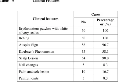
Kalanjacha Padai in relation with Ennvagai thervugal
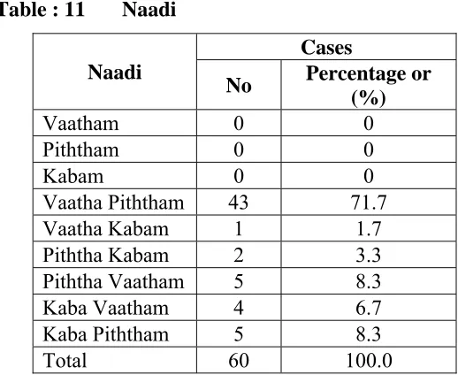
1. Naadi (Pulse)
clypy; capH jhpj;jpUg;gjw;Ff; fhuzkhd rf;jp vJNth mJNt jhJ my;yJ ehb vdg;gLk;.
“ehb vd;why; ehbay;y, euk;gpy; jhNd eykhfj; Jbf;fpd;w JbjhDky;y
ehb vd;why; mz;l Nguz;lnky;yhk; ehb vOtifj; Njhw;wj;Js;sha; epd;w ehbaJah uha;e;J ghHj;jhuhdhy; ehbAWk; nghUs; njhpe;J ehL thNu”.
- gjpndz; rpj;jH rjf ehb E}y;
Naadi is responsible for the existence of life and can be felt one inch proximal to the wrist on the radial side by means of palpation with the timps of index, middle and ring fingers corresponding vaatham, Piththam and kabam respectively.
The three humours vaatham, Piththam and kabam exists in the ratio 1: ½ : ¼ normally. Derangement in these rations leads to various disease entities.
The three “Uyir thathukal” are formed by the combination of three naadigal with three Vaayu.
a) Edakalai + Abaanan = Vaatham b) Pinkalai + Piraanan = Piththam c) Suzhumunai + Samaanan = Kabham
In kalanchaha padai the following types of naadi can be seen commonly. They are,
a) Vaatha Piththam b) Piththa vaatham c) Kaba Piththam
II. Sparism
In case of Kalanchaga Padai well defined macules, papules, thickening, roughness, pain and white silvery scaling of the skin can be noticed at the affected area.
III. Naa
No abnormality is seen in Naa
IV. Niram
White patches with silvery scales can be noticed at affected areas.
V. Mozhi
VI. Vizhi
No abnormality is seen in vizhi.
VII. Malam
Constipation was reported in some cases.
VIII. Moothiram
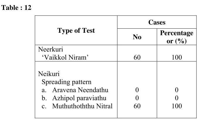
Collection of urine for the determination of Neerkuri and Neikuri, a special diagnotic method.
Neerkuri and Neikuri
“mUe;J khwpujKk; mtpNuhkjha;
m/fy mytHjy; mfhyT+d; jtpHe;jow; Fw;ws tUe;jp cwq;fp itfiw
Mbf; fyrj; jhtpNa fhJnga; NjhU K$Hj;jf; fiuFl; gLePHpd; epwf;Fwp nea;f;Fwp epUkpj;jy; flNd”.
- rpj;j kUj;Jthq;f RUf;fk;
Prior to the day of urine examination the patient is instructed to take a balanced diet and quantities of food must be proportionate to his routine in take. The patient could have no disturbed sleep. After waking up in the morning, the first urine voided is collected in a clear wide mouthed glass container and is subjected to analysis of “neerkuri and neikuri” with in one and a half an hour. Then, neerkuri is to be found out by
Neerkuri
“te;j ePHf;fhp iail kzk; Eiu vQ;rnyd; iwe;jpa Ysit aiwFJ KiwNa”
- rpj;j kUj;Jthq;f RUf;fk; Voided urine has the following characters
1. Niram - Colouration 2. Edai - Specific gravity 3. Manam - Smell
5. Enjal - Quantity of urine voided
Apart from these, the frequency of urination, abonormal constituents, such as sugar, protein, presence of blood, pus, renal calculus crystals also be to found out.
In Kalanchaga padai patient straw or hay coloured urine is noticed.
Neikuri:
The speciality of neikuri is stated in the following verse. “Iaf;Fwp nfhLtl thdpo ykHe;NjhH
iff;Fwp njhpj;j eq;flTisj; Jjpj;Nj nka;f;Fwp epwe;njhdp tpopeh ,Ukyk; iff;Fwp KotJ} cq;fw;whH jk;kpDk; ngha;f;Fwp nka;f;Fwp GfY nktHf;Fk; nea;f;Fwp mjid ,e;ePzpyj; Jiug;gghk;”.;
- rpj;j kUj;Jt Neha; ehly; Neha; Kjdhly; jpul;L
The Process of dropped gingely oil indication “epf;Fwpf; Fiuj;j epUkhz ePhpw;
rpwf;f ntz;nza;NahH rpWJsp eLtpLj; njd;Wwj; jpwe;njhyp Vfhjikj;jjp dpd;wjptiy Nghk; newptpopawpaTk; nrd;wJ GfYQ; nra;jpia AzNu”
- rpj;j kUj;Jt Neha; ehly; Neha; Kjdhly; jpul;L The collected specimen as said above is to be analysed by following method. The specimen is kept open in a glass dish or hina clay container. It is to be examined under direct sunlight, without any shaking of the vessel. Then add on drop of gingely oil by at a distance of ½” or ¾” height observe keenly the direction it spreads with in few minutes, and conclude the diagnosis as follows.
“muntd ePz;bd; m/Nj thjk; Mop Nghy; gutpd; m/Nj gpj;jk; Kj;njhj;J epw;fpd; nkhoptnjd; fgNk mutpyhopAk; Mopapy; muTk;
mutpd; Kj;Jk; Mopapy; Kj;Jk;”.
Sl.No Kalam Kuttram State of Kuttram Suvai 1. Kaar Kaalam
(Aavani – Puratasi) (Aug 16 – Oct 15)
Vaatham↑↑ Piththam↑ Vettrunilai Valarchi Thannilai Valarch Enippu Pulippu Uppu 2. Koothir kaalam
(Iypasi – Karthigai) (Oct 16 – Dec 15)
Vaatham (-) Piththam ↑↑
Thannilai Adaithal Vettrunilai Valarchi Enippu Kaippu Thuvarppu 3. Munpanikaalam
(Markazhi – Thai) (Dec 19 – Feb 15)
Piththam (-) Thannilai Adaithal Enippu Pulippu Uppu 4. Pinpanikaalam
(Masi – Panguni) (Feb 16 – Apr 15)
Kabam ↑ Thanniklai Valarchi Enippu Pulippu
Thuvarppu 5. Elevenil kaalam
(Chithirai – Vaikasi) (Apr 16 – Jun 15)
Kabam ↑↑ Vetrunilai Valarchi Kaippu Karppu Thuvarppu 6. Mudhuvenil kaalam
(Aani – Aadi) (Jun 16 – Aug 15)
Vaatham ↑
Kabam (-)
Thannilai Valarchi Thannilai Adaithal
Enippu
Five Types of Lands
It is divided in to five types.
1. Kurinji : Mountain regions and surroundings 2. Mullai : Forest regions and surroundings 3. Marutham : Cultivating regions and surroundings 4. Neithal : Sea and coastal region
5. Palai : Desert land only
Udal Kattugal
Sl.No Udal
kattugal
Functions
1. Saaram It gives strength to the body and mind
2. Senneer Saram after absorption is converted into senneer. It is
responsible for knowledge, strength, boldness and healthy complexion.
3. Oon Gives structure and shape to the body and is responsible for
the movements of the body.
4. Kozhuppu Lubricates the organs and proceed on its own works.
5. Enbu Protects the vital organs and used for movements and
nominates body structure
6. Moolai Present inside the bones and it gives strength and maintains
the normal condition of the bone. 7. Sukkilam
(or)
Suronitham
Responsible for the reproductive function of species.
Sl.No Udal
Kattugal
Increased Conditions Decreased Conditions
1. Saaram Leads to disease identical to the
increase in Kabha like loss of appetite, excessive salivation
Loss of weight, tiredness, dryness of skin, laziness, diminished activity of the sense organs
2. Senneer Boils and tumous in different parts
of the body, splenomegaly Colic pain, increased blood pressure, reddish eye and skin, jaundice, leprosy, haematuria etc.
Tiredness, Lassitude, anaemia
the neck, face, abdomen, thigh, genitalia etc.
4. Kozhuppu Identical to that of increased oon
associated with dysponea and loss of activity
Pain
5. Enbu Strong bones and teeth Weak bones, teeth, nails and
hairs 6. Moolai Heaviness, swollen eyes, swollen
phalanges, oliguria and non – healing ulcers
Ostoporosis and shunken eyes
7. Sukkilam
(or)
Suronitham
Increased sexual activity and signs identical to urinary calculi
Failure to reproduce, pain in genitalia etc.
In the case of Kalanchaga Padai out of seven udalkattugal saaram, senneer, oon and enbu commonly affected.
Saaram : Dryness, roughness, tiredness Senneer : Dryness, paleness of the skin
Oon : Weakness of the sense organs Enbu : Pain in the knee joints
Mukkutram
Human body is influenced by Three Thodam (ie) Vaatham, Piththam and Kabam. They are responsible for normal physiological condition of the body.
Vaatham
Vaatham is a kinetic energy, which influences all movements.
Vaatham is located in abaanan, idakalai, feces, spermatic cord, illiac bone, skin, nerves, joints, hair folicles, muscles, bone, ear and thigh.
Sl.No Name Locations Physiologic functions 1. Piranan Heart and Lower and
Upper Respiratory Tracts
2. Abanan Lower abdomen and extremities
Responsible for urination, expels faeces and foetus, discharge sperm and menstruation.
3. Viyanan Mainly at heart Responsible for movement of all parts of the body and used to fell the sensation
4. Uthanan Chest Responsible for vomiting cough, hiccough, sneezing
5. Samanan Stomach Aids for proper digestion. It controls the activity of other Vaayus
6. Naagan Eyes Responsible for opening and closing of the eyes
7. Koorman Heart and Eyes Responsible for vision and yawning and controls lacrimation 8. Kirukaran Throat Responsible for salivation nasal
secretion and appetite
9. TheVaathathan Eruvaai & Karuvaai For laziness, sleeping and anger 10. Thananjeyan Nose Responsible for bloating of the
body after death. It escapes on the third day after death through the cranium when it bursts.
In the case of Kalanchaga Padai
1. Abaanan - Habitual constipation
2. Viyaanan - Erythematous in the affected lesions of skin 3. DeVaathathan - Insomnia like condition
The above Vaayus are affected commonly
Piththam
Piththam is responsible for all the transformation. Piththam is located in urinary bladder, heart, head, umbilicus, abdomen, blood, sweat, skin and eye.
Piththam is classified into 5 types. They are,
2. Ranjaga Piththam - Responsible for colour of blood
3. Saathagam - Located in heart and is responsible for normal activities of the body.
4. Aalosagam - Responsible for normal vision
5. Praasagam - Responsible for the complexion of skin.
In case of Kalanchaga padai
1. Anala Piththam - Indigestion of food
2. Ranjagam - Paleness of the conjuctiva and tongue 3. Saathagam - Difficulty to do the routine works
properly & sluggishness 4. Praasagam - Dryness and roughness of skin
Kabam
Stabilizes, maintains and lubricates all movements.
Kabam is found in samaanan, semen, brain, head, tongue, nose, bones, bone marrow, fat, nerves, chest, blood, large intestine, eye, stomach and pancreas.
Kabam is classified in to 5 types, they are
1. Avalambagam : Heart is the center for avalambagam. It controls all other forms of kabam
2. Kilethagam : Stomach is the center for kilethagam. It give moisture and softness to the ingested food and helps for digestion
3 Pothagam : Tongue is the center for pothagam and it is responsible for the sense of taste
5. Santhigam : It lies in the joints and is responsible for the locomotive action of movable bony joints.
In case of kalanchaga padai,
Santhigam : Pain in knee joints & elbow and interphalangeal joints are affected.
Abnormal functions of Vaatham
Pain in the wholebody, twitching, piercing pain, inflammation, redness of the complexion also roughness of the skin. Hardness of the limbs, astringent taste, sweating, sleep contraction and numbness or paralysis of the limb, tremors, muscular wasting, severe pain, decrease in the amount of excretion of stools and urine, thirst, blackish discolouration of the skin, stools, urine and muddy conjuctiva.
Abnormal Functions of Kabham
Pale skin complexion, cold, itching, dullness, heaviness, oilyness, loss of sensation, a sense of sweetness in mouth.
Humor Increase Decrease Vaatham Distended abdomen,
Constipation, Weakness, Insomnia, Tremors Breathlessness, Blackish discoloration
Body pain, Feeble Voice, Syncope,
Diminished capability of brain
eyes, skin, urine and motion
Polyphagia, Polydypsia,
Burning sensation all over the body,
Sleeplessness
Pallor,
Decreased appetite
Symptoms associated with growth of kabam
Kabam Loss of appetite, Excessive salivation, Heaviness, Dyspnoea, Excessive sleeping, Whiteness, Diminished activity
Prominence of bone edges, Dry cough,
Lightness,
Profuse sweating, Palpitation, Giddiness, Dryness of joints
Relation between Suvai, Panjabootha and Mukkutram
Sl.No Suvai Panjabootha Mukkutram 1. Enippu
(Sweet)
Piruthivi + Appu Kabha ↑
Vaatha ↓ (-) Piththa ↓ (-) 2. Pulippu (Sour) Piruthivi + Theyu Kabha ↑
Piththa ↑
Vaatha ↓ (-) 3. Uppu (Salty) Appu + Theyu Kabha ↑
Piththa ↑
Vaatha ↓ (-) 4. Kaippu
(Bitter)
Vaayu + Space Vaatha ↑
Piththa ↓ (-) 5. Karppu
(Pungent)
Vaayu + Theyu Vaatha ↑
Piththa ↑
Kabha ↓ (-) 6. Thuvarppu
(Astringent)
Piruthivi + Vaayu Vaatha ↑
Kabha ↓ (-) Piththa ↓ (-)
↑ - Valarchi
↓ - Samappaduthuthal
Udal Vanmai – Body Immunity
The Udal Vanmai is classified in to 3 types. They are, 1. Iyarkkai Vanmai
2. Seyarkkai Vanmai 3. Kaala Vanmai
1. Iyarkkai Vanmai :
Natural immunity of the body itself by birth.
2. Seyarkkai Vanmai:
Improving the health by intake of nutritious food materials, activities and medicines.
3. Kaala Vanmai:
Development of immunity according to age and environment
When Udal vanmai is affected there may be a possibility affection in Kalanchaga padai.
Gnanenthiriyam.
Gnanenthiriyam are Mei, Vaai, Kan, Mookku and Sevi
1. Mei : Feels all types of sensation
2. Vaai : For recognize taste
3. Kann : Meant for vision
5. Sevi : For hearing
In case of ‘Kalanchaga Padai’.
1. Mei : Roughness of the skin, white silvery scales are
affected generally. Other are not affected
Kanmenthriyam
Kanmenthriyam are kai, kaal, vaai, eruvaai and karuvaai
1. Kai : Majority of normal works done by hands
2. Kaal : For Walking
3. Vaai : For Speaking
4. Eruvaai : For defaecation
5. Karuvaai : For reproduction
In case of ‘Kalanchaga padai’
1. Kai, Kaal : Difficult to use the limbs in this stage of
kalanchaga Vaatham
Line of Treatment
In Siddha system, the main aim of the treatment is cure Udarpini (due to Mukkuttram) and Manappini (due to changes in Mukkunam). Treatment is not only for perfect healing but also for the prevention and rejuvenation.
Thiruvalluvar says about physicians duty, study the disease, study the cause, seek subsiding ways and do what is proper and effective.
“Neha; ehb Neha; Kjy; ehb mJ jzpf;Fk; tha; ehb tha;g;gr; nray;”.
- jpUf;Fws;
So it is essential to know the disease, the aetiology, the nature of the patient, severity of the illness, the seasons and the time of occurrence must be observed clearly.
Line of treatment is as follows: Kaappu (prevention) Neekkam (Treatment) Niraivu (Retoration)
Kaappu: (Prevention)
As per Siddha system even during the time of conception the vinaipayan is transferred into the fertilized embryo, which is aetilogy for certain diseases may be cured not only by medicines but by teaching the following habits.
1. Teaching good moral habits. 2. Avoid Stress and strain.
3. Taking purgatives once in 6 months.
4. Always have good mental thoughts by doing meditation. 5. Yoga
Yoga:
Skin is the reflex of mind and so we should treat not only the physical but also treat mind and soul. Thereby patients were advised to do yoga practice i.e. Pranayamam, Aasanas like Pathmaasana Sarvaangasana and Poorna savasanthi aasanam. These aasanas relieve patient’s stress and strain and also useful in kalanchagapadai disease.
gj;khrdk;
rkjsj;jpy; rk;kzkpl;L cl;fhHe;J tyg;ghjj;ij ,lj; njhil kPJk;, ,lg;ghjj;ij tyJ njhil kPJk; Vw;wp ,uz;L iffisAk; Kd;Gsuk; xd;wd; kPJ xd;whf kyHe;jpUf;FkhW ,Uj;jy;. ,jdhy; cly; eyKk; kd kfpo;r;rpAk; Vw;gLk;.
ky;yhe;J gLj;J fhy;fis nkJthf xl;bagbNa NkNy J}f;fpg; gpd; Gl;l ghfj;ijAk; ,Lg;Gg; ghfj;ijAk; NkNy J}f;fpf; iffshy; KJFg; Gwj;jpy; jhq;fp epw;wy;.
tPjd Nfhsk; J}z;lg;gLfpwJ (Chin with sternum) eiu, jpiu, %g;G khwp ,sikAz;lhFk;
ngUNeha; jPUk;
clypd; vy;yh cWg;GfSk; gyg;gLk;.
G+uz rtrhe;jp Mrdk;
ky;yhe;J gLj;Jf; fhy;fis NeuhAk; iffis clNyhL gf;fthl;bYk; itj;J Neuhfg; gLj;J ,Uj;jy;
,J fisg;igg; Nghf;Fk; Gj;JzHr;rpia cz;lhf;Fk;
All the patients were also avised to follow Siddhars preventive measures which would give immortality of body and soul, quoted in Pathartha Guna Chinthamani as
follows.
“jpz;z kpuz;Ls;ns rpf;f tlf;fhkw;
ngz;zpd;gh nyhd;iwg; ngUf;fhky; - cz;Zq;fhy; ePhRUf;fp NkhH ngUf;fp, nea;AUf;fp Az;gtHjk; NgUiuf;fpw; NghNk gpzp”
“ghYz;Nghk;; vz;nza;ngwpd; nte;ePhpw; Fspg;Nghk; ; gfw;GzNuhk;; gfw;WapNyhk; ; gNahjuK %j;j
VyQ;NrH FoypaNuh bsntapYk; tpUg;Nghk;
,uz;llf;Nfhk; ; xd;iwtpNlhk; ; ,lJ ifapw; gLg;Nghk; %yQ;NrH fwp EfNuhk; %j;j japH cz;Nghk; ;
Kjdhspw; rikj;jfwp aKnjdpD kUe;Njhk; ; Qhye;jhd; te;jpbDk; grpj;njhopa Tz;Nzhk; ekdhhf;fpq; NfJfit ehkpUf;F kplj;Nj.”
thioapsk; gpQ;nrhopaf; fdpae;jy; nra;Nthk; ; ez;Gngw Tz;lgpd;G FweilAq; nfhs;Nthk; ekdhHf;fpq; NfJfit ehkpUf;Fkplj;Nj”.
“MW jpq;fl; nfhU jlit tkd kUe; japy;Nthk; mlH ehd;F kjpf;nfhUfhw; NgjpAiw EfHNthk; NjWkjp nahd;wiuf;NfhH juerpak; ngWNthk; jpq;fsiuf; fpuz;LjuQ; rtstpUg;GWNthk; tPWrJH ehl;nfhUfhy; nea;nfhUfh ypLNthk; ehWfe;jk; Gl;kpit eLeprpapd; KfNuhk;
ekdhHf;fpq; NfJfit ehkpUf;F kplj;Nj
“gfj;njhOf;F khjurq; fue;Jilg;g kpit e;J}l; glneUq;Nfhk; ; jPg ike;jH kuepoypy; trpNahk; ;
Rfg;GzHr;rp ardgf dj;jUzQ; nra;Nahk; ; JQ;rYz tpUkyQ;ir NahfkOf; fhil tFg;ngLf;fpw; rpe;Jfr kpit khiy tpUg;Nghk;
tw;rye;nja; tk;gpJHrw; Fuit tpl khl;Nlhk; efr;ryK Kisr;ryKe; njwpf;Fkpl kZNfhk; ekdhHf;fpq; NfJfit ehkpUf;F kplj;Nj”
- gjhHj;j Fz rpe;jhkzp
Neekkam (Treatment)
The aim of treatment is based on
a) To bring the three thodams in to normal equilibrium state by purgation with. “Poovarasam pattai ennai”
b) To treat the patient according to symptoms, by internal medicine “Shenkottai Nei” as well as external medicine Sivappu ennai. For normalizing three thodams,
“tpNurdj;jhy; thjk; jhOk; tkdj;jhy; gpj;jk; jhOk;
Here Kalanchaga padai, Vaatha humor is deranged mainly. Administration of laxatives or purgatives to the patient brings the vitiate Vaatha into normal. The patients were derived to use “Nalunguma” instead of soap.
The patients were also advised to wear clean cotton clothes and to avoid cosmetics for allergic reactions.
Restriction Regarding Food Habits
1. Avoid Karappan food items.
“ngUFQ; Nrhs kpWFk; ngUk; fk;G tuF fhUld; thioapd; fhnahL
ciunfhs; ghfw; nfspw;W kPd; cz;bby; tphptjha;f; fug;ghD kpFe;jNj”
- gjhHj;j Fz rpe;jhkzp
2. Avoid bitter Millet, Maize, Unriped banana, Bitter guard, Pumpkin, Tubers, Banana, Citrus Fruits, Brinjal, Tomta, Ladies Finger, Tamarind, Spices.
3. Avoid chicken, Fish, Dry fish, Egg, Artificial Food colours. 4. Obese must be restricted.
5. Avoid Alcohol, Cigarette / Beedi, Betal nut, Tabaco.
6. Avoid Narcotic drugs, Medications like penicillin etc, Chemicals, paints, fertilizers, cold air, occupational allergens.
7. Should not be stressed and strained.
8. Adapt regular exercise keep regular bowel movement.
9. Since it is chronic and not a life threatening disease, it should not be loaded with heavy drugs.
10.The medications and meditations should calm the mind just free from stress and strain.
11.The patches should be washed with lukewarm water to remove the scales every day early morning and after the bath when moisture of the lesions gets dried, the external applications are to be applied there after.
Treatment for karma of previous birth incarnation. Agasthiyar quoted as follows;
fd;kk; jPu
eaq;fhk yhba khthirad;W
ed;ikAs;s njspePhpy; \;ehdk; gz;zp Kaq;fhkw; rptjyj;ijf; Nfhhp te;J Kj;jpAldhapuk; NgH NtjpaHf;F
Rak;ghfj;jhd; nfhLj;J etf;fpufe; jd;idj; Njhj;jhpj;J ml;rijAk; thq;fpf; nfhs;Ns.”
“thq;fpNa nghjpiff;Fj; njd;fpof;F %iy thpiaAs;s thdkh kiyf;Fr; nrd;why; J}q;fpNa ahypiyNw; gs;sp nfhz;l Rthkpapl re;epjpapy; jpahdQ; nra;J Njq;fp NaAjf nkd;w fpzuq; Fz;L jpukhf xU nrk;G nea;ia thNu”.
“thHj;JNk kWnrk;G japynkhz;L ike;jNd NtLfl;bf; nfhz;L Ngha; eP Mw;WNk ae;jpre;jp fhnrilaha;f; nfhs;s mg;gNd jpq;fsJ %d;Wf;Fs;Ns
Njhw;WNk NjfkJ nghd;NghNy jhd; Jaukhd fd;kNdha; J}uNthLq; fhj;JNk apj;j apyq; nfhs;stpl;lhy;
fd;kNeha; jPuhJ apd;dq;fNs.”
- nra;As; - 217
- Agasthiyar Pari Pooranam 400
MODERN ASPECTS
The Skin Anatomy
The integument , or skin (cutis) is an anatomically and physiologically specialized boundary lamina essential to life. It is major organ of the body, forming about 8% of its total mass and having an area of between 1.2 to 2.2 m2. In total thickness ranges from about 1.5 to 4mm. Skin covers the entire external surface of the body including the external auditory meatus and lateral aspect of tympanic membrane. It is continuous with the mucosa of the alimentary, respiratory and urogenital tract at their respective orifices, where the specialised skin of mucocutaneous junction occurs; it also fuses with the conjunctiva at the lacrimal puncta.
Structurally skin is a compex and highly specialized as might be expected of the major interspace between the body and its environment. Microscopically it is formed as an intimate association between two distinct tissues.
Because of this combination; it is within limits a most effective barrier against microbial invasion and dehydration and against mechanical, chemical osmotic, thermal, photic damage.
Epidermis
The epidermis is formed of non – vascular startified epithelium. It’s usual thicknesss is between 0.07mm and 0.12 mm. The epidermis is mainly two divisible into two main systems, they are keratinising (or) malpighian system (keratinocytes) which forms the bulk and the pigmentary system (melanocytes) which produces the pigments.
There are seven layers in the eipidermis.
Stratum Germinativum (or) Stratum Basale
This is the deepest portion of the epidermis and is composed of a columar cells placed perpendicular to the skin surface. The whole of the epidermis germinates from this stratum, hence the name ‘stratum germinativum’
Stratum malpigii (or) The Prickle Cell Layer
It is superficial to the basal cell layer, and is composed of several layers of polyhedral cells connected to each other by intercellular bridges.
Stratum Granulosum
It is superficial to the stratum malpighii. It is composed of flat, fusiform cells which are one to three layers thick. These cells contain irregular, granules of keratohyalin and lysosomal enzymes and cystine rich proteins.
Stratum Lucidum
It is superficial to the stratum granulosum, is the pale, wavy – looking layer. This layer contains refractile droplets of eleidin.
Stratum Corneum
This is the most superficial layer, the outer surface of which is exposed to the atmosphere. It consists of many layers of non – nucleated, flattened and cornified cells.
Basal lamine (Basement membrane)
Dermis
The dermis which is bounded distally by it junction with the epidermis and proximally by the subcutaneous fat. The basis of the dermis is a supporting matrix (or) ground substnce in which polysaccharides, the matrix contains two kinds of proteins. There are
(a) Collagen - Which has great tensile strength (b) Elastin - Which has considerable elasticity.
Hair follicles, various types of sebaceous and sweat glands, plain muscle fibres snesory and organs like pacinian and adipose tissue are seen in the microscopic section of dermis.
The dermis contains few cells, which are fibro blasts, mast cells, histocytes (or) macrophages, lymphocytes (or) other leucocytes and melanocytes. In the deeper layer of dermis, then in arterio – venous anastomosis surrounded by sphincter, the group of smooth muscles under autonomic nervous control.
Sebaceous glands
They are situated in the upper half of the corium. The sebaceous glands are derived from the peithelial cells of the hair follicles and are present everywhere in the skin except on the palms and soles.
They are multilobulated and covered by a connective tissue capsule within, which is a layer of small epithelial cells. As these cells mature towards the centre of the lobules they enlarge, their cytoplasm becoming arranged in a delicate network surrounding globular of fat (sebum). Towrds the duct the whole cell disintegrates, lobulating its fat, the glands therefore being classified as a Nolocrine glands.
Sweat glands
Apocrine glands
They occur in the axillae, areola and nipples of breasts, umbilicus, around the anus and the genitalia. The myo – epithelial cells are highly developed and more abundant in these glands. They are specialized sweat glands, and their secetion is odoriferous with a secondary sexual significance.
Hair
Hair is found on almost every part of the body surface except on the palms and the soles, the dorsal surface of the terminal phalanges, the inner surface of the labia, inner surface of the prepuce and the glans penis. Hair growth and development is under enoocrine control.
Hair is made of hard keratin and is analogous to nail. It is formed by the hair matrix, a layer of specified epidermal cells, capping the papilla, the two structures making up the hair bulb. Melanocytes are present in the matrix and form the pigment f hair. The portion of the hair below the surface of the scalp is know as he hair root. Above the surface of the scalp is known as the hair root. Above the surface of the scalp, the hair is composed of the medulla, cortex, cuticle. The medulla consists f seven rows of soft keratin, but it is discontinous 9or) even absent in most human hairs. The cortex is the main structual component and is made up of tightly packed fusiform keratinished cells.
Nails
These are semi – transparent, plates – like horny structures, covering the dorsal surfaces of the distal phalanges of the fingers and toes. The nail is composed of many layers of flattened keratinized cells fused into a homogenous mass.
Blood Vessels
The blood supply of the skin originates from a large number of arterioles forming anastamosis in the deepest part of the cortex. From here single vessles run upwards and form a second network in the upper cortex. Finally terminal arterioles ascend into the papillae ending in capillary loops; which drain into connecting venules. The blood is returned to the large veins in the subcutaneous tussue.
Lymphatics
The skin contains a rich network of lymphatics, which drain into a few larger vessels in the hypodermis.
Nerve Supply
The nerve supply of the skin consists of a motor sympathetic portion dervied from the sympathetic ganglia and sensory spinal portion arising from the dorsal root ganglia. The sympathetic fibres innervate the blood vesse, erector piporum muscles and apocrine duct, where the fibres are adrenergic and cause contraction.
PHYSIOLOGY
1. Protective Function
The epidermis and subcutaneous fat play roles in the protective functions, the mechanical properties of the skin depends mainly on the dermis. It protects the penetration of harmful substances and bacterial invasions. Another is to protect against sumlight by synthesis of melanin pigment.
2. Immunological Function
The skin is the front line of the defences of the body. In essence the defence involvesm the protection of antibody – comples, multi hair proteins which bind with the offensive antigens. Langerhan cells probably play a crucial role in the contact sensitiztion, surveillance against viral infections and neoplasms.
3. Sensory Functions
specialized sensory receptors located at the bases of the hair follicles which serve to enhance sensory appreciation.
4. Secretion and Excretion
The skin possesses various types of glands, which pour secretions on the surface. The more important glands are sweat and sebaceous glands. The ecdrine glands which are scattered alll over the body surface secrete a thin, transparent watery fluid, known as true sweat; while the apocrine glands secrete a thicker, rather milky and odoriferous solution.
Sweat in its composition consists of 1.2% solids and 98.8% water. The important substances excreted in it are sodium chloride, sodium phosphate, sodium bicarbonate, kertain and a small amount of urea. The skin can also excrete certain drugs administered to the individual, for example mercury, arsenic, iodine etc.
The sebaceous glands of the skin secrete sebum, which is composed of fatty acids, cholesterol, alcohols etc. fatty acids have a mild fungistatic activity. The sebum acts as a lubricant for the drying for the drying effects of the atmosphere.
5. Synthesis of Vitamin D
Vitamin d is synthesised in the skin as a result of exposure to ultra violet ‘B’ (UVB) radiation and, since it is carried in the blood attached to a binding protein to exercise a specific affect at a different sits. Vitamin D5 is essential for skeletal development, and it contains antirachitic properties. Vitamin D3 is formed prinicipally in the stratum spinosum and the stratum basale, from the precursor 7- dehydrocholesterol by way of a provitamin D3 (2,5).
6. Body Heat Regulation
The skin plays the most important role in the regulation of heat loss. It loses heat to the external environment in three ways: by conduction, by conduction, by radiation and by evaporation. Heat loss by the first two mechanisms take place when the environmental temperature is lower than that of the skin. Heat loss by evaporation mainly means the amount of heat spent by the body to evaporate the sweat from the surface of the skin. About 90% of the total heat loss of the body is regulated by the skin. The heat loss through the skin is regulated by various physiological mechanisms which include
2. the reaction of the smoooth muscle fibres of the skin and 3. persipration.
7. Endocrine Function
Hair follicles and sebaceous glands are the targets ofr androgenic steroids secreted by the gonads and the adrenal cortex and melanocytes and directly influenced by polypeptide of the pituitary.
8. Storage Function of Skin
Blood is stored in the rich sub papillary plexus of the dermis, about one litre. The skin is also a good store house of ergosterol which is irradiated, by the ultra violet light of the sum and converted into vitamin D.
The junction between dermis and hypodermis has a considerable capacity for storing fat and permanent store of subcutaneous adipose tissue. Certain substances like glucose and chloride also acts as a reservoir for topically applied corticosteroids (or) other hormones which absorb slowly for many days from the skin surface.
9. Absorption
The skin can absorb substances dissolved in fatty solvents like sitamins and hormones. Inflammation greatly increases the skin permeability substances that are completely insoluble in water and lipids do not penerate.
10. Gaseous exchange through Skin
A small amount of gaseous exchange occurs through the skin. In man the amount of Co2 exchanged through the skin is negligible compared to the amount exhaled from
lungs.
AUTO IMMUNE DISORDERS
Auto immunity is a condition, in which structural or functional damage is produced by action of immunologically component cells or antibodies against normal components of body. Auto immunity literally means “Protection against self” and therfore it has been criticised as contadicion in terms.
Antibodies forms against these antigens and T cells become sensitized to them but the reaction does neither good nor harm
Occasionally, an autoimmune reaction is beneficial. One theory suggests that tumour cells are destroyed by immune respones against abnormal antigens on their surface before they can grow into a big enough to be recorgnised. It is usually believed that destructioni is essential if the body is to be cleared of virus.
Other auto immune reactions cause cause serious injury. The destruction of red cells by antibodies that fix complement in the auto immune anaemia the hyperthyroidsm caused by the attachment of an antibody to the cells of the thyroid gland in Graves disease, the glomerular basement membrance in Good pasture’s syndrome and the many other conditions in which immune system plays an important role.
Mechanism of autommunizations
1. Clonal elimination 2. Suppressor cells
3. Blocking antibodies 4. Anti-idiotype network control
Pathogenesis of Autoimmunity
The mechanism by which the immune tolerance of body is broken causes autoimmunity. These mechanisms may be immunological genetic and microbial all of which may be interacting.
1. Immunological factors:
Failure of immunological mechanism of tolerance initiates auto immunity.
These machanisms are as follows:
1. Polyclonal activation of B cells.
2. Generatin of self-reacting B cell clones.
5. Sequestered antigen released from tissues.
2. Genetic Factors
1. Ther is increased expression of class –II HLA Antignes on tissues involved in auto immunity.
2. There is increased familial incidence of some of auto immune disorders.
3. Microbial factors
4. Particularly virus (eg. EBV infaction) and less often becterial (eg: Streptococci, Klebsiella) and mycoplasma.
Types and examples of Autoimmune diseases
1. Organ specific diseases
In these the auto antibodies formed react specifically against an organ or target tissue component and cause its chronic inflammatory destruction. The tissues affected are endocrine glands (thyroid, pancreatic islets of langerhans, adrenal cortex), alimentary tract, blood cells and various other tissues and organs.
2. Non organ specific diseases
These are disease in which a number of auto antibodies are formed which react with antigens in many tissues and thus cause systemic lesions.
Non organ specific (systemic)
1. Systemic lupus erythematosis 5. Poly anteritis nodosa (PAN) 2. Rheumatoid arthritis 6. Jogren’s syndrome
3. Scleroderma 7. Reiters syndrome
4. Polymyositis – Dermatomyositis 8. Mixed connective tissue disease organ specific (Localised)
1. Heshimotos (auto immune) thyroiditis 2. Graves diseases
3. Insulin dependent diabetes diabetes mellitus 4. Idiopathic addisons disease
2.Alimentary Tract
3. Crohns disease
3.Blood cells
1. Auto immune haemolytic anaemia. 2. Auto immune thrombocytopenia 3. Others
1. Myasthenia gravis 5. Primary biliary cirrhosis 2. Auto immune orchitis 6. Chronic active hepatitis
3. Auto immune encephalomyelitis 7. Membranous glomerulonephritis 4. Good pasture’s syndrome 8. Auto immune skin disease
Diseases of anti immune origin usually exhibit the following features: 1. An elevated level of immunoglobulins
2. Demonstrable auto antibodies
3. Deposition of immunoglobulins or their derivatives at site of election such as renal glomeruli.
4. Accumulative of lymphocytes and plasmsa cells at the sites of lesions.
5. Temporary or lasting benefit from corticosteroid or other immuno suppressive therapy.
6. the genetic predisposition auto immunity.
PSORIASIS
Definiton
Synonyms
The designation “Psoriasis” has been preferred for many years and there are no synonyms of importance.
Basic facts about the Genetics of Psoriasis
Among, the benefits expected to derive from the Human Genome Project is the identification of specific genes associated with specific diseases. While there is always hope that identification will be simple as “one gene of disease” this is unlikely to be the case for most major diseases that have genetic component. For most diseases including psoriasis there are probably multiple genes involved in producing the sequence of events that results in the expression of disease complicating the picture still further is the probability that genes found to be associated with psoriasis may or may not cause psoriasis in an individual person, depending on the activity of the genes in relation, to one another. The activity of genes in relation to one another is often determined by the pattern in which they were inherited from parents.
Psoriasis a disease with Genetic Predisposition
Numerous studies over many years support the finding that genetic predisposition an inherited tendency to develop the disease has a major role in the pathogenesis of psoriasis. Genetic predisposition does not mean a 100 percent gurantee that the disease, will appear; other initiating factors such as injury or infection may act together with genetic predisposition to the disease process in motion supporting evidence for genetic predisposition includes
¾ There is a higher – than – average incidence of psoriasis in relatives of people with psoriasis, indicating “familial tendency” to develop the disease; however in some people with psorasis no family history is evident.
¾ In studies of identical and non identical twins. Psoriasos is much more likely to appear in both identical twins than in both non identical twins a finding that also confirms that more than one gene must be inherited to establish predisposition for psoriasis.
¾ There is higher than expected frequency of certain white cell antigens (class I human Leucocyte Antigents or HLA’s ) on cells of people with psoriasis and their close relatives; this finding also supports psoriasis inheritability and also suggests that the gene(s) involved in psoriasis may be on the some chromosome that holds the genes for HLA. There are many types of HLA in the HLA complex and studies have shown that HLA type may be associated in some degree with timing of disease onset, type of psoriasis and diseases severity.
Sites of Predilection
The lesion in ordinary case have a predilection for the scalp, nails, extensor surfaces of the limbs ( especially the shins ) the elbows, the knees and the sacral region.
Age of onset
The onset of Psoriasis constitutes a Lifelong threat. As demonstrated by several studies most patients develop the initial lesions of psoriasis in the third decade of life. First signs appear in males at mean age of 29, and in females at age27,a recent study of the onset of psoriasis in 2400 patients showed a peak incidence at 22.5 years of age; a second peak of onset around the age of 55 was found in 11.8 percent of the patients.
Epidemiology
Incidence
Historical Aspects of Psoriasis
The earliest descriptions of what appears to represent psoriasis are given at the beginning of medicine in the corpus Hippocratium. This work was edited in Alexandria 100 years after the death of Hippocrates (460- 377 B.C) who presumably was the authour Hippocrates used the terms Psora and lepra for conditions that can be recognized as
Psoriasis.
Pathogenesis:
Accelerated epidermopoiesis has been considered to be the fundamental pathologic event in psoriasis. The transit rate of psoriatic keratinocye is increased and the deoxyribo nuclic acid synthesis time is decreased. It has been suggested that it is the heightened proportion of epidermal cells participating in the proliferative process rather than the actual rate of epidermopoiesis, that is the basic fault in psoriatic lesions. The result in either case is greatly increased production of keratin.
The earliest histologic change was inflammatory perivascular upper dermal infiltrate, with only epidermal acanthosis and parakeratosis after the transformation of the lesion into a scaly papule.
Polyamines are significantly increased in Psoriatic lesions. There is an increased production of leucotrienes and 12-hydroxy eicosatetraenoic acid, both of which are chemotactic for polymorphonuclear leukocytes.
The Arachidonic acids cascade:
Psoriasis is associated with different HLA antigens. They are B13 or B17 has a five
fold risk of developing psoriasis. In pustular Psoriasis HLA-B27 may be seen whereas
B13 or B17 are increased in guttate and erythrodermic psoriasis. In palmoplantar psoriasis,
there is an increased proportion of persons having HLA-B8, Bw, 35-Cw7 and DR3.
Farber affirms an abnormal nucleoprotein metabolism in the incomplete Keratinization process in psoriasis. In the statum corneum the free amino acids are low, with an accumulation of MPS and of free and esterified choline. DNA and RNA accumulate and pentose, purins, uracil and organic phosphates are also increased.
There is prominent neutrophil response in psoriatic lesion. How this relates to findings of increased plasminogen activatory activity in psoriatic skin or to the enhanced physiologic functions that have been attributed to psoriatic polymorpho nuclear leucocytes or to the immunologic findings of IgG and complement in the stratum corneum is not yet known. Both psoriasis and Reiter’s disease occur with increased frequency in patients with AIDs. Also interleukin – 2 therapy for malignancy may induce psoriasis by what mechanism is not clear (Reference – Text book of Dermatology – fitz patrik)
Weddell showed that there is a profuse hyperplasia of nerve endings beneath and into the lesion of psoriasis.
Pathology and pathogenesis
The histopathological appearance of psoriasis is distinctive but not specific. The main features may be subdivided into (1) the epidermal thickening, (2) the inflammatory component and (3) the vascular component, but of course all are closely interlinked.
The epidermal thickening:
are labelled in DNA synthesis by exposing them to a radiolabelled procursor of DNA in vitro or in vivo and an auto radiographic technique to demonstrate the labelled cells, there is an obvious increase of cells in divison in psoriasis compared to normal. The turnover time of psoriatic epidermis and stratum corneum is consequenly very much shortened. Normally it takes some 28 days for newly born cells to ascend from the basal layer and travel through the epidermis and the stratum corneum and reach the sruface and desquamate off. In psoriasis it takes some four days!
Surmounting the thickened epidermis is a stratum corneum in which is epidermal nuclei have not disappeared during differentiatin (’parakeratosis’)
The inflammatory Component
Interpersed between the ‘parakeratotic’ horns cells are collection of desivvated polymorphonuclear leukocytes known as Munro microabscesses. The epidermis is oedematous and itself infiltrated by inflammatory cells. The dermis immediately below the epidermis also contains many inflammatory cells, mostly lymphocytes. In pustulr psoriasis the epidermal component is much less in evidence and there are collections of inflmmatory cells with in the epidermis. It has been suggested that chemotactic influence attract the inflammatory cells in to the epidermis and stratum corneum and both complement components and eicosanoids – particularly leukotrience B4 – have been identified in psoriatic stratum corneum.
The Vascular Component
These abnormal capillaries are the last of the features to go during resoultion and it has been claimed that abnormal capillaries can be detected in the apparently normal uninvolved skin of psoriatic subjects.
Cell systems involved in pathogenesis of Psoriasis
Keratinocytes;
A characteristic feature of involved skin of psoriatic subjects is hyper proliferation. There is two fold increase in proliferative cell population and 100 percent of the germinative cells of the epidermis appear to enter the growth fraction compared width 60 to 70 percent for normal subjects.
T cells
Numerous T cells are present in psoriatic lesions predominantly surrounding the vessels of the upper derml plexus.
Granulocytes
Formation of spongiform microabscesses (Munromicro abscesses) filled with granulocyte is a hallmark of psoriasis. The presence of these cells in psoriatic lesions is variable and becomes more pronounced with disease activity (eg) in acute guttate or pustular psoriasis.
Resident cells
Endothelial cells
Changes in the dermal capillary endothelium have repeatedly been implicates at the site of primary defect. Hyper proliferation of endothelial cells is most pronounced in the tortuous dilated capillaries at the advancing edges of a lesion.
Mast cells
Mast cell densities are increased in lesional psoriatic skin compared with normal or un involved psoriatic skin.
Fibroblasts
Terms Describing Morphologic Features of Psoriasis
Psoriasis Ostracea
Old patches may be thickened tough and covered with lamellar scales like outside of an oyster shell.
Psoriasis gutata
The lesions are the size of water drops.
Psoriasis follicularis
Tiny scaly lesions are located at the orifices of the pilosebaceous follicles.
Psoriasis figurata, Psoriasis annulata & Psoriasis gyrata
The lesions have curved linear patterns produced by central involution.
Psoriasis Discoidea
In this type central involution does not occur and solid patches persists..
Psoriasis rupoides
In this type crustaceous lesions occur resembling syphilitic rupia.
Psoriasis Flexura
Better known as inverse psoriasis and is found in intertriginous area.
Volar Psoriasis
Seen in palms and soles
Trigger factors for Psoriasis
External factors may provoke manifestation of psoriatic skin lesions, they may be called trigger factors.
Natural incidence studies suggest that environment plays a predisposing role. Hot weather and sunlight are reported to be beneficial, whereas cold weather appears to have an opposite effect.
Physical trauma – Koebner’s Phenomena


![Bromination of bis(pyridin 2 yl) diselenide in methylene chloride: the reaction mechanism and crystal structures of 1H pyridine 2 selenenyl dibromide and its cycloadduct with cyclopentene (3aSR,9aRS) 2,3,3a,9a tetrahydro 1H cyclopenta[4,5][1,3]sel](data:image/gif;base64,R0lGODlhAQABAIAAAP///wAAACH5BAEAAAAALAAAAAABAAEAAAICRAEAOw==)