23
Review
Abdominal compartment syndrome
Jeffrey Bailey and Marc J Shapiro
Saint Louis University, St Louis, Missouri, USA
Abstract
Intra-abdominal hypertension (IAH) associated with organ dysfunction defines the abdominal compartment syndrome (ACS). Elevated intra-abdominal pressure (IAP) adversely impacts pulmonary, cardiovascular, renal, splanchnic, musculoskeletal/integumentary, and central nervous system physiology. The combination of IAH and disordered physiology results in a clinical syndrome with significant morbidity and mortality. The onset of the ACS requires prompt recognition and appropriately timed and staged intervention in order to optimize outcome. The history, pathophysiology, clinical presentation, and management of this disorder is outlined.
Keywords:compartment, abdomen, syndrome, hypertension Received: 4 January 2000
Accepted: 5 January 2000 Published: 24 January 2000
Crit Care2000, 4:23–29 © Current Science Ltd
ACS = abdominal compartment syndrome; IAH = intra-abdominal hypertension; IAP = intra-abdominal pressure; ICP = intracranial pressure; ICU = intensive care unit; PAWP = pulmonary artery wedge pressure.
Introduction
Intra-abdominal pressure (IAP) and its effects on respira-tion and the abdominal contents has been the subject of scientific study since the 19th century. Marey hypothe-sized a reciprocal relationship between intra-thoracic pres-sure and IAP [1]. Bert obtained prespres-sure meapres-surements from anesthetized animals and concluded that diaphrag-matic descent caused a rise in IAP, supporting Marey’s hypothesis [1]. The potentially profound effect of IAP on organ function was also of interest to early investigators. Wendt inferred IAP from rectal measurements and noted a progressive decline in urine output with increasing IAP [1]. Bradley and Bradley [2] measured renal plasma flow and glomerular filtration rate, and monitored pressures in the inferior vena cava and renal veins while manipulating IAP, and concluded that the decreased renal plasma flow and glomerular filtration rate seen with increased IAP was a
function of elevated renal venous pressure. Heinricius noted a steady decline in inspired air with respiratory failure and death occurring with IAP above 27–46 cmH2O in anesthetized cats and guinea pigs [1]. Emerson, follow-ing a series of elaborate experiments, concluded that excessive IAP diminished venous return to the heart, resulting in cardiovascular failure [1]. Coombs [3] demon-strated the additive effect of hemorrhage and diminished circulating blood volume on cardiovascular compromise from elevated IAP.
associ-24
ated with analogous procedures in adults with high-tension repairs of acquired abdominal wall defects. Refer-encing earlier investigations, he concluded that death was a result of respiratory dysfunction. Baggot coined the phrase ‘acute tension pneumoperitoneum’, believing that trapping a large volume of air within the abdomen during wound closure caused the elevation in IAP. He recom-mended that tight abdominal closures and dressings be abandoned in favor of loose dressings placed on the open abdomen, primarily to prevent entry of microbes. Interest-ingly Ogilvie [5], more than a decade earlier, described a ‘dodge that has twice helped me out’ in order to avoid closing a ‘burst abdomen’ under tension. He describes the use of Vaseline impregnated canvas or cotton cloth sutured to the wound edges in order to cover abdominal contents. After this he enhanced epithelialization with ‘pinch grafts … liberally sprinkled’ on the granulating wound surface. He recommended a waiting period of several months to allow for wound contracture before any attempt at repair of the resultant ventral hernia.
Despite these early contributions, the clinical and patho-physiologic significance of elevated IAP went largely unrecognized. Given the significant mortality associated with repair of congenital abdominal wall defects, pediatric surgeons developed the prosthetic silo technique for gradual reduction of abdominal viscera. This methodology resulted in a marked reduction in mortality in these patients and revisited the topic of the adverse conse-quences of compressed abdominal viscera and elevation in IAP [6]. Also, the advent of laparoscopy renewed inter-est in the physiologic consequences of elevated IAP asso-ciated with pneumoperitoneum. Several investigators demonstrated altered hemodynamics associated with ele-vation in IAPs above 20 cmH2O. Although these investiga-tions demonstrated alteration in various cardiovascular indices, no adverse clinical effects occurred. In keeping with the findings of Coombs [3], the authors of one such study [7] recommended caution with the use of laparoscopy in patients with impaired cardiovascular func-tion, anemia, or hypovolemia.
The 1980s ushered in a renewed interest in the patho-physiologic effects of elevated IAP. Several authors pub-lished reports of impaired organ function (particularly renal) associated with presumed elevated IAP, with clini-cal improvement after abdominal decompression. Kron et al[8], in 1984, reported the first series in which IAP was measured and used as a criterion for abdominal decom-pression, followed by improvement in organ function. Kron
et alwere the first to use the phrase ‘abdominal compart-ment syndrome’ (ACS).
Pathophysiology
The ‘normal’ barometric environment of the abdominal compartment and its regulation has long been a subject of
interest. Hammermilk is credited with providing the first definitive statement on normal IAP. In 1858 he concluded that the normal intra-abdominal environment was a vacuum and believed that the visceral surfaces of its con-tents were opposed by a ‘horror vacui’. Measurement of IAP was described by Braune in 1865; he attempted to measure positive IAP by the use of rectal bougies. He found the pressures within the abdomen varied with posi-tion (lowest horizontal and highest vertical) and contrac-tion of abdominal musculature. His studies were criticized because the measurements were based on barometric conditions within hollow viscera. Odebrecht in 1875 tested pressures within the urinary bladder and confirmed the findings of Braune [1]. Multiple investigators have since confirmed the normal pressure environment of the abdomen to be atmospheric or subatmospheric, and to vary inversely with intra-thoracic pressure during normal spontaneous ventilation [1,3,9].
Measurement of intra-abdominal pressure
Contemporary measurement of IAP outside of the labora-tory is accomplished by a variety of means. These include direct measurement of IAP by means of an intra-peritoneal catheter, as is done during laparoscopy. Bedside mea-surement of IAP has been accomplished by transduction of pressures from indwelling femoral vein, rectal, gastric, and urinary bladder catheters. Of these methods, mea-surement of urinary bladder and gastric pressures are the most common clinical applications [8–12]. In 1984 Kron
et al [8] reported a method by which to measure IAP at the bedside with the use of an indwelling Foley catheter Sterile saline (50–100 cm3) is injected into the empty bladder through the indwelling Foley catheter. The sterile tubing of the urinary drainage bag is cross-clamped just distal to the culture aspiration port. The end of the drainage bag tubing is connected to the Foley catheter. The clamp is released just enough to allow the tubing proximal to the clamp to flow fluid from the bladder, then reapplied. A 16-gauge needle is then used to Y-connect a manometer or pressure transducer through the culture aspiration port of the tubing of the drainage bag. Finally, the top of the symphysis pubic bone is used as the zero point with the patient supine (Fig. 1).
An alternative bedside technique has been described in which intragastric pressure measurements are taken from an indwelling nasogastric tube. This method has been vali-dated and found to vary within 2.5 cmH2O of urinary bladder pressures [12]. Of these techniques, measure-ment of urinary bladder pressure appears to have gained widest clinical acceptance and application [9,13,14].
para-25 meter. This parameter has generally been set at between
20 and 25 mmHg [10,13]. ACS exists when IAH is accompanied by manifestations of organ dysfunction, with reversal of these pathophysiologic changes upon abdomi-nal decompression [9,10,13–15].
The adverse physiologic effects of IAH impact multiple organ systems. These include pulmonary, cardiovascular, renal, splanchnic, musculoskeletal/integumentary (abdomi-nal wall), and central nervous system [9,13–15].
Pulmonary dysfunction
Elevated IAP has a direct effect on pulmonary function. Pul-monary compliance suffers with resultant progressive reduction in total lung capacity, functional residual capacity and residual volume [9]. This is manifested clinically by ele-vated hemidiaphragms on chest radiography. These changes have been demonstrated with IAP above 15 mmHg [16]. Respiratory failure secondary to hypoventi-lation results from progressive elevation in IAP. Pulmonary vascular resistance increases as a result of reduced alveo-lar oxygen tension and increased intrathoracic pressures. Ultimately, pulmonary organ dysfunction is manifest by hypoxia, hypercapnia and increasing ventilatory pressure. Decompression of the abdominal cavity results in nearly immediate reversal of respiratory failure [9].
Cardiovascular dysfunction
Elevated IAP is consistently correlated with reduction in cardiac output. This has been demonstrated with IAP above 20 mmHg [17]. Reduction in cardiac output is a result of decreased cardiac venous return from direct compression of the inferior vena cava and portal vein. Increased intrathoracic pressure also results in reduced
inferior and superior vena cava flow. Maximal resistance to vena cava blood flow occurs at the diaphragmatic caval hiatus. This is related to the abrupt pressure gradient between the abdominal and chest cavities. Elevated intrathoracic pressure causes cardiac compression and reduction in end-diastolic volume. Elevations in systemic vascular resistance result from the combined effect of arteriolar vasoconstriction and elevated IAP. These derangements result in reduced stroke volume that is only partly compensated for by increases in heart rate and con-tractility. The Starling curve is thus shifted down and to the right, and cardiac output progressively falls with increasing IAP [9,16,17]. These derangements are exacerbated by concomitant hypovolemia [3].
Increased intrapleural pressures resulting from transmitted intra-abdominal forces produce elevations in measured hemodynamic parameters. including central venous pres-sure and pulmonary artery wedge prespres-sure (PAWP). Sig-nificant hemodynamic changes have been demonstrated with IAP above 20 mmHg [9,16]. Animal models have shown that approximately 20% of IAP is transmitted to the chest cavity from upward bulging of the hemidiaphragms [17]. Accurate prediction of end-diastolic filling pressures by means of equations that subtract a component of pleural pressure from PAWP have not been demonstrated to be consistently reliable, however [16,18]. Recent tech-nologic advances have allowed measurement of right ven-tricular end-diastolic volumes by means of a rapid thermistor flow-directed pulmonary artery catheter. This technology has been shown to be a more accurate predic-tor of left ventricular end-diastolic volume and cardiac index than PAWP measurements [18,19]. The cardiovas-cular environment produced by elevated IAP may be more reliably discerned by reliance on this methodology for hemodynamic measurements.
Renal dysfunction
Graded elevations in IAP are associated with incremental reductions in measured renal plasma flow and glomerular filtration rate. This results in a decline in urine output, beginning with oliguria at IAP of 15–20 mmHg and pro-gressing to anuria at IAP above 30 mmHg [2,9,20]. The mechanism by which renal function is compromised by elevated IAP is multifactorial. Early investigations [2] pointed to elevated renal venous pressure as a means that is sufficient to account for renal insufficiency associated with IAH. Later investigators criticized these studies for failure to establish the effect of direct ureteral compres-sion on renal dysfunction. Subsequent investigations showed no significant difference in renal dysfunction when ureteral stents were used in a subgroup of patients [20].
[image:3.612.58.298.94.267.2]The adverse renal physiology associated with IAH is pre-renal and pre-renal. Prepre-renal derangements result from altered cardiovascular function and reduction in cardiac output
Figure 1
with decreased renal perfusion. Reduced cardiac output is not solely responsible for renal insufficiency associated with elevated IAP because correction of cardiac indices does not completely reverse impairment in renal function. Renal parenchymal compression produces alterations in renal blood flow secondary to elevated renal vascular resistance. This occurs by compression of renal arterioles and veins. Resistance changes have been measured with graded elevation in IAP. Renal vascular resistance ranges from 500% or greater at 20 mmHg to 1500% or greater at 40 mmHg, and is many times greater than simultane-ously measured systemic vascular resistance [20].
The combined effect of prerenal and renal derangements produces progressive reduction in renal plasma flow and glomerular filtration. This results in elevated levels of circu-lating renin, antidiuretic hormone, and aldosterone, which further elevate renal and systemic vascular resistance. The result is azotemia with renal insufficiency and renal failure that is only partly correctable by improvement in cardiac output [2,9,20].
Portosystemic visceral dysfunction
Splanchnic blood flow abnormalities that result from IAH are not limited to the kidneys. Impaired liver and gut perfu-sion have also been demonstrated with elevation in IAP. Severe progressive reduction in mesenteric blood flow has been shown with graded elevation in IAP from approx-imately 70% of baseline at 20 mmHg, to 30% at 40 mmHg. Intestinal mucosal perfusion as measured by laser flow probe has been shown to be impaired at IAP above 10 mmHg, with progressive reductions in flow cor-responding to increased measured abnormalities in mesenteric perfusion. Metabolic changes that result from impaired intestinal mucosal perfusion have been shown by tonometry measurements that demonstrate worsening aci-dosis in mucosal cells with increasing IAH [21]. Similarly, measured abnormalities in intestinal oxygenation have been shown with elevations of IAP above 15 mmHg. Impairment in bowel tissue oxygenation occurs without corresponding reductions in subcutaneous tissue oxy-genation, indicating the selective effect of IAP on organ perfusion [22]. Not surprisingly, reductions in mesenteric flow have been shown to be greatly exacerbated in the setting of resuscitation after hemorrhagic shock [23].
Impaired bowel perfusion has been linked to abnormalities in normal physiologic gut mucosal barrier function, result-ing in a permissive effect on bacterial translocation. This may contribute to later septic complications associated with organ dysfunction and failure [24].
Adverse effects of IAP on hepatic arterial, portal, and microcirculatory blood flow have also been shown with pressures above 20 mmHg. A progressive decline in per-fusion through these vessels occurs as IAP increases,
despite cardiac output and systemic blood pressure being maintained at normal levels. Splanchnic vascular resis-tance is a major determinant in the regulation of hepatic arterial and portal venous blood flow. Elevated IAP can become the main factor in establishing mesenteric vascu-lar resistance and ultimately abdominal organ perfusion [25]. These abnormalities are amplified in the setting of hypovolemia and hemorrhage, and are only partly cor-rectable by physiologic and resuscitative improvements in cardiac output [21–25].
Although technically not a component of the abdominal cavity itself, the abdominal wall is also adversely impacted by elevations in IAP. Significant abnormalities in rectus muscle blood flow have been documented with progres-sive elevations in IAP. These perfusion abnormalities are roughly on par with changes in abdominal visceral perfu-sion with graded increases in IAP. Clinically, this derange-ment is manifest by complications in abdominal wound healing, including fascial dehiscence, and surgical site infection [26].
Central nervous system dysfunction
Elevations in intracranial pressure (ICP) have been shown in both animal and human models with elevated IAP. These pressure derangements have been shown to be independent of cardiopulmonary function and appear to be primarily related to elevations in central venous and pleural pressures. The exact mechanism of elevated ICP associated with IAH remains to be definitively elucidated, but appears to be a function of impaired cranial venous outflow. Elevated IAP has been demonstrated to coexist with obesity and increased abdominal girth. This is pro-posed as a chronic form of IAH and has been hypothe-sized as a mechanism for benign ICP, which is also referred to as pseudotumor cerebri. Abdominal decom-pression and weight loss via bariatric surgery have been shown to reverse benign ICP associated with IAH [9,27].
Clinical presentation
ACS exists when elevated IAP or IAH is associated with organ dysfunction. Mechanistically this occurs when there is a pressure–volume disparity between the abdominal cavity and its contents. The result is elevated IAP, causing the adverse physiologic consequences described above.
Incidence and risk factors
these patients [28]. Additionally, circumferential abdomi-nal burn eschars cause extrinsic compression of the abdominal wall, leading to increases in IAP [9]. Among the trauma population, the group that is especially at risk includes those patients undergoing abbreviated or ‘damage control’ laparotomy, especially with intra-abdomi-nal packing [9,28]. In one prospective series of 145 patients who were identified as being at risk for develop-ment of the ACS [10] the incidence was reported as 14%. The incidence following primary closure after repair of ruptured abdominal aortic aneurysm is reported in one series as 4% [9].
Risk factors for ACS are summarized in Table 1.
Diagnosis
The ACS exists when IAH is associated with organ dys-function that is reversible upon abdominal decompression. The patients at risk have been previously described. Organ dysfunction occurs in multiple systems, as previ-ously mentioned.
Clinical manifestations of organ dysfunction include respi-ratory failure that is characterized by impaired pulmonary compliance, resulting in elevated airway pressures with progressive hypoxia and hypercapnia. Extremely high driving pressures may be required to maintain minimally sufficient tidal volumes, often with loss of delivered tidal volume by distension of ventilatory tubing. Some authors report pulmonary dysfunction as the earliest manifestation of ACS [14]. Chest radiography may show elevated hemidiaphragms with loss of lung volume [29].
Hemodynamic indicators include elevated heart rate, hypotension, normal or elevated PAWP and central venous pressure, reduced cardiac output and elevated systemic and pulmonary vascular resistance [9,29]. Measurement of right ventricular end-diastolic volume may be a more accurate pre-dictor of a patient’s position on the Starling curve [18,19].
Impairment in renal function is manifest by oliguria pro-gressing to anuria with resultant azotemia. Renal insuffi-ciency as a result of IAH is only partly reversible by fluid resuscitation. Renal failure in the absence of pulmonary dysfunction is not likely to be the result of IAH [14,29].
Elevated ICP is an additional clinical manifestation of ACS [29]. Clinical confirmation of IAH requires bedside mea-surements indicative of IAP. These techniques include transduction of gastric, rectal, and bladder pressures [8,11,12]. A technique for measurement of bladder pres-sure has been described by Kron et al [8] (discussed above). Experimental and clinical data indicate that IAH is present above an IAP of 20 mmHg [10,13].
Management
Definitive management of ACS is based on optimal timing and staging of abdominal decompression and is predi-cated on early identification of at-risk patients.
Surveillance for IAH and ACS requires close monitoring of relevant physiologic parameters, including indicators of IAP. The decision to intervene surgically is based on the clinical decision that improvement in organ dysfunction can best be accomplished by abdominal decompression, which is the treatment required [9,14].
Prevention
[image:5.612.314.552.123.214.2]The earliest and potentially most effective means of addressing this disorder is by recognition of patients who are at risk and pre-emptive interventions designed to mini-mize the chances for development of IAH. These deci-sions are primarily made during laparotomy and involve choices regarding the decision to terminate an operation because of overwhelming nonoperative disorders in patient physiology (hypothermia, acidosis, coagulopathy) and the method of abdominal wound closure [30]. Various types of mesh closures of the abdominal wall and other alternative means of abdominal content coverage have been described [5,9,13,14,31,32]. There is evidence [31] that ACS may be preventable by use of absorbable mesh in high-risk injured patients undergoing laparotomy. Achieving optimal resuscitation rather than over-resuscita- 27
Table 1
Risk factors for abdominal compartment syndrome
Severe penetrating and blunt abdominal trauma Ruptured abdominal aortic aneurysm
Retroperitoneal hemorrhage Pneumoperitoneum Neoplasm Pancreatitis Massive ascites Liver transplantation Abdominal wall burn eschar
Table 2
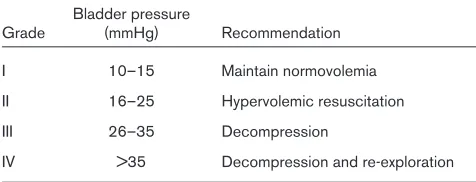
Abdominal compartment grading system
Bladder pressure
Grade (mmHg) Recommendation I 10–15 Maintain normovolemia II 16–25 Hypervolemic resuscitation
III 26–35 Decompression
tion is a potentially preventable complication in intensive care management. Multiple indicators of effective resusci-tation have been evaluated. Lactate, base deficit, and gastric mucosal pH appear to be reliable indicators to guide resuscitative interventions [33].
Surgical intensive care unit management
Identifying patients in the intensive care unit (ICU) at risk for developing ACS with constant surveillance can help lead to prevention. A further strategy is based on recogni-tion of IAH and resultant organ dysfuncrecogni-tion. A four-stage grading scheme base on IAP has been developed, tested, and proposed as a useful ACS management tool (Table 2) [10]. These stages are based on measured bladder pres-sures. This methodology correlates worsening organ dys-function with increasing bladder pressures, with 100% of patients showing pulmonary, cardiovascular, and renal dysfunction with pressures greater than 35 mmHg. Meldrum et al [10] perform simple bedside decompres-sion for bladder pressures from 26 to 35 mmHg, but rec-ommend formal abdominal exploration with pressures greater than 35 mmHg in anticipation of significant intra-abdominal ischemia. This is based on impaired bowel cap-illary perfusion at IAP greater than 35 mmHg.
Alternative means for surgical decision making are based on clinical indicators of adverse physiology, rather than on a single measured parameter. In the setting of IAH, abdominal decompression has been recommended with any coexisting deterioration in pulmonary, cardiovascular, or renal function. Additionally, with IAH that is unrespon-sive to standard intervention and with indicators of bowel ischemia (acidosis by tonometry or dusky bowel seen through transparent coverage material), decompression is recommended [9,34]. Worsening hypercapnia and pul-monary compliance have been identified as critical indica-tors of pulmonary failure that warrant emergent abdominal decompression in the setting of IAH [13].
Abdominal decompression and wound management
Once the decision is made to proceed to surgical decom-pression and the need for intervention is established, the location and possibly transportation requirements for per-forming this procedure must be decided. A decision to perform the decompression in the ICU is a function of the ventilatory requirements of the patient and the risk associ-ated with transport to the operating room. Although optimal respiratory support may be available in the ICU, this location is generally suboptimal for controlling surgical bleeding. The potential for major intra-abdominal hemor-rhage varies, but it can be significant in patients with ACS. Operative planning must include contingencies for man-agement of surgical bleeding encountered when decom-pression is performed in the ICU, which may require repacking and immediate transport to the operating room. It is mandatory that an operating room be immediately
available and appropriately staffed before beginning an ICU abdominal decompression. Patients who require high airway pressures for adequate gas exchange require transport on a high-flow pressure ventilator powered by a battery source [14].
Abdominal decompression may itself precipitate adverse physiologic and metabolic events that should be antici-pated. These include a large increase in pulmonary com-pliance with resultant elevation in minute ventilation and respiratory alkalosis unless appropriate ventilatory changes are instituted. ‘Washout’ of accumulated intra-abdominal products of anaerobic metabolism may result in a bolus of acid and potassium systemically delivered to the heart. This may result in an adverse cardiac event such as an arrhythmia or asystole. Anticipating, recognizing, and treating these effects is of critical importance [9,14].
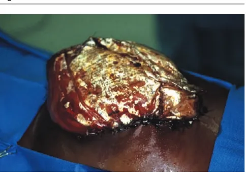
Under most circumstances following abdominal decom-pression, immediate primary fascial closure is obviated. Alternative means for coverage of the abdominal contents include skin closure with towel clips or suture, abdominal wall advancement flaps, plastic or silicone coverage, and mesh interposition grafts (Fig. 2). Patients undergoing decompressive laparotomy are by definition at risk for future redevelopment of ACS, and strong consideration should be given to providing for re-exploration and a staged closure. This may include fascial closure after a period of 7–10 days versus placement of split thickness skin grafts on a granulating surface followed by delayed repair of the resulting abdominal wall hernia after several months [9,13,14,30–32]. Finally, early management of the open abdomen must include recognition for significant fluid losses and fluid replacement [14].
[image:6.612.315.557.95.265.2]28
Figure 2
Outcomes
The ACS is a condition with a potentially high lethality that must be recognized early and effectively managed in order to optimize outcome. Most deaths associated with ACS are due to sepsis or multiple organ failure. Mortality asso-ciated with this condition has been reported in 10.6–68% of patients [9,10,14,28]. In one series [14], nonsurvivors tended toward a more fulminant course, with the majority of deaths occurring within the first 24 h of injury. There is some evidence that the syndrome may be prevented in high-risk patient groups by selective mesh closure of the abdominal wall after laparotomy [28,31].
Further study is needed to better establish the incidence, long-term and short-term morbidity, and mortality of this condition.
Conclusion
The abdominal compartment syndrome is defined as intra-abdominal hypertension associated with organ dysfunc-tion. Adverse physiology has been demonstrated in pulmonary, cardiovascular, renal, musculoskeletal/integu-mentary, and central nervous system function. Identifica-tion of patients at risk, early recogniIdentifica-tion, and appropriately staged and timed intervention is key to effective manage-ment of this condition.
References
1. Emerson H: Intra-abdominal pressures.Arch Intern Med1911, 7: 754–784.
2. Bradley SE, Bradley GP: The effect of increased abdominal pres-sure on renal function.J Clin Invest1947, 26:1010–1015. 3. Coombs HC: The mechanism of regulation of intra-abdominal
pressure.Am J Physiol1920, 61:159–163.
4. Baggot MG: Abdominal blow-out: a concept.Curr Res Anesthesia Analg1951, 30:295–299.
5. Ogilvie WH: The late complications of abdominal war wounds. Lancet1940, 2:253–256.
6. Hrabovsky EE, Boyd JB, Savrin RA: Advances in the management of gastroschisis.Ann Surg1980, 192:244–248.
7. Lenz RJ, Thomas TA, Wilkins DG: Cardiovascular changes during laparoscopy.Anaesthesia1976, 31:4–12.
8. Saggi BH, Sugerman HJ, Ivatury RR, Bloomfield GL: Abdominal com-partment syndrome.J Trauma1998, 45:597–609.
9. Kron IL, Hartman PK, Nolan SP: The measurement of intra-abdomi-nal pressure as a criterion for abdomiintra-abdomi-nal re-exploration.Ann Surg
1984, 199:28–30.
10. Meldrum DR, Moore FA, Moore EE, et al: Prospective characteriza-tion and selective management of the abdominal compartment syndrome.Am J Surg 1997, 174:667–673.
11. Shafik A, El-Sharkawy A, Sharaf WM: Direct measurement of intra-abdominal pressure in various conditions.Eur J Surg1997, 163: 883–887.
12. Collee GG, Lomax DM, Ferguson C, et al: Bedside measurement if intra-abdominal pressure via an indwelling naso-gastric tube: clin-ical validation of the technique. Intensive Care Med 1993, 19: 478–483.
13. Ivatury RR, Diebel L, Porter JM, Simon RJ: Intra-abdominal hyperten-sion and the abdominal compartment syndrome.Surg Clin North Am1997, 77:783–801.
14. Eddy V, Nunn C, Morris JA: Abdominal compartment syndrome. Surg Clin North Am 1997, 77:801–811.
15. Burch JM, Moore EE, Moore FA, Franciose R: The abdominal com-partment syndrome.Surg Clin North Am 1996, 76:833–841. 16. Ridings PC, Bloomfield GL, Blocher CR, et al: Cardiopulmonary
effects of raised intra-abdominal pressure before and after intravascular volume expansion.J Trauma1995, 39:1071–1075.
17. Barnes GE, Laine GA, Giam PY, et al: Cardiovascular responses to elevation of intra-abdominal hydrostatic pressure. Am J Physiol
1985, 248:R208–R213.
18. Diebel LN, Wilson RF, Tagett MG, Kline RA: End-diastolic volume. Arch Surg1992, 127:817–822.
19. Durham R, Neunaber K, Vogler G, Shapiro M, Mazuski J: Right ven-tricular end-diastolic volume as a measure of preload.J Trauma
1995, 39:218–223.
20. Harman PK, Kron IL, McLachlan HD, et al: Elevated intra-abdominal pressure and renal function.Ann Surg1982, 196:594–597. 21. Diebel LN, Dulchavsky SA, Wilson RF: Effect of increased
intra-abdominal pressure on mesenteric arterial and intestinal mucosal blood flow.J Trauma1992, 33:45–49.
22. Bongard F, Pianim N, Dubecz S, Klein SR: Adverse consequences of increased intra-abdominal pressure on bowel tissue oxygen. J Trauma1995, 39:519–525.
23. Friedlander MH, Simon RJ, Ivatury RR, et al: Effect of hemorrhage on superior mesenteric artery flow during increased intra-abdominal pressures.J Trauma1998, 45:433–439.
24. Diebel LN, Dulchavsky SA, Brown WJ: Splanchnic ischemia and bacterial translocation in the abdominal compartment syndrome.J Trauma1997, 43:852–855.
25. Diebel LN, Wilson RF, Dulchavsky SA, et al: Effect of increased intra-abdominal pressure on hepatic arterial, portal venous, and hepatic microcirculatory blood flow.J Trauma1992, 33:279–282. 26. Diebel LN, Saxe J, Dulchavsky S: Effect of intra-abdominal pressure
on abdominal wall blood flow.Am Surg1992, 58:573–576. 27. Sugerman H, Windsor A, Bessos M, Wolfe L: Intra-abdominal
pres-sure, sagittal abdominal diameter and obesity comorbidity. J Intensive Med1997, 241:71–79.
28. Ivatury RR, Porter JM, Simon RJ, et al: Intra-abdominal hypertension after life-threatening penetrating abdominal trauma: prophylaxis, incidence, and clinical relevance of gastric mucosal pH and abdominal compartment syndrome.J Trauma1998, 44:1016–1021. 29. Williams M, Simms HH: Abdominal compartment syndrome: case reports and implications for management of critically ill patients. Am Surg1997, 63:555–558.
30. Morris JA, Eddy VA, Blinman TA, Rutherford EJ, Sharp KW: The staged celiotomy for trauma.Ann Surg1993, 217:576–586. 31. Mayberry JC, Mullins RJ, Crass RA, Trunkey DD: Prevention of
abdominal compartment syndrome by absorbable mesh prosthe-sis closure.Arch Surg1997, 132:957–961.
32. Sherck J, Seiver A, Shatney C, Oakes D, Cobb L: Covering the open abdomen: a better technique.Am Surg1998, 64:854–857. 33. Porter JM, Ivatury RR: In search of optimal end points of trauma
patients: a review.J Trauma 1998, 44:908–914.
34. Bloomfield GL, Dalton JM, Sugerman HJ, et al: Treatment of increas-ing intracranial pressure secondary to the acute abdominal com-partment syndrome in a patient with combined abdominal and head trauma.J Trauma1995, 39:1168–1170.
Authors’ affiliation:Department of Surgery at Saint Louis University, St Louis, Missouri, USA
Correspondence:Marc J Shapiro, MD, FACS, Department of Surgery, Saint Louis University, 3635 Vista @ Grand, St Louis, Missouri 63110-0250, USA. Fax: +1 314 268 5194; e-mail: [email protected]