THE STUDY ON
THYROID FUNCTIONS IN
CHRONIC LIVER DISEASE
This dissertation is submitted to
THE TAMILNADU DR.M.G.R. MEDICAL UNIVERSITY
in partial fulfillment of the requirement of the award
for the degree of
M.D BRANCH I
GENERAL MEDICINE
STANLEY MEDICAL COLLEGE,
CHENNAI – 600001
This is to certify that this dissertation entitled
‘
THYROID FUNCTIONS IN CHRONIC LIVER DISEASE
’
submitted by
Dr. S.SRIRAM
to
THE
TAMILNADU DR. MGR
MEDICAL UNIVERSITY,CHENNAI
is in partial fulfillment of the
requirement of the award of M.D degree Branch-I (General Medicine)
and is a Bonafide Research work carried out by him under direct
supervision and guidance
Signature of the Unit Chief Signature of the HOD
DECLARATION
I solemnly declare that the dissertation titled, ‘THYROID FUNCTIONS IN CHRONIC LIVER DISEASE’ was done by me at Stanley Medical College and Hospital during 2007 – 2010 under the guidance and supervision of my Unit
Chief Prof.Dr.A.GOWRISHANKAR.,M.D.
The dissertation is submitted to THE TAMILNADU DR.M.G.R.
MEDICAL UNIVERSITY towards the partial fulfillment of requirement for the award of M.D. Degree (Branch I) in General Medicine.
Place:
Date:
DR.S.SRIRAM
I owe my thanks to the Dean, Stanley Medical College and Hospital
Dr.A.Priya M.S, D.O, for allowing me to avail the facilities needed for this study. I have great pleasure in expressing my gratitude and respect for
Prof.Dr.S.Ramasamy,M.D., Professor and Head of the Department of Medicine, Stanley Medical College and Hospital for permitting me to do the study and for his
encouragement.
I am extremely thankful to my respected teacher and unit chief
Prof.Dr.A.Gowrishankar M.D., Professor of medicine , for his guidance and encouragement without which this would not have been possible.
I am extremely thankful to my Assistant Professors Dr.Nalini Kumaravelu M.D., and Dr.S.Chadrasekar M.D., for their valuable guidance and constant encouragement.
I am extremely thankful to the hospital laboratory and its staff members for
their immense help in conducting the study.
Finally, I would like to thank my colleagues who offered valuable help to
me during the study, and the patients without whose patience, this study would not
CHAPTER NO. TITLE PAGE NO.
1. INTRODUCTION 1
2. AIM OF THE STUDY 3
3. REVIEW OF LITERATURE 4
4. MATERIALS AND METHODS 38
5. RESULTS 43
6. DISCUSSION 54
7. CONCLUSION 56
8. BIBLIOGRAPHY
INTRODUCTION
Thyroxine and tri-iodothyronine are essential for normal organgrowth,
development and function. These hormones regulate thebasal metabolic rate of all
cells, including hepatocytes, andthereby modulate hepatic function. The liver plays
an important role in thyroid hormone metabolism being involved in their
conjugation, excretion, peripheral deiodination and in the synthesis of thyroid
binding globulin. Thyroid dysfunction may perturb liverfunction, liverdisease
modulates thyroid hormone metabolism, and a varietyof systemic diseases affect
both organs.
Although almost all patients with liver disease are clinically euthyroid,
some abnormalities in the circulating hormone concentrations have been shown in
previous studies. These data, however, are still controversial as the discrepant
results reported may depend on the different analytical methods used as well as
different groups of patients investigated.
The total and free thyroxine have been reported as normal, increased or
because of a hypothetical circulating inhibitor, have also been reported. Moreover,
total and free triiodothyronine concentration are often decreased, sometimes
profoundly and their levels correlate well with severity of liver dysfunction. In
order to further evaluate the thyroid function in liver disease, this study measures
T3, T4, FT3, FT4 serum levels in patients with chronic liver disease
AIM OF THE
AIM OF THE STUDY
To evaluate the following :
1. thyroid functions in patients with chronic liver disease
2. To assess the severity of liver dysfunction in relation with interpretation of
thyroid functions
REVIEW
OF
REVIEW OF LITERATURE
The relationship between the thyroid gland and the liver
Thyroxine and tri-iodothyronine are essential for normal organgrowth,
development and function. These hormones regulate thebasal metabolic rate of all
cells, including hepatocytes, andthereby modulate hepatic function; the liverin
turn metabolizesthe thyroid hormones and regulates their systemic endocrine
effects. Thyroid dysfunction may perturb liver function, liverdisease modulates
thyroid hormone metabolism, and a varietyof systemic diseases affect both organs
Intracellular signaling
The thyroid gland secretes two iodine containing amine hormonesderived
from the amino acid tyrosine, L-thyroxine (T4) and 3,5,3'-L-tri-iodothyronine(T3).
Free T3 and T4 enter all cells through the plasma membraneand bind to a nuclear
T3 receptor.
The thyroid receptor is partof the nuclear super family group of receptors
(retinoic acid,retinoid X, vitamin D and peroxisome proliferator receptor).These
receptors all possess six similar domains, two of whichare a ligand-binding region
The main function of the thyroid receptor is to act as a ligand-activated
transcription factor that regulates target gene expression directlythrough DNA
response elements (thyroid response elements).
However, an important property of these receptors is that theybind thyroid
response elements constitutively, independent ofligand occupancy.
REGULATION OF THYROID HORMONES
Thyroid hormone metabolism
The thyroid gland secretes 110 nmol of thyroxineand 10 nmol of
tri-iodothyronine each day. Tri-tri-iodothyroninehas a ten times greater affinity and ten
times greater efficacythan thyroxine for the nuclear receptor, thus even though
thyroxineis quantitatively secreted at much higher levels, it shouldbe regarded as a
pro-hormone that requires deiodination andconversion to T3 to become
biologically active. There arethree groups of enzymes that regulate thyroid
hormone metabolism,forming part of the iodothyronine seleno-deiodinase enzyme
system. They are responsible forthe activation of T4 to T3, inactivation of T4 to rT3
and theconversion of rT3 and T3 to T2
Peripheral conversion of thyroid hormones
The conversion of T4 to T3 in extra thyroidal tissue occurs througha rapidly
equilibrating pool. The type 1 deiodinaseis mainly found in the liver and kidney,
and accounts forapproximately 30–40% of extra thyroidal production of T3(12
nmol). The type 2 deiodinase is found in the pituitary,the CNS, and skeletal
muscle and contributes 60–70% ofthe extra thyroidal production of T3 (30 nmol).
Although thisenzyme system performs similar actions to the type 1 de-iodinase
activity group of enzymes,its kinetics, regulation and susceptibility to
Although both the type 1 de-iodinase and type2 de-iodinase system can also
inactivate T4 and T3, the major inactivator is the type 3 deiodinasesystem, which
primarily exhibits inner-ring deiodination (unlikethe other systems). It is found in
the liver, skin and CNS,where it catalyses the conversion of T4 to rT3 and T3 to T2,
both inactive metabolites; it also converts rT3 to rT2. Thisenzyme system is also
expressed in placenta, where it protectsthe fetus from maternal thyroid hormones.
In addition to the central role in deiodination to activateand deactivate
thyroid hormones, the liver performs specificfunctions relating to thyroid hormone
transport and metabolism.
The liver extracts 5–10% of plasma T4 during a singlepassage, as shown by
studies using [131I]T4. This value is muchhigher than can be accounted for by the
amount of free T4 deliveredto the liver, indicating that a substantial amount of
bound T4 is available for uptake. An active stereo-specific transportmechanism has
been identified for transporting T4 and T3 acrossthe hepatocyte membrane. The
intracellular concentrations ofthe free hormone are higher than the plasma levels,
and theprocess is energy-dependent
The liver synthesizes a number of plasma proteins that bindthe
lipophilic thyroid hormones and thereby provide a large,rapidly exchangeable pool
of circulating hormone. The thyroidhormones are >99% bound to thyroxine-
binding globulin, thyroxine-bindingprealbumin and albumin in plasma. The free
hormone componentwithin plasma is in equilibrium with the protein-bound
hormone,and it is this free fraction which accounts for the hormone'sbiological
activities. The plasma concentrations of free T4and T3 are at a steady
concentration, so that the tissues areexposed to the same concentrations of the free
hormone. However,the free hormone concentrations in different tissues vary
accordingto the transport and deiodinase activity within specific tissues.
Thus tissue thyroid status depends not only on thyroxine secretionbut also
on normal thyroid hormone metabolism, delivery of T3to nuclear receptors and on receptor distribution and function.Normal thyroid function, which is essential for
normal growth,development and the regulation of energy metabolism withincells,
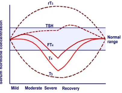
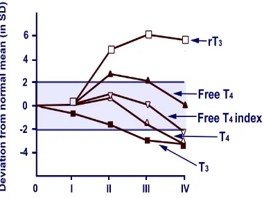
Thyroid metabolism in chronic illness
In most chronic illness, defects arise inthyroid hormone metabolism,
resulting in the sick euthyroid syndrome. This is characterizedby a normal total T4,
normal/high free T4, low total T3, lowfree T3 and an elevated rT3. These changes
reflect a reductionin type 1 de-iodinase activity, an increase in type 3 de-iodinase
activity and changes inthe plasma concentration of thyroid-binding proteins and
freefatty acids (which displace thyroid hormones from binding proteins).There are
also non-thyroidal influences on the hypothalamic-pituitary-thyroidaxis, e.g.
cortisol inhibiting TSH secretion
It has been suggested that this syndrome may confer a survivaladvantage,
which adapts an organism to chronic illness by reducingthe basal metabolic rate
within cells and thereby reducing caloricrequirements.These changes in thyroid
function test results are observed in most of the acute and chronic illnesses.
Examples of illness include the following:
• Gastrointestinal diseases
• Pulmonary diseases
• Cardiovascular diseases
• Infiltrative and metabolic disorders
• Inflammatory conditions
• Myocardial infarction
• Starvation
• Sepsis
• Burns
• Trauma
• Surgery
• Malignancy
Pathophysiology
Proposed mechanisms explaining abnormalities in thyroid hormone levels
• Accuracy of test assays in nonthyroidal illness
Abnormalities of thyroid function test results might represent test artifacts or
true abnormalities. According to one proposition, the assays would indicate
reference range thyroid hormone levels in the blood if appropriate tests were
applied.
Inhibition of thyroid hormone binding to thyroid-binding proteins and
tissues
Serum thyroid hormone abnormalities are due to inhibition of thyroid
hormone binding to proteins, thus preventing tests from appropriately reflecting
free hormone levels. This binding inhibitor can be present both in the serum and in
body tissues and might inhibit uptake of thyroid hormones by cells or prevent
binding to nuclear T3 receptors, thus inhibiting the action of the hormone. This
inhibitor is associated with the nonesterified fatty acid (NEFA) fraction in the
serum.
Contrary to this proposition, substantial evidence indicates that, in an in vivo
circulating levels of free T4, even in patients who are severely ill. Also, some
studies have failed to demonstrate an existing binding inhibitor.
• Cytokines:
Cytokines are thought to play a role in NTI—particularly interleukin (IL)-1,
IL-6, tumor necrosis factor (TNF)-alpha, and interferon-beta. Cytokines are
thought to affect the hypothalamus, the pituitary, or other tissues, inhibiting
production of TSH, thyroid-releasing hormone (TRH), thyroglobulin, T3, and
thyroid-binding globulins. Cytokines are also thought to decrease the activity of
type I deiodinase and to decrease the binding capacity of T3 nuclear receptors.
• Deiodination
Peripheral deiodination of T4 to T3 is impaired, largely secondary to
decreased activity of type I deiodinase enzyme, which deiodinates T4 to T3.
Diminished enzyme activity accounts for decreased deiodination of T4 to T3.
An alternative explanation is that reduced tissue uptake of T4 secondary to
deficiency of cytosolic cofactors (e.g, nicotinamide adenine dinucleotide phosphate
[NADPH], glutathione) results in decreased substrate for type I deiodinase
common in critically ill patients, some propose that selenium deficiency may
contribute to type I deiodinase malfunction.
Cytokines (e.g, IL-1 beta, TNF-alpha, interferon-gamma) decrease type I
deiodinase messenger RNA (mRNA) in vitro. Type I deiodinase does not exist in
the pituitary, where T3 levels are within the reference range, because of enhanced
local deiodination. This indicates that an enhancement of intrapituitary T4 to T3
conversion exists due to pituitary-specific and brain-specific type II deiodinase.
• Inhibition of thyroid-releasing hormone and thyroid-stimulating hormone secretion:
Cytokines, cortisol, and leptin, as well as changes in brain thyroid hormone
metabolism, affect inhibition and secretion of TRH and TSH.
• Inhibition of plasma membrane transport of iodothyronines:
Serum factors, such as bilirubin, NEFA, furanoic acid, hippuric acid, and
indoxyl sulphate, which are present in various Non Thyroidal Illness (NTIs), have
• Thyroxine-binding globulin decrease and desialation:
T4-binding globulin (TBG) is a member of the serine protease inhibitors.
Diminished T4 in NTI has been proposed to be due to low TBG caused by protease
cleavage at inflammatory sites in acute inflammatory conditions. One other
hypothesis for the cause of disproportionately low serum T4 concentrations in
patients with NTI is the presence of abnormal serum binding due to desialation of
TBG.
The effects of nonthyroidal illness
:• Triiodothyronine and reverse triiodothyronine
In healthy people, 20% of T3 production comes from thyroidal secretion and
80% from peripheral deiodination from T4. In NTI, thyroidal production of T3 is
normal, but the peripheral production of T3 is decreased. The fractional rate of
transport of T3 to tissues is unaltered. Production of T3 is decreased, but its
clearance is unchanged. Production of rT3 is unchanged, while its clearance is
diminished.
In rat hepatocytes, rT3 and T4 have been demonstrated to be transported in
the same mechanism, which implies that a diminished transport of rT3 to the liver
calorie deprivation). Because the liver is the main site of disposal of T3, this leads
to a diminished metabolic clearance rate of rT3 and T4.
Another explanation could be reduced 5'-deiodinase tissue activity, resulting
in decreased T3 production from T4 and reduced breakdown of rT3. The decreased
production of T3 during early and late starvation has been explained as either a
diminished activity of the enzyme (deiodinase) itself or a deficiency of cytosolic
cofactors, such as NADPH or glutathione.
Specific deiodinative enzymes, 3 of which have been identified, affect
deiodination of iodothyronines. Type I deiodinase is present in the liver, kidney,
and thyroid and affects both 5 and 5' deiodination of T3. Type II deiodinase is
present in the brain, pituitary, and brown adipose tissue and is active only in 5'
deiodination. Type III deiodinase is found particularly in the brain, skin, and
placenta, and it deiodinates iodothyronines at the 5 locations.
Both type II and type III enzymes are insensitive to 6-propylthiouracil
(PTU). Alterations of serum thyroid hormone parameters in cases of calorie
deprivation exhibit similarities to the changes observed in NTI. Fasted animals had
decreased 5'-deiodinase activity. The activity of type I deiodinase is inhibited by
6-PTU. Because it is a selenoprotein and selenium deficiency is common in critically
Cytokines, such as IL-1 beta, TNF-alpha, and interferon-gamma, decrease
type I deiodinase mRNA in vitro. Infusion of TNF-alpha decreases serum T3 and
increases rT3. Soluble TNF-alpha, soluble TNF-alpha receptor, soluble IL-2
receptor antagonist, and IL-6 are inversely correlated with serum T3 levels. The
elevations of soluble TNF-alpha receptor and IL-6 were independent determinants
of serum T3 and accounted for 35% and 14%, respectively, of the change in T3.
These cytokine changes can be concluded to occur concomitantly with
changes in T3 and may play a pathogenic role through mechanisms that are not
clearly defined. The increase of endogenous cortisol during illness apparently is
not involved in inhibition of type I deiodinase.
Using an adenovirus model in mice hepatocyte primary cultures, it was
demonstrated that forced expression of steroid receptor co-activator 1 (SRC-1)
prevented the cytokine induced inhibition of type 1 deiodinase activity, suggesting
the involvement of receptor co-activators in the nonthyroidal illness.
• Free triiodothyronine:
• Thyroxine
The decrease in the T4 binding of TBG has been used as an explanation for the
low plasma T4 concentration in patients with NTI. The existence of a binding
inhibitor could explain the observed alterations in T4 and free T4 fraction. TBG
levels usually are within the reference range in patients with NTI and are
somewhat lower in critically ill patients with low serum T4. Low TBG levels can
be explained, according to some proposals, by rapid protease cleavage at
inflammatory sites, particularly in acute inflammatory states (in which the decrease
in TBG is too rapid to be accounted for by inhibition of synthesis).
In patients with NTI, serum T4 concentration has been demonstrated to be
low because much of the circulating TBG in these patients is desialated. In NTI,
the fractional rate of T4 transport from serum to tissues is reduced to 50% of the
reference range value. This decrement in fractional rate of T4 transport is not
related to the serum levels of total or free T4. Because in illness the reduction in
the fractional rate of T4 transport from serum to tissues cannot be attributed to
alterations in serum T4 binding, consider other causes such as an impairment of
transport into tissues.
In nonuremic critical illness, it has been demonstrated that elevated
partially responsible for the T4 transport inhibition in T3-producing tissues (e.g,
the liver).
A correlation exists between the probability of death and the levels of
total T4. When serum T4 levels drop below 4 mcg/dL, the probability of death
is about 50%; with serum T4 levels below 2 mcg/dL, the probability of death
reaches 80%.
• Free thyroxine
Evaluating thyroid function in patients with NTI has considerable
challenges. No consensus exists as to whether free T4 levels are within the
reference range, low, or high. Free T4 is believed to represent the hormone
available to tissues.
Measurement of total serum T4 has only limited value because nearly all
(99.97%) of the circulating T4 is bound to TBG, T4-binding prealbumin (TBPA),
and albumin. The rest of the circulating T4 (0.2-0.03%) is free T4. The circulating
concentration of these binding proteins is understood to affect the total T4
concentration without necessarily changing the amount of free T4. Usually, TBG
levels are within the reference range in patients with NTI and somewhat lower in
Decreased concentrations of one or more of the binding proteins would
explain low levels of total T4 but does not explain a significant increase in free T4
fraction, which some patients with NTI exhibit.
Various explanations for the existence of inhibitors of T4 binding have been
reported. Although low levels of TBPA and albumin may occur in patients with
NTI, even complete inhibition of T4 binding to these proteins has been
demonstrated to produce only about a 30% increase in free T4 fraction. Because
free T4 fraction is increased above this level in many patients, other factors must
be present.
The observations of reduced total T4 and free T4 have been explained
alternatively as either a fall in TBG levels or an inhibition of thyroid hormone
binding to TBG. Some studies have shown a decrease in the T4 binding of TBG,
which has been used as an explanation for the low plasma T4 concentration and,
perhaps, the high free T4 fractions, in patients with NTI. Other studies postulate
the existence of a binding inhibitor that could explain the observed alterations in
free T4 fraction.
The inhibitor also has been demonstrated to interfere with the binding of
iodothyronines to solid matrices, thus interfering with the T3 resin uptake and
extractable with ether and was associated with the NEFA fraction in the serum.
Furthermore, the extracted inhibitor from sera of patients with NTI reduced
conversion of T4 to T3 in rat liver homogenates. The inhibitor could be extracted
from extrathyroidal tissues as well.
The addition of NEFA to normal serum is able to raise the free T4 fraction
only if total NEFA concentration is higher than 3 millimoles in normal serum,
representing a NEFA-to-albumin molar ratio greater than 5:1. Because this high
NEFA-to-albumin ratio is not reached even in severely ill patients, NEFA is
unlikely to influence the circulating free T4 concentration in vivo. Inhibitors of
binding were also observed during equilibrium dialysis assay in patients treated
with heparin. This is due to an in vitro artifact that is not present in vivo.
Cytokines also can elevate free T4. When TNF-alpha was infused, it was
observed that free T4 could elevate transiently in association with a significant rise
in free fatty acids. However, other studies question the role of NEFA inhibition or
whether any thyroid hormone–binding inhibitor exists at all.
• Thyroid hormone receptor expression and DNA binding:
In experimental mouse liver models, infection decreased thyroid hormone
TR-alpha and TR-beta protein levels were both decreased when lipopolysaccharide
was administered, particularly at 16 hours. Lipopolysaccharide exposure was also
shown to reduce RXR protein levels in the liver.
• Thyroid-stimulating hormone and thyroid-releasing hormone
Some patients with Non Thyroidal Illness have slightly elevated serum TSH,
which is thought to have reduced biological activity. After recovery from severe
Non Thyroidal Illness, transient elevation of TSH to above-normal limits
commonly occurs. Some authors interpret this TSH elevation as a sign of recovery
from a hypothyroid state. Despite the distortion of TSH in some euthyroid patients
with Non Thyroid Illness, patients with Non Thyroidal Illness who have significant
elevation of TSH usually have underlying primary hypothyroidism.
Responsiveness of the pituitary to TRH during Non Thyroidal Illness varies;
some patients respond normally, while many have a less-than-normal response.
Normal responsiveness in the presence of low TSH may suggest that a
hypothalamic abnormality is causing the low TSH and low T4. The
down-regulation at the hypothalamus-pituitary level provides an explanation for the
decreased sensitivity of TSH secretion to low serum T3 and T4 concentrations in
TSH also occurs, and some studies have produced evidence for a reduction of TSH
glycosylation with lower TSH bioactivity.
That TSH is not elevated in the presence of low T4 indicates that the patients
are not hypothyroid. Diminished release of TRH also is thought perhaps to result in
low TSH and, thus, low output of thyroid hormones by the thyroid. Low TRH
mRNA in hypothalamic paraventricular nuclei also has been demonstrated.
The role of cytokines, especially IL-1 beta, in the activity of the
hypothalamic-pituitary-adrenal axis is well known. Cytokines also affect TRH in
rats. IL-1 beta decreases the release of TSH in cultured rat anterior pituitary cells,
but the role of TNF-alpha on TSH release is disputed. IL-6 decreases TSH
secretion. In rodents, leptin has been demonstrated as a major mediator of changes
in hypothalamic-pituitary-thyroid function during fasting. However, TSH secretion
and thyroid gland function are less affected during Non Thyroidal Illness in
humans than they are in animals.
The role of leptin in patients with Non Thyroidal Illness is unclear. Leptin
concentrations often are elevated during critical illness and increase acutely in
response to administration of TNF-alpha or IL-1; however, the leptin increase is
Thyroid
abnormalities
in
liver
disease
Inthe different types ofliver disease, similar processes mayoccur to those seen in the sick euthyroidsyndrome, butin additiona number of changes specific
to the type or stage of liverdiseaseis also found.
Cirrhosis
The most consistent thyroid hormoneprofile in patients with cirrhosis are a
low total and freeT3 and an elevated rT3, similar changes to those in thesick
euthyroid syndrome, probably reflecting a reduced deiodinasetype 1 activity,
resulting in reduced conversion of T4 to T3.This results in an increase in
conversion of T4 to rT3 by thedeiodanase type 3 system, and an increase in the rT3
to T3 ratio.
The plasma T3:rT3 ratio has a negative correlation with theseverity of
cirrhosis when assessed in non-alcoholic cirrhotics.Since T3 and rT3 bind to the
same plasma proteins, the T3/rT3ratio provides a parameter of liver function that is
largelyindependent of protein binding. Both the T3/rT3 ratio and freeT3 levels in
plasma thus provide a correlate of liver functionin cirrhosis, and are of prognostic
The low total and free T3 levels may be regarded as an adaptivehypothyroid
state that serves to reduce the basal metabolicrate within hepatocytes and preserve
liver function and totalbody proteinstores. Indeed, a recent study in cirrhotic
patientsshowed that the onset of hypothyroidism from intrinsic thyroiddisease of
various etiologies during cirrhosis resulted ina biochemical improvement inliver
function (e.g. coagulationprofiles) as compared to cirrhotic controls.
Hypothyroidismhas also been associated with lesser degrees of decompensationin
cirrhosis. Controlled induction of hypothyroidism mighttherefore be beneficial in
cirrhotic patients, but further studiesare required to test this hypothesis.
Acute hepatitis and acute liver failure
In acute hepatitis of mild or moderate severity, patients haveelevated serum
levels of total T4, due to increased thyroid-bindingglobulin, which is synthesized
as an acute-phase reactant, butnormal levels of free T4. In more severe cases with
impendingliver failure, the data is variable, and low total T4 levelsmay reflect
reduced hepatocellular synthesis of thyroid-bindingglobulin.
Serum T3 levels are extremely variable, but thefree T3:T4 ratio correlates
negatively with the severity ofthe liver disease and has prognostic value. Again
this probablyreflects diminished type 1 deiodinase activity, resulting ina reduced
Some series have describedpatients with acute hepatic failure (especially viral
hepatitis)as having goiters that resolved with improvement in liverfunction.
Specific forms of chronic liver disease
In patients with chronic hepatitis associated with primary biliarycirrhosis
(PBC) or chronic autoimmune hepatitis, there is anincreased prevalence of
autoimmune thyroid disease. Thusabnormalities may arise from thyroid gland
dysfunction or asa consequence of the liver disease. Autoimmune hypothyroidism
is a prominent feature in PBC, occurring in 10–25% ofpatients. There is often an
increase in total T4 in PBC, dueto an increase in thyroid-binding globulin levels,
and thismay mask hypothyroidism, emphasizing the need to perform a freeT4 and
TSH assay. Anti-thyroidmicrosomal antibodies are commonin PBC (34%), as are
anti-thyroglobulin antibodies (20%).Thyroid dysfunction may precede or follow
the diagnosis of PBC.In autoimmune hepatitis, both Grave's disease (6%) and
autoimmunehypothyroidism (12%) are relatively common. Primary sclerosing
cholangitis is associated with an increased incidence of Hashimoto'sthyroiditis,
Graves's disease and Riedel's thyroiditis.
In patients with chronic hepatitis who do not have co-existingautoimmune
often increased, but TSH and free T4 levelsare usually normal, and patients are
clinically euthyroid.
Currently the treatment of viral hepatitis with alpha interferonhas added
another dimension to the abnormalities of thyroidfunction seen inchronicliver
diseases. In different studiesassessing patients treated with alpha interferon for
hepatitisC, 2.5–10% developed thyroiddysfunction, with boththyrotoxicosis (due
to acute thyroiditis) and hypothyroidismbeing observed. Although the reason is not
altogether clear,the induction of an autoimmune reaction has been postulated,
resulting in the development of anti-thyroid and anti-thyrotrophinreceptor
antibodies. However, a distinct effect on intrathyroidalorganification of iodine has
also been suggested.
The riskfactors for developing thyroid dysfunction with alpha interferon
(which may persist after discontinuation of the drug) are femalesex, underlying
malignancy, high doses of long duration, combinationimmunotherapy (especially
Il-2), and the presence of anti-thyroidperoxidase antibodies prior to commencing
treatment.
It should be noted that interferon therapy causes weakness andmuscle
aching, and in this setting the myopathy of hypothyroidismmay be missed. It is
are performed prior totherapy, and subsequently monitored at 3–6 month intervals
during interferon therapy.
Liver abnormalities in thyroid disease
Hypothyroidism
Hypothyroidism may have features that mimic liver disease (pseudo-liver
disease): examples include myalgias, fatigue and muscle crampsinthe presence of
an elevated aspartate aminotransferase froma myopathy, coma associated with
hyperammonaemia in myxoedemacoma, and myxoedema ascites.
Hyperthyroidism
Liver injury caused by thyrotoxicosisis relatively common, and can be
conveniently divided into hepatiticor cholestatic types.
Hepatitic injury
An increase in the aspartate aminotransferase (AST) and alanine
aminotransferase (ALT) is seen, although the majority of these patients showedno
other clinical or biochemical features of liver impairment.The mechanism of injury
appears to be relative hypoxia in theperivenular regions, due to an increase in
Cholestatic injury
An elevated serum alkaline phosphatase is seen in of patientswith
thyrotoxicosis
Anti thyroid drugs Increasedserum levels of aspartate aminotransferase and alanine aminotransferase
occur in about 30% of patients treated with propylthiouracil. The rise in AST
appears to be dose-related, so that AST andALT levels are highest during the first
few weeks of treatment,falling rapidly with a dose reduction
Other thyroid and liver interactions
Physiological
The liver is the major site for cholesterol and triglyceridemetabolism, and the
thyroidhormones play an integral part inhepatic lipid homeostasis. Thyroid
hormones increase the expressionof LDL receptors on the hepatocytes, and
increase the activityof lipid-lowering liver enzymes, resulting in a reduction in
low-density lipoprotein levels. Thyroid hormones also increasethe expression of
apolipoprotein A1, a major component of high-densitylipoprotein.
Pathological
thyroid gland simultaneously. The autoimmune diseases, organ infiltration such as
malignancy, amyloid, orin secondary haemachromatosis, drugs like amiodarone,
mefloquine, carbamazepine
Recent work investigating the use of tri-iodothyronine as ahepatic growth
factor has shown it to be a primary mitogen forthe liverin animal models (i.e. it
induces hepatocyte proliferationand increases liver mass when administered at
high doses inthe absence of hepatic injury). The ability to increase livermass inthe
absence of liverdamage, and to enhance proliferationduring compensatory
hyperplasia after liver damage, could betherapeutically valuable if applicable to
man. More generally,the ability to manipulate liver cell proliferation in vivo may
be helpful in designing cell transplantation and gene therapyapproaches to liver
diseases.
Diagnosis of Thyroid Disease in Euthyroid Sick Syndrome
During starvation and mild illness, a low T3 concentration, or low T3 and
low T4 levels in a patient with a low-normal TSH level, is the hallmark of
euthyroid sick syndrome. However, nonthyroidal illness may present with a
spectrum of abnormalities in thyroid function that may complicate the diagnosis of
Low T3 levels with normal T4 and TSH levels are the most common
abnormality seen in euthyroid sick syndrome. Serum TSH levels are typically
normal or reduced. The TSH levels are normal/subnormal in approximately 80% of
patients, and are markedly suppressed (<0.1 µU/mL) in <10% of patients. Thus, in
a patient with a systemic illness, low T4 and T3 levels and a normal or low-normal
TSH level most likely indicate euthyroid sick syndrome.
In the recovery phase of illness, mild elevation of TSH levels can be
observed; however, serum level of TSH >30 µU/mL is rarely seen in euthyroid
sick syndrome and strongly suggests the diagnosis of primary hypothyroidism.
Levels of TSH above 20 µU/mL are found in <3% of patients with nonthyroidal
illness.
Differentiation between secondary hypothyroidism (pituitary or
hypothalamic) and euthyroid sick syndrome may be difficult. Both conditions
present with decreased levels of total T4, T3, and TSH. Many chronically ill
patients are edematous, have associated infections, or have cardiopulmonary
disorders that could easily mask evidence of thyroid disorders. Additional tests,
including obtaining basal and/or stimulated cortisol, serum gonadotropin, and
prolactin levels may be of help in such cases. If the serum cortisol level is normal
is probably the cause, rather than pituitary dysfunction. If serum cortisol and
gonadotropin levels are low, pituitary dysfunction should be suspected, and
treatment with corticosteroids and thyroid hormone supplementation is indicated.
In some instances, it may be difficult to exclude hyperthyroid patients, who
may present with suppressed TSH levels and normal T4 and T3 levels in the
presence of infection or other catabolic illness. Hyperthyroid patients who are
chronically ill or malnourished may have hypoproteinemia and low levels of TBG
that lower their T4 and T3 levels. In such patients, an elevated free-T4 level and a
low or undetectable TSH level will confirm the diagnosis of hyperthyroidism. A
previous history of thyroid illness, a history of external radiation, or the presence
of goiter and/or a midline neck scar may indicate a primary thyroid condition.
Certain pharmacologic agents may alter the serum concentration of thyroid
hormones and should be taken into account in the evaluation of patients with
nonthyroidal illness. The concentrations of total T3, free-T4, and TSH are reduced
in patients treated with dopamine or corticosteroids, due to suppression of pituitary
TSH release and/or inhibition of conversion of T4 to T3. Levels of total and free-T4
may be increased in patients treated with amiodarone or iodinated radiocontrast
free-T4 levels, due to in vitro interference with the laboratory assay; however, most
such patients remain clinically euthyroid and have normal total T4 and TSH levels.
It is not prudent to rely solely on a single thyroid test in the evaluation of
thyroid function of patients with critical illness. In such patients, a careful
assessment of multiple tests may be needed to distinguish patients with euthyroid
sick syndrome. In many instances, it is reasonable to delay the final diagnosis for
several days to weeks, or after recovery from the acute illness, to determine the
correct thyroid status.
Faber J et al kinetic studies of thyroxine, 3,5,3'-triiodothyronine,
3,3,5'-triiodothyronine, 3',5'-diiodothyronine, 3,diiodothyronine, and
3'-monoiodothyronine in patients with liver cirrhosis found that serum T4, T3, and
3,3'-T2 levels were reduced in patients withliver cirrhosis, whereas serum rT3 and
3',5'-T2 levels wereincreased. Serum 3'-T1 levels were unaltered.
Chopra IJ et al showed alterations in circulating thyroid hormones and
thyrotropin in hepatic cirrhosis: evidence for euthyroidism despite subnormal
serum triiodothyronine
Nomura S et al showed reduced peripheral conversion of thyroxine to
Bermudez F et al observed a high incidence of decreased serum
triiodothyronine concentration in patients with nonthyroidal disease.
Bianchi GP et al measured Thyroid volume at ultrasound in 118 consecutive
patients with cirrhosis of different etiology and 48 healthy subjects matched for
age and sex. No subjects had evidence of overt thyroid disease. The mean volume
was increased by 17% (from 16.0 [SD 5.2] ml in controls to 18.8 [7.6] in cirrhosis;
P less than 0.025), and thyroid enlargement (antero-posterior diameter greater than
20 mm) was present in 38% of cases, in the presence of hormone values indicative
of low-T3 syndrome. No significant differences in thyroid gland size were
observed in relation to the extent of liver dysfunction or to the etiology of liver
disease. The prevalence of thyroid nodules was similar in controls and in patients
with cirrhosis
M Borzio et al studied thyroid function tests in chronic liver disease and
confirmed the existence of severalabnormalities of thyroid function tests in
patients with chronicliver disease, although showing that euthyroidism is almost
L'age M, Meinhold H, Wenzel KW, Schleusener H measured Serum levels
of TSH, thyroxine-binding globulin (TBG), T4, T3 and reverse T3 (rT3) in 36
patients with fatty liver disease, 11 patients with chronic persistent hepatitis, 17
patients with chronic active hepatitis, and 29 patients with liver cirrhosis. TBG was
significantly above normal levels in both groups of chronic hepatitis, the slight
concomitant T4 and T3 increase was significant only for T4 in chronic persistent
hepatitis. A significant decrease in T4 and T3 concentration was found in fatty
liver disease and in hepatic cirrhosis. A shift in T4 conversion to rT3 could
exclusively be demonstrated for the group of hepatic cirrhosis, reflected by a
significant increase in rT3
Hitomi TAKAHASHI et al studies on changes of Thyroid Hormones in
Various Liver Diseases showed usefulness of free Thyroid Hormones as Liver
Function Test.
R. MALIK and H. HODGSON studiedthe relationship between the thyroid
gland and the liver and observed that thyroxine and tri-iodothyronine hormones
modulate hepatic function; the liver in turn metabolizesthe thyroid hormones and
function, liverdisease modulates thyroid hormone metabolism, and a varietyof
systemic diseases affect both organs.
Van Thiel DH et alassayed T4, T3, rT3 and TSH in 134 adult patients
evaluated and accepted as potential liver transplant candidates at the University of
Pittsburgh from March, 1981 to December, 1983. The subsequent course of these
patients was evaluated with respect to the levels of these hormones obtained at the
time of acceptance for transplantation. T4 levels were increased significantly while
their T3 levels were reduced (both p less than 0.01) in those who survived and
were discharged home as compared to either those who died waiting to be
transplanted or died following the procedure. As a result, the ratio of T3/T4 was
reduced markedly (p less than 0.01) in those who were transplanted and survived
as compared to those not transplanted or dying following transplantation
Van Steenbergen W, et al studied thyroid hormones and the hepatic handling
of bilirubin and effects of hypothyroidism and hyperthyroidism on the hepatic
transport of bilirubin
Malik R et al studied the effects of thyroidhormone on the liver and
MATERIALS
AND
MATERIALS AND METHODS
SETTING : 40 patients with symptoms, signs with biochemical and radiological evidence of chronic liver disease who were admitted in the general medical ward
of Government Stanley Medical College and Hospital were enrolled for this study
after prior written and informed consent
The age group of these patients ranged from 25 years to 75 years
PERIOD OF STUDY : JANUARY 2009 – OCTOBER 2009
ETHICAL COMMITTEE APPROVAL : The present study was approved by the ethical committee
CONSENT : Study group thus identified was informed about the nature of the study and willing participants were included in the study after getting written
informed consent
Out of these 40 cases, 30 patients were males and 10 patients were females.
Out of the 30 males, 25 had had alcohol related chronic liver disease and out
of the remaining 5, 2 patients had Wilson’s disease, 3 had post viral chronic liver
disease.
Out of the 10 females, 5 had post viral chronic liver disease, 2 patients had
alcoholic liver disease and remaining had cryptogenic cirrhosis.
All subjects were hospitalized because of signs and symptoms of
decompensated liver disease.
INCLUSION CRITERIA :
• Patients with symptoms, signs with biochemical and radiological evidence
of chronic liver disease
• Those patients willing to participate in the study
EXCLUSION CRITERIA
• Patients with upper gastro intestinal bleeding,
• Patients with acute hepatic encephalopathy
• Patients with renal failure
Our patients did not show clinical signs or symptoms of thyroid dysfunction
and did not receive medications that might have affected the radio immuno assay
performed in the study.
As a control group 40 healthy subjects (30 men; 10 women) aged 25 – 75
years, matched for their age, sex was investigated.
All the patients were assessed for the duration of chronic liver disease and
were also asked about past history of jaundice, blood transfusion, marital and
sexual history and duration of alcoholism (if present).
Physical examination in search of stigmata of chronic liver disease was
routinely done in all patients. Ophthalmologic examination was done to look for
KF ring. Detailed cardiovascular and neurological examination was done.
Each patients’ complete history was recorded in a proforma. Every patient
was investigated in the following order after the completion of physical
examination
Blood:
Hemoglobin
ESR
Random blood sugar
Blood urea
Serum creatinine
Liver function tests
Ultrasound abdomen and pelvis
Portal vein Doppler
Serum T3, T4, TSH FT3, FT4
Serum T3, T4 was determined by standard radio immuno assay. Serum free
T3 (FT3) and free T4 (FT4) were measured by direct radio immuno assay.
Patients were deferred liver biopsy owing to clinical conditions or
coagulation abnormalities.
The normal ranges for thyroid functions in our laboratory are as follows:
T3 : 80 – 200 ng / dl
FT3 : 2.7 – 6.6 pg / ml
FT4 : 6.3 – 16.4 pg / ml
Blood samples were also collected from 40 control subjects for thyroid and
liver function analysis.
All the vital data and blood reports for the study and control group were
entered into a master chart and analysed
The statistical analysis was carried out using unpaired t test. Data were
expressed with respect to ‘p’ value, mean and standard deviation.
RESULTS
Total no. of patients in the study group : 40
Male : 30
Female : 10
Total no. of controls in the study group : 40
Male : 30
[image:53.612.63.546.471.628.2]Female : 10
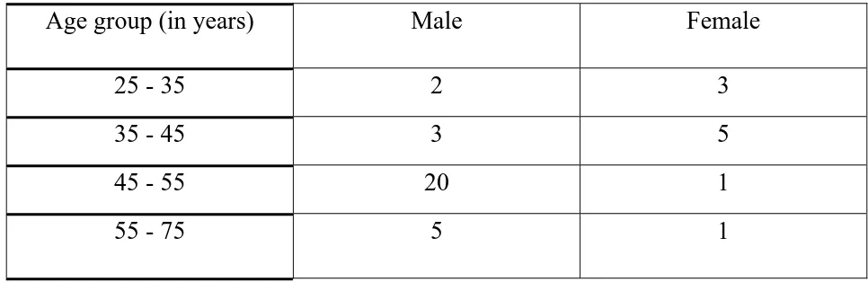
Table I
Age wise distribution of patients in the study group
Age group (in years) Male Female
25 - 35 2 3
35 - 45 3 5
45 - 55 20 1
0
5
10
15
20
MALE
FEMALE
ALCOHOLIC
NON-ALCOHOLIC
0
20
40
60
80
100
120
140
160
180
200
0
2
4
6
8
10
12
T4 Vs T3
T3
(ng/dl)
T4 (mcg/dl)
- zone represents normal values of T3 and T4
LOW(42%)
NORMAL(58%)
LOW(12.5%)
NORMAL(87.5%)
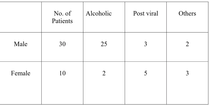
Table II
Clinical diagnosis and etiological factors:
No. of Patients
Alcoholic Post viral Others
Male 30 25 3 2
Female 10 2 5 3
Table III
Biochemical indices of liver function of patients:
SGPT BILIRUBIN ALBUMIN PROTHROMBIN
TIME
[image:58.612.67.511.142.365.2]Table IV
Indices of thyroid function in patients with chronic liver disease and
sex and age matched healthy controls (mean values with standard deviation)
T3(ng/dl) T4(mcg/dl) FT3(pg/ml) FT4(pg/ml) TSH(m U/ml)
Cases 84.1
±
9.6 6.2±
1.3 3.7±
0.9 11.5±
2.6 3.2±
0.9INTERPRETATION OF THE VALUES
:
• Patients with chronic liver disease showed significantly reduced serum
levels of T3.
• 4 patients had low normal levels of FT3.
• 5 patients had low T4 values.
• All patients had normal FT4 and TSH values.
• Simple correlation analysis showed that the serum T3 concentration
significantly correlated with serum bilirubin, albumin and prothrombin in
STATISTICAL ANALYSIS :
T3 :
• Out of 40 cases, T3 value ranged from a minimum of 76 to maximum of
112.2 ng/dl
• Mean value of T3 among cases was 84.1 ng/dl
• Standard deviation of T3 among cases was ± 9.6
• Out of 40 controls, T3 value ranged from a minimum of 82 to maximum of
118.0 ng/dl
• Mean value of T3 among controls was 92.7 ng/dl
• Standard deviation of T3 among cases was ± 8.1
‘p’ value calculated for T3 is <0.001 which is statistically significant.
T4 :
• Out of 40 cases, T4 value ranged from a minimum of 3.6 to maximum of 9.7
mcg/dl
• Mean value of T4 among cases was 6.2 mcg/dl
• Out of 40 controls, T4 value ranged from a minimum of 4.6 to maximum of
10.2 mcg /dl
• Mean value of T4 among controls was 7.1 mcg / dl
• Standard deviation of T4 among cases was ± 1.2
‘p’ value calculated for T4 is 0.0013 which is statistically significant.
Free T3:
• Out of 40 cases, FT3 value ranged from a minimum of 2.55 to maximum of
6.1 pg/ml
• Mean value of FT3 among cases was 3.7pg/ml
• Standard deviation of FT3 among cases was ± 0.9
• Out of 40 controls, FT3 value ranged from a minimum of 2.8 to maximum
of 6.4 pg/ml
• Mean value of FT3 among controls was 4.2 pg/ml
• Standard deviation of FT3 among cases was ± 0.7
• ‘p’ value calculated for FT3 is 0.0042 which is statistically significant
FT4 :
• Out of 40 cases, FT4 value ranged from a minimum of 6.3 to maximum of
• Mean value of FT4 among cases was 11.5 pg/ml
• Standard deviation of FT4 among cases was ± 2.6
• Out of 40 controls, FT4 value ranged from a minimum of 8.75 to maximum
of 16.4 pg/ml
• Mean value of FT4 among controls was 12.9 pg/ml
• Standard deviation of FT4 among cases was ± 2.4
• ‘p’ value calculated for FT4 is 0.008 which is statistically significant
TSH :
• Out of 40 cases, TSH value ranged from a minimum of 1.73 to maximum of
5.4 mU/ml
• Mean value of TSH among cases was 3.2 mU/ml
• Standard deviation of TSH among cases was ± 0.9
• Out of 40 controls, TSH value ranged from a minimum of 2.0 to maximum
of 5.5 mU /ml
• Mean value of TSH among controls was 3.7 mU/ml
• Standard deviation of T4 among cases was ± 0.9
DISCUSSION
The existence of low T3 syndrome i.e., low total T3 with normal total T4 in
the absence of clinical hypothyroidism has been frequently reported in patients
with chronic liver disease and it has been shown to depend on impaired liver
conversion of T4 to T3.
The liver plays an important role in thyroid hormone metabolism being
involved in their conjugation, excretion, peripheral deiodination and in the
synthesis of thyroid binding globulin.
This study confirms a highly significant decrease in T3 serum concentration
in liver disease, the lowest values correlate with sever disease
In a large group of alcoholic patients Israel et al reported a significant
correlation between serum T3 concentration and severity of liver dysfunction as
well as progressive T3 increase in those subjects eventually displaying favourable
outcome suggesting that T3 concentration in patients with advanced liver disease
This study found a good correlation between T3 concentration and serum
albumin, bilirubin, prothrombin time while no correlation has been found with
hepatic inflammatory indices like transaminases
This result suggests that T3 concentration should be considered as a
sensitive index of hepatic function in liver disease.
Green et al found normal FT3 and FT4 in a small group of cirrhotic patients
while low FT4 and normal FT3 concentrations were present in alcoholic fatty liver.
Many studies performed on equilibrium dialysis, however showed decreased
FT3 and normal or frequently increased FT4 concentration. These findings are
confirmed by present study with direct radioimmunoassay of FT3 and FT4 in
chronic liver disease.
These data suggest that in a patient with chronic liver disease, euthyroidism
is maintained by a subtle equilibrium between low FT3 and increased FT4
CONCLUSION
• The present investigation in which thyroid function has been evaluated with
all the clinically available indices, confirms the existence of several
abnormalities in thyroid function test in chronic liver disease, although
showing that euthyroidism is maintained virtually in all patients, probably
as a result of low normal FT3 and high normal FT4.
• Furthermore serum T3 concentration appear to parallel the severity of liver
1. Bianchi GP, Zoli M, Marchesini G, Volta U, Vecchi F, Iervese T, Bonazzi
C, Pisi E. Thyroid gland size and function in patients with cirrhosis of the
liver. Liver1991; 11:71–7.
2. L'age M, Meinhold H, Wenzel KW, Schleusener H. Relations between
serum levels of TSH, TBG, T4, T3, rT3 and various histologically classified
chronicliver diseases. J Endocrinol Invest1980; 3:379–83.
3. Faber J, Thomsen HF, Lumholtz IB, Kirkegaard C, Siersbaek-Nielsen K,
Friis T. Kinetic studies of thyroxine, 3,5,3'-triiodothyronine,
3,3,5'-triiodothyronine, 3',5'-diiodothyronine, 3,diiodothyronine, and
3'-monoiodothyronine in patients with liver cirrhosis. J Clin Endocrinol
Metab1981; 53:978–84.
4. Guven K, Kelestimur F, Yucesoy M. Thyroid function tests in non-alcoholic
cirrhotic patients with hepatic encephalopathy. Eur J Med1993; 2:83–5. 5. Borzio M, Caldara R, Borzio F, Piepoli V, Rampini P, Ferrari C. Thyroid
function tests inchronic liverdisease: evidence for multiple abnormalities
despite clinical euthyroidism Gut1983; 24:631–6.
6. Huang MJ, Liaw YF. Clinical associations between thyroid and liver
Gut2000; 34 (Suppl. 1):78.
8. Van Thiel DH, Udani M, Schade RR, Sanghvi A, Starzl TE. Prognostic
value of thyroid hormone levels in patients evaluated for liver
transplantation. Hepatology1985; 5:862–6.
9. Chopra IJ, Solomon DH, Chopra U, Young RT, Chua Teco GN. Alterations
in circulating thyroid hormones and thyrotropin in hepatic cirrhosis:
evidence for euthyroidism despite subnormal serum triiodothyronine. J Clin
Endocrinol Metab. 1974 Sep;39(3):501–511.
10.Nomura S, Pittman CS, Chambers JB, Jr, Buck MW, Shimizu T. Reduced
peripheral conversion of thyroxine to triiodothyronine in patients with
hepatic cirrhosis. J Clin Invest. 1975 Sep;56(3):643–652.
11.Green JR, Snitcher EJ, Mowat NA, Ekins RP, Rees LH, Dawson AM.
Thyroid function and thyroid regulation in euthyroid men with chronic liver
disease: evidence of multiple abnormalities. Clin Endocrinol (Oxf). 1977
binding globulin in chronic alcoholism. Clin Endocrinol (Oxf). 1981
Feb;14(2):113–118.
13.Chopra IJ, Solomon DH, Hepner GW, Morgenstein AA. Misleadingly low
free thyroxine index and usefulness of reverse triiodothyronine measurement
in nonthyroidal illnesses. Ann Intern Med. 1979 Jun;90(6):905–912.
14.Woeber KA, Maddux BA. Thyroid hormone binding in nonthyroid illness.
Metabolism. 1981 Apr;30(4):412–416.
15.Chopra IJ, Teco GN, Nguyen AH, Solomon DH. In search of an inhibitor of
thyroid hormone binding to serum proteins in nonthyroid illnesses. J Clin
Endocrinol Metab. 1979 Jul;49(1):63–69.
16.Israel Y, Walfish PG, Orrego H, Blake J, Kalant H. Thyroid hormones in
alcoholic liver disease. Effect of treatment with 6-n-propylthiouracil.
Gastroenterology. 1979 Jan;76(1):116–122.
17.Larsen PR. Direct immunoassay of triiodothyronine in human serum. J Clin
and follicle stimulating hormones. J Lab Clin Med. 1967 Dec;70(6):973–
980.
19.Levy RP, Marshall JS, Velayo NL. Radioimmunoassay of human
thyroxine-binding globulin (TBG). J Clin Endocrinol Metab. 1971 Mar;32(3):372–381.
20.Romelli PB, Pennisi F, Vancheri L. Measurement of free thyroid hormones
in serum by column adsorption chromatography and radioimmunoassay. J
Endocrinol Invest. 1979 Jan–Mar;2(1):25–40.
21.Bermudez F, Surks MI, Oppenheimer JH. High incidence of decreased
serum triiodothyronine concentration in patients with nonthyroidal disease. J
Clin Endocrinol Metab. 1975 Jul;41(1):27–40.
22.Hepner GW, Chopra IJ. Serum thyroid hormone levels in patients with liver
disease. Arch Intern Med. 1979 Oct;139(10):1117–1120.
23.Schussler GC, Schaffner F, Korn F. Increased serum thyroid hormone
binding and decreased free hormone in chronic active liver disease. N Engl J
chronic liver diseases. J Endocrinol Invest.1980 Oct–Dec;3(4):379–383.
25.Fischer JE, Baldessarini RJ. False neurotransmitters and hepatic failure.
Lancet. 1971 Jul 10;2(7715):75–80.
26.Borzio M, Caldara R, Ferrari C, Barbieri C, Borzio F, Romussi M. Growth
hormone and prolactin secretion in liver cirrhosis: evidence for
dopaminergic dysfunction. Acta Endocrinol (Copenh). 1981 Aug;97(4):441–
447.
27.Scanlon MF, Pourmand M, McGregor AM, Rodriguez-Arnao MD, Hall K,
Gomez-Pan A, Hall R. Some current aspects of clinical and experimental
neuroendocrinology with particular reference to growth hormone,
Name Age Sex
O.P/ I.P No :
Occupation :
Address :
Per capita income :
Presenting complaints :
H/o present illness :
onset
duration
course
H/o suggestive of hypo- or hyperthyroidism
Family history :
Personal history :
Diet :
Sleep :
Appetite :
Bowel & bladder habits :
Habits : smoking / alcoholism
H/o high risk behaviour
Menstrual history (in females):
Menarche/menopause; cycles; flow
Obstetric history (in females):
H/o pregnancy / lactation
H/o intake of OCPs
Treatment history :
Built & nourishment
Febrile/afebrile
Pallor / cyanosis / clubbing / icterus / lymphadenopathy / pedal edema
Pulse rate : Peripheral pulses :
Blood pressure :
Respiratory rate :
Temperature :
Thyroid examination :
Systemic Examination :
Cardiovascular system :
Respiratory system :
Abdomen (including genital and per rectal examination):
Central nervous system :
• Hb%, TC, DC, ESR, Platelet
• Urine – albumin, sugar, deposits • Blood urea
• Serum creatinine
• Liver function tests
• Coagulation profile
• Ultrasound abdomen and pelvis
• Portal vein Doppler
• Serum T3, T4, TSH FT3, FT4
• Portal vein Doppler
• Ultrasonogram abdomen & pelvis
S.No
Age
Sex
T3
T4
FT3
FT4
TSH
Bilirubin
Albumin
PT
SGPT
1
27
M
79.2
4.2
2.55
6.7
1.73
3.3
2.1
45
46
2
30
M
81.4
4.5
3.8
7.9
2.5
2.1
2.8
56
62
3
40
M
79
5.2
3.81
6.9
2.6
2
2.4
36
58
4
37
M
90.6
6
5.6
12.2
4.6
1.7
3.1
44
51
5
42
M
76.5
3.7
2.7
9
3.11
3.2
2
46
48
6
48
M
83
4.8
3.82
7.7
2.81
1.8
1.9
52
59
7
47
M
82.4
4
3.8
10.7
3.4
1.6
2.1
55
47
8
55
M
110.6
7.3
6.1
16.4
3.5
1.9
2.4
51
65
9
52
M
81.4
6.1
3.2
12
5
2.3
3
49
40
10
50
M
98.2
7
6
14
5.2
2
2.7
57
54
11
49
M
78
4.9
2.9
6.3
2.8
2.6
2
52
57
12
50
M
80
3.9
3.8
12.2
2.7
1.3
2.6
50
46
13
52
M
77
3.6
2.62
11.8
3.2
3
2.8
42
62
14
47
M
76
5.4
2.6
12.4
3.51
3.8
3.3
53
58
15
45
M
100.4
8.1
4.4
10.6
5.4
1.7
3.1
50
49
16
54
M
84
6.3
3.1
8.6
3.27
3.2
2.7
57
47
17
51
M
79
5.7
2.7
8.4
2.9
2.7
2.2
48
40
18
48
M
84.8
6.5
3.1
10.8
4.1
3.3
2.5
44
72
19
46
M
78.6
5.8
2.82
11.9
2.84
3.2
3
52
49
S.No
Age
Sex
T3
T4
FT3
FT4
TSH
Bilirubin
Albumin
PT
SGPT
21
55
M
81.6
7.5
3.15
13.2
3.31
2.2
2.1
44
35
22
48
M
82.4
6.4
3.8
13
3.2
2
2.4
47
45
23
60
M
83.6
7.4
3.9
10.62
2.7
2.4
2.5
43
67
24
72
M
78.2
6.2
3.2
8
3
2.1
2
39
56
25
52
M
79.3
6.4
3.12
10.2
3.1
3.4
2.2
52
49
26
70
M
112.2
9.7
4.8
15.2
5.3
1.9
3.2
56
39
27
54
M
114
8.2
4.45
15
3.5
2
3
42
58
28
48
M
84.2
7.2
3.1
11.9
3.1
2.2
2
40
49
29
53
M
76
6
3
7.2
1.82
2.7
2.8
36
40
30
59
M
77.9
7.1
4.5
10
2
3
1.9
47
50
31
32
F
85.7
6.3
3.9
15.9
1.96
1.7
2
41
62
32
42
F
77.4
6.1
2.9
10.9
1.89
2.9
3
34
49
33
48
F
82.6
6.6
4.2
12.6
2.5
2.3
2.8
39
47
34
56
F
79.9
6.2
2.8
12.3
2.5
2.5
3
30
54
35
34
F
78.7
7
2.91
12.4
2
2.6
2.7
43
55
36
38
F
86.2
6.7
4.1
15.8
3.41
1.9
2.3
45
50
37
30
F
78.2
6.8
2.94
10.8
2.7
3
2.6
47
43
38
45
F
82.8
6.9
3.95
14
3.38
1.8
2.9
34
45
39
39
F
83.4
6.8
3.8
13.4
3.3
3.2
2.8
39
58
S.No
Age
Sex
T3
T4
FT3
FT4
TSH
Bilirubin
Albumin
PT
SGPT
1
28
M
84
5.4
3.95
14.4
2.4
0.9
4.7
21
23
2
32
M
90.2
8.2
3.8
13.7
3.2
1.1
3.2
24
19
3
39
M
94
8.2
3.82
12.4
4.2
1.2
3.3
30
16
4
37
M
96.9
7.3
3.9
8.9
4
0.9
4.2
30
21
5
40
M
97.2
7.9
4
15.5
3.2
1.1
3
25
30
6
46
M
100.4
9.4
4.41
10.8
4.1
0.7
4
28
31
7
46
M
92.6
5.4
5
15
5.42
1.2
3.7
17
32
8
54
M
94.1
7.4
3.4
10.2
5.5
0.7
3.6
15
34
9
51
M
83
8.4
6.4
9.8
4.32
0.9
4.1
19
23
10
52
M
94.2
8.7
4.2
16
3.9
1.3
3.2
21
19
11
47
M
97.6
6
4.9
10.2
3.2
1.1
3.9
24
39
12
49
M
104.2
7.2
4.3
9.7
3.14
0.8
4.9
22
15
13
52
M
84.3
6.4
3.7
15
3.72
1
4
29
28
14
45
M
86.2
7.5
4.23
9.9
5.48
1.2
4.2
30
19
15
47
M
94.7
7.2
3.7
16.24
4.8
0.7
4.8
24
36
16
52
M
91.8
8.1
4.6
12
2
0.8
4.1
30
17
17
54
M
108.4
5.8
4.2
9.7
2.62
1.1
3.7
28
35
18
49
M
93.8
7.4
4.29
12.5
3.4
0.9
3.9
30
20
19
47
M
88.4
6.9
4.28
12.