Copyright © 1999, American Society for Microbiology. All Rights Reserved.
Characterization of V3 Sequence Heterogeneity in Subtype C Human
Immunodeficiency Virus Type 1 Isolates from Malawi:
Underrepresentation of X4 Variants
LI-HUA PING,1,2JULIE A. E. NELSON,1IRVING F. HOFFMAN,1,2JODY SCHOCK,1,3SUZANNA L. LAMERS,4
MELISSA GOODMAN,1,2PIETRO VERNAZZA,5PETER KAZEMBE,6MARTIN MAIDA,6DICK ZIMBA,6
MAUREEN M. GOODENOW,4JOSEPH J. ERON, JR.,1,2SUSAN A. FISCUS,1,3
MYRON S. COHEN,1,2ANDRONALD SWANSTROM1,7*
UNC Center For AIDS Research,1Department of Medicine,2Department of Microbiology and Immunology,3and Department of Biochemistry and Biophysics,7University of North Carolina at Chapel Hill, Chapel Hill, North
Carolina; Department of Pathology, Immunology, and Laboratory Medicine, University of Florida College of Medicine, Gainesville, Florida4; Institute for Clinical Microbiology and Immunology,
St. Gallen, Switzerland5; and Lilongwe Central Hospital, Lilongwe, Malawi6
Received 19 February 1999/Accepted 20 April 1999
We have examined the nature of V3 sequence variability among subtype C human immunodeficiency virus type 1 (HIV-1) sequences from plasma-derived viral RNA present in infected men from Malawi. Sequence variability was assessed by direct sequence analysis of the V3 reverse transcription-PCR products, examination of virus populations by a subtype C V3-specific heteroduplex tracking assay (V3-HTA), and selected sequence analysis of molecular clones derived from the PCR products. Sequence variability in V3 among the subtype C viruses was not associated with the presence of basic amino acid substitutions. This observation is in contrast to that for subtype B HIV-1, where sequence variability is associated with such substitutions, and these substitutions are determinants of altered coreceptor usage. Evolutionary variants in subtype C V3 sequences, as defined by the V3-HTA, were not correlated with the CD4 level in the infected person, while such a correlation was found with subtype B V3 sequences. Viruses were isolated from a subset of the subjects; all isolates used CCR5 and not CXCR4 as a coreceptor, and none was able to grow in MT-2 cells, a hallmark of the syncytium-inducing phenotype that is correlated with CXCR4 usage. The overall sequence variability of the subtype C V3 region was no greater than that of the conserved regions of gp120. This limited sequence variability was also a feature of subtype B V3 sequences that do not carry the basic amino acid substitutions associated with altered coreceptor usage. Our results indicate that altered coreceptor usage is rare in subtype C HIV-1 isolates in sub-Saharan Africa and that sequence variability is not a feature of the V3 region ofenv
in the absence of altered coreceptor usage.
Human immunodeficiency virus (HIV-1) isolates are phylo-genetically clustered into distinct groups based on sequence analysis of the viral genome (reviewed in reference 45). These groups have been termed subtypes or clades and given alpha-betical designations (e.g., subtype or clade A). Partial-se-quence analysis of a portion of a viral genome derived from a tissue isolate taken in 1959 suggests that the different subtypes represent a fairly recent radiation (93), perhaps with each subtype representing the early establishment of independent focal infections. The major HIV-1 subtype found in the United States and western Europe, and the most extensively studied subtype, is subtype B. Subtype C virus is part of an expanding epidemic in sub-Saharan Africa and India and is now the most abundant subtype of HIV-1 worldwide (81).
A dramatic feature of subtype B HIV-1 infection is the de novo evolution of a more pathogenic variant in up to 50% of infected people (6, 10, 12, 20, 68, 70, 77). This variant repre-sents an altered form of the virus whose appearance corre-sponds to an accelerated decrease in the number of circulating CD4⫹T helper cells (42, 69). Typically, transmitted virus uses
the CCR5 chemokine receptor as a coreceptor for entry into
cells (3, 16, 26, 31, 32) and has also been characterized as nonsyncytium inducing (NSI), slow/low, and macrophage tropic (65, 84, 94); this type of virus has been named R5 (8). The new variant that has evolved from the initial R5 virus is able to use an alternate chemokine receptor, typically CXCR4 (36), and has been characterized as syncytium inducing (SI), rapid/high, and T-cell-line tropic (reviewed in reference 7). Variants that use CXCR4 are now called X4 (8). Primary X4 isolates can be dually tropic for CXCR4 and CCR5 (73).
Sequence alignments of theenvgene revealed multiple vari-able regions (2, 76, 86), which were subsequently named V1 to V5 (57). The major determinant of specificity in coreceptor usage by subtype B isolates is within the V3 loop domain of the viral Env protein (16, 17, 75; reviewed in reference 71). Se-quence changes within V3 are largely responsible for deter-mining coreceptor specificity, probably through a direct inter-action with surface residues of the chemokine receptor (9, 17, 79, 88).
There are distinctive sequence changes in V3 that are asso-ciated with the change in coreceptor usage. These changes include substitutions that result in the presence of increased numbers of basic amino acids at discrete positions within V3 (13, 24, 38, 55, 72). Other changes in the V3 sequence are strongly associated with the presence of the basic amino acid substitutions (56), and some of these changes play a direct role in coreceptor specificity (14). Overall, subtype B V3 sequences * Corresponding author. Mailing address: CB7295, Rm 22-006
Lineberger Bldg., University of North Carolina at Chapel Hill, Chapel Hill, NC 27599. Phone: (919) 966-5710. Fax: (919) 966-8212. E-mail: [email protected].
6271
on November 9, 2019 by guest
http://jvi.asm.org/
that contain basic amino acid substitutions display twice as much amino acid variability from the consensus sequence as do sequences without the basic amino acids (13, 55). This in-creased variability is in addition to the basic amino acid sub-stitutions. The presence of this additional sequence variability has allowed the heteroduplex tracking assay (HTA) (25) to be used to rapidly identify V3 evolutionary variants that are strongly correlated with the more pathogenic (X4) form of the subtype B HIV-1 (60).
While the evolution of X4 variants among patients infected with subtype B virus is a striking and well-documented phe-nomenon, its impact on people infected with viruses of other subtypes is not known. Viruses that use the CXCR4 receptor (SI phenotype) have been identified among most of the HIV-1 subtypes (23, 28, 63, 80, 89–92). In cases where the X4 viruses of other subtypes have been examined, these variants encode increased numbers of basic amino acids in their V3 loops (23, 28, 63, 89, 92), suggesting that common mechanisms are de-termining changes in coreceptor interactions. However, the potential for differences in patterns of coreceptor usage among the different subtypes was suggested by the observation that subtype C SI/X4 variants were rare among a group of 16 people under care in Sweden (80) and among a group of 22 French military personnel infected during overseas deploy-ment (63), although subtype C SI/X4 variants have been ob-served (78, 80, 91).
Understanding the differences in the evolution of subtype C virus is becoming increasingly important because the domi-nance of this subtype in the worldwide HIV epidemic will lead to the inevitable expansion of subtype C vaccine development. Because of our ongoing clinical studies in Malawi (18, 33), we had an opportunity to examine this question in a study of a large cohort of men infected primarily with subtype C HIV-1. We adapted the V3-HTA (60) to detect V3 evolutionary variants of subtype C virus. In a group of plasma samples from 80 HIV-1-infected men from Malawi, we found that 31% of the samples showed evidence of evolutionary variants within the V3 region. However, the presence of these variants was not related to low levels of CD4⫹ T cells, as is seen in people
infected with subtype B HIV-1. Sequence analysis of viral RNA revealed a virtual absence of basic amino acid substitu-tions in the V3 regions of the evolutionary variants. Virus isolates were established from a subset of the Malawi subjects, and all of them preferentially used the CCR5 receptor for virus entry. Total sequence variability in subtype C V3 sequences was comparable to the reduced variability seen with sequences from subtype B viruses that do not contain basic amino acid substitutions in the V3 loop and was similar in magnitude to the variability in the conserved regions ofenv. These results indicate that X4 variants are rare among men in sub-Saharan Africa infected with subtype C virus and that V3 variability is a feature of viruses with altered coreceptor usage.
MATERIALS AND METHODS
Source of patient samples.Plasma and peripheral blood mononuclear cells (PBMC) were collected and processed from subjects in the sexually transmitted disease and dermatology clinics of the Lilongwe Central Hospital in Lilongwe, Malawi, as described previously (18). HIV-1 seropositivity was determined by two enzyme immunoassays (Genetic Systems HIV-1/HIV-2 EIA; Genetics Sys-tems Corp., Redmond, Wash.; and Murex HIV-1⫹2; Murex Diagnostics Ltd., Dartford, United Kingdom) and Western blot analysis (Organon-Teknika, Durham, N.C.), after which the HIV-1 serotype was determined by an envelope V3 peptide immunoassay with antigens specific for clades A through F (33, 62). Plasma samples from subjects infected with HIV-1 subtype B were chosen from the samples that were collected as part of AIDS Clinical Trial Group Clinical Trial 201 (37) with Institutional Review Board approval. Subtype B samples were chosen from subjects with fewer than 400 CD4⫹T cells/l.
Virus isolation and test for coreceptor usage.The following reagents were obtained from the National Institutes of Health AIDS Research and Reference Reagent Program: the MT-2 cell line was from Douglas Richman, the U373-MAGI cell lines (85) were from Michael Emerman, the YU-2 molecular clone (51) was from Beatrice Hahn and George Shaw, and the HIV-189.6viral isolate (19) was from Ronald Collman. Malcolm Martin provided the AD8 (ADA) (15) and NL4-3 molecular clones (1), and Nathaniel Landau provided the HIV-1 clone of HXB with theenvgene derived from Ba-L (39). PBMC from infected individuals were cocultured with phytohemagglutinin-stimulated PBMC from uninfected donors in qualitative HIV cultures as described previously (82). Cul-ture supernatants were tested twice a week for p24 antigen production (Or-ganon-Teknika). Virus isolates were assayed in triplicate for syncytium formation in MT-2 cells by using a previously described method (41). Briefly, 50l of PBMC coculture (including the cells) was added to 5⫻104MT-2 cells in 150l of medium in a 96-well plate. The cultures were monitored for syncytia twice a week; they were scored positive when there were three to five syncytia per high-power field under light microscopy.
Coreceptor usage was determined by using three U373-MAGI cell lines that express no coreceptor (U373-MAGI), the CCR5 coreceptor (U373-MAGI-CCR5), or the CXCR4 coreceptor (U373-MAGI-CXCR4) (85). The cell lines were maintained and infected as described previously (85). Briefly, each of the cell lines was plated in separate wells of a 48-well plate 1 day prior to infection. An aliquot of 90l of diluted viral supernatant from PBMC coculture was added to the cells and adsorbed for 2 h at 37°C under 5% CO2. The subtype C viral isolates were used at a 1:3 dilution in medium or undiluted. Positive control viruses (YU-2, Ba-L, ADA, NL4-3, and 89.6) in infected-cell supernatants were used at a 1:6 dilution. An aliquot of 0.5 ml of culture medium was then added to each well, and the plate was incubated for 40 to 48 h at 37°C under 5% CO2. The medium was then removed, and the cells were fixed for 5 min with 0.5 ml of fixing solution (1% formaldehyde, 0.2% glutaraldehyde) per well. The cells were washed twice with phosphate-buffered saline, 200l of staining solution (4 mM potassium ferricyanide, 4 mM potassium ferricyanide, 2 mM MgCl2, and 0.4 mg of 5-bromo-4-chloro-3-indolyl--D-galactopyranoside [X-Gal] per ml in PBS)
was added to each well, and the plate was incubated at 37°C for 2 h. The cells were then washed with phosphate-buffered saline twice, and the blue cells were counted.
Viral RNA isolation, RT-PCR, and DNA sequence determination.Viral RNA was isolated from 140l of patient plasma with a QIAamp viral RNA kit (Qiagen); the RNA was eluted with 50l of RNase-free water. Primers for reverse transcription-PCR (RT-PCR) were designed to correspond to the HIV-1 subtype C V3 consensus sequence present in the Human Retroviruses and AIDS database (44). The upstream (C⫹V3) primer was 5⬘-ATAGTACATCTTAATC AATCTGTAGAAATT-3⬘, and the downstream (C⫺V3) primer was 5⬘-CCAT TTATCTTTACTAATGTTACAATGTGC-3⬘; these primers generate a 159-bp product. RT-PCRs were performed by the method described by Nelson et al. (60) with the following modifications. RT reaction mixtures of 20l consisted of 5l of the viral RNA eluate, 1⫻Expand HF buffer (Boehringer Mannheim), 2.5 mM MgCl2, 1l of 10 mM deoxynucleoside triphosphate mix (U.S. Biochemi-cal), 10 U of RNase inhibitor (Boehringer Mannheim), 15 pmol of primer C-V3, and 10 U of avian myeloblastosis virus reverse transcriptase (Boehringer Mann-heim). Reverse transcription was done at 42°C for 30 min, followed by 2 min at 95°C to inactivate the enzyme. A 30-l aliquot of PCR mix (1⫻Expand HF buffer, 2.5 mM MgCl2, 15 pmol of primer C⫹V3, 2 U of Expand High Fidelity enzyme mix [Boehringer Mannheim]) was added to each RT reaction mixture. PCR was carried out in a Stratagene Gradient-40 Robocycler with the following program: one cycle at 95°C for 2 min 45 s; then 40 cycles at 95°C for 45 s, 49°C for 45 s, and 68°C for 1 min (after the first 10 cycles, 1 min was added to the 68°C step for 10 cycles, 2 min was added for the next 10 cycles, and 3 min was added for the last 10 cycles). RT-PCR of subtype B samples was performed as described previously (60). The RT-PCR products were purified by using QIAquick PCR purification columns (Qiagen), and the PCR products were sequenced with an ABI PRISM dye terminator cycle-sequencing kit (Perkin-Elmer). Alternatively, the PCR product was cloned into the pT7Blue(R) vector (Novagen), individual clones were screened by HTA, and examples of each HTA-defined species were sequenced by using an ABI PRISM dye terminator cycle-sequencing kit.
V3-HTA.Probe construction, probe labeling, and HTA conditions for subtype C samples were adapted from those described by Nelson et al. (60) and Delwart et al. (25). Based on direct sequencing of several RT-PCR products, the product from patient C128 was chosen for probe construction. The C128 RT-PCR prod-uct was cloned into the pT7Blue(R) vector. Several clones were sequenced, and a probe plasmid (D516-11) was chosen that had only three nucleotide differences from the subtype C V3 consensus. The subtype C V3 probe was labeled by first digesting 1g of D516-11 plasmid withBamHI. The plasmid was end labeled for 15 min at room temperature in a reaction mixture of 50l containing 12.5Ci of35S-dATP (1,250 Ci/mmol; NEN Life Science Products), unlabeled dGTP at final concentration 1 mM, and 2 U of the Klenow fragment of DNA polymerase I; this was followed by heat inactivation of the enzyme. The plasmid was further digested withSpeI to release the probe from the vector. The labeled probe was purified by using a QIAquick PCR purification column and recovered in a final volume of 50l. Heteroduplex formation reactions were done in 10-l reaction mixtures consisting of 8l of purified RT-PCR product, 1l of 10⫻annealing buffer (1 M NaCl, 100 mM Tris-HCl [pH 7.5], 20 mM EDTA), 1M primer
on November 9, 2019 by guest
http://jvi.asm.org/
C⫹V3, and 0.8l of labeled D516-11 probe. The reaction mixtures were dena-tured at 95°C for 2 min and allowed to anneal at room temperature for 15 min. The heteroduplexes were separated in nondenaturing 12% polyacrylamide gels. V3-HTA of subtype B RT-PCR products was performed as described previously (60).
A unified linear regression model in rank scale was used to assess the rela-tionship between the CD4 cell count and the HTA mobility ratio of the samples with single and multiple bands for both the subtype B and subtype C data sets. When multiple bands were present, the mobility ratio value of the slowest-migrating band was used. Calculations were done with SAS (version 6.12) pro-grams rk0101.sas and rk0102.sas.
Analysis of sequence heterogeneity.Total amino acid variability within the newly determined V3 sequences was analyzed as follows. The total number of times a nonconsensus sequence amino acid was present was tallied, this number was divided by the total number of V3 sequences times 35 (the number of amino acid positions in V3), and the final number was multiplied by 100 to give the percentage of amino acid substitutions.
For analysis of evolutionary distance, envelope sequences spanning V1 through V5 were obtained from the Los Alamos HIV-1 database (44). A total of 33 subtype C and 69 subtype B sequences were used. Subtype B sequences were classified into two groups, either R5-like or X4-like, based on known biological properties of the viruses or the charge characteristics of amino acids at position 11 or 25 in the V3 loop. Nucleotide sequences from all viruses were aligned by using DNA (Harvard University Molecular Biology Computer Resources). The alignments were optimized manually to ensure that codons remained intact and gaps were minimized (50). Sequences were divided into segments, which ranged in length from 78 to 150 nucleotides or 26 to 50 codons, based on the location of hypervariable and conserved domains. Phylogenetic analysis to assess the rela-tionship among the sequences and between subtype C and subtype B sequences in V3 was performed by using the neighbor-joining method (66) in the PHYLIP package (34, 35). Estimation of total distance in each segment among the three groups of viruses was based on the method of Nei and Gojobori (59) in MEGA (48). Distances within each segment among the three groups of sequences were evaluated by a one-way analysis of variance (SigmaStat; Jandel). A pairwise multiple comparison procedure was used to analyze the significance of relation-ships between groups (SigmaStat). APvalue of⬍0.05 was considered significant. Nucleotide sequence accession numbers.The nucleotide sequences for the V3 region described here have been assigned GenBank accession no. AF153129 to AF153190.
RESULTS
Application of V3-HTA to subtype C virus. As a starting point to study the nature of V3 sequence variability in subtype C virus, we used a collection of plasma samples taken from 80 separate subjects participating in a clinical trial (18, 33). Each of the samples was taken from an HIV-1-infected subject from Malawi, and the virus from each subject was characterized as subtype C by using a V3 peptide immunoassay (33, 62). The viral RNA level in plasma and CD4⫹-T-cell count for each of
the subjects are presented in Table 1. The range of values for the CD4⫹-T-cell count was 36 to 1,253 cells/l, with the
me-dian value being 273 cells/l; the range of values for viral RNA load in plasma was 1.6⫻103by 5,700⫻103copies/ml.
Subtype C-specific primers were designed based on con-served regions of subtype C virus sequences in the Human Retroviruses and AIDS database (44) to amplify a 159-bp region of the env gene encompassing V3. Initially, RT-PCR products amplified from viral RNA in plasma from 20 subjects were analyzed by direct sequencing. Samples that gave largely unambiguous sequencing results (implying an absence of a significant mixture of V3 sequences) were used to construct a consensus V3 sequence for subtype C.
A clone from the RT-PCR product derived from the plasma of subject C128 was chosen for the V3-HTA probe because it was the closest to the consensus sequence; it varies from the consensus sequence at only three widely spaced positions, and all three of the differences are G-to-A transitions. The probe was labeled by first cleaving the V3 insert at one junction with the plasmid by using a unique restriction enzyme site in the plasmid. The overhanging end was filled in with a radioactive nucleotide to label one strand of the probe, allowing detection of heteroduplexes formed with only one of the probe strands. The probe insert was then released from the plasmid by
cleav-age at a second unique cleavcleav-age site at the other insert/plasmid junction. The inclusion of several flanking nucleotides from the plasmid in the probe results in distinct migrations for the reannealed probe strands versus the heteroduplexes formed between the labeled probe strand and complementary RT-PCR products (60).
Characterization of viral RNA in plasma by V3-HTA. V3-HTA is able to identify many sequence variants within the region ofenv encoding V3 (60). The magnitude of sequence evolution that leads to a change in coreceptor usage is, in most cases, significant enough to score in the V3-HTA. Such se-quence variants cause the heteroduplex formed between the PCR product and the probe to bend and therefore to be re-tarded during polyacrylamide gel electrophoresis. As a first step in characterizing subtype C V3 sequence variability, we applied this assay to the RT-PCR product from each of the 80 plasma samples. Such an analysis provides two pieces of infor-mation: first, the presence of variants with sequences distinct from the probe can be identified; second, the presence of mixtures of cocirculating viral sequences is revealed. Examples of V3-HTA patterns from subjects with homogeneous V3 se-quences (single band) or heterogeneous V3 sese-quences (multi-ple bands) are shown in Fig. 1. A summary of the V3-HTA results for the plasma samples is shown in Table 1.
Of the 80 plasma samples, 55 had single bands in the V3-HTA analysis, indicating that the virus populations in these samples were homogeneous within V3. The remaining 25 sam-ples had multiple bands in the V3-HTA analysis, revealing the presence of multiple virus species with different V3 sequences. Of the samples with single bands, 53 had bands that migrated near the bottom of the gel, indicating similarity to the consen-sus sequence. The other two samples (S018 and S051) had single bands that migrated more slowly, indicative of significant sequence differences with respect to the probe (Table 1). Se-quence analysis of the bulk PCR product was done for 46 of the samples that gave a single band in V3-HTA and for 7 samples that gave multiple bands but for which there was a predominant band with rapid mobility and unambiguous se-quence. In addition, seven samples that gave multiple bands were subjected to molecular cloning, and individual clones were screened by V3-HTA and sequenced. In total, sequence information was determined for 62 of the 80 samples.
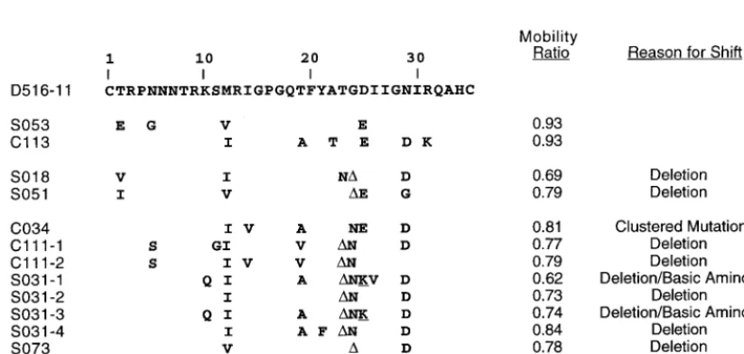
We previously found a strong correlation between subtype B HIV-1 samples that had slowly migrating bands in the V3-HTA analysis, the presence of sequence changes that would encode basic amino acid substitutions in the V3 loop, and the presence of SI viruses in the virus isolate (60). Therefore, we were interested in determining the nature of the sequence changes in the subtype C isolates that resulted in slow migration in the V3-HTA analysis. A summary of the amino acid substitutions encoded by the nucleotide changes for the shifted bands rep-resenting viruses from nine patients is shown in Fig. 2. This includes the two samples that had a single band with low mobility (S018 and S051) and seven of the samples with mul-tiple bands. Bands were considered “shifted up” if the mobility ratio (the distance migrated by the heteroduplex divided by the distance migrated by the probe homoduplex) was less than 0.85, and by this definition only 10 of the samples with multiple bands included bands that were considered significantly shifted. Seven of the nine sequenced examples of shifted bands were due to three nucleotide deletions, usually corresponding to position 24 or 25 of V3. One of the sequences with a deletion (S031) and one other sequence (S134) had basic amino acid substitutions that are associated with the SI/X4 phenotype in subtype B viruses. The ninth sequence, C034, shifted because of clustered mutations away from the
on November 9, 2019 by guest
http://jvi.asm.org/
sus, but these did not include basic amino acid substitutions. These results suggest that clustered mutations that include basic amino acid substitutions, which are characteristic fea-tures of subtype B X4 variants, are not a prominent feature of V3 sequence variability among subtype C sequences.
Comparison of evolutionary variants in subtype B and sub-type C. X4 viruses typically evolve late in the course of an HIV-1 infection, usually when the levels of CD4⫹ T helper
cells fall below 400/l (42). We carried out an analysis of the appearance of evolutionary variants among subtype B versus subtype C HIV-1 as a function of the patient CD4⫹
-T-helper-cell level (Fig. 3). Samples were obtained from subjects in-fected with subtype B virus who were participants in an ACTG study. As expected, among the subtype B viruses, V3-HTA (with the subtype B probe) revealed slowly migrating bands and/or multiple bands with increasing frequency as patient CD4⫹-T-helper-cell levels declined. In contrast, the presence
of slowly migrating bands derived from the subtype C viruses (detected with the subtype C probe) showed no relationship
with the patient CD4⫹-T-helper-cell level. The association was
tested by using a unified linear-regression model in rank scale (n⫽83). The subtype B samples with multiple bands showed an association between decreasing mobility ratio and decreas-ing CD4 counts that approached statistical significance even with this small sample size (slope⫽0.462;P⫽0.06). This was not the case for the subtype B samples with single bands (slope⫽0.158;P⫽0.35) or the subtype C samples with either multiple bands (slope ⫽ 0.011; P ⫽ 0.94) or single bands (slope⫽0.006;P⫽0.96). Thus, there appears to be a differ-ence between these two subtypes in the nature of V3 sequdiffer-ence variants as detected by V3-HTA. The difference was tested statistically by a comparison of slopes (0.426 versus 0.011;P⫽
0.13). A plausible explanation for the lack of statistical signif-icance is the small sample size for subtype B (n⫽ 26). The power of the test comparing slopes was approximately 0.324, or 32.4%.
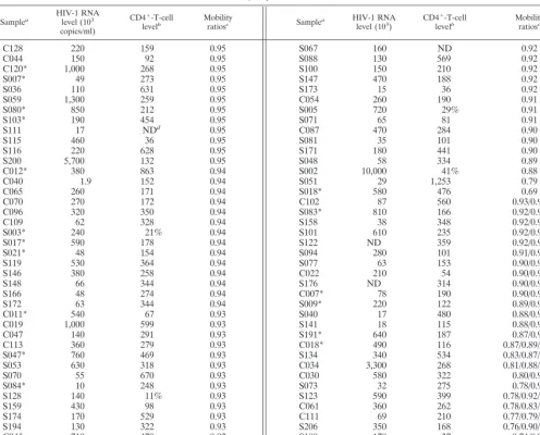
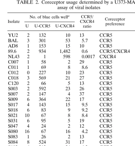
[image:4.612.56.552.83.483.2]Coreceptor usage of subtype C viruses.Both the near ab-sence of basic amino acid substitutions in samples scoring in TABLE 1. Subject profiles and V3-HTA results
Samplea HIV-1 RNAlevel (103 copies/ml)
CD4⫹-T-cell
levelb Mobilityratiosc Samplea HIV-1 RNAlevel (103) CD4 ⫹-T-cell
levelb Mobilityratiosc
C128 220 159 0.95 S067 160 ND 0.92
C044 150 92 0.95 S088 130 569 0.92
C120* 1,000 268 0.95 S100 150 210 0.92
S007* 49 273 0.95 S147 470 188 0.92
S036 110 631 0.95 S173 15 36 0.92
S059 1,300 259 0.95 C054 260 190 0.91
S080* 850 212 0.95 S005 720 29% 0.91
S103* 190 454 0.95 S071 65 81 0.91
S111 17 NDd 0.95 C087 470 284 0.90
S115 460 36 0.95 S081 35 101 0.90
S116 220 628 0.95 S171 180 441 0.90
S200 5,700 132 0.95 S048 58 334 0.89
C012* 380 863 0.94 S002 10,000 41% 0.88
C040 1.9 152 0.94 S051 29 1,253 0.79
C065 260 171 0.94 S018* 580 476 0.69
C070 270 172 0.94 C102 87 560 0.93/0.95
C096 320 350 0.94 S083* 810 166 0.92/0.95
C109 62 328 0.94 S158 38 348 0.92/0.95
S003* 240 21% 0.94 S101 610 235 0.92/0.94
S017* 590 178 0.94 S122 ND 359 0.92/0.94
S021* 48 154 0.94 S094 280 101 0.91/0.95
S119 530 364 0.94 S077 63 153 0.90/0.95
S146 380 258 0.94 C022 210 54 0.90/0.94
S148 66 344 0.94 S176 ND 314 0.90/0.93
S166 48 274 0.94 C007* 78 190 0.90/0.92
S172 63 344 0.94 S009* 220 122 0.89/0.91
C011* 540 67 0.93 S040 17 480 0.88/0.91
C019 1,000 599 0.93 S141 18 115 0.88/0.90
C047 140 291 0.93 S191* 640 187 0.87/0.91
C113 360 279 0.93 C018* 490 116 0.87/0.89/0.91
S047* 760 469 0.93 S134 340 534 0.83/0.87/0.91
S053 630 318 0.93 C034 3,300 268 0.81/0.88/0.91
S070 55 670 0.93 C030 580 322 0.80/0.95
S084* 10 248 0.93 S073 32 275 0.78/0.95
S128 140 11% 0.93 S123 590 399 0.78/0.92/0.94
S159 430 98 0.93 C061 360 262 0.78/0.83/0.87
S174 170 529 0.93 C111 69 210 0.77/0.79/0.88
S194 130 322 0.93 S206 350 168 0.76/0.90/0.92
C045 710 479 0.92 S180 170 37 0.74/0.93
S032 1.6 881 0.92 S031* 43 333 0.62/0.73/0.74/0.84
aSamples for which a virus isolate was recovered are indicated by asterisks. bCD4⫹-T-cell count per microliter or percentage of CD4⫹T cells among all T cells.
cMobility ratios are shown for all heteroduplexes in each sample. dND, not determined.
on November 9, 2019 by guest
http://jvi.asm.org/
the V3-HTA analysis and the lack of correlation between V3 sequence variants and CD4⫹-T-helper-cell levels suggested
that subtype C viruses do not evolve to utilize different core-ceptors in a manner analogous to that seen with subtype B viruses. To determine the nature of coreceptor usage among these viruses, PBMC corresponding to 79 of the 80 plasma samples were used in coculture assays to recover virus. Virus isolates were obtained from only 19 of the 79 samples. Four-teen of these isolates were from patients with less than 400 CD4⫹T cells/l, the point at which X4 viruses start appearing
in people infected with subtype B viruses (42). The isolates were tested for coreceptor usage in U373-MAGI cell lines (85) and for syncytium induction in MT-2 cells, a frequent feature of subtype B X4 variants (43). As shown in Table 2, all of the isolates used CCR5 as a coreceptor, did not use CXCR4, and did not induce syncytia in MT-2 cells. An additional 12 subtype C isolates that were not associated with the 80 plasma samples
were tested in the MT-2 assay, and all were NSI (data not shown), although this additional group has not been subjected to sequence, V3-HTA, or coreceptor usage analysis. To date we have not found a single example of CXCR4 coreceptor usage or SI phenotype among the 31 subtype C virus isolates in our collection.
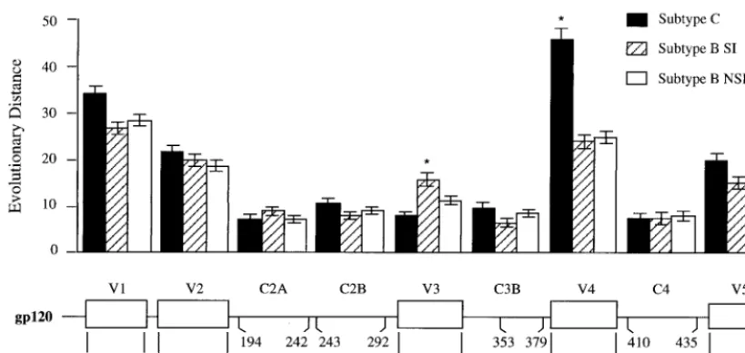
Analysis of V3 sequence variability in subtype C HIV-1.We combined the sequence data from the 62 samples analyzed in this study with 64 sequences available for subtype C viruses in the Human Retroviruses and AIDS database (44) to create a data set that would allow an estimate of total sequence vari-ability within V3. Five subtype C sequences that had deletions were removed from the data set and not included in this anal-ysis, leaving 121 sequences. Each deletion was of 3 nucleotides, i.e., one codon, and appeared at position 24 or 25, suggesting that these are positions that can accommodate a deletion. These deletions do not appear to be involved in altered core-ceptor usage, since three of the viruses with these deletions were included in the analysis of coreceptor usage and all were specific for CCR5 (Fig. 2, Table 2).
The pattern of sequence variability for subtype C, as shown in Fig. 4, was compared to the patterns of sequence variability seen in the two sets of subtype B viruses, those without basic amino acid substitutions (NSI/R5-like) and those with basic amino acid substitutions (SI/X4-like) (56). We previously noted that NSI/R5-like sequences, presumed to represent R5 variants, have half as much sequence variability as do SI/X4-like sequences, presumed to represent X4 variants (56), and this pattern can be seen in Fig. 4. Total sequence variability among the subtype C viruses was nearly identical to that of the NSI/R5-like V3 loop sequences of the subtype B viruses and was approximately one-half of the variability of the subtype B viruses with SI/X4-like V3 loop sequences. Also, there was a virtual absence of nonconservative basic amino acid substitu-tions among subtype C sequences, with single examples at positions 11 and 25, the two positions most commonly substi-tuted with a basic amino acid among subtype B sequences.
[image:5.612.56.292.72.191.2]There are five amino acid positions within V3 where the subtype C consensus sequence differs substantially from the subtype B consensus sequence: positions 13, 18, 19, 22, and 25.
FIG. 1. Examples of V3-HTA patterns obtained with the subtype C probe. RT-PCR products were generated from viral RNA isolated from the plasma of subjects infected with HIV-1 subtype C. Lanes: 1, probe without PCR product; 2, probe with PCR product generated from the D516-11 probe plasmid; 3 to 18, examples of RT-PCR products from subjects with either a single dominant heteroduplex band or multiple heteroduplex bands. Starting with lane 3, the order is as follows: S115, C011, S081, C044, S071, S200, S021, C065, S180, C111, C034, S073, S031, S123, S134, and S191. The positions in the gel of the single-stranded probe (A), probe homoduplex (B), and heteroduplexes with the most rapid migration (C) are shown.
FIG. 2. Alignment of inferred V3 amino acid sequences corresponding to V3-HTA bands with mobility ratios of less than 0.85. Each sequence represents a single clone from the RT-PCR product from that subject. The mobility of each clone was verified by V3-HTA. Basic amino acid substitutions at SI-associated positions are underlined; deletions are designated by⌬.
on November 9, 2019 by guest
http://jvi.asm.org/
[image:5.612.95.507.504.700.2]At position 13, a basic amino acid appears in the subtype C consensus sequence while a basic amino acid is characteristic of the X4 variants among subtype B viruses (55). At position 19, threonine is the consensus amino acid in the subtype C virus, with alanine being a common substitution, while alanine is the consensus amino acid in the subtype B viruses. Similarly, at position 22, alanine is the consensus amino acid in the subtype C viruses while threonine is the consensus amino acid in the subtype B viruses, with alanine being a frequent substi-tution. In these last two cases, a pair of amino acids is allowed in one subtype while one of these is essentially fixed in the other subtype. Although the consensus amino acid at position 25 is different in these two subtypes, in both cases it is an acidic amino acid with both acidic amino acids being frequently rep-resented in both viruses. Finally, the glutamine at position 18 represents the common amino acid at this position outside of the subtype B viruses (44).
Is V3 really variable?The original description of five vari-able regions flanked by conserved regions in theenvgene was based on sequence comparisons of subtype B viruses that in-cluded R5-like and X4-like sequences (57). We considered the possibility that the excess variability in V3 is either the result of or associated with change in coreceptor usage. To determine the level of V3 variability in the absence of altered coreceptor use, we compared a collection of subtype Cenvsequences to a group of subtype Benvsequences that were sorted into R5-like and X4-like groups by using a conservative criterion based on the presence or absence of a basic amino acid substitution at either position 11 or position 25. Between 30 and 39env se-quences spanning V1 through V5 were available for each group from the Los Alamos HIV-1 sequence database (44). The length of each sequence was divided into segments of 26 to 50 codons, representing discrete variable and conserved regions, and the evolutionary distance for each segment was determined. The distances between the groups of viral se-quences within each segment and between segments were com-pared. The results are shown in Fig. 5. As expected, the V3 region of the X4-like subtype B sequences is more variable than the V3 region of the subtype B R5-like sequences (P⬍
0.01). The V3 region of subtype C sequences is similar in its
variability to the R5-like sequences of subtype B and less variable than the subtype B X4-like sequences (P ⬍ 0.05). Finally, while the V3 domain of the subtype B X4-like se-quences is more variable than the flanking conserved domains (P ⬍ 0.01), the V3 regions of both the subtype B R5-like sequences and the subtype C V3 sequences are no more
vari-FIG. 3. V3-HTA mobility ratios plotted against the CD4⫹-T-cell count. Only the lowest mobility ratio was used for samples with multiple bands. The data for the
[image:6.612.139.464.75.281.2]subtype C viruses were taken from Table 1. The data from the subtype B viruses were generated from samples described in Materials and Methods.
TABLE 2. Coreceptor usage determined by a U373-MAGI assay of viral isolates
Isolate No. of blue cells well
a CCR5/
CXCR4 ratio
Coreceptor preference
Syncytia in MT-2 cells
U U-CCR5 U-CXCR4
YU2 2 132 10 13 CCR5 NDb
BAL 3 301 53 5.7 CCR5 ND
AD8 1 153 15 10 CCR5 ND
89.6 2 934 1,482 0.6 CCR5/CXCR4 ND
NL43 2 1 598 0.0017 CXCR4 ⫹
C007 1 58 2 29 CCR5 ⫺
C011 1 69 8 8.6 CCR5 ⫺
C012 0 227 10 23 CCR5 ⫺
C018 3 569 21 27 CCR5 ⫺
C120 2 66 5 13 CCR5 ⫺
S003 2 592 23 26 CCR5 ⫺
S007 2 147 4 37 CCR5 ⫺
S009 6 364 22 17 CCR5 ⫺
S017 4 143 15 9.5 CCR5 ⫺
S018 6 83 9 9.2 CCR5 ⫺
S021 10 67 8 8.4 CCR5 ⫺
S031 6 95 5 19 CCR5 ⫺
S047 4 24 2 12 CCR5 ⫺
S080 16 67 16 4.2 CCR5 ⫺
S083 1 26 2 13 CCR5 ⫺
S084 8 524 31 17 CCR5 ⫺
S103 6 213 34 6.3 CCR5 ⫺
S180 1 76 9 8.4 CCR5 ⫺
S191 3 23 3 7.7 CCR5 ⫺
aAssays were done in duplicate; the value given is the average from two wells. U, U373-MAGI; U-CCR5, CCR5; U-CXCR4, U373-MAGI-CXCR4.
bND, not determined.
on November 9, 2019 by guest
http://jvi.asm.org/
[image:6.612.312.549.439.696.2]FIG.
4.
Sequence
variability
of
subtype
C
V3
sequences
compared
to
the
variability
of
NSI/R5-like
and
SI/X4-like
subtype
B
V3
sequences.
The
consensu
s
sequence
for
subtype
C
was
generated
from
a
data
set
of
121
sequences.
The
NSI/R5-like
and
SI/X4-like
variability
patterns
are
from
reference
(56).
Substitution
percentages
were
calculated
by
dividing
the
sum
of
the
substitutions
away
from
the
consensus
by
the
total
number
of
amino
acids.
on November 9, 2019 by guest
http://jvi.asm.org/
able than the flanking conserved regions. Thus, in the absence of sequence changes associated with change in coreceptor use (i.e., X4 viruses), the V3 domain is no more variable than the conserved regions ofenv.
We extended the analysis of the extent of sequence variabil-ity between the groups of sequences to the other regions ofenv. With the exception of V4, each group of viral sequences (sub-type C, sub(sub-type B X4-like, and sub(sub-type B R5-like) showed comparable levels of variability within each segment that was compared, including both the variable and conserved regions. However, within the V4 region, the subtype C sequences were significantly more variable than the comparable sequences from subtype B. A previous analysis of small regions immedi-ately flanking V3 also found comparable levels of sequence variability among different subtypes (29). Thus, it is not the case that overall subtype Cenvgenes are less divergent but, rather, that V3 is not more divergent.
DISCUSSION
In up to 50% of people infected with subtype B HIV-1, a distinctive variant of the virus evolves with the ability to use an alternative coreceptor. We have used five criteria to evaluate the presence of equivalent variants among subtype C HIV-1. Based on the results of experiments examining all five criteria, we conclude that X4 variants of HIV-1 are rare among subtype C viruses, in contrast to their frequent appearance among subtype B viruses. First, basic amino acid substitutions were rare among a large collection of V3 sequences from subtype C viruses (Fig. 2 and 4). Such substitutions are the hallmark of X4 variants among subtype B viruses (13, 24, 38, 55, 72). Second, total sequence variability among subtype C V3 se-quences was low, comparable to the V3 variability of sese-quences representative of subtype B R5 variants and lower than the V3 variability of sequences representative of X4 variants (Fig. 4 and 5). The absence of the variability in subtype C sequences that would be associated with X4 variants probably explains, at least in part, the previous observation that subtype C V3
se-quences are more highly conserved than in other subtypes (29, 46). Third, there was a lack of correlation between the appear-ance of multiple V3 sequence variants and more extensive V3 sequence evolution as a function of decreasing CD4⫹
-T-help-er-cell levels (Fig. 3). Such a correlation is seen with subtype B viruses (Fig. 3), and the appearance of multiple divergent V3 species is correlated with the presence of SI variants (60). Fourth, when a subset (19 of 80) of the subtype C viruses were isolated by culture of primary PBMC, none of the viral isolates was able to form syncytia in MT-2 cells (Table 2); replication in transformed T-cell lines is another feature of SI/X4 variants (43). Fifth, a direct test of coreceptor usage demonstrated that these isolates preferentially used CCR5 and had not evolved to use CXCR4 for entry (Table 2). Thus, by all five criteria, subtype C viruses appear to be predominantly R5 variants of HIV-1 with, by comparison to subtype B HIV-1, a significant underrepresentation of X4 variants. X4 variants among differ-ent subtypes appear to select basic amino acid substitutions in V3 as a generalizable strategy for evolving to use CXCR4 as a coreceptor (23, 28, 63, 89, 92). Why, then, is evolution to use CXCR4 not a prominent feature of infection with subtype C HIV-1?
There is as yet too little information to answer this question, but there is a range of possibilities that can be considered. Although subtype C viruses can evolve to use CXCR4, perhaps the ability of a subtype C Env protein to accommodate the required amino acid changes is lower than that of a subtype B Env protein. Given the ability of HIV-1 to evolve, this possi-bility seems unlikely but remains unproved. The detection of at least some SI/X4 variants with a subtype C Env (78, 80, 91) demonstrates that there is no absolute block to the evolution of these variants. Another possibility is that subtype C viruses have not had sufficient time to evolve into X4 variants. HIV-1-infected people in developing countries can go through a more rapid disease course (5). X4 variants generally appear after a number of years of infection with a subtype B virus, typically when levels of CD4⫹T helper cells are below 400/l.
Therefore, a shorter disease course in people living in
devel-FIG. 5. Evolutionary distances of segments of gp120. Groups of sequences of the gp120 coding region for subtype C, subtype B R5-like, and subtype B X4-like viruses were assembled from the Los Alamos HIV-1 Sequence Database. A description of the sequences used is available on request. The sequences were aligned and then divided into segments of between 26 and 50 codons. These segments are indicated in the figure below the line, using the HIV-1JR-FLnumbering. The region in C3 just downstream of V3 was omitted because of its recently described position as an external loop (loop E) in the protein structure with associated sequence variability (49). Evolutionary distance was calculated for each segment for each group of sequences. The distance of each segment within a group was compared to the equivalent distance in the other two groups to determine if they were significantly different. The two cases where a statistically significant difference was observed are noted with an asterisk. The vertical thin lines show standard error.
on November 9, 2019 by guest
http://jvi.asm.org/
[image:8.612.97.507.72.266.2]oping countries may preclude X4 variants from appearing. A corollary of this scenario is the possibility that the evolution of X4 variants is comparable to the appearance of an opportu-nistic infection. However, if one succumbs to an initial oppor-tunistic infection, subsequent opporoppor-tunistic infections are pre-cluded. The lack of linkage between low CD4⫹-T-helper-cell
levels and the presence of V3 sequences characteristic of X4 variants suggests that it is not the absence of a longer disease course that precludes the appearance of subtype C X4 variants. One limitation of our study is that our subject population consisted of relatively healthy persons, although they did have a wide range of CD4⫹-T-helper-cell levels (Table 1). Also, it is
clear that other subtypes, for example subtype D, are readily evolving SI/X4 variants in a similar population (reviewed in reference 29).
Several genetic polymorphisms affect the disease course and the evolution of SI/X4 virus in people infected with HIV-1. A deletion in the CCR5 gene (22, 27, 40, 52, 54, 67) and a genetic polymorphism in the CCR2 gene (4, 47, 64, 74, 83) have been associated with slowed disease progression in infected people, although it has been suggested that the CCR2 effect occurs predominantly in people of African descent (58). The absence of these allelic variants in a given population might increase the apparent rate of disease progression and perhaps influence the frequency of SI/X4 evolution. The CCR5 mutation is rare among African-American and African populations (22, 52, 53, 67), but the CCR2 polymorphism is common (4, 74), and its presence contrasts with the more rapid disease progression that is a feature of HIV-1 infection in sub-Saharan Africa (5). These mutations also select for an increased incidence of SI/X4 variants (21, 83), again in contrast to what is observed in the Malawi cohort. An allelic variant of the SDF-1 gene has been reported to reduce HIV-1 disease progression, perhaps through elevated levels of SDF-1 that protect against SI/X4 variants, but this allele is not common, at least among African-Americans (87), although the opposite effect has also been reported (58). Thus, the near absence of SI/X4 variants among the cohort of men in Malawi infected with subtype C HIV-1 cannot easily be ascribed to known genetic variation in the host population.
The reduced variability of R5-like viral sequences prompted the question whether this represented significant variability within theenvgene (Fig. 5). We found that V3 in the absence of the sequence variability associated with changes in corecep-tor use is no more variable than the conserved regions of gp120. V3 variability among X4-like viral sequences is about twice that among R5 sequences (13, 55). Half of this variability is linked to the presence of basic amino acid substitutions, and another quarter of the variability is represented by another set of substitutions that are biased in their presence in X4-like viral sequences (56). It is not clear what role this additional variability plays in the biology of the subtype B Env protein, i.e., whether it plays a direct role in altered coreceptor usage or whether this variability accumulates for other as yet unknown reasons that are linked to altered coreceptor usage. Similarly, simian immunodeficiency virus isolates most often use CCR5 (reviewed in reference 30), and where it has been examined, V3 is not variable (11, 61).
The lack of coreceptor switching in subtype C virus may affect transmission. Virus that is transmitted is almost always CCR5 dependent, regardless of the variants present in the transmitting donor (65, 84, 94). If the subtype C virus continues to use the CCR5 coreceptor throughout infection, persons carrying the subtype C virus may be more infectious through-out their entire infection than persons carrying subtype B virus who evolve X4 variants.
ACKNOWLEDGMENTS
We thank Malcolm Martin for molecular clones of the AD8 and NL4-3 HIV-1 genomes and Nathaniel Landau for the molecular clone of the Ba-L HIV-1 genome.
P.V. is supported by the Swiss National Science Foundation (3233-48902.96). In addition, this work was supported by the following grants from the National Institutes of Health: AI44667 (to R.S.), R01-DK381 (to M.S.C.), R01-HD32259 (to M.M.G.), the UNC Center for STD Research (U01-AI31496), and the UNC Center For AIDS Re-search (P30-HD37260), including the help of Rakhi Kilaru and Paul Stewart.
REFERENCES
1.Adachi, A., H. E. Gendelman, S. Koenig, T. Folks, R. Willey, A. Rabson, and M. A. Martin.1986. Production of acquired immunodeficiency syndrome-associated retrovirus in human and nonhuman cells transfected with an infectious molecular clone. J. Virol.59:284–291.
2.Alizon, M., S. Wain-Hobson, L. Montagnier, and P. Sonigo.1986. Genetic variability of the AIDS virus: nucleotide sequence analysis of two isolates from African patients. Cell46:63–74.
3.Alkhatib, G., C. Combadiere, C. C. Broder, Y. Feng, P. E. Kennedy, P. M. Murphy, and E. A. Berger.1996. CC CKR5: a RANTES, MIP-1a, MIP-1b receptor as a fusion cofactor for macrophage-tropic HIV-1. Science272: 1955–1958.
4.Anzala, A. O., T. B. Ball, T. Rostron, S. J. O’Brien, F. A. Plummer, S. L. Rowland-Jones, and University of Nairobi Collaboration for HIV Research. 1998. CCR2-64I allele and genotype association with delayed AIDS progres-sion in African women. Lancet351:1632–1633.
5.Anzala, O. A., N. J. Nagelkerke, J. J. Bwayo, D. Holton, S. Moses, E. N. Ngugi, J. O. Ndinya-Achola, and F. A. Plummer.1995. Rapid progression to disease in African sex workers with human immunodeficiency virus type 1 infection. J. Infect. Dis.171:686–689.
6.Asjo, B., L. Morfeldt-Manson, J. Albert, G. Biberfeld, A. Karlsson, K. Lid-man, and E. M. Fenyo.1986. Replicative capacity of human immunodefi-ciency virus from patients with varying severity of HIV infection. Lancet ii:660–662.
7.Berger, E. A.1997. HIV entry and tropism: the chemokine receptor connec-tion. AIDS11:S3–S16.
8.Berger, E. A., R. W. Doms, E. M. Fenyo, B. T. Korber, D. R. Littman, J. P. Moore, Q. J. Sattentau, H. Schuitemaker, J. Sodroski, and R. A. Weiss.1998. A new classification for HIV-1. Nature391:240.
9.Bieniasz, P. D., R. A. Fridell, I. Aramori, S. S. G. Ferguson, M. G. Caron, and B. R. Cullen.1997. HIV-1-induced cell fusion is mediated by multiple regions within both the viral envelope and the CCR-5 co-receptor. EMBO J. 16:2599–2609.
10. Bjorndal, A., H. Deng, M. Jansson, J. R. Fiore, C. Colognesi, A. Karlsson, J. Albert, G. Scarlatti, D. R. Littman, and E.-M. Fenyo.1997. Coreceptor usage of primary human immunodeficiency virus type 1 isolates varies according to biological phenotype. J. Virol.71:7478–7487.
11. Burns, D. P. W., and R. C. Desrosiers.1991. Selection of genetic variants of simian immunodeficiency virus in persistently infected rhesus monkeys. J. Virol.65:1843–1854.
12. Cheng-Mayer, C., D. Seto, M. Tateno, and J. A. Levy.1988. Biologic features of HIV-1 that correlate with virulence in the host. Science240:80–82. 13. Chesebro, B., K. Wehrly, J. Nishio, and S. Perryman.1992.
Macrophage-tropic human immunodeficiency virus isolates from different patients exhibit unusual V3 envelope sequence homogeneity in comparison with T-cell-tropic isolates: definition of critical amino acids involved in cell tropism. J. Virol.66:6547–6554.
14. Chesebro, B., K. Wehrly, J. Nishio, and S. Perryman.1996. Mapping of independent V3 envelope determinants of human immunodeficiency virus type 1 macrophage tropism and syncytium formation in lymphocytes. J. Vi-rol.70:9055–9059.
15. Cho, M. W., M. K. Lee, M. C. Carney, J. F. Berson, R. W. Doms, and M. A. Martin.1998. Identification of determinants on a dualtropic human immu-nodeficiency virus type 1 envelope glycoprotein that confer usage of CXCR4. J. Virol.72:2509–2515.
16. Choe, H., M. Farzan, Y. Sun, N. Sullivan, B. Rollins, P. D. Ponath, L. Wu, C. R. Mackay, G. LaRosa, W. Newman, N. Gerard, C. Gerard, and J. Sodroski.1996. The beta-chemokine receptors CCR3 and CCR5 facilitate infection by primary HIV-1 isolates. Cell85:1135–1148.
17. Cocchi, F., A. L. DeVico, A. Garzino-Demo, A. Cara, R. C. Gallo, and P. Lusso.1996. The V3 domain of the HIV-1 gp120 envelope glycoprotein is critical for chemokine-mediated blockade of infection. Nat. Med.2:1244– 1247.
18. Cohen, M. S., I. F. Hoffman, R. A. Royce, P. Kazembe, J. R. Dyer, C. C. Daly, D. Zimba, P. L. Vernazza, M. Maida, S. A. Fiscus, J. J. Eron, Jr., and AIDSCAP Malawi Research Group.1997. Reduction of concentration of HIV-1 in semen after treatment of urethritis: implications for prevention of sexual transmission of HIV-1. Lancet349:1868–1873.
on November 9, 2019 by guest
http://jvi.asm.org/
19.Collman, R., J. W. Balliet, S. A. Gregory, H. Friedman, D. L. Kolson, N. Nathanson, and A. Srinivasan.1992. An infectious molecular clone of an unusual macrophage-tropic and highly cytopathic strain of human immuno-deficiency virus type 1. J. Virol.66:7517–7521.
20.Connor, R. I., K. E. Sheridan, D. Ceradini, S. Choe, and N. R. Landau.1997. Change in coreceptor use coreceptor use correlates with disease progression in HIV-1-infected individuals. J. Exp. Med.185:621–628.
21. D’Aquila, R. T., L. Sutton, A. Savara, M. D. Hughes, V. A. Johnson, and NIAID AIDS Clinical Trials Group Protocol 241 Virology Team. 1998. CCR5/delta(ccr5) heterozygosity: a selective pressure for the syncytium-inducing human immunodeficiency virus type 1 phenotype. J. Infect. Dis. 177:1549–1553.
22. Dean, M., M. Carrington, C. Winkler, G. A. Huttley, M. W. Smith, R. Allikmets, J. J. Goedert, S. P. Buchbinder, E. Vittinghoff, E. Gomperts, S. Donfield, D. Vlahov, R. Kaslow, A. Saah, C. Rinaldo, R. Detels, and S. J. O’Brien.1996. Genetic restriction of HIV-1 infection and progression to AIDS by a deletion allele of the CKR5 structural gene. Science273:1856– 1862.
23. de Jong, J., F. Simon, G. van der Groen, E. Baan, S. Saragosti, F. Brun-Vezinet, and J. Goudsmit.1996. V3 loop sequence analysis of seven HIV type 1 group O isolates phenotyped in peripheral blood mononuclear cells and MT-2 cells. AIDS Res. Hum. Retroviruses12:1503–1507.
24. de Jong, J. J., A. de Ronde, W. Keulen, M. Tersmette, and J. Goudsmit.1992. Minimal requirements for the human immunodeficiency virus type 1 V3 domain to support the syncytium-inducing phenotype: analysis by single amino acid substitution. J. Virol.66:6777–6780.
25. Delwart, E. L., E. G. Shpaer, J. Louwagie, F. E. McCutchan, M. Grez, H. Rubsamen-Waigmann, and J. I. Mullins.1993. Genetic relationships deter-mined by a DNA heteroduplex mobility assay: analysis of HIV-1envgenes. Science262:1257–1261.
26. Deng, H., R. Liu, W. Ellmeier, S. Choe, D. Unutmaz, M. Burkhart, P. Di Marzio, S. Marmon, R. E. Sutton, C. M. Hill, C. B. Davis, S. C. Peiper, T. J. Schall, D. R. Littman, and N. R. Landau.1996. Identification of a major coreceptor for primary isolates of HIV-1. Nature381:661–666.
27. de Roda Husman, A.-M., M. Koot, M. Cornelissen, I. P. Keet, M. Brouwer, S. M. Broersen, M. Bakker, M. T. Roos, M. Prins, F. De Wolf, R. A. Coutinho, F. Miedema, J. Goudsmit, and H. Schuitemaker.1997. Associa-tion between CCR5 genotype and the clinical course of HIV-1 infecAssocia-tion. Ann. Intern. Med.127:882–890.
28. De Wolf, F., E. Hogervorst, J. Goudsmit, E.-M. Fenyo, H. Rubsamen-Waig-mann, H. Holmes, B. Galvao-Castro, E. Karita, C. Wasi, S. D. Sempala, E. Baan, F. Zorgdrager, V. Lukashov, S. Osmanov, C. Kuiken, M. Cornelissen, and the WHO Network For HIV Isolation and Characterization.1994. Syn-cytium-inducing and non-synSyn-cytium-inducing capacity of human immunode-ficiency virus type 1 subtypes other than B: phenotypic and genotypic char-acteristics. AIDS Res. Hum. Retroviruses10:1387–1400.
29. Dighe, P. K., B. T. Korber, and B. T. Foley.1997. Global variation in the HIV-1 V3 region. Human retroviruses and AIDS 1997: a compilation and analysis of nucleic acid and amino acid sequences, p. III–75 to III–207. Theoretical Biology and Biophysics Group, Los Alamos National Labora-tory, Los Alamos, N.M.
30. Doms, R. W., A. L. Edinger, and J. P. Moore.1998. Coreceptor use by primate lentiviruses. Human retroviruses and AIDS 1998: a compilation and analysis of nucleic acid and amino acid sequences, p. III-20–III-35. Theoret-ical Biology and Biophysics Group, Los Alamos National Laboratory, Los Alamos, N.Mex.
31. Doranz, B. J., J. Rucker, Y. Yi, R. J. Smyth, M. Samson, S. C. Peiper, M. Parmentier, R. G. Collman, and R. W. Doms.1996. A dual-tropic primary HIV-1 isolate that uses fusin and the beta-chemokine receptors CKR-5, CKR-3, and CKR-2b as fusion cofactors. Cell85:1149–1158.
32. Dragic, T., V. Litwin, G. P. Allaway, S. R. Martin, Y. Huang, K. A. Na-gashima, C. Cayanan, P. J. Maddon, R. A. Koup, J. P. Moore, and W. A. Paxton.1996. HIV-1 entry into CD4⫹cells is mediated by the chemokine receptor CC-CKR-5. Nature381:667–673.
33. Dyer, J. R., P. Kazembe, P. L. Vernazza, B. L. Gilliam, M. Maida, D. Zimba, I. F. Hoffman, R. A. Royce, J. L. Schock, S. A. Fiscus, M. S. Cohen, and J. J. Eron, Jr.1998. High levels of human immunodeficiency virus type 1 in blood and semen of seropositive men in sub-Saharan Africa. J. Infect. Dis.177: 1742–1746.
34. Felsenstein, J.1993. PHYLIP version 3.5c. University of Washington, Seat-tle.
35. Felsenstein, J. 1989. PHYLIP—phylogeny inference package. Cladistics 5:164–166.
36. Feng, Y., C. C. Broder, P. E. Kennedy, and E. A. Berger.1996. HIV-1 entry cofactor: functional cDNA cloning of a seven-transmembrane, G protein-coupled receptor. Science272:872–877.
37. Fiscus, S. A., A. Heggem-Snow, L. Troiani, E. Wallmark, J. D. Folds, B. Sheff, and C. M. van der Horst.1995. Transient high titers of HIV-1 in plasma and progression of disease. J. Acquired Immune Defic. Syndr.9:51– 57.
38. Fouchier, R. A., M. Groenink, N. A. Kootstra, M. Tersmette, H. G. Huisman, F. Miedema, and H. Schuitemaker.1992. Phenotype-associated sequence
variation in the third variable domain of the human immunodeficiency virus type 1 gp120 molecule. J. Virol.66:3183–3187.
39. Gartner, S., P. Markovits, D. M. Markovitz, M. H. Kaplan, R. C. Gallo, and M. Popovic.1986. The role of mononuclear phagocytes in HTLV-III/LAV infection. Science233:215–219.
40. Huang, Y., W. A. Paxton, S. M. Wolinsky, A. U. Neumann, L. Zhang, T. He, S. Kang, D. Ceradini, Z. Jin, K. Yazdanbakhsh, K. Kunstman, D. Erickson, E. Dragon, N. R. Landau, J. Phair, D. D. Ho, and R. A. Koup.1996. The role of a mutant CCR5 allele in HIV-1 transmission and disease progression. Nat. Med.2:1240–1243.
41. Japour, A. J., S. A. Fiscus, J. M. Arduino, D. L. Mayers, P. S. Reichelderfer, and D. R. Kuritzkes.1994. Standardized microtiter assay for determination of syncytium-inducing phenotypes of clinical human immunodeficiency virus type 1 isolates. J. Clin. Microbiol.32:2291–2294.
42. Koot, M., I. P. Keet, A. H. Vos, R. E. de Goede, M. T. Roos, R. A. Coutinho, F. Miedema, P. T. Schellekens, and M. Tersmette.1993. Prognostic value of HIV-1 syncytium-inducing phenotype for rate of CD4⫹cell depletion and progression to AIDS. Ann. Intern. Med.118:681–688.
43. Koot, M., A. H. Vos, R. P. Keet, R. E. de Goede, M. W. Dercksen, F. G. Terpstra, R. A. Coutinho, F. Miedema, and M. Tersmette.1992. HIV-1 biological phenotype in long-term infected individuals evaluated with an MT-2 cocultivation assay. AIDS6:49–54.
44. Korber, B., B. Hahn, B. Foley, J. W. Mellors, T. Leitner, G. Myers, F. McCutchan, and C. L. Kuiken.1997. Human retroviruses and AIDS 1997: a compilation and analysis of nucleic acid and amino acid sequences. Theo-retical Biology and Biophysics Group, Los Alamos National Laboratory, Los Alamos, N.M.
45. Korber, B. T., E. E. Allen, A. D. Farmer, and G. L. Myers.1995. Heteroge-neity of HIV-1 and HIV-2. AIDS9:S5–S18.
46. Korber, B. T., K. MacInnes, R. F. Smith, and G. Myers.1994. Mutational trends in V3 loop protein sequences observed in different genetic lineages of human immunodeficiency virus type 1. J. Virol.68:6730–6744.
47. Kostrikis, L. G., Y. Huang, J. P. Moore, S. M. Wolinsky, L. Zhang, Y. Guo, L. Deutsch, J. Phair, A. U. Neumann, and D. D. Ho.1998. A chemokine receptor CCR2 allele delays HIV-1 disease progression and is associated with a CCR5 promoter mutation. Nat. Med.4:350–353.
48. Kumar, S., K. Tamura, and M. Nei.1994. MEGA: Molecular Evolutionary Genetics Analysis software for microcomputers. CABIOS10:189–192. 49. Kwong, P. D., R. Wyatt, J. Robinson, R. W. Sweet, J. Sodroski, and W. A.
Hendrickson.1998. Structure of an HIV gp120 envelope glycoprotein in complex with the CD4 receptor and a neutralizing human antibody. Nature 393:648–659.
50. Lamers, S. L., J. W. Sleasman, and M. M. Goodenow.1996. A model for alignment of Env V1 and V2 hypervariable domains from human and simian immunodeficiency viruses. AIDS Res. Hum. Retroviruses12:1169–1178. 51. Li, Y., H. Hui, C. J. Burgess, R. W. Price, P. M. Sharp, B. H. Hahn, and
G. M. Shaw.1992. Complete nucleotide sequence, genome organization, and biological properties of human immunodeficiency virus type 1 in vivo: evi-dence for limited defectiveness and complementation. J. Virol.66:6587– 6600.
52. Liu, R., W. A. Paxton, S. Choe, D. Ceradini, S. R. Martin, R. Horuk, M. E. MacDonald, H. Stuhlmann, R. A. Koup, and N. R. Landau.1996. Homozy-gous defect in HIV-1 coreceptor accounts for resistance of some multiply-exposed individuals to HIV-1 infection. Cell86:367–377.
53. Martinson, J. J., N. H. Chapman, D. C. Rees, Y. T. Liu, and J. B. Clegg.1997. Global distribution of the CCR5 gene 32-basepair deletion. Nat. Genet. 16:100–103.
54. Michael, N. L., G. Chang, L. G. Louie, J. R. Mascola, D. Dondero, D. L. Birx, and H. W. Sheppard.1997. The role of viral phenotype and CCR-5 gene defects in HIV-1 transmission and disease progression. Nat. Med.3:338–340. 55. Milich, L., B. Margolin, and R. Swanstrom.1993. V3 loop of the human immunodeficiency virus type 1 Env protein: interpreting sequence variability. J. Virol.67:5623–5634.
56. Milich, L., B. H. Margolin, and R. Swanstrom.1997. Patterns of amino acid variability in NSI-like and SI-like V3 sequences and a linked change in the CD4-binding domain of the HIV-1 Env protein. Virology239:108–118. 57. Modrow, S., B. H. Hahn, G. M. Shaw, R. C. Gallo, F. Wong-Staal, and H.
Wolf.1987. Computer-assisted analysis of envelope protein sequences of seven human immunodeficiency virus isolates: prediction of antigenic epitopes in conserved and variable regions. J. Virol.61:570–578. 58. Mummidi, S., S. S. Ahuja, E. Gonzalez, S. A. Anderson, E. N. Santiago, K. T.
Stephan, F. E. Craig, P. O’Connell, V. Tryon, R. A. Clark, M. J. Dolan, and S. K. Ahuja.1998. Genealogy of the CCR5 locus and chemokine system gene variants associated with altered rates of HIV-1 disease progression. Nat. Med.4:786–793.
59. Nei, M., and T. Gojobori.1986. Simple methods for estimating the numbers of synonymous and nonsynonymous nucleotide substitutions. Mol. Biol. Evol.3:418–426.
60. Nelson, J. A. E., S. A. Fiscus, and R. Swanstrom.1997. Evolutionary variants of the human immunodeficiency virus type 1 V3 region characterized by using a heteroduplex tracking assay. J. Virol.71:8750–8758.
61. Overbaugh, J., L. M. Rudensey, M. D. Papenhausen, R. E. Benveniste, and