0022-538X/95/$04.0010
Copyrightq1995, American Society for Microbiology
Function of a 5
9
-End Genomic RNA Mutation
That Evolves during Persistent Mouse
Hepatitis Virus Infection In Vitro
WAN CHEN
ANDRALPH S. BARIC*
Program in Infectious Diseases, Department of Epidemiology and Department
of Microbiology and Immunology, University of North Carolina at
Chapel Hill, Chapel Hill, North Carolina 27599-7400
Received 17 March 1995/Accepted 23 August 1995
Persistently infected cultures of DBT cells were established with mouse hepatitis virus strain A59
(MHV-A59), and the evolution of the MHV leader RNA and 5
*
end of the genome was studied through 119 days
postinfection. Sequence analysis of independent clones demonstrated an overall mutation frequency
approach-ing 1.2
3
10
23to 6.7
3
10
23. The rate of fixation of mutations was about 1.2
3
10
25to 7.6
3
10
25per
nucleotide (nt) per day. In contrast to finding in bovine coronavirus, the MHV leader RNA sequences were
extremely stable and did not evolve significantly during persistent infection. Rather, a 5
*
untranslated region
(UTR) A-to-G mutation at nt 77 in the genomic RNA emerged by day 56 and accumulated until 50 to 80% of
the genome-length molecules retained the mutation by 119 days postinfection. Although other 5
*
-end mutations
were noted, only the nt 77 mutation was significantly associated with viral persistence in vitro. Mutations were
also found in the 5
*
end of the p28 coding region, but no specific alterations accumulated in genome-length
molecules through 119 days postinfection. The 5
*
UTR nt 77 mutation resulted in an 18-amino-acid open
reading frame (ORF) upstream of the ORF 1a AUG start site. By in vitro translation assays, the small ORF
was not translated into detectable product but the mutation significantly enhanced translation of the
down-stream p28 ORF about 2.5-fold. Variant viruses, containing either the nt 77 A-to-G mutation (V16-ATG
1) or
wild-type sequences at this locus (V1-ATG
2), were isolated at 119 days postinfection. The variant viruses
replicated more efficiently than wild-type virus and were extremely cytolytic in DBT cells, suggesting that the
A-to-G mutation did not encode a nonlytic or attenuated phenotype. Consistent with the in vitro translation
results, a significant increase (
;
3.5-fold) in p28 expression was also observed with the mutant virus
(V16-ATG
1) in DBT cells compared with that in wild-type controls. These data indicate that MHV persistence was
significantly associated with mutation and evolution in the 5
*
-end UTR which enhanced the translation of the
ORF 1a and potentially ORF 1b polyproteins which function in virus transcription and replication.
Mouse hepatitis virus (MHV), a member of the family
Coro-naviradae, contains a 32-kb single-stranded, nonsegmented,
positive-sense genomic RNA that is bound in a helical
nucleo-capsid structure constructed from multiple bound copies of a
60-kDa basic protein, N (77, 80). The nucleocapsid is
sur-rounded by a lipid envelope bearing three or four structural
glycoproteins, including a 180-kDa–90-kDa peplomer
glycop-rotein (S), a 23-kDa membrane glycopglycop-rotein (M), and a
10.5-kDa SM glycoprotein (41, 87). Some strains of MHV contain a
65-kDa hemagglutinin-esterase glycoprotein (55, 76), which
has an esterase activity similar to the receptor-destroying
en-zyme of influenza C virus (85), and a hemagglutinin activity
(63, 85). The MHV genomic RNA contains 8 to 10 open
reading frames (ORFs). The 5
9
-most 22 kb of the genomic
RNA contain two large ORFs, designated ORF 1a (encoding
440 kDa) and ORF 1b (encoding 310 kDa). Expression of
ORF 1b occurs by a ribosomal frameshifting mechanism (15).
While the function of the ORF 1a and ORF 1b polyproteins is
uncertain, the ORF 1a product contains membrane-rich
do-mains, cysteine-rich dodo-mains, and papaine and poliovirus
3C-like protease motifs. The ORF 1b product contains
metal-binding domains, helicase, and RNA polymerase sequence
motifs, and temperature-sensitive mutants mapping in ORF 1b
contain defects in positive- and negative-strand RNA synthesis
(7, 14, 15, 31, 34, 52, 63, 73, 78). ORF 1a is probably expressed
as a large precursor which is subsequently processed into
sev-eral viral proteins, including a 220-kDa and an N-terminal
28-kDa protein (6, 78).
The RNA-dependent RNA polymerase directs the synthesis
of both full-length and subgenomic-length mRNAs which are
arranged in a nested set structure from the 3
9
end of the
genome (20, 49, 50, 53, 66). All MHV mRNAs also contain a
leader RNA sequence at the 5
9
end which is derived from the
5
9
end of the genome (8, 9, 49). In addition, transcriptionally
active full-length and subgenomic-length negative strands
which act as templates for the synthesis of the equivalently
sized mRNAs are present in infected cells (71, 73, 75). Several
discontinuous-transcription models have been proposed to
ex-plain the presence of leader RNA sequences in the mRNA and
antileaders in the negative-stranded RNA, including the
lead-er-primed transcription, transcription attenuation, and
loop-ing-out models (8, 9, 56, 71, 73).
Although MHV and other coronaviruses readily initiate
per-sistent infections in vitro and in vivo, the mechanism by which
these viruses establish and maintain a persistent infection is
unclear (10, 29, 39, 47, 64, 81). In MHV, previous studies have
suggested that virus evolution and mutation result in the
pro-duction of attenuated temperature-sensitive, cold-sensitive,
small-plaque, and fusion-defective viral variants during
persis-tent infection (35, 37, 42, 81), but the function of these virus
variants in the establishment or maintenance of MHV
persis-* Corresponding author. Phone: (919) 3895. Fax: (919) 966-2089.
7529
on November 9, 2019 by guest
http://jvi.asm.org/
tence is not clear. During persistent infection with bovine
coro-navirus (BCV), a group II corocoro-navirus similar to MHV,
mu-tations and evolution in the BCV leader RNA resulted in an
intraleader ORF which potentially attenuated the translation
of downstream ORFs in each BCV mRNA (38).
Unfortu-nately, little information is available concerning the molecular
mechanisms by which MHV and other coronaviruses persist
and evolve in vitro and in vivo. We have studied the evolution
of the MHV-A59 genome during persistent infection in DBT
cells. In contrast to findings in BCV, the MHV leader RNA
sequences were extremely stable and did not evolve
signifi-cantly during persistent infection. Rather, MHV persistence
was significantly associated with mutation and evolution in the
5
9
untranslated region (UTR) which enhanced translation of
the downstream ORF 1a p28 polyprotein in the MHV
genome-length RNA.
MATERIALS AND METHODS
Virus, cell lines, and preparation of viral RNA.MHV-A59 was used through-out the course of the studies. Virus was propagated and plaqued in DBT cells as previously described (74). Persistently infected cultures of DBT cells were es-tablished by infection at a multiplicity of infection (MOI) of 5 with MHV-A59. After acute cytolytic infection, cells that survived infection (,5%) were cultured into stably infected cell lines that continuously released infectious virus. All cell lines were cultured and passaged under identical treatment conditions in
mini-mum essential medium containing 5% fetal calf serum and 3% newborn calf serum, supplemented with 5% tryptose phosphate broth and 1% penicillin and streptomycin. Intracellular RNA was extracted from acutely and persistently infected cells with RNA STAT-60 reagents (total RNA-mRNA isolation re-agent) following the manufacture’s directions (Tel-TEST ‘‘B,’’ Inc., Friendwood, Tex.).
Cloning and sequencing the 5*leader RNA on MHV mRNAs.For cloning and sequencing the 59leader RNA of mRNAs 3 and 7, the 59RACE system kit (GIBCO-BRL) was used throughout the course of these studies. Primer 1 (59 -GCCAGAAAACAAGGAGTAATG-39), which was complementary to nucleoti-des (nt) 193 to 213 in the N gene (nucleocapsid protein), and primer 3 (59 -AAACCCTATAAGCTTATTACC-39), which was complementary to nt 511 to 531 in the S gene (spike protein), were used as primers to synthesize a cDNA from mRNAs 7 and 3, respectively, with reverse transcriptase (72) (Fig. 1A). After purification of the cDNA and 59-end tail with dCTP and terminal de-oxynucleotidyl transferase as described by the manufacturer, the products were mixed with internal antisense primers (39primers) and the 59-G-tailed anchor primer (59primer) for PCR amplification. The internal 39primers, encoding sequences spanning nt 26 to 47 in the N gene coding region (59-CAUCAUCAU CAUAGGAGCTTCTGCCACCGGCAT) (primer 2) or spanning nt 9 to 32 in the S gene coding region (59-CAUCAUCAUCAUGAGGGCAAAAATAGAA TAAACACG-39) (primer 4) (Fig. 1A), were used for 25 to 30 PCR cycles (the uracil DNA glycosylase cloning site sequence is underlined in primers 2 and 4). The PCR products were purified from 0.8% agarose gels with a QIAEX gel extraction kit (Qiagen Inc., Chatsworth, Calif.) and then ligated into the pAMP1 vector (Bethesda Research Laboratories). The leader primer L31(59-TAAGAG TGATTGGCGTCCGTACG-39), containing nt 3 to 25 in the leader RNA se-quence, was used to identify colonies containing leader RNA sequences (70). The plasmid DNA from positive clones was prepared for sequencing with an INSTA-MINI-PREP kit (5 prime3 3 prime, Inc., Boulder, Colo.) and se-FIG. 1. Cloning strategy and structures of the 59end of the MHV-A59 genome, mRNA 3, and mRNA 7. The 59end of the MHV genome was cloned by using specific oligomers encompassing the first 670 nt in the genomic RNA. For cloning the 59leader of mRNAs 7 and 3, primers 1 and 3 were used to synthesize a cDNA from each mRNA. The internal 39primers 2 and 4 were used with the 59-G-tailed anchor primer for PCR amplification as described in Materials and Methods (A). The comparison of the MHV-A59 and BCV leader RNA sequences reveals that an A-to-U mutation at position 5 could produce an intraleader ORF in MHV-A59 (B).
on November 9, 2019 by guest
http://jvi.asm.org/
[image:2.612.135.475.90.425.2]quenced with the Sequenase version 2.0 DNA sequencing kit (U.S. Biochemi-cals) with the Sp6 primer.
Sequencing the 5*end of genomic RNA.To clone the 59end of the genomic RNA, cDNA synthesis was first accomplished with reverse transcriptase and random primers (72). Following cDNA synthesis, primers L31and GIA 670(2) (59-AAGTTGAAAGGCCACG-39), representing nt 655 to 670 in gene 1a, were used for PCR amplification to clone the 59end of the MHV-A59 genome (Fig. 1A). The appropriately sized PCR products were cloned into pGEM-T vector (Promega), and positive clones were identified with the L31oligomer probe (70). To clone the 59end of the genomic RNA from extracellular virus, virions in supernatants were concentrated by ultracentrifugation (40,000 rpm) for 2 h in a Beckman 70TI rotor and virion RNA was extracted with RNA STAT-60 re-agents. All sequence analysis was performed with the PC Gene program (Intel-liGenetics, Inc., Mountain View, Calif.) as previously described (63).
In vitro transcription, translation, and immunoprecipitation.Two MHV-A59 59-end genomic clones, A59 G1A-2, derived from input virus at 6 h postinfection, and P16 G1A-16, isolated from persistently infected DBT cells at 56 days postin-fection, were chosen for in vitro translation studies. These clones contain nt 3 through 670 at the 59end of the genome and should encode a truncated 17-kDa N-terminal fragment of the ORF 1a p28 protein. P16 G1A-16 differed from A59 G1A-2 by a single mutation of A to G at nt 77 in the 59UTR. To increase efficiency of p28 protein translation from transcripts derived from these clones, a portion of the leader sequences (nt 3 to 24) was deleted by AatII and SnaBI digestion. Following Klenow fill-in and ligation, plasmids A59 G1A-2-L2and P16 G1A-16-L2were linearized with PstI and used for in vitro transcription and translation studies.
To detect the putative 1.9-kDa intra-UTR ORF peptide, the A59 G1A-2 and P16 G1A-16 plasmids were linearized by NarI digestion, which cleaves the 59
UTR at nt 181 downstream of the termination codon from the wild-type and mutant 59intra-UTR ORFs. Prior to in vitro transcription, digested plasmids were isolated from 0.8% agarose gels with the QIAEX gel extraction kit (Qiagen Inc.).
Transcripts were generated from 1mg of the linear DNA template in the presence of 5 mM 7mG(59)ppp(59)G cap structure analog (New England Bio-labs) by T7 RNA polymerase runoff transcription as described by the manufac-turer (Promega). The DNA templates were removed by digestion with RNase-free DNase (10 U; Stratagene), and the transcripts were purified by phenol-chloroform and phenol-chloroform extraction (twice) and precipitated with 2 to 3 volumes of ethanol. One microgram of capped in vitro-transcribed RNA was translated in rabbit reticulocyte lysates (Promega) containing 180 mM potassium acetate, 1.5 mM magnesium acetate, and a 1 mM concentration of each amino acid except methionine. [35
S]methionine (1,000 mCi/mmol; Amersham) was added at 20mCi/50ml of reaction mix, and the reaction mixtures were incubated for 90 min at 308C. Reactions without RNA transcripts were used as a control. Translation products were analyzed by electrophoresis on sodium dodecyl sulfate (SDS)–18 or 15% polyacrylamide gels as described elsewhere (70).
Immunoprecipitation was performed witha-p28 (anti-p28 serum), kindly pro-vided by Susan Baker, Loyola University (6). Briefly, in vitro-translated products were diluted to 1 ml in radioimmunoprecipitation assay buffer (50 mM Tris [pH 7.4], 0.3 M NaCl, 4 mM EDTA, 0.5% Triton X-100, 0.1% SDS) and incubated with 4ml ofa-p28 with top-to-bottom inversion at 48C overnight. Following the addition of 50ml of protein A-Sepharose (Sigma), the reaction mix was incu-bated for an additional 2 h at 48C and subsequently washed four times with 1 ml of radioimmunoprecipitation assay buffer. The immunoprecipitated proteins were denatured at 1008C for 4 min in Laemmli loading buffer, and the protein A-Sepharose beads were removed by centrifugation at 14,000 rpm for 5 min in an Eppendorf centrifuge. The proteins were analyzed by electrophoresis on SDS– 15% polyacrylamide gels (70), fixed in 5% methanol and 7% acetic acid solution for 30 min, and then soaked overnight in a 10% acetic acid and 1.7% glycerol solution. The gels were impregnated with Enlightening (Amersham), dried, and exposed to Kodak X-ray film at2708C.
Isolation of virus variants, virus growth curves, and detection of intracellular viral RNA.Virus variants were collected at 119 days postinfection from persis-tently MHV-A59-infected cultures. Individual viruses were plaque purified twice on DBT cells as previously described, and stock viruses were grown in DBT cells (74). Intracellular RNA was extracted from infected DBT cells by using RNA STAT-60 at 6 h postinfection. Following cDNA synthesis, the 59 end of the genomic RNA was amplified by PCR using the L31and G1A 670(2) primers, and PCR products were cloned and sequenced to identify variants (V-ATG1) that contained the A-to-G mutation at the 59-end UTR or that had the wild-type sequence at this locus (V-ATG2). Variant viruses V16-ATG1and V1-ATG2 were chosen for future study.
After isolation and identification of virus variants, cultures of DBT cells were infected at a MOI of 5 for 1 h at room temperature with wild-type and variant viruses. The inoculum was removed, and samples were harvested at different times postinfection and stored at2708C for plaque assay. Intracellular RNA was also isolated at different times postinfection, and equivalent amounts of ex-tracted RNA were bound to a nitrocellulose filter and hybridized with N gene cDNA subclone IBI 76N probe (74). The32
P-radiolabeled DNA probe was synthesized with a random primer DNA labeling system (GIBCO-BRL). After the filters were exposed to Kodak X-ray film, the dots were localized, excised, and counted. To further quantitate levels of viral mRNA synthesis, cultures of
cells (5 3105) were incubated overnight in 90% phosphate-free minimum
essential medium and inoculated with MHV-A59 or V16-ATG1at a MOI of 10 for 1 h. The infected cultures were incubated in 99% phosphate-free medium for 6 h, treated with 10mg of actinomycin D per ml at 3 h postinfection, and radiolabeled with 300mCi of32P
iper ml from 6 to 7 h postinfection. Intracellular
RNAs were extracted as previously described (73). Radiolabeled RNAs were separated on 0.9% agarose gels, dried, and exposed to Kodak X-ray film. Levels of each mRNA were quantitated by the AMBIS radioanalytic imaging system (Ambis, San Diego, Calif.).
Immunoprecipitation of p28 translation products from whole-cell lysates.The preparation of whole-cell lysates and immunoprecipitation of p28 translation products synthesized in vivo were performed as described by Denison et al. (24). Briefly, DBT cells were infected with MHV-A59 or V16-ATG1(an nt 77 mutant virus) in 60-mm-diameter petri dishes at a MOI of 10. After the virus inoculum was removed, the cells were incubated in methionine- and cysteine-free Eagle’s minimum essential medium containing 2% fetal calf serum. Actinomycin D was added at 3 h postinfection (final concentration, 10mg/ml), and polypeptides were labeled with [35
S]methionine-cysteine (Tran35
S-label; ICN) at a concentration of 200mCi per plate. The cultures were radiolabeled at 5 h postinfection for 2 h. Then radiolabel was removed, the cells were washed twice with 150 mM Tris (pH 7.4) and lysed with 300ml of 1% Nonidet P-40 lysis solution (1% Nonidet P-40, 1% sodium deoxycholate, 150 mM NaCl, 10 mM Tris, pH 7.4), and the nuclei were removed by centrifugation at 13,000 rpm for 10 min at 48C in an Eppendorf centrifuge. The supernatant (200ml) was immunoprecipitated witha-p28 anti-serum, and 100ml of the supernatant was incubated with A1.10, an anti-M protein monoclonal antibody obtained from John Fleming, University of Wis-consin (30). The antibodies were first bound to protein A-Sepharose beads by rocking at 48C for 2 h and washed three times with the lysis solution containing 0.1% SDS. After immunoprecipitation at 48C overnight, the supernatants were discarded and the beads were washed four times with alternating high- and low-salt solutions (the lysis solution containing 0.1% SDS and either 150 mM or 1 M NaCl). After rinsing, the proteins were separated by electrophoresis on SDS–15% polyacrylamide gels, and the gels were fixed and dried as described in Materials and Methods. Gels were scanned with an AMBIS radioanalytical imaging system for 12 h, and individual protein bands were imaged and quanti-tated. The quantity of p28 expression was first standardized to the level of M gene expression and statistically analyzed by using the standard deviation6the Student t test.
Statistical tests.To determine if individual mutations were significantly asso-ciated with persistence in vitro, the one- and two-tailed Fisher exact tests were performed (69). Chi-square analysis for trend (biostatistic program Epi Info 6; Centers for Disease Control and Prevention, Atlanta, Ga., and World Health Organization, Geneva, Switzerland) was also used to determine whether a spe-cific mutation is increased in successive groups compared with the baseline level.
RESULTS
[image:3.612.315.556.624.694.2]Establishment of persistently infected DBT cells.
Inocula-tion of MHV-A59 into DBT cells rapidly results in an acute
cytolytic infection in which
.
95% of the cells are destroyed
within 16 h postinfection. The surviving cells replicated to
confluence over a 3- to 4-day period (42), and cultures
contin-uously shed infectious virus at titers approaching 10
6PFU/ml
through 119 days postinfection. About 15 to 30% of the cells
were positive for viral antigen as detected by an
immunofluo-rescence assay with polyclonal or monoclonal sera, and all viral
mRNAs were expressed through 119 days postinfection. The
detailed characterization of MHV gene expression and
tran-scription in these cultures will be reported at a later date. In
this study, we have examined the evolution of the leader RNA
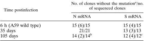
TABLE 1. Evolution of the 59-end leader RNA sequence in mRNAs 3 (S gene) and 7 (N gene) during persistent infection
Time postinfection
No. of clones without the mutationa
/no. of sequenced clones
N mRNA S mRNA
6 h (A59 wild type) 15 (6)/15 15 (4)/15
35 days 21/21 13 (3)/13
105 days 14 (2)/14b 12 (4)/12c
a
Numbers in parentheses indicate the number of clones with truncated first and/or second bases.
b
One clone with a U-to-A mutation at nt 49.
c
One clone with a G-to-A mutation at nt 25.
on November 9, 2019 by guest
http://jvi.asm.org/
sequences, 5
9
UTR, and ORF 1a p28 protein during persistent
MHV-A59 infection.
Intraleader ORFs are absent during persistent MHV-A59
infection.
It has been suggested that mutations within the 5
9
leader RNA of BCV mRNAs function in maintaining
persis-tence in vitro by attenuating translation of downstream ORFs
in each mRNA (38). Since MHV-A59 and BCV mRNAs have
relatively similar 5
9
-end leader RNA sequences, an A-to-U
point mutation at nt 5 could result in a similar intraleader ORF
in MHV-A59 (Fig. 1B). To address this question, the
full-length leader RNA sequences of mRNAs 3 and 7 were cloned
and sequenced with the 5
9
RACE system. Intracellular RNA
was isolated at 6 h, 35 days, and 105 days postinfection.
Fol-lowing cDNA synthesis and PCR amplification of the 5
9
ends
of mRNA 3 and 7 as described in the Materials and Methods,
individual clones were isolated and sequenced.
In contrast to findings reported during persistent BCV
in-fection, no 5
9
-terminal leader mutations were evident in the
MHV-A59 mRNAs until 105 days postinfection (Table 1). At
105 days postinfection, only 7 and 8.3% of clones from mRNAs
7 and 3 contained leader RNA mutations (1 of 14 and 1 of 12
clones, respectively) (Fig. 2). One mRNA 7 clone had a U-to-A
mutation at nt 49, while an mRNA 3 clone had a G-to-A
mutation at nt 25. Neither mutation resulted in intraleader
ORFs (Table 1). Interestingly, the extensive polymorphism
and deletion noted at the 5
9
end of BCV mRNAs were not
detected in the MHV-A59 mRNAs, although several clones
contained a 1- or 2-nt truncation at the 5
9
end which was
probably associated with premature termination during reverse
transcription. Consequently, the MHV-A59 leader RNA
se-quence and the leader-mRNA junction sese-quences were
ex-tremely stable and did not evolve significantly through the first
105 days postinfection.
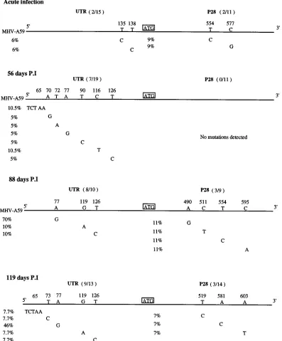
Evolution and mutation in the MHV-A59 genomic RNA.
Since our data indicated that intraleader mutations and ORFs
were not significantly associated with either the establishment
or maintenance of MHV persistence in DBT cells through 105
days postinfection, we cloned and sequenced the 5
9
end of the
genomic RNA because this domain contains critical cis- and
trans-acting sequences which regulate mRNA, genome RNA,
and leader RNA synthesis, as well as the expression of the
MHV polymerase (5, 8, 48, 88). The 5
9
-most 670 bp of genomic
RNA were cloned by reverse transcriptase and PCR
amplifi-cation using two 5
9
-end-specific primers, L3
1and G1A 670(
2
)
(Fig. 1A). Individual plasmid clones containing the 5
9
end of
MHV-A59 were isolated at 6 h and 56, 88, and 119 days
postinfection. An A-to-G mutation at nt 77 was first detected
at 56 days postinfection in 1 of 19 clones (Fig. 3A). This
mutation created an ORF encoding 16 amino acids (aa) in the
5
9
UTR of the genomic RNA. Interestingly, the new ATG start
site at nt 75 enlarged the preexisting small ORF (encoding 8
aa) between nt 99 and 125 in wild-type virus (Fig. 3B). In
addition to this mutation, other changes were also noted at the
5
9
end of the genome in a small number of clones at different
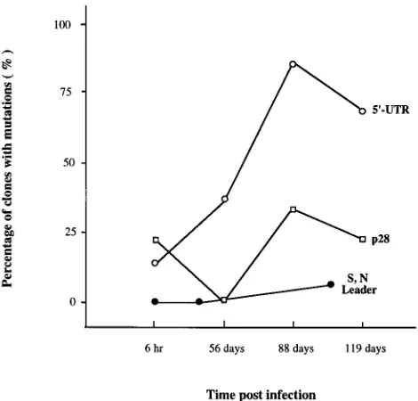
FIG. 2. Evolution of the MHV-A59 leader RNA, p28, and 59UTR sequences [image:4.612.60.295.70.295.2]during persistent infection. Cultures of cells were infected with MHV-A59, and intracellular RNA was isolated at 6 h or 35, 56, 88, 105, or 119 days postinfection. The 59ends of mRNAs 7, 3, and 1 were cloned and sequenced. The percentages of the clones containing mutations were shown. S, N Leader, mutations found in leader sequence of the S (mRNA 3) and N (mRNA 7) subgenomic RNAs; 59-UTR and p28, mutations found in the 59UTR and p28 coding region, respec-tively.
FIG. 3. A predominant 59UTR mutation in the genomic RNA during MHV-A59 persistent infection. The A-to-G mutation at nt 77 in the genomic RNA was first detected at 56 days postinfection (A). (B) The putative 59UTR ORF for 16 aa created by the nt 77 A-to-G mutation extends the ORF for 8 aa in MHV-A59. The new AUG start site is encoded at nt 75. The gels are read from the bottom.
on November 9, 2019 by guest
http://jvi.asm.org/
passages. For example, the A-to-G, U-to-A, and U-to-C
mu-tations at nt 70, 72, and 90, respectively, were detected at 56
days postinfection (Fig. 4). However, some evolutionary
ad-vantage was clearly associated with the A-to-G mutation at nt
77, because 50 to 80% of the genome-length molecules
con-tained this mutation by 119 days postinfection (Fig. 4). In
contrast, most other mutations (7 of 44 clones) were neutral or
deleterious and lost with subsequent passage. The only other
mutations that appeared to confer some evolutionary
advan-tage were the C-to-U and U-to-C double mutations at nt 119
and 126 seen on days 88 and 119 and a UCUAA insertion at nt
65 seen on days 56 and 119 (Fig. 4). Statistical analysis,
how-ever, demonstrated no significant association between these
mutations and MHV-A59 persistence (data shown in next
sec-tion).
[image:5.612.109.506.74.553.2]Interestingly, the A-to-G mutation is located within
poten-tially important cis- and trans-acting sequences at the 5
9
end of
the genome. As previously reported, a relatively stable hairpin
loop structure (
D
G
5 2
35.2 kcal [ca.
2
147 kJ]) may exist at
the 5
9
end of the MHV genome (63). Compared with the wild
type, in the region between nt 72 and 80, the nt 77 mutation did
cause some modulation in the putative secondary structure.
The overall stability and the structure of the 5
9
end of the
genome in the mutant virus, however, were very similar to
FIG. 4. Evolution and mutation at the 59end of the MHV-A59 genome during persistent infection. Intracellular RNA was isolated at 6 h and 56, 88, and 119 days postinfection (P.I) and sequenced as described in Materials and Methods. The numbers of clones containing mutations in the 59UTR and p28 coding sequences (in parentheses) and the location of each nucleotide change are shown. The percentages of clones containing a specific mutation are also indicated.on November 9, 2019 by guest
http://jvi.asm.org/
those of the wild-type virus (
D
G
5 2
35.5 kcal [ca.
2
149 kJ])
(data not shown).
Molecular evolution of MHV-A59 during persistent
infec-tion.
Among the RNA viruses, the rate at which mutations
become fixed in different portions of the viral genome varies
during persistent infection (18, 19, 26). To compare the
muta-tion rates in different pormuta-tions of the MHV genome during
persistent infection, we also sequenced the 5
9
end of the p28
nonstructural protein coding region in some of the same clones
that contained the 5
9
-end UTR. The number of mutations in
the sequenced p28 coding region was significantly low. During
acute infection (6 h postinfection), the numbers of clones
con-taining mutations in both regions were similar: 18% (2 of 11
clones) contained mutations in p28 and 13% (2 of 15 clones)
contained mutations in the 5
9
-end UTR. After passage, the
rates at which mutations accumulated in these two regions
were significantly different. In p28, no mutations were detected
at 56 days postinfection but 30% (3 of 9) and 21% (3 of 14) of
the clones contained mutations by 88 and 119 days
postinfec-tion, respectively. In contrast, 37% (7 of 19), 80% (8 of 10),
and 69% (9 of 13) of the clones contained mutations in the
5
9
-end UTR at 56, 88, and 119 days postinfection (Fig. 4).
Statistical analysis for trend indicated a specific increase in the
number of mutations detected within the 5
9
UTR (P
,
0.001)
but not for the mutations in the p28 domain (P
,
0.43) over
time. Among the mutations found in the 5
9
end of the p28
coding region, 30% were silent and only one (1 of 45 clones
sequenced), at position 603 (A to T), resulted in a premature
stop codon by 119 days postinfection. No mutations in the p28
coding region appeared to contribute evolutionary advantages,
since they did not accumulate through 119 days postinfection.
The rate of fixation of mutations was 4.3
3
10
25to 7.58
3
10
25and 1.2
3
10
25to 3.37
3
10
25/nt/day, and the mutation
fre-quency ranged from 3.3
3
10
23to 6.7
3
10
23and 1.2
3
10
23to 2.96
3
10
23substitutions per nt in the 5
9
UTR and p28
coding regions (8,550 and 6,750 nt sequenced in each region),
respectively.
Since infectious clones are not available to evaluate the role
of a particular mutation in MHV-A59 persistence, we have
used biostatistical techniques to determine if a particular
mu-tation was significantly associated with MHV-A59 persistence
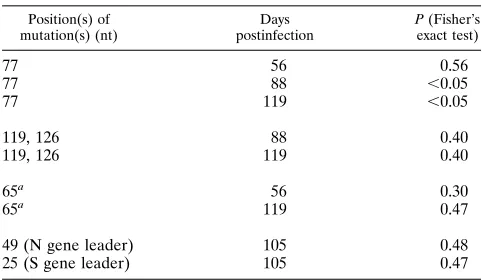
in vitro (69). Statistical analysis has clearly demonstrated a
strong association (P
,
0.05) between mutation and evolution
at nt 77 in the genomic RNA during MHV-A59 persistence at
days 88 and 119 postinfection (Table 2). Similar associations
were not detected among other 5
9
end genomic mutations on
days 56, 88, and 119 postinfection or among mutations
de-tected in p28 (P
.
0.1). Importantly, statistical analysis for
trend also demonstrated a significant increase in the number of
clones containing the A-to-G mutation over time (P
,
0.001)
compared with baseline levels. No significant evolution was
present within either the leader RNA or intergenic sequences
as well (P
.
0.1). These data indicate that the 5
9
UTR A-to-G
mutation was significantly associated with the maintenance of
the MHV-A59 genome in persistently infected DBT cells.
In vitro translation studies.
As the 5
9
UTR A-to-G mutation
is located near the leader sequence at the 5
9
end of the
ge-nome, it might alter the virus replication cycle by affecting the
transcription of leader RNA or mRNA or altering the
effi-ciency of translation of the genomic RNA. Alternatively, the 5
9
UTR ORF may be translated into a small,
;
1.9-kDa peptide
which modulates the acute cytolytic potential of MHV-A59.
To test whether the intra-UTR ORF is translated into the
putative 1.9-kDa product, two clones were translated in rabbit
reticulocyte lysates in vitro (Fig. 5A). Clone A59 G1A-2 was
isolated from input wild-type A59 acute infection (6 h
postin-fection), and its sequence was identical to the reported
MHV-A59 sequence (63). A putative 8-aa peptide of about 1 kDa is
encoded in the 5
9
-end UTR between nt 99 and 125 in wild-type
virus (63). The P16 G1A-16 clone was obtained from
persis-tently infected cells at 56 days postinfection and was identical
to clone A59 G1A-2 except for a single A-to-G mutation at nt
77, encoding a potential 16-aa ORF product of about 1.9 kDa
between nt 75 and 125 at the 5
9
end of the genome. The
plasmids were linearized with NarI, which cleaves downstream
from the termination codon for the 8- and 16-aa ORF products
(nt 181), and in vitro transcription and translation were
per-formed as described in Materials and Methods. No
radiola-beled polypeptide of the predicted size for the intra-UTR ORF
product (1 or 1.9 kDa) was detected from either A59 G1A-2 or
P16 G1A-16 transcripts, suggesting that both ORFs were in a
poor context for translation in vitro (data not shown).
We then addressed whether the nt 77 mutation altered the
level of p28 protein expression in the in vitro translation assay.
As the presence of the leader sequences greatly reduces the
efficiency of p28 translation in vitro (4a), a small portion of
leader RNA sequences (nt 3 to 23) in both plasmids was
deleted by enzymatic digestion to enhance p28 expression. The
5
9
-truncated plasmids A59 G1A-2-L
2and P16 G1A-16-L
2were linearized with PstI, and equivalent amounts of in vitro
transcripts were translated in vitro. In vitro translation of both
plasmids resulted in the synthesis of a 17-kDa protein,
equiv-alent in size to the N-terminal 153 aa of p28 that is encoded in
each plasmid. Antiserum
a
-p28, which is directed against the N
terminus of p28, immunoprecipitated the 17-kDa product,
demonstrating that it contained p28 coding sequences (Fig.
5B). The radioactive protein products were analyzed by gel
electrophoresis and quantitated by AMBIS. Compared with
that in the MHV-A59 wild-type control, a significant 2.37
6
0.54-fold (mean
6
standard deviation; n
5
3) increase in p28
expression was observed when the intra-UTR ORF was
present, suggesting that this mutation increased the
transla-tional efficiency of the MHV-A59 genomic RNA (P
,
0.05)
(Fig. 5B).
[image:6.612.57.298.90.230.2]Role of the 5
*
-end UTR mutation in MHV persistence.
The
accumulation of the 5
9
-end nt 77 mutation in the MHV-A59
genome by day 56 postinfection suggests that the mutation
functions in establishing or maintaining MHV-A59 persistence
in vitro. To test this hypothesis, we isolated intracellular viral
RNA, extracellular virion RNA, and virion RNA from 12
twice-plaque-purified infectious virus clones at 119 days
postin-fection. Sequence analysis indicated that the nt 77 mutation
TABLE 2. Association between a particular mutation and MHV persistence
Position(s) of mutation(s) (nt)
Days postinfection
P (Fisher’s
exact test)
77 56 0.56
77 88 ,0.05
77 119 ,0.05
119, 126 88 0.40
119, 126 119 0.40
65a 56 0.30
65a 119 0.47
49 (N gene leader) 105 0.48
25 (S gene leader) 105 0.47
aUCUAA insertion.
on November 9, 2019 by guest
http://jvi.asm.org/
was present in 46% (6 of 13) of the cloned intracellular
genomic RNAs, 33% (4 of 12) of the cloned extracellular
virion RNAs (infectious and noninfectious), and 50% (6 of 12)
of the plaque-purified isolates at 119 days postinfection (Table
3). Virus isolates with (V16-ATG
1) or without (V1-ATG
2)
the A-to-G mutation at 119 days postinfection were extremely
virulent, fusigenic, and cytolytic and destroyed
.
99% of the
DBT monolayers within 16 h postinfection. Thus, the variant
viruses were not more efficient than wild-type virus in
estab-lishing a persistent infection in the parental cell lines. These
data suggested that neither the A-to-G mutation nor the new
5
9
-end ORF encoding the putative 16-aa peptide attenuated
the cytopathic effect of MHV-A59 or enhanced its ability to
establish a persistent infection in vitro. Rather, virus growth
curves demonstrated that V16-ATG
1and V1-ATG
2replica-tion was increased compared with that of wild-type virus in
DBT cells under identical conditions, suggesting that
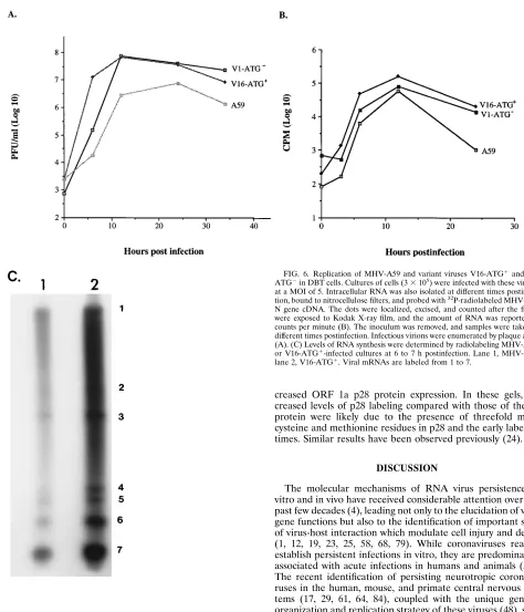
persis-tence selected for more-virulent virus variants (Fig. 6A). By 24
h postinfection, V16-ATG
1infection resulted in 123 PFU per
cell, compared to 136 PFU per cell for V1-ATG
2and 26 PFU
per cell for MHV-A59. Interestingly, V16-ATG
1replication
was significantly more efficient at early times in the infection
compared with that of MHV-A59 and V1-ATG
2(Fig. 6B). At
6 h postinfection, the production of infectious virus averaged
43.3 PFU per cell for V16-ATG
1, 0.06 PFU per cell for
MHV-A59, and 0.5 PFU per cell for V1-ATG
2. Consistent with this
hypothesis, levels of viral intracellular RNA were increased
throughout V16-ATG
1infection, as well as V1-ATG
2infec-tion, compared with that in wild-type MHV-A59 infection (Fig.
6B). To provide additional evidence that more-vigorous virus
variants evolve during MHV persistence, cultures of DBT cells
were infected with MHV-A59 and V16-ATG
1at a MOI of 10
and radiolabeled with
32P
i
from 6 to 7 h postinfection.
In-creased levels of mRNA 1 to 7 synthesis were clearly evident in
V16-ATG
1-infected cultures compared with those in the
wild-type control (Fig. 6C). AMBIS scans indicated that the relative
percent molar ratio of each mRNA was similar, but
V16-ATG
1transcribed about three times more RNA than
MHV-A59 at this time postinfection.
[image:7.612.167.460.83.388.2]To determine whether viral infection was characterized by
increased expression of the ORF 1a products in vivo, the levels
of p28 expression were monitored during acute MHV-A59 and
V16-ATG
1infection in DBT cells. Cultures of the cells were
infected and radiolabeled for 2 h with [
35S]methionine-cysteine
FIG. 5. Effect of the nt 77 A-to-G mutation on the expression of the ORF 1a p28 protein. In vitro translation of transcripts synthesized from the clones depicted in Fig. 1B was performed as described in Materials and Methods. (A) These clones encode the N-terminal 17 kDa of the p28 nonstructural protein. (B) Immuno-precipitation of the truncated p28 in vitro. Lanes 1 and 4, translation products from A59 G1A-2-L2; lanes 2 and 5, translation products from P16 GIA-16-L2; lane 3, no RNA transcripts (control).TABLE 3. Distribution of the nt 77 mutation in cloned intracellular genomic RNAs, extracellular virion RNAs, and plaque-purified
isolates at 119 days postinfection
Source of virus
No. of clones with ATG mutation/no. of clones sequenced
% with the mutation
Intracellular 6/13 46
Extracellular 4/12 33
Plaque purified 6/12 50
on November 9, 2019 by guest
http://jvi.asm.org/
[image:7.612.58.297.662.727.2]at 5 h postinfection. The MHV p28 and M proteins were
immunoprecipitated with p28 antiserum or monoclonal
anti-body to the M glycoprotein and separated on SDS–15%
poly-acrylamide gels as described in Materials and Methods. The
amount of p28 expression was quantitated by AMBIS scans
relative to M gene expression. Consistent with the in vitro
translation results, a 3.21
6
1.03-fold increase in p28
expres-sion was observed with the mutant virus V16-ATG
1compared
with that in the control (Fig. 7) (P
,
0.05, n
5
4). Thus, these
data were consistent with the notion that the 5
9
-end UTR
A-to-G mutation in V16-ATG
1was likely associated with
creased ORF 1a p28 protein expression. In these gels,
in-creased levels of p28 labeling compared with those of the M
protein were likely due to the presence of threefold more
cysteine and methionine residues in p28 and the early labeling
times. Similar results have been observed previously (24).
DISCUSSION
The molecular mechanisms of RNA virus persistence in
vitro and in vivo have received considerable attention over the
past few decades (4), leading not only to the elucidation of viral
gene functions but also to the identification of important sites
of virus-host interaction which modulate cell injury and death
(1, 12, 19, 23, 25, 58, 68, 79). While coronaviruses readily
establish persistent infections in vitro, they are predominately
associated with acute infections in humans and animals (59).
The recent identification of persisting neurotropic
coronavi-ruses in the human, mouse, and primate central nervous
sys-tems (17, 29, 61, 64, 84), coupled with the unique genetic
organization and replication strategy of these viruses (48),
sug-gests that novel virus-host interactions evolve to modulate the
cytolytic potential of these viruses.
[image:8.612.67.541.63.616.2]Although many groups have established persistent
corona-virus infections in vitro, the precise mechanisms by which this
cytoplasmic RNA virus establishes and maintains a persistent
infection are uncertain. It is also unclear how rapidly the viral
genome evolves under these conditions. Coronaviruses, like
other positive-polarity RNA viruses, have high mutation rates
probably caused by the lack of proofreading capabilities in the
RNA polymerase (26). MHV polymerase error rates estimated
by reversion frequencies of temperature-sensitive mutants
FIG. 6. Replication of MHV-A59 and variant viruses V16-ATG1and V1-ATG2in DBT cells. Cultures of cells (33105) were infected with these virusesat a MOI of 5. Intracellular RNA was also isolated at different times postinfec-tion, bound to nitrocellulose filters, and probed with32P-radiolabeled MHV-A59
N gene cDNA. The dots were localized, excised, and counted after the filters were exposed to Kodak X-ray film, and the amount of RNA was reported as counts per minute (B). The inoculum was removed, and samples were taken at different times postinfection. Infectious virions were enumerated by plaque assay (A). (C) Levels of RNA synthesis were determined by radiolabeling MHV-A59-or V16-ATG1-infected cultures at 6 to 7 h postinfection. Lane 1, MHV-A59; lane 2, V16-ATG1. Viral mRNAs are labeled from 1 to 7.
on November 9, 2019 by guest
http://jvi.asm.org/
range from 10
23to 10
25substitution per site (31), very similar
to mutation frequencies of 10
23to 10
24substitution per nt
measured during MHV persistence through 119 days
postin-fection. While these values may be slightly elevated because of
high error rates associated with the reverse transcriptase and
Taq polymerases, these values are very similar to rates
mea-sured for other positive-stranded RNA viruses, including
trans-missible gastroenteritis virus (27, 40, 86). The majority of
evo-lutionary changes detected in this study probably represented
the random fixation of selectively neutral or nearly neutral
mutations under continued selection (46). Deleterious
muta-tions were lost, but rare advantageous mutamuta-tions (A to G at nt
77) rapidly spread through the population, readily explaining
the increased percentage of genomic clones that contained the
5
9
-end mutation at later times. In MHV, the p28 protein and
leader RNA evolved at much lower rates compared with the 5
9
UTR, consistent with the notion that viral sequences evolve at
different rates during persistent infection (18, 19, 33). Like
influenza virus evolution in the face of host immune selection,
our data also suggest that MHV undergoes positive Darwinian
selection during persistent infection (28).
Persistence of RNA viruses in vitro is associated with gene
evolution, mutations in critical domains that function in the
normal virus replication cycle, and/or the coevolution of host
cells that resist viral cytopathology (2, 4, 12, 23, 25, 45, 58, 68).
During BCV infection, persistence was associated with the
evolution of 5
9
-end intraleader RNA mutations which
attenu-ated the translation of downstream ORFs in each mRNA (38).
Unfortunately, the 5
9
-end mutation, extensive hypervariability,
and polymorphism in the BCV leader RNA sequences did not
develop during MHV persistence. Rather, through 105 days
postinfection, the MHV leader RNA sequences and intergenic
start sites were extremely stable and highly conserved. As the
clones containing leader RNA sequences were identified by
the L3
1(nt 3 to 25) probe, our method may not have detected
extreme polymorphisms in 5
9
-end leader RNA if nt 3 to 25
sequences in the leader RNA were deleted or significantly
altered. However, clones containing point mutations and
in-traleader ORFs should have been detected. While it is possible
that intraleader mutations evolve later during MHV
persis-tence, such mutations were evident within 4 days after BCV
infection and subsequently accumulated with passage. These
data suggest that the establishment and maintenance of MHV
persistence are mediated by different host-virus interactions
that are uncoupled from the presence of intraleader
polymor-phisms, mutations, and translation-attenuating intraleader
ORFs. In addition, if leader-body junction sequences encode
critical cis- and trans-acting elements that are required for virus
transcription as predicted by the leader-primed and
transcrip-tion attenuatranscrip-tion models for discontinuous transcriptranscrip-tion of
coronavirus RNAs, stable leader-body junction sequences
should be maintained during persistent infection (39, 54, 71,
73, 75, 88). Since leader RNA sequences in mRNA and
ge-nome evolve at similar low rates, these data suggest that these
sequences are critical elements in virus transcription.
The difference in the evolution of BCV and MHV mRNAs
is intriguing and suggests that different mechanisms of
persis-tence occur among the group II coronaviruses. Interestingly,
BCV infection in HRT cells is noncytolytic, dramatically
dif-ferent from the extremely cytolytic MHV infection in DBT
cells (38). It is well established that the host cell environment
dramatically regulates the cytopathic potential of MHV. For
example, MHV infection in LM-K cells or primary mouse glial
cells occurs in the absence of significant cytopathology, fusion,
and cell killing (51, 60). In contrast, the infection in 17 Cl-1, L2,
and DBT cells is extremely cytolytic (37). While additional
studies must be performed, the most likely explanation for
these different findings is that viral strains and host cell
geno-types exert different selection pressures on the coronavirus
genome, resulting in the evolution and accumulation of distinct
mutations that initiate and maintain viral persistence (1, 13, 22,
25). In support of this hypothesis, it has been reported that
organ-specific selection of viral variants has been
demon-strated during chronic lymphocytic choriomeningitis virus
in-fection in carrier mice (3) and that the host cell environment
imposes significant evolutionary constraints in the selection of
precise silent mutations in the poliovirus genome (13).
While intraleader ORFs did not develop in mRNAs from
persistently infected DBT cells, MHV persistence was
signifi-cantly associated with the evolution and accumulation of a
specific A-to-G mutation that resulted in the appearance of a
new ORF for 16 aa in the 5
9
UTR of the genomic RNA. The
selection for precise mutations is not without precedent and
has been reported during poliovirus and foot-and-mouth
dis-ease virus (FMDV) persistence (13, 26). Persistent
noncyto-lytic MHV infection in primary mouse glial cells also selects for
fusion-defective MHV-A59 variants that contain precise
mu-tations in the spike glycoprotein gene (35). In our study, the nt
77 A-to-G mutation was significantly associated with increased
expression of the ORF 1a p28 polyprotein in vitro, suggesting
that the 5
9
-UTR mutation enhances expression of the genes at
the 5
9
end of the MHV genome. The 5
9
end of the MHV
genome contains two large ORFs, designated ORF 1a and
ORF 1b, which are probably translated into two large
polypro-teins of 440 and 330 kDa by a ribosomal frameshifting
mech-anism (11, 15, 52). These large polyproteins are then processed
by viral and/or host proteases into the MHV polymerase
pro-teins and other propro-teins functioning in RNA synthesis (52).
Since the p28 protein is encoded at the N terminus of ORF 1a,
enhanced p28 expression is likely associated with an increased
expression of all viral gene 1 products, including the putative
viral RNA polymerase. Consonant with these findings, virus
replication, RNA synthesis, and gene expression were also
increased in variant viruses containing the mutation,
suggest-ing that the 5
9
-end mutation contributed to MHV persistence
by increasing polymerase gene expression. Additional genetic
alterations, however, must also occur in the genomes of viruses
isolated during persistent infection, since variants lacking the
A-to-G mutation also replicated more efficiently than wild-type
virus. Definitive structure-function analysis of the 5
9
-end
mu-tation and element and ORF must be accessed upstream of
FIG. 7. Immunoprecipitation of p28 and M protein from DBT cells infectedwith MHV-A59 and V16-ATG1variant virus. Duplicate cultures of cells were infected at a MOI of 10 and incubated in methionine-cysteine-free media. Polyproteins were labeled with 200mCi of [35S]methionine-cysteine per plate,
immunoprecipitated, and separated on SDS–15% polyacrylamide gels. Lanes 1 and 5, p28 protein from MHV-A59-infected cells; lanes 2 and 6, p28 protein from V16-ATG1-infected cells; lanes 3 and 7, M protein from MHV-A59-infected cells; lanes 4 and 8, M protein from V16-ATG1-infected cells; lanes 9 and 10, uninfected cells witha-p28 antisera or A1.10 M protein monoclonal anti-body; lane M, molecular weight markers. Arrowheads indicate p28 (28-kDa) and M (23-kDa) proteins.
on November 9, 2019 by guest
http://jvi.asm.org/
reporter genes to definitively ascertain its affects on secondary
structure and in the stimulation of translation. Similar
quanti-tative assays have been reported in picornavirus (57).
The 5
9
UTR in many positive-strand RNA viruses has been
demonstrated not only to regulate viral protein expression but
also to function in the replication and transcription of viral
positive-stranded RNA (16, 44, 54, 57, 67, 79). Systemic
move-ment of a hordeivirus is regulated by 5
9
-end substitutions in the
genome (65). A single nucleotide mutation in the ribosome
entry site of FMDV has been demonstrated to enhance
cap-independent translation in vitro two- to fourfold (57). During
acute MHV infection, a 13-nt element (UCUAAUCCAAA
CA) containing the UCUAA pentanucleotide repeat within
leader RNA sequences enhances the translation of viral
mRNAs. Enhanced translation of viral mRNAs may also
re-quire an interaction between specific viral proteins and this
pentanucleotide repeat as well (82). Since the A-to-G mutation
resides within a similar pentanucleotide repeat element
(UAUAA to UAUGA) just 3
9
to the leader RNA
pentanucle-otide repeat motifs in the genomic RNA, this alteration may
enhance translation of genome-length molecules by a similar
cis-recessive mechanism (82). The 5
9
UTR mutation might also
enhance MHV replication by regulating the level of
sub-genomic mRNA synthesis, since the mutation resides within a
9-nt domain (UUUAUAAA) that has been suggested to
reg-ulate the initial synthesis of the viral subgenomic mRNAs by
promoting or attenuating leader RNA switching between
tem-plates (88). We have not, however, detected any significant
difference in the relative percent molar ratio of the V16-ATG
1or type mRNAs during infection. Alternatively, in
wild-type virus, a putative small 8-aa peptide which might function
to downregulate expression of the ORF 1a and 1b polyproteins
is encoded in the MHV-A59 5
9
UTR. Since the nt 77 mutation
expands this ORF product to a 16-aa peptide, this may alter
the regulation of MHV polymerase gene expression. This
mechanism does not, however, explain the increased
replica-tion efficiency of variant viruses lacking the A-to-G mutareplica-tion in
DBT cells, and in vitro translation studies suggest that the
putative 1.9-kDa protein is poorly expressed at best. Thus, the
most likely explanation for the nt 77 mutation’s function in
MHV persistence is that it enhances the persistence of the
MHV genome by enhancing translation of the ORF 1a and 1b
polyproteins. Consistent with this hypothesis, the nt 77
muta-tion appears to alter the secondary structure of the 5
9
end of
the genomic RNA, and similar enhancing mutations have been
described in piconavirus (57).
Viral persistence likely involves alterations in critical
host-virus interactions which initiate a persistent infection and
sub-sequently lead to selection for mutations which maintain the
persistent state by attenuating or enhancing virus replication or
by altering host susceptibility to infection (19, 57, 58). Distinct
mechanisms which function in the establishment and
mainte-nance of a persistent poliovirus, FMDV, and reovirus infection
have been described elsewhere (3, 12, 25, 58). Like the BCV
intraleader ORF (38), the MHV 5
9
-end mutation probably
functions in the maintenance of a persistent infection, since it
does not evolve until after 56 days postinfection. Thus, it seems
likely that other genetic changes in the virus or host must
evolve to initially establish and maintain a persistent MHV
infection. The evolution of a specific 5
9
-end enhancing
muta-tion as well as other potential mutamuta-tions which probably
con-tributes to the maintenance of MHV persistence by increasing
the efficiency of virus replication and virulence is surprising in
view of the well-documented evidence demonstrating that the
attenuation of viral gene functions or downregulation of
spe-cific viral genes is the principle mechanism limiting cell killing
(1, 4, 43, 62). Our findings are not, however, unique, since
variant viruses, which were more cytolytic and replicated more
efficiently than the wild type have been isolated from persistent
reovirus and FMDV cultures (1, 23, 58). Persistently coxsackie
A9 virus-infected HeLa cell cultures also selected for viruses
with increased virulence, while echovirus 6 variant viruses
which could not replicate in the parental cells evolved during
persistent infection (21, 32, 83). Functional alterations that
favored measles virus persistence have also been demonstrated
(36). Since the evolution of virulent virus variants in persistent
cultures seems to be related to the coevolution of host cell
variants that resist the cytopathic effects of wild-type viruses,
these data suggest that MHV persistence is mediated by
sim-ilar coevolutionary mechanisms in vitro (22, 23, 58).
ACKNOWLEDGMENTS
We thank Boyd Yount, Jr., for excellent technical assistance. This research was supported by a research grant from the National Institutes of Health (AI 23946) and a fellowship from the Public Health Service (5 T32 A107151-16) to W.C. This research was con-ducted during the tenure of an established investigator award from the American Heart Association (89-0193) to R.S.B.
REFERENCES
1. Ahmed, R., M. Canning, R. S. Kauffman, A. H. Sharpe, J. V. Hallum, and
B. N. Fields.1981. Role of the host cell in persistent viral infection: coevo-lution of L cells and reovirus during persistent infection. Cell 25:325–332. 2. Ahmed, R., and B. N. Fields. 1982. Role of the S4 gene in the establishment
of persistent reovirus infection in L cells. Cell 28:605–612.
3. Ahmed, R., C. S. Hahn, T. Somasundaram, L. Villarette, M. Matloubian,
and J. H. Strauss.1991. Molecular basis of organ-specific selection of viral variants during chronic infection. J. Virol. 65:4242–4247.
4. Ahmed, R., and J. G. Stevens. 1990. Viral persistence, p. 241–265. In B. N. Fields and D. M. Knipe (ed.), Virology, 2nd ed. Raven Press, New York. 4a.Baker, S. Personal communication.
5. Baker, S. C., and M. M. C. Lai. 1990. An in vitro system for the leader-primed transcription of coronavirus mRNAs. EMBO J. 9:4173–4179. 6. Baker, S. C., C.-K. Shieh, L. H. Soe, M.-F. Chang, D. M. Vannier, and
M. M. C. Lai.1989. Identification of a domain required for autoproteolytic cleavage of murine coronavirus gene A polyprotein. J. Virol. 63:3693–3699. 7. Baric, R. S., K. Fu, M. C. Schaad, and S. A. Stohlman. 1990. Establishing a genetic recombination map for murine coronavirus strain A59 complemen-tation groups. Virology 177:646–656.
8. Baric, R. S., S. A. Stohlman, and M. M. C. Lai. 1983. Characterization of replicative intermediate RNA of mouse hepatitis virus: presence of leader RNA sequences on nascent chains. J. Virol. 48:633–640.
9. Baric, R. S., S. A. Stohlman, M. K. Razavi, and M. M. C. Lai. 1985. Char-acterization of leader-related small RNAs in coronavirus infected cells: fur-ther evidence for leader-primed mechanism of transcription. Virus Res.
3:19–33.
10. Baybutt, H. N., H. Wege, M. J. Carter, and V. Ter Meulen. 1984. Adaptation of coronavirus JHM to persistent infection of murine Sac(2) cells. J. Gen. Virol. 65:915–924.
11. Bonilla, P. J., A. E. Gorbalenya, and S. R. Weiss. 1994. Mouse hepatitis virus strain A59 RNA polymerase gene ORF 1a: heterogeneity among MHV strains. Virology 198:736–740.
12. Borzakian, S., T. Couderc, Y. Barbier, G. Attal, I. Pelletier, and F.
Colbere-Garapin.1992. Persistent poliovirus infection: infection establishment and maintenance involve distinct mechanisms. Virology 186:398–408. 13. Borzakian, S., I. Pelletier, V. Calvez, and F. Colbere-Garapin. 1993. Precise
missense and silent point mutations are fixed in the genomes of poliovirus mutants from persistently infected cells. J. Virol. 67:2914–2917.
14. Boursnell, M. E. G., T. D. K. Brown, I. J. Foulds, P. F. Green, F. M. Tomley,
and M. M. Binns.1987. Completion of the sequence of the genome of the coronavirus avian infectious bronchitis virus. J. Gen. Virol. 68:57–77. 15. Bredenbeek, P. J., C. J. Pachuk, A. F. H. Noten, J. Charite, W. Luytjes, S. R.
Weiss, and W. J. M. Spaan.1990. The primary structure and expression of the second open reading frame of the polymerase gene of the coronavirus MHV-A59; a highly conserved polymerase is expressed by an efficient ribo-somal frameshifting mechanism. Nucleic Acids Res. 18:1825–1832. 16. Brown, E. A., S. P. Day, R. W. Jansen, and S. M. Lemon. 1991. The 59
nontranslated region of hepatitis A virus RNA: secondary structure and elements required for translation in vitro. J. Virol. 65:5828–5838. 17. Burks, J. S., B. L. Devald, L. D. Jakovsky, and J. C. Gerdes. 1980. Two
coronaviruses isolated from central nervous system tissue of two multiple sclerosis patients. Science 209:933–934.