Copyright © 2004, American Society for Microbiology. All Rights Reserved.
Entry and Transcription as Key Determinants of Differences in
CD4 T-Cell Permissiveness to Human Immunodeficiency
Virus Type 1 Infection†
Angela Ciuffi,
1Gabriela Bleiber,
1Miguel Mun˜oz,
1Raquel Martinez,
1Corinne Loeuillet,
1Manuela Rehr,
2Marek Fischer,
3Huldrych F. Gu¨nthard,
3Annette Oxenius,
2Pascal Meylan,
1Sebastian Bonhoeffer,
4Didier Trono,
5and Amalio Telenti
1*
Institute of Microbiology, University of Lausanne, Lausanne,1Institute for Microbiology2and Ecology and Evolution,4
ETH Zurich, and Division of Infectious Diseases and Hospital Epidemiology, University Hospital Zurich,3Zurich,
and Department of Genetics and Microbiology, University of Geneva, Geneva,5Switzerland
Received 25 March 2004/Accepted 13 June 2004
Isolated primary human cells from different donors vary in their permissiveness—the ability of cells to be infected and sustain the replication of human immunodeficiency virus type 1 (HIV-1). We used replicating HIV-1 and single-cycle lentivirus vectors in a population approach to identify polymorphic steps during viral
replication. We found that phytohemagglutinin-stimulated CD4ⴙCD45ROⴙCD57ⴚT cells from healthy blood
donors (nⴝ128) exhibited a 5.2-log-unit range in virus production. For 20 selected donors representing the
spectrum of CD4 T-cell permissiveness, we could attribute up to 42% of the total variance in virus production to entry factors and 48% to postentry steps. Efficacy at key intracellular steps of the replicative cycle (reverse transcription, integration, transcription and splicing, translation, and budding and release) varied from 0.71 to 1.45 log units among donors. However, interindividual differences in transcription efficiency alone accounted for 64 to 83% of the total variance in virus production that was attributable to postentry factors. While vesicular stomatitis virus G protein-mediated fusion was more efficacious than CCR5/CD4 entry, the latter resulted in greater transcriptional activity per proviral copy. The phenotype of provirus transcription was stable over time, indicating that it represents a genetic trait.
The genetic makeup of individuals plays a role in suscepti-bility to human immunodeficiency virus type 1 (HIV-1) infec-tion and progression of disease. Some of the observed varia-tions have been attributed to immunogenetic diversity (major histocompatibility complex homozygosity and specific HLA types) and polymorphisms in chemokines, chemokine recep-tors, and cytokine genes (those for CCR5, CCR2, CX3CR1, SDF1, MIP1␣, RANTES, interleukin-10 [IL-10], and IL-4) (5, 35). Isolated primary human cells from different donors also vary in their permissiveness—the ability of cells to be infected and sustain the replication of HIV-1 (41). Spira and Ho in-fected peripheral blood mononuclear cells (PBMCs) from 10 healthy donors with a wide array of viral isolates and labora-tory strains and reported a 40-fold range of differences in infection among individuals (31). Eisert et al. analyzed param-eters influencing the susceptibility of cells of the monocyte/ macrophage lineage isolated from 30 healthy donors (11). The proportion of infected cells ranged from 0.03 to 99%, and the reverse transcriptase activity ranged from undetectable to 5⫻
106 cpm/ml over 90 min. Infection of macrophages derived
from pairs of identical twins displays a high concordance in the kinetics of HIV-1 replication (6, 23), underscoring the role of genetic factors in determining individual cell susceptibility to infection.
The HIV-1 life cycle is characterized by numerous interac-tions with host cellular proteins (14, 25). While restriction at entry plays a key role in determining infection kinetics (4, 26), there is limited knowledge on whether host proteins involved in other steps of the viral life cycle contribute to interindividual susceptibility to HIV-1 infection. Certain proteins are neces-sary for infection and for sustaining viral replication, while others represent antiviral factors. Some cellular antiviral fac-tors can be selectively suppressed by viral proteins, as shown by the interaction between APOBEC3G, a cytidine deaminase, and Vif (15, 18–20, 30). An early interplay between incoming retroviral preintegration complexes and the nuclear proteins integrase interactor 1 and promyelocytic leukemia protein cre-ates an antiviral state that interferes with the immediate-early steps of HIV-1 infection (37). ZAP, a zinc finger protein, inhibits the production of retroviral RNA (12). A number of antiretroviral factors target the capsid protein, imposing a postentry block (16, 32). This growing list of specific antiret-roviral factors adds to current knowledge on antiviral innate defense mechanisms implicating interferon responses medi-ated by double-stranded RNA-dependent protein kinase, the MX proteins, and RNase L-mediated degradation of viral RNAs (13). Genetic polymorphisms in antiviral genes or in host genes participating in the viral life cycle could thus result in differences in the levels of expression or in the functional potential of protein variants and thus lead to differences in permissiveness to HIV-1 infection.
To address these issues, we (i) defined the variability in the permissiveness of CD4 T cells in a human population, (ii) expanded T cells from individuals representing the spectrum of * Corresponding author. Mailing address: Institute of Microbiology,
1011 Lausanne, Switzerland. Phone: 41 21 314 0550. Fax: 41 21 314 4095. E-mail: [email protected].
† Supplemental material for this article may be found at http://jvi .asm.org/.
10747
on November 8, 2019 by guest
http://jvi.asm.org/
cellular susceptibility to HIV-1 infection, (iii) used assays to measure the progress of the virus through the different repli-cation steps in the cells and the variability of each step in a population, and (iv) estimated the contributions of various intracellular blocks to the overall permissiveness of the cells.
MATERIALS AND METHODS
Cells.CD4 T cells from 128 healthy Caucasian blood donors were isolated with anti-CD4 magnetic beads (Miltenyi Biotech) and cultured in vitro in RPMI 1640 (Gibco-Invitrogen) supplemented with 20% fetal calf serum (FCS), human IL-2
(Roche) at 20 U/ml, and gentamicin at 50g/ml (R-20) following stimulation
with phytohemagglutinin (PHA) at 2g/ml for 2 days. CD4 T cells were
ex-panded in R-20 in the presence of irradiated PBMCs and PHA at 2g/ml.
Immunotypic characterization of the collection included flow cytometric analysis
of the expression of RANTES (R&D Systems), MIP1(R&D Systems), tumor
necrosis factor alpha (Becton Dickinson [BD]), gamma interferon (BD), IL-2 (BD), IL-4 (BD), IL-10 (BD), CCR5 (BD), HLA-DR (BD), CD25 (BD), CD57
(BD), and CD69 (BD). Cells were genotyped for the CCR5⌬32 deletion.
The collection was assessed for permissiveness to HIV-1 infection in vitro
under standardized conditions with NL4-3BaLenv, an R5 infectious clone
gen-erated by substituting the SalI-BamHI fragment of pNL4-3 (encompassing most
of theenvopen reading frame) with the corresponding fragment of pBaL (21).
CD4 T cells (106cells) were infected with 1,000 pg of p24 antigen of
NL4-3BaLenvin a 1-ml final volume for 2 h at 37°C in 5% CO2. Cells were washed and
cultured in R-20 for 10 days. Virus-containing supernatant was harvested, and p24 antigen production was monitored by an enzyme-linked immunosorbent assay (ELISA) (Abbott). Temporal trends in permissiveness were investigated for three donors by isolating CD4 T cells at three time points over a 9-week period.
Vectors and viral production.pHIV⌬Env was generated by deleting a 1,206-bp
KpnI-BsaBI fragment in the gp120 coding region of NL4-3BaLenv.
pHIV/RT-minus was generated by introducing a D185E mutation in the reverse transcrip-tase active site. Viral production was carried out with 293T cells. For
pseudotyp-ing, cells were cotransfected by the calcium phosphate technique with pHIV⌬Env
and with a plasmid carrying the vesicular stomatitis virus (VSV) G protein or the HIV-1 R5-tropic JRFL envelope. The lentivirus LvGFP was produced by
co-transfecting the Gag-Pol construct (pCMV.⌬R8.92), the Rev expression plasmid
(pRSVRev), the VSV G protein envelope construct (pMD.G), and the packaged construct (pWPT-GFP) expressing green fluorescent protein (GFP) (see www .tronolab.unige.ch for vector details).
Analysis of intracellular steps.CD4 T cells (600,000 cells) were transduced
with 10,000 pg of p24 of HIV⌬Env/VSV or 100,000 pg of p24 of HIV⌬Env/R5 in
the presence of Polybrene (Sigma, Buchs, Switzerland) at 2g/ml in R-20 by
spinoculating cells for 3 h at 1,500⫻gand 22°C (24). Cells were washed and
harvested 12 h and 4 days posttransduction for the extraction of nucleic acids and for the assessment of cell-associated and extracellular p24. RNA was extracted by using an RNeasy kit (Qiagen) and reverse transcribed by using Expand reverse transcription-PCR (Roche) with random hexamers. DNA was extracted by using a QiaAmp DNA extraction kit (Qiagen). cDNA and DNA were used for Taqman
quantitative PCR (Applied Biosystems) of total viral RNA (nefprimers),
un-spliced RNA (gag primers), and 12-h and 4-day HIV-1 DNA (gagprimers),
normalized with the housekeeping genes and transcripts of-actin and
cyclo-philin A. Multiply spliced RNAs (tat,rev, andnefprimers) were quantified by a
direct one-step reverse transcription-PCR protocol (11a) and normalized to
-actin mRNA. The primers and probes used are shown in Table S1 in the
supplemental material.
8E5/LAV cells carrying a single defective proviral copy of HIV-1 were used as a standard to quantify HIV-1 integrated copies. Synthetic RNA and cDNA dilution series from one donor were used to establish standard curves for RNA. For experiments with LvGFP, 600,000 cells were transduced with increasing amounts of the LvGFP construct as described above. GFP expression was as-sessed by flow cytometry 3 and 6 days posttransduction.
Postentry restriction.To examine the possibility that expanded CD4 T cells present a postentry block due to a saturable restriction factor (16), we preex-posed CD4 T cells (300,000 cells) to a high dose of defective HIV/RTminus prior
to infection with NL4-3BaLenv. CD4 T cells were exposed to HIV/RTminus
(300,000 pg of p24) for 4 h, washed, and infected with 300 pg of p24 of
NL4-3BaLenvfor 2 h. Supernatant was harvested 5 and 7 days postinfection and
monitored by a p24 ELISA. Similarly, CD4 T cells were transduced with LvGFP after preexposure to HIV/RTminus, and GFP expression was assessed by flow cytometry.
Supernatant from day 7 replication kinetic assays was har-vested and used in infectivity assays. Ghost-CCR5 cells (30,000 cells) (Central Facility for AIDS Reagents, NIRSC, MRC, London, United Kingdom) were infected by spinoculation with 2,000 pg of HIV-1 p24. At 24 h postinfection, Ghost-CCR5 cells were washed with phosphate-buffered saline, trypsinized, fixed, and analyzed for GFP expression by flow cytometry.
CC-chemokine secretion.Culture supernatants were harvested from stimu-lated CD4 T cells before and 10 days after HIV-1 infection. Supernatants were
filtered (0.22-m-pore-size filters) and heat inactivated (30 min at 60°C), and
RANTES, MIP1␣, and MIP1levels were assessed by an ELISA (R&D
Sys-tems).
Apoptosis.CD4 T cells (500,000 cells), uninfected or HIV-1 infected for 5 days, were stained with annexin V-fluorescein isothiocyanate (BD) according to the manufacturer’s instructions and analyzed by flow cytometry.
Statistical analysis and modeling.Multiple regression analysis was performed by using the statistics package R (www.r-project.org). All values were log
trans-formed. For both viral constructs, HIV⌬Env/R5 and HIV⌬Env/VSV, we first
regressed extracellular p24 against all predictors (i.e., cDNA, proviral DNA, viral mRNA, and cell-associated p24) simultaneously. The predictors with least sig-nificance then were removed by stepwise backward elimination as implemented by the function step in statistics package R. The best model was chosen based on the Akaike information criterion, which balances the quality of the fit against the number of predictors included in the model.
RESULTS
Interindividual differences in permissiveness to HIV-1
in-fection. After 7 days of infection, CD4 T cells displayed a
5.2-log-unit range in p24 antigen production, i.e., in permis-siveness to HIV-1 infection (Fig. 1A). CD4 T cells from 20 individuals chosen to represent the extremes and the spectrum of permissiveness were expanded in vitro to 108 cells.
Ex-panded CD4 T cells consisted of a⬎97% pure population of CD45RO⫹CD57⫺T lymphocytes—the cells most susceptible
to HIV-1 (1). These purified populations also displayed a 5-log-unit range in susceptibility to HIV-1 infection (3.5 to 4.8 log units when homozygous carriers of the CCR5⌬32 allele were excluded) and, overall, presented the same permissive-ness phenotype as the original native cells (Fig. 1B). Although there were differences among the various CD4 T-cell popula-tions in growth rates (mean and standard error of the mean [SEM], 1.5-fold ⫾ 0.2-fold after 3 days postinfection) or in apoptosis (mean and SEM, 7.6%⫾0.7% apoptotic cells 5 days postinfection), we did not observe significant contributions of these factors to the permissiveness phenotype (for growth rates:R2, 0.16, andP, 0.11; for apoptosis:R2, 0.02, andP, 0.45)
(see Fig. S1 and S2 in the supplemental material).
The cells were characterized on the basis of the expression of cytokines and chemokines. There were differences in the frequencies of cells expressing RANTES (0.04%⫾ 0.007%), MIP1(11.1%⫾2.2%), tumor necrosis factor alpha (20.7%⫾
3.0%), gamma interferon (20.7% ⫾ 2.4%), IL-2 (9.0% ⫾
0.78%), IL-4 (3.4%⫾0.32%), and IL-10 (0.24%⫾0.05%) (all reported as mean and SEM). The activation status of the cells was assessed by determining the frequencies of cells expressing HLA-DR (80.9%⫾2.6%), CD25 (4.4%⫾0.8%), and CD69 (96.2% ⫾0.4%) (all reported as mean and SEM). For all of the above markers, there was no association between flow cytometry results and permissiveness phenotype (Fig. 2).
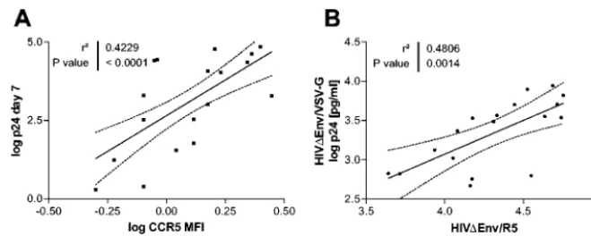
Flow cytometry of CCR5 expression on uninfected CD4 T cells demonstrated a 0.4- to 0.6-log-unit range in mean fluo-rescence intensity. Differences in CCR5 density correlated sig-nificantly with cell permissiveness (R2, 0.42;P,⬍0.0001) (Fig.
3A). As CC-chemokine levels could compete with HIV-1 for
on November 8, 2019 by guest
http://jvi.asm.org/
binding to CCR5 and thus impair HIV-1 replication, we looked for differences in RANTES, MIP1␣, and MIP1 levels in supernatants of CD4 T-cell cultures before or 10 days after HIV-1 infection. We observed a 0.9- to 1.5-log-unit range in CC-chemokine levels that did not correlate with differences in permissiveness (see Fig. S3 in the supplemental material). These data are consistent with prior reports (11) and with the observation that although CC-chemokines are generally viewed as inhibitory molecules, they block R5 viruses only at very high concentrations (8).
To identify differences in permissiveness that are not attrib-utable to entry variability, we infected cells with viral constructs that differ only in the entry pathway. For this, HIV⌬Env was pseudotyped with the R5-tropic JRFL envelope or with the VSV G protein envelope and transduced into selected CD4 T-cell populations. A comparison of amounts of p24 antigen released from a single round of infection for the two viruses revealed that shared intracellular steps explained 48% of the total variance in virus p24 antigen production (R2, 0.48; P,
0.0014) (Fig. 3B).
Native CD4 T cells
10-1
104
104
105
106
p24 [pg
/ml]
Seven day results
A
B
2X Expanded CD4 T cells
4 5 6 7 8 9 10
100
101
102
103
104
105
106
Time [days]
p
24 [
p
g
/m
l]
Native CD4 T cells
10-1
104
104
105
106
p24 [pg
/ml]
Seven day results
A
B
2X Expanded CD4 T cells
4 5 6 7 8 9 10
100
101
102
103
104
105
106
Time [days]
p
24 [
p
g
/m
l]
FIG. 1. Replication kinetics for HIV-1 in native or expanded CD4 T cells. (A) Cells isolated from 128 healthy blood donors were stimulated in vitro for 2 days with PHA and infected with 1,000 pg of p24 of R5-tropic NL4-3BaLenv. Infection was monitored as the p24 antigen level in the supernatant. (B) CD4 T-cell populations (n⫽20) representative of the spectrum of permissiveness to HIV-1 were expanded twice in vitro with irradiated PBMCs, PHA, and IL-2 and infected. Error bars indicate SEMs. Shown are hyperpermissive (red), normally permissive (black), and hypopermissive (green) cells.
A
B
0 1 2 3 4 5 6
0 10
RANTES MIP1b TNFalpha IFNgamma IL-2 IL-4 IL-10
20 30 40 50 60 70
log p24 [pg/ml]
%
posi
ti
ve cel
ls
0 1 2 3 4 5 6
0 10 20 30
HLA-DR CD69 CD25
50 100
log p24 [pg/ml]
%
posi
ti
ve cel
ls
FIG. 2. Immunological characterization of expanded CD4 T-cell pools. The expression of selected cytokines and chemokines (A) and surface activation markers (B) was analyzed by flow cytometry. CD4 T-lymphocyte pools represented a⬎97% pure population of CD45RO⫹CD57⫺T
cells. All correlations (R2) were⬍0.15, andPvalues were⬎0.1. TNF, tumor necrosis factor; IFN, interferon.
on November 8, 2019 by guest
http://jvi.asm.org/
Variations in viral life cycle steps.We quantified viral nu-cleic acids and proteins that accumulate at different times during the viral life cycle in order to assess efficiency at each infection step in cells from different individuals. We found that all postentry steps—reverse transcription (cDNA copies at 12 h postinfection), integration (proviral copies at 4 days), transcription (viral mRNA copies at 4 days), translation (cell-associated p24 antigen at 4 days), and budding and release (extracellular p24 and the ratio of extracellular p24 to cell-associated p24)—were variable. Interindividual differences ranged from 0.77 to 0.80 log units for the reverse transcriptase step up to 1.45 log units for the number of integrated copies (Fig. 4A). HIV-1 integration events were estimated by gag PCR (total HIV-1 DNA) 4 days postinfection, as it has been
shown that integrated DNA accounts for all HIV-1 DNA de-tected by 72 h postinfection (2, 3, 33, 34).
VSV G protein-mediated fusion was more efficacious than R5-tropic vector transduction (Fig. 4A, reverse transcription and integration), despite the use of a 1-log-unit-smaller inoc-ulum. However, when results were evaluated per proviral DNA copy, we observed greater transcriptional activity after CD4/ CCR5 entry than after VSV G protein-mediated fusion (Fig. 4B), consistent with a role for envelope-mediated signaling in determining the activation and transcriptional status of the cells (8).
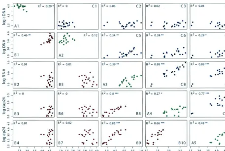
Variations in reverse transcription and integration steps were explained to a large extent by random effects, as indicated by the low concordance of results for the two viruses (Fig. 5A1, FIG. 3. Contributions of entry and nonentry host factors to cell permissiveness. (A) CCR5 receptor density, expressed as mean fluorescence intensity (MFI), correlated with p24 antigen production measured 7 days after infection with R5-tropic NL4-3BaLenv(R2, 0.42;P,⬍0.0001).
Similar estimates were obtained for percentages of CCR5⫹cells. (B) Assessment of entry-independent factors influencing cell permissiveness.
Viral vectors differing only in the mechanism of entry—HIV⌬Env pseudotyped with the R5-tropic JRFL envelope (105pg of p24) and with the
VSV G protein envelope (104pg of p24)—were used to infect 6⫻105CD4 T cells. The correlation (R2, 0.48;P,⬍0.0014) represents the shared
[image:4.603.128.458.67.199.2]contributions of intracellular steps to the cell permissiveness phenotype, measured as the amount of p24 in the supernatant 4 days posttransduction. Not shown in this analysis are values for cells from two donors homozygous for CCR5⌬32. Each point represents the mean of triplicate values.
FIG. 4. Variations in intracellular steps. (A) Single-cycle experiments with HIV⌬Env pseudotyped with the R5-tropic JRFL envelope or with the VSV G protein envelope. Expanded CD4 T cells (6⫻105cells) were infected with 105pg of p24 of HIV⌬Env/R5 or 104pg of p24 of
HIV⌬Env/VSV. DNA samples were taken at 12 h and 4 days (4d) postinfection, and HIV-1gagcopies were normalized to the cyclophilin A copy number to quantify the efficiencies of reverse transcription and integration per cell. RNA samples were taken at 4 days postinfection, reverse transcribed with random hexamers, and assessed for HIV-1 mRNA copies normalized to-actin mRNA. Cell-associated p24 and extracellular p24 were assessed at 4 days postinfection by an ELISA. (B) Reporting postintegration events relative to the number of proviral copies underscored the higher efficiency of transcription after CD4/CCR5 entry than after VSV G protein-mediated fusion. Not shown for R5 values are the results for two donors homozygous for CCR5⌬32. Each point represents the mean of triplicate values for one donor.
on November 8, 2019 by guest
http://jvi.asm.org/
reverse transcription R2, 0.29; Fig. 5A2, integrationR2, 0.12)
and the poor reproducibility for the same vector over time (data not shown). These results were observed despite the optimal intratest reproducibility of triplicate values, with a test variation coefficient of less than 1%. In contrast, transcription, translation, and budding exhibited a higher concordance of results for the two viruses (Fig. 5A3 to A5, transcription R2,
0.39; translation R2, 0.27; buddingR2, 0.48) and good
repro-ducibility for the same vector over time (data not shown).
Contributions of intracellular steps to the permissiveness
phenotype.Variations in given intracellular steps of the viral
life cycle were assessed by pairwise correlation plots of cDNA, proviral DNA, viral RNA, cell-associated p24, and extracellu-lar p24 for both HIV⌬Env/R5 and HIV⌬Env/VSV (Fig. 5). For viruses pseudotyped with VSV G protein, variations in preintegration and integration steps correlated poorly with late-phase events or with interindividual differences in permis-siveness (Fig. 5B1 to B7). This result was confirmed by using LvGFP to transduce CD4 T cells, where the vector proviral copy number was poorly correlated with the levels of GFP expression (R2, 0.07;P, 0.095) (see Fig. S4 in the supplemental
material). There was a better correlation between the proviral copy number and later steps for viruses with the R5-tropic envelope, driven in part by results from two CCR5⌬32 indi-viduals (Fig. 5C5 to C7) and consistent with a gate effect of
CCR5 entry (4). In contrast, variations in transcription, trans-lation, and budding between individuals were highly correlated with the outcome of the infection, both for R5-tropic viruses (Fig. 5C8 to C10) and for viruses with a VSV G protein enve-lope (Fig. 5B8 to B10).
The HIV-1 transcription phenotype of each CD4 T-cell pool was common to HIV-1 vectors (transcribed from the long terminal repeat promoter) and to the LvGFP construct (tran-scribed from a cytomegalovirus promoter); the correlation (R2)
for HIV⌬Env/VSV mRNA and LvGFP expression was 0.49 (P,⬍0.0001) (see Fig. S4 in the supplemental material). -Ac-tin mRNA levels were lower in cells expressing higher levels of viral transcripts (for HIV⌬Env/VSV mRNA: R2, 0.16, and
P, 0.08). Analysis of unspliced and multiply spliced viral tran-scripts demonstrated an excellent correlation between different RNA species (R2, 0.92;P,⬍0.0001), indicating the absence of
significant polymorphisms in the host splicing machinery. We examined the possibility that expanded CD4 T cells present a postentry block due to a saturable restriction factor. For this, we preexposed CD4 T cells to a high dose of defective HIV/RTminus prior to infection with NL4-3BaLenv. Consis-tent with previous reports that human cells do not restrict HIV-1 in a saturable fashion (16), we observed no inhibition (i.e., overcoming the postentry block generated by an inhibi-tory factor), as the levels of HIV-1 replication were compara-FIG. 5. Contributions of intracellular steps of the viral life cycle to cell permissiveness. Shown are pairwise correlation plots of measurements of 12-h viral cDNA (cDNA), proviral DNA (DNA), viral mRNA (RNA), cell-associated p24 (cap24), and extracellular p24 (ep24). The plots below and above the diagonal correspond to HIV⌬Env/VSV (red dots; B1 to B10) and HIV⌬Env/R5 (blue dots; C1 to C10), respectively. The diagonal plots (green dots; A1 to A5) represent the concordance between the viral constructs, but with the results for two donors homozygous for CCR5⌬32 excluded. Each point represents the mean of triplicate values for each donor. The correlations are displayed in the plots. Single, double, and triple asterisks indicatePvalues of⬍0.05,⬍0.01, and⬍0.001, respectively.
on November 8, 2019 by guest
http://jvi.asm.org/
[image:5.603.64.522.67.374.2]ble in cells preexposed and in cells not preexposed to the reverse transcriptase-defective virus (see Fig. S5 in the supple-mental material).
Infectiousness of viral progeny.Differences in the activities
of cellular antiviral factors or interindividual differences in particle maturation could have resulted in modifications of the ability of viral progeny to infect a reporter cell line. CD4 T-cell populations were infected with NL4-3BaLenv, and the virus-containing supernatant was harvested 7 days postinfection, standardized for p24 antigen content, and used to infect Ghost-CCR5 cells. This procedure was possible only for 11 CD4 T-cell pools, the other cells not providing sufficient levels of virus in the supernatant to allow for testing under standard-ized conditions. A maximum twofold difference in infectious-ness was observed and was correlated poorly with the overall cell permissiveness phenotype (R2, 0.09;P, 0.046). However,
the limited number of donors contributing to this analysis precludes a definitive conclusion regarding a role for particle infectiousness in differences in replication kinetics.
Modeling variations in the viral life cycle.Multiple
regres-sion of extracellular p24 against cDNA, proviral DNA, viral mRNA, and cell-associated p24 indicates that over 90% of the variance in p24 production is explained by the inclusion of these four factors. However, viral mRNA contributes 0.64 and 0.83 to the R2 values for HIV⌬Env/R5 and HIV⌬Env/VSV,
respectively, indicating that it is the most important single explanatory factor in the variance of extracellular p24. Model selection on the basis of the Akaike information criterion confirms that extracellular p24 resulting from infection by HIV⌬Env/R5 is best explained by the inclusion of only viral cDNA and mRNA as explanatory variables. The regression coefficient for log10viral mRNA is 1.13 (P,⬍10⫺11), and that
for log10viral cDNA is⫺0.71 (P, 0.005). Model selection for
extracellular p24 produced by HIV⌬Env/VSV indicates that, in addition to viral mRNA and cDNA, proviral DNA is a signif-icant explanatory factor. The regression coefficient for log10
viral mRNA is 1.03 (P,⬍10⫺9), that for viral cDNA is 0.87 (P,
0.002), and that for proviral DNA is ⫺0.31 (P, 0.012). Note that although the corresponding plots in Fig. 5C10 and B10 show a correlation between cell-associated p24 and extracellu-lar p24 for both viral constructs, cell-associated p24 was
nev-ertheless eliminated during model selection, since its inclusion did not significantly improve the model, a reflection of its strong dependence on viral mRNA levels.
Temporal trends in permissiveness.To assess whether
per-missiveness and transcription would represent stable traits of the individual, three healthy individuals donated cells at 3-week intervals for a total of three time points. Primary pu-rified CD4 T cells were transduced with HIV⌬Env/R5 and HIV⌬Env/VSV. Interday variations in p24 antigen release were very constant for viruses pseudotyped with the ampho-tropic VSV G protein envelope, with maximal observed differ-ences of 0.07 to 0.29 log pg of p24/ml (Fig. 6A). Transcription per proviral copy remained markedly stable for all three do-nors over time and for both transduced clones (Fig. 6B). In contrast, transduction with viruses pseudotyped with the R5-tropic JRFL envelope was less reproducible over time, with maximal observed differences of 0.40 to 0.57 log pg of p24/ml for the various donors (Fig. 6A). As in experiments with ex-panded CD4 T cells, we observed greater transcriptional ac-tivity after CD4/CCR5 entry than after VSV G protein-medi-ated fusion.
DISCUSSION
We sought to create a cellular system to measure interindi-vidual differences in permissiveness to HIV-1 infection by us-ing purified and in vitro-expanded CD4 T cells from healthy HIV-1-negative blood donors. This system allowed us to con-centrate on the investigation of (intra)cellular factors modify-ing HIV-1 infection, thus removmodify-ing confoundmodify-ing by immuno-logical and immunogenetic factors. Cells were infected under highly standardized conditions with a laboratory clone to elim-inate the variability associated with viral strain diversity (36). Thereafter, we used a number of techniques and viral vectors to assess the contributions of selected steps in the viral life cycle to the overall permissiveness phenotype.
This work identified a 5-log-unit range in p24 antigen pro-duction by CD4 T cells from healthy blood donors (3.5 to 4.8 log units when homozygous carriers of the CCR5⌬32 allele were excluded). Overall, 50% of the observed interindividual variations in CD4 T-cell permissiveness were derived from
0 10 20 30 40
103.5 104.0 104.5 105.0
Time [days]
p2
4 [
p
g/ml
]
0 10 20 30 40
10-1 100 101 102
Time [days]
H
IV RN
A/
H
IV DN
A
A
B
FIG. 6. Temporal trends in permissiveness. CD4 T cells were isolated from three donors (1, blue; 2, green; 3, red) at three different times (days 0, 21, and 42). Cells were transduced with HIV⌬Env/VSV (broken lines; 104pg of p24) and HIV⌬Env/R5 (solid lines; 105pg of p24) to infect 6⫻
105CD4 T cells. At 4 days postinfection, p24 antigen was measured in the supernatant (A), and real-time PCR was used to determine transcription
per integrated provirus (B).
on November 8, 2019 by guest
http://jvi.asm.org/
entry-independent host factors. Analysis suggested that 64 to 83% of the variance in viral production was due to interindi-vidual differences in provirus transcription. Results were de-rived from polyclonal infections, where transcription repre-sents the outcome of several thousands of independent integration events, thus ruling out a positional bias (28).
Analysis of the transcriptome after infection with HIV-1, exposure of the cell to the viral envelope, or the expression of Nef has identified the up-regulation of a significant number of transcription activation genes (7, 8, 38, 39), including the nu-clear factor of activated T cells (NFAT). The expression of NFAT is enhanced by PHA stimulation, and NFAT is sufficient as a cellular factor to induce a highly permissive state for HIV-1 replication (17). Given the number of transcription factors participating in the cellular response to HIV-1, we speculate that there is opportunity for the expression of genetic polymorphisms in one or more of the implicated genes. De-spite the more efficient transduction for viruses pseudotyped with VSV G protein, which delivers HIV-1 into the endocytic compartment, CD4/CCR5 entry and envelope-mediated sig-naling (8) resulted in higher levels of transcription per proviral copy. As the viral clones differed solely in pseudotype, we can only conclude that the mode of entry determines the transcrip-tional status of the cell. Disappointing levels of infection by the R4 clone NL4-3 after the CD4 T cells were expanded, mainly because of down-regulation of the cell surface CXCR4 recep-tor, precluded a comparative analysis of CXCR4 restriction.
The experimental approach is challenging because of the complexity of using primary cells in a multitude of independent analyses. Maintaining cell viability and subjecting the cells to two cycles of expansion can lead to oligoclonal selection of CD4 T-cell subpopulations with heterogeneity in their permis-siveness phenotypes. Although Mikovits et al. did not report significant differences between Th1 and Th2 CD4 cell clones with respect to permissiveness to HIV-1 (22), subpopulations of cells with reduced susceptibility to infection have been de-scribed in other settings (40). We used CD4 T-lymphocyte populations comprising CD45RO⫹CD57⫺T cells—the cells
most susceptible to HIV-1 (1). Extensive analyses of intracel-lular and surface markers did not reveal profound differences among cells from the various donors, and the levels of cell activation markers in response to PHA stimulation did not correlate with differences in the permissiveness of the various CD4 T-cell pools. We are now establishing immortalized pop-ulations of cells from the various donors by using herpesvirus saimiri (27). Here, multiple clones can be established per in-dividual, thus ensuring better control of intraindividual vari-ability.
For most donors, the permissiveness phenotype of the pri-mary cells was retained upon cellular expansion in vitro. In addition, we assessed the permissiveness and transcription phenotypes for cells obtained from the same individuals at separate (three) time points. Results were particularly repro-ducible when VSV-pseudotyped virions were used and when the number of viral transcripts per copy of proviral DNA was analyzed. These data would be in line with the results of studies of cells from twins, which underscore the stability and major contribution of genetic factors to cell permissiveness to HIV-1 (23).
We found that differences in the capacity of cells to
prolif-erate, levels of apoptosis, and levels of expression of chemo-kines or cytochemo-kines did not correlate with the permissiveness phenotype. Additional characterization of the cells will be completed in the future by using different cell activation pro-tocols, e.g., IL-7 stimulation (10). In addition, the degree of permissiveness may vary depending on the choice of viral strain for testing. Viruses that grow to a high level in cells from one donor do not necessarily do so in cells from another donor (6, 31). These data are fully consistent with the hypervariable nature of viral genomes, as polymorphisms in viral proteins may modify interactions with host factors.
As shown by the paradigm of the 32-nucleotide deletion in CCR5 (9), by the absence of APOBEC3G in a number of lymphoid and nonlymphoid cell lines (30), and by the relevance of protein variations in determining interspecies restriction of retroviruses (29, 32), identification of polymorphisms in rele-vant genes is key to unraveling the basis of HIV-1 pathogen-esis. Thus, the observation that, after maximal cell activation, transcription polymorphism is just as important as entry vari-ation should trigger a detailed analysis of genes participating in the activation, transcriptional status, and transcriptional ma-chinery of cells.
ACKNOWLEDGMENTS
This study was funded by the Swiss National Science Foundation and by research awards from the Leenaards Foundation to D.T. and A.T. and from the Hartmann-Mueller Stiftung to M.F. and H.F.G.
We thank F. Bushman for helpful discussions.
REFERENCES
1. Brenchley, J. M., B. J. Hill, D. R. Ambrozak, D. A. Price, F. J. Guenaga, J. P. Casazza, J. Kuruppu, J. Yazdani, S. A. Migueles, M. Connors, M. Roederer, D. C. Douek, and R. A. Koup.2004. T-cell subsets that harbor human immunodeficiency virus (HIV) in vivo: implications for HIV pathogenesis.
J. Virol.78:1160–1168.
2. Brussel, A., and P. Sonigo.2003. Analysis of early human immunodeficiency virus type 1 DNA synthesis by use of a new sensitive assay for quantifying
integrated provirus. J. Virol.77:10119–10124.
3. Butler, S. L., M. S. Hansen, and F. D. Bushman.2001. A quantitative assay
for HIV DNA integration in vivo. Nat. Med.7:631–634.
4. Butticaz, C., A. Ciuffi, M. Munoz, J. Thomas, A. Bridge, S. Pebernard, R. Iggo, P. Meylan, and A. Telenti.2003. Protection from HIV-1 infection fo primary CD4 T cells by CCR5 silencing is effective for the full spectrum of
CCR5 expression. Antivir. Ther.8:373–377.
5. Carrington, M., G. Nelson, and S. J. O’Brien.2001. Considering genetic profiles in functional studies of immune responsiveness to HIV-1. Immunol.
Lett.79:131–140.
6. Chang, J., H. M. Naif, S. Li, J. S. Sullivan, C. M. Randle, and A. L. Cunningham.1996. Twin studies demonstrate a host cell genetic effect on productive human immunodeficiency virus infection of human monocytes
and macrophages in vitro. J. Virol.70:7792–7803.
7. Chun, T. W., J. S. Justement, R. A. Lempicki, J. Yang, G. Dennis, Jr., C. W. Hallahan, C. Sanford, P. Pandya, S. Liu, M. McLaughlin, L. A. Ehler, S. Moir, and A. S. Fauci.2003. Gene expression and viral prodution in latently
infected, resting CD4⫹T cells in viremic versus aviremic HIV-infected
individuals. Proc. Natl. Acad. Sci. USA100:1908–1913.
8. Cicala, C., J. Arthos, S. M. Selig, G. Dennis, Jr., D. A. Hosack, D. Van Ryk, M. L. Spangler, T. D. Steenbeke, P. Khazanie, N. Gupta, J. Yang, M. Daucher, R. A. Lempicki, and A. S. Fauci.2002. HIV envelope induces a cascade of cell signals in non-proliferating target cells that favor virus
rep-lication. Proc. Natl. Acad. Sci. USA99:9380–9385.
9. Dean, M., M. Carrington, C. Winkler, G. A. Huttley, M. W. Smith, R. Allikmets, J. J. Goedert, S. P. Buchbinder, E. Vittinghoff, E. Gomperts, S. Donfield, D. Vlahov, R. Kaslow, A. Saah, C. Rinaldo, R. Detels, S. J. O’Brien, et al.1996. Genetic restriction of HIV-1 infection and progression to AIDS
by a deletion allele of the CKR5 structural gene. Science273:1856–1862.
10. Ducrey-Rundquist, O., M. Guyader, and D. Trono. 2002. Modalities of interleukin-7-induced human immunodeficiency virus permissiveness in
qui-escent T lymphocytes. J. Virol.76:9103–9111.
11. Eisert, V., M. Kreutz, K. Becker, C. Konigs, U. Alex, H. Rubsamen-Waig-mann, R. Andreesen, and H. von Briesen.2001. Analysis of cellular factors influencing the replication of human immunodeficiency virus type I in human
on November 8, 2019 by guest
http://jvi.asm.org/
macrophages derived from blood of different healthy donors. Virology 31–44.
11a.Fischer, M., B. Joos, B. Hirschel, G. Bleiber, R. Weber, H. F. Gu¨nthard, and the Swiss HIV Cohort Study.Levels of multiply spliced HIV-1 RNA encod-ing nef predict cellular viral rebound after cessation of potent antiretroviral therapy. J. Infect. Dis., in press.
12. Gao, G., X. Guo, and S. P. Goff.2002. Inhibition of retroviral RNA
produc-tion by ZAP, a CCCH-type zinc finger protein. Science297:1703–1706.
13. Goff, S. P.2003. Death by deamination: a novel host restriction system for
HIV-1. Cell114:281–283.
14. Greene, W. C., and B. M. Peterlin.2002. Charting HIV’s remarkable voyage through the cell: basic science as a passport to future therapy. Nat. Med.
8:673–680.
15. Harris, R. S., K. N. Bishop, A. M. Sheehy, H. M. Craig, S. K. Petersen-Mahrt, I. N. Watt, M. S. Neuberger, and M. H. Malim.2003. DNA
deami-nation mediates innate immunity to retroviral infection. Cell113:803–809.
16. Hatziioannou, T., S. Cowan, S. P. Goff, P. D. Bieniasz, and G. J. Towers.
2003. Restriction of multiple divergent retroviruses by Lv1 and Ref1. EMBO
J.22:385–394.
17. Kinoshita, S., B. K. Chen, H. Kaneshima, and G. P. Nolan.1998. Host control of HIV-1 parasitism in T cells by the nuclear factor of activated T
cells. Cell95:595–604.
18. Lecossier, D., F. Bouchonnet, F. Clavel, and A. J. Hance.2003.
Hypermu-tation of HIV-1 DNA in the absence of the Vif protein. Science300:1112.
19. Mangeat, B., P. Turelli, G. Caron, M. Friedli, L. Perrin, and D. Trono.2003. Broad antiretroviral defence by human APOBEC3G through lethal editing
of nascent reverse transcripts. Nature424:99–103.
20. Mariani, R., D. Chen, B. Schrofelbauer, F. Navarro, R. Konig, B. Bollman, C. Munk, H. Nymark-McMahon, and N. R. Landau.2003. Species-specific
exclusion of APOBEC3G from HIV-1 virions by Vif. Cell114:21–31.
21. Meylan, P. R., M. Baumgartner, A. Ciuffi, M. Munoz, and R. Sahli.1998. The nef gene controls syncytium formation in primary human lymphocytes
and macrophages infected by HIV type 1. AIDS Res. Hum. Retrovir.14:
1531–1542.
22. Mikovits, J. A., D. D. Taub, S. M. Turcovski-Corrales, and F. W. Ruscetti.
1998. Similar levels of human immunodeficiency virus type 1 replication in
human TH1 and TH2 clones. J. Virol.72:5231–5238.
23. Naif, H. M., S. Li, M. Alali, J. Chang, C. Mayne, J. Sullivan, and A. L. Cunningham.1999. Definition of the stage of host cell genetic restriction of replication of human immunodeficiency virus type 1 in monocytes and
mono-cyte-derived macrophages by using twins. J. Virol.73:4866–4881.
24. O’Doherty, U., W. J. Swiggard, and M. H. Malim.2000. Human immuno-deficiency virus type 1 spinoculation enhances infection through virus
bind-ing. J. Virol.74:10074–10080.
25. Peterlin, B. M., and D. Trono.2003. Hide, shield and strike back: how
HIV-infected cells avoid immune eradication. Nat. Rev. Immunol.3:97–107.
26. Reeves, J. D., S. A. Gallo, N. Ahmad, J. L. Miamidian, P. E. Harvey, M. Sharron, S. Pohlmann, J. N. Sfakianos, C. A. Derdeyn, R. Blumenthal, E. Hunter, and R. W. Doms.2002. Sensitivity of HIV-1 to entry inhibitors correlates with envelope/coreceptor affinity, receptor density, and fusion
kinetics. Proc. Natl. Acad. Sci. USA99:16249–16254.
27. Saha, K., P. Sova, W. Chao, L. Chess, and D. J. Volsky.1996. Generation of
CD4(⫹) and CD8(⫹) T-cell clones from PBLs of HIV-1 infected subjects
using herpesvirus saimiri. Nat. Med.2:1272–1275.
28. Schroder, A. R., P. Shinn, H. Chen, C. Berry, J. R. Ecker, and F. Bushman.
2002. HIV-1 integration in the human genome favors active genes and local
hotspots. Cell110:521–529.
29. Schrofelbauer, B., D. Chen, and N. R. Landau.2004. A single amino acid of APOBEC3G controls its species-specific interaction with virion infectivity
factor (Vif). Proc. Natl. Acad. Sci. USA101:3927–3932.
30. Sheehy, A. M., N. C. Gaddis, J. D. Choi, and M. H. Malim.2002. Isolation of a human gene that inhibits HIV-1 infection and is suppressed by the viral
Vif protein. Nature418:646–650.
31. Spira, A. I., and D. D. Ho.1995. Effect of different donor cells on human immunodeficiency virus type 1 replication and selection in vitro. J. Virol.
69:422–429.
32. Stremlau, M., C. M. Owens, M. J. Perron, M. Kiessling, P. Autissier, and J. Sodroski.2004. The cytoplasmic body component TRIM5alpha restricts
HIV-1 infection in Old World monkeys. Nature427:848–853.
33. Suzuki, Y., N. Misawa, C. Sato, H. Ebina, T. Masuda, N. Yamamoto, and Y. Koyanagi.2003. Quantitative analysis of human immunodeficiency virus type 1 DNA dynamics by real-time PCR: integration efficiency in stimulated and
unstimulated peripheral blood mononuclear cells. Virus Genes27:177–188.
34. Swiggard, W. J., U. O’Doherty, D. McGain, D. Jeyakumar, and M. H. Malim.
2004. Long HIV type 1 reverse transcripts can accumulate stably within
resting CD4⫹T cells while short ones are degraded. AIDS Res. Hum.
Retrovir.20:285–295.
35. Telenti, A., V. Aubert, and F. Spertini.2002. Individualising HIV
treat-ment—pharmacogenetics and immunogenetics. Lancet359:722–723.
36. Trkola, A., H. Kuster, C. Leeman, C. Ruprecht, B. Joos, A. Telenti, B. Hirschel, R. Weber, S. Bonhoeffer, H. F. Gu¨nthard, et al.2003. Human immunodeficiency virus type 1 fitness is a determining factor in viral rebound
and set point in chronic infection. J. Virol.77:13146–13155.
37. Turelli, P., V. Doucas, E. Craig, B. Mangeat, N. Klages, R. Evans, G. Kalpana, and D. Trono.2001. Cytoplasmic recruitment of INI1 and PML on incoming HIV preintegration complexes: interference with early steps of
viral replication. Mol. Cell7:1245–1254.
38. Vahey, M. T., M. E. Nau, L. L. Jagodzinski, J. Yalley-Ogunro, M. Taubman, N. L. Michael, and M. G. Lewis.2002. Impact of viral infection on the gene expression profiles of proliferating normal human peripheral blood
mono-nuclear cells infected with HIV type 1 RF. AIDS Res. Hum. Retrovir.18:
179–192.
39. van ’t Wout, A. B., G. K. Lehrman, S. A. Mikheeva, G. C. O’Keeffe, M. G. Katze, R. E. Bumgarner, G. K. Geiss, and J. I. Mullins.2003. Cellular gene
expression upon human immunodeficiency virus type 1 infection of CD4⫹
-T-cell lines. J. Virol.77:1392–1402.
40. Vyakarnam, A., J. Eyeson, I. Teo, M. Zuckerman, K. Babaahmady, H. Schuitemaker, S. Shaunak, T. Rostron, S. Rowland-Jones, G. Simmons, and P. Clapham.2001. Evidence for a post-entry barrier to R5 HIV-1 infection
of CD4 memory T cells. AIDS15:1613–1626.
41. Williams, L. M., and M. W. Cloyd.1991. Polymorphic human gene(s) de-termines differential susceptibility of CD4 lymphocytes to infection by
cer-tain HIV-1 isolates. Virology184:723–728.