Rochester Institute of Technology
RIT Scholar Works
Theses
Thesis/Dissertation Collections
5-1-1995
Preparation of enzyme - analyte conjugates
containing linker arms
Tong Zhu
Follow this and additional works at:
http://scholarworks.rit.edu/theses
This Thesis is brought to you for free and open access by the Thesis/Dissertation Collections at RIT Scholar Works. It has been accepted for inclusion
in Theses by an authorized administrator of RIT Scholar Works. For more information, please contact
Recommended Citation
Preparation
of
Enzyme • Analyte Conjugates
Containing Linker Arms
Clinical Chemistry Candidate: Tong Zhu
May
t1995
Submitted in Partial Fulfillment of the
Requirements for the Degree of Master of Science
Approved:
Paul Craig
James Aumer
JohnWaud
Rochester Institute of Technology
Title of Thesis
Preparation
of Enzyme - Ana/vie Conjugates
Containing Linker Arms
I, Tong Zhu, hereby grant permission to the Wallace Memorial Library
of R. I. T., to produce my thesis in whole or in part. Any reproduction
will not be for commercial profit.
Acknowledgments
/
wouldlike
to
sincerely
thank
my
advisorDr.
Paul
Craig
for
his
contribution
of
expertise,
time,
patience,
and supportAlso I
wouldlike
to
thank the
membersof my
graduatecommittee,
Dr. John Waud
andMr.
Abstract
In
this
study,
we areexamining
the
effect ofthe
length
andpolarity
of peptidelinker
arms onthe
performance of enzyme - analyte conjugatesin
areagent-limited
heterogeneous
immunoassay
which utilizes a solid phaseformat.
In
ourmodel
system,
our goalis
to
attachbenzoylecgonine,
the
primary
urinary
metabolite of
cocaine,
to
glucose oxidaseby
a series of peptidelinker
arms.Peptide linker
armswere synthesizedusing
the fluorenylmethoxycarbonyl
(Fmoc)
solid phase peptide synthesisstrategy described
by
Stewart
andYoung
(1). We
attachedthe
BEC
to the
exposed amino -terminal
end of
the
peptidelinker
armby
the
free
carboxylgroup
ofBEC
priorto
removal ofthe
peptidefrom
the
solid phaseto
avoidthe
requirementfor isolation
ofthe
peptidebefore
reacting
withthe
analyte.The
BEC-peptide
willbe
activatedby
the
mixedanhydride method
(2)
and coupledto
glucose oxidase.The
BEC
- peptide canalso
be
conjugatedto
the
carbohydrate portion of glucose oxidaseusing
peptides
containing carboxy
-terminal
hydrazides.
The
enzyme-analyte conjugates willbe
characterizedimmunochemically
to
determine
the
effect ofthe
linker
arm on conjugate performancein
animmunoassay.
We
willfocus
mainly
onthe
following
three
factors:
binding
ofthe
conjugate
to the
antibody in
the
absence ofbenzoylecgonine;
displacement
ofthe
bound
enzyme - analyte conjugateby
the
physiological concentrations ofthe
analyte;
and non - specificbinding.
It
is
expectedthat the
enzyme analyte conjugatecontaining
the
linker
armsystem should give
better
binding
ofthe antibody
to
the
enzyme-analyte
than
the
onewithoutany linker
arm.This
systemfor
binding
an analyteto
an enzymethrough
a peptidelinker
arm couldbe
expandedto
avariety
of applicationsin
TABLE
OF
CONTENTS
PAGE
LIST
OF FIGURES
CHAPTER
I.
INTRODUCTION
1
II.
EXPERIMENTAL
DESIGN
A.
MATERIALS
1 1
1
.Reagents
1 1
2. Instruments
12
B. METHODS
1
.Synthesis
ofthe
Benzoylecgonine
-Peptide
Conjugate
1 2
2. Purification
ofBEC
- peptide15
III.
Results &
Discussion
A.
Synthesis
ofthe
Benzoylecgonine
-Peptide Conjugate
17
B. Purification
ofBEC
-peptide
1
8
IV.
Plans & Thoughts
A.
Solid
Phase
Synthesis
ofthe
Benzoylecgonine
-Peptide
20
Conjugate
with an enzyme cleavablelinker
armB.
Conjugation
ofthe
Benzylecgonine
-Peptide
to the
Enzyme
20
C.
Purification
ofBEC
- peptide-GO
by
Immunoaffinity
21
Chromatography
D. Characterization
ofEnzyme
-Analyte
Conjugate
21
LIST OF REFERENCES
23
FIGURES
24
LIST
OF FIGURES
FIGURE
PAGE
1.
Structure
ofBEC
24
2.
Proposed
BEC
-GO
Conjugates
with
Peptide Linker Arm
25
3.
Enzyme
-Analyte
Conjugate
26
4.
Attaching
BEC
to Enzyme
by
Linker Arm
27
5.
Attaching
BEC
-peptide
Hydrazide
to
enzyme28
6.
Synthesis
ofBEC
-gly
-gly
-gly
(
method1
)
29
7.
Synthesis
ofSymmetrical
Anhydride
30
8.
Synthesis
ofHOBt Active Esters
31
9.
Synthesis
ofBEC
-gly
-gly
-gly
(
method2
)
32
1 0.
Synthesis
ofBEC
-GO Conjugate
33
CHAPTER I
INTRODUCTION
In
this
study,
we areexamining
the
effect ofthe length
andpolarity
of peptidelinker
arms onthe
performance of enzyme - analyte conjugatein
areagent-limited heterogeneous
immunoassay
which utilizes a solid phaseformat.
In
ourmodel
system,
our goalis
to
attachbenzoylecgonine
(see fig. 1
),
the
primary
urinary
metabolite ofcocaine, to
glucose oxidaseby
a series of peptidelinker
arms
(see
fig. 2).
The
concentration of small moleculesin
physiologicalfluids is
oftendetermined
by
a competitiveimmunoassay
-an -analytical procedure
based
onthe
reversiblebinding
of an analyteto
an antibody.The labeled
analyteis
mixed withthe
test
solution
containing
an unknown amount ofthe
analyte.The
solutioncontaining
the
labeled
and unlabeled analyte reacts with alimited
amount ofantibody
bound
to
a solid surface.A variety
of methodshave been developed
to
measure eitherthe
bound
orunbound
forms
ofthe
labeled
analytes.A homogenous
assay
approachdoes
notrequire
the
separation ofthe
bound
andfree
labeled
analytebecause
they
canbe
directly
distinguished
from
one another.The
enzymatic activities ofthe
label
analyte.
In
mostinstances,
binding
ofantibody
to
the
enzyme - analyteconjugate
inhibits
the
enzymeby limiting
the
substrate's accessto
the
catalyticsite.
A
good exampleis
the
gentamicinassay (4). The
measurement ofthyroxin
(T4)
is
based
onthe
competitionbetween
serumT4
and enzyme-labeledT4
for
the
limited
binding
sites onthe thyroxin
antibody.The
enzymelabel,
malatedehydrogenase,
becomes
inactive
onceit binds
to
T4
because
the
active siteis
blocked
by
T4
.The
activity
ofthe
enzymeincreases in
the
presence ofT4
antibody
because
T4
binds
to the
antibody
andtherefore
is displaced from
the
active sites
(4).
On
the
otherhand,
aheterogeneous assay
requiresthe
separation ofthe
bound
and
free
labeled
analyte.Some
techniques,
such as nonspecificabsorbents,
specific
absorbents,
chromatography,
saltprecipitation,
anddouble
antibody
precipitation
have been
usedto
performthe
separations.The
concentration ofthe
analytethen
is
inversely
proportionalto
the
signal producedby
the
labeled
analyte
that
stillbinds
to the
antibody.In
someassays, the
labeled
enzyme-analyte
does
not produce signals.Therefore,
the
concentration of analyteis
determined
by
the free form
ofthe
labeled
analyte.Consequently,
successful conjugationbetween
analytes ofinterest (such
asdrugs, hormones,
environmentaltoxins,
cell surfaceantigens,
andfragments
ofparticles,
or colloidalgold) is
essentialto
the
development
of new analyticalmethods,
newtherapeutic
reagents,
andmany
other areas ofbiomedical
research.
Because
it
will provide a greatbody
ofinformation
which could nothave been
obtainedby
other methods.The
mostcommonly
used signal generatorsfor
animmunoassay
areradioisotopes,
enzymes,
andfluorophores.
During
recentyears,
competitiveimmunoassays based
on enzymes andfluorescent labels
have become
morepopular
due
to
the
increasing
awareness ofthe
inherent difficulties in
generationand
disposal
of radioactive waste.Signal
amplificationby
an enzymelabel
is
achieved
by
rapidturnover
of substrateto
form
colored,
fluorescent,
orluminescent
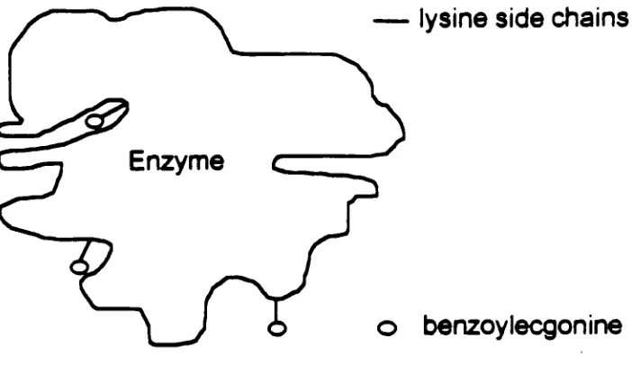
products.The
reactions canbe
photometrically
monitored.When using
an enzymelabel
for
small analytes such asdrugs
ofabuse,
the
challenge
is
to
insure that
the
attached analyteis
notsterically hindered from
interaction
with antibodies.Usually,
the
analyte attachedto
an enzymeis
sosmall
that
it may be buried
withinthe
enzyme andtherefore
cannotinteract
withantibodies
(see fig. 3).
Hence,
the thought
is
that
the
conjugated analyte couldbe
attachedto
the
enzymethrough
alinker
armto
be
extendedaway
from the
surface of
the
enzymes.As
aresult,
it
could moreeffectively
compete withthe
Linker
armshave been
usedin
a widevariety
ofbiological
applications.They
are often
necessary
to
preservethe
biological
activity
oftwo
proteinsthat
arecross
-linked.
This
wasdemonstrated
in
cases of a carbohydratelinker
armthat
was used
to
conjugatetwo
enzymes(5). The Chitin Lash
was usedto
crosslinkstaphylococcal nuclease
to
ribonucleaseA
with retention of75%
and78%,
respectively,
ofthe starting
enzyme activities.Different
peptidelinker
armshave been
usedto
influence the local
environmentof
immobilized
enzymes(6).
In
this
study,
polypeptide arms ofknown
composition were
quantitatively
graftedto
alcoholdehydrogenase.
The
properties of
the
microcavity
ofthe gel, in
whichthe
enzymeis immobilized
by
multipoint covalent
linkages,
arefully
determined
by
the
linker
arms.According
to
the article, the
results obtainedusing
the
soluble preparationindicate that
the
chemical activation
by
itself (alcohol
dehydrogenase)
does
notinduce
any
modification of
the
activationenergy
ofthe
reaction,
whereasthe
grafting
of acomplete peptide
induces
a rise ofthis
valuefor
the two
hydrophilic
peptides.The grafting
ofonly
one amino acid(Alcohol
dehydrogenase
-Homocystine)
induces
aslightly lower
value.This
canbe
explainedby
the
possibledynamic
interactions
between
the two
hydrophilic
peptides andthe
outerlayer
ofthe
enzyme.
These
interactions
could stablizethe
outerlayer
ofthe
protein .As
aresult, the
transconformationalmobility
ofthe
protein couldbe inhibited
to
aPeptide
linker
armshave
alsobeen
found
to
affectthe
metabolism andbiodistribution
ofantibody
-analyte conjugates
(7). Studer
etal.,
comparedtwo
kinds
of antibody-conjugatedbenzyl
-EDTA:
one
had
a peptidelinker arm:( Ala
-Leu
-Ala
-Leu)
whereasthe
other conjugatedid
not.Digestion
ofthe benzyl
-EDTA
withlinker
arm and withoutlinker
armin
vivo showedthat the
conjugatecontaining
the
linker
arm was cleaved atthe
liver
morerapidly
by
the
liver
protease cathepsin
B1
(T1/2=6
h)
than the
otherconjugate,
whichhad
79%
radioactivity
still attachedto the
protein after97h
of exposuresto
cathepsinB1.
Indeed,
the
resultindicated
that the
rate of clearanceby
liver
depends
onthe
linker.
Furthermore,
the
excretion rate ofthe
conjugatecontaining
the
linker
armwas
faster
than
the
one whichlacked this feature.
Optimal
linker
armlengths have been identified for
improving
the
efficacy
ofinhibitors
ofhuman
leukocyte
elastase(8)
andinhibitors
of chloride-transporting
systemsin
membranes(9). It
wasdemonstrated
that the
human
leukocyte
elastaseinhibitory
potency
ofderivatives
ofbenzisothiazolinone
1,1-dioxide
(saccharin)
N
- acetylated with aliphatic andaromatic substituted acyl
groups
increased
as afunction
oftheir
carbon chainlength. The
resultindicated
that
macromolecular conjugates weredemonstrably inhibitory
to
redblood
cellanion exchangewhen
the ligands
wereappropriately
coupled:inhibitory
efficacy
strongly depended
onthe
chemical structure ofthe
coupledligand
andthe
Similarly,
by increasing
the
length
ofthe
alkyllinker
armsin
derivatives
ofthe
(3
-adrenergic
drug, isoproterenol,
the
potency
ofthese derivatives
wasincreased
in
anin
vitroassay (10). Reitz
etal.,
found
that
abenzyl
carbonatederivative
containing
abranched,
seven-carbon spacergroup
was40
times
more potentthan
isoproterenol in
the
in
vitro assay.Finally,
linker
armshave
been
usedto
improve
the
sensitivity
of a quantitativeassay
for
specific ribonucleic acid sequences(11).
Improved
hybridization
behavior
was notedby increasing
the
length
ofthe
alkyl spacer ofthe
oligonucleotide-alkaline phosphatase conjugate.
The
enzymemoiety
wasseparated
from
the
nucleic acidby
ethylene orhexene. A
two
-fold increase in
signal
-to
-background
ratio was observed
for
the
detection
ofthe
probehaving
a six-carbon spacer
between
the
oligonucleotide andthe
enzyme.Peptide
chains are chosen aslinker
armsbecause
numerous possibilities ofcomposition and properties
(polar,
non-polar,
positively
charged,
negatively
charged,
flexible
andrigid)
canbe
achievedthrough the
choice of amino acidsand
because
solid phase peptide synthesis methods arewell established .Selection
of appropriate enzymefor
use as alabel
for the
immunochemical
reagents
is
very
oftenempirical,
and eachhas
distinct
advantages andbinding
reaction canmodify
their
activity.Consequently,
enzymes canbe
usedas
labels only
whenthey
satisfy
the
following
criteria(4):
1
.The
enzymes musthave
avery high
specific activity.The
signal amplificationseen with enzymes
is
relatedto the
amount of substrate convertedto
productduring
the time
ofincubation. Enzymes
withthe
highest
specific activities arepotentially
capable ofgiving
the
greatest amplification.Assays using
suchenzymes are able
to
detect very low
concentrations ofanalytes.2.
The labels
mustbe
stableduring
the
assay
and under refrigerated storageconditions.
3.
The
enzymes must notbe
presentin
the
biological fluid
ortissue
samplethat
is
to
be
analyzed.Contaminating
label
in the biological
fluid
wouldincrease
background,
decrease the sensitivity,
and givefalse
positives.4.
The
enzymes must retain most oftheir
activity
when attachedto the
antibody.Glucose
oxidase(GO1
)
is
considered anideal
enzymefor
biosensor
development
(12)
andhas
been
used asthe
enzymelabel
in
steroidimmunoassays (13).
This
enzymehas been
used asthe
basis for
glucosesensors
in
some glucosedetection
kits. It
catalyzesthe
oxidation of(3-D-glucose
to
glucono-8-lactone andthe
reduction of molecular oxygento
hydrogen
peroxide
(equation 1).
glucose oxidase
glucose +
02
> gluconic acid +H202
(equ. 1
)
1
BEC.benzoylecgonine; DCC.
dicyclohexylcarbodiimide;
DCM, dichloromethane; DIEA.The
hydrogen
peroxide producedfrom
the
glucose oxidase reactionis
consumed
by
a peroxidase-dyeindicator
reactionin
whichthe
oxidizeddye is
colored,
allowing
the
reactionto
be
photometrically
monitored(equation
2).
peroxidaseH202
+ reduceddye
< > oxidizeddye
+H20
(equ.
2)
In
ourresearch,
the
substrate(reduced
dye)
3,3',5,5'-tetramethylbenzidine
willbe
oxidizedby
horseradish
peroxidase.The
reaction canbe
detected
at410
nm.Since
a single molecule of enzyme can catalyzethe turnover
ofmany
moleculesof
substrate, the
sensitivity
ofthe
assay
couldbe improved
.Glucose
oxidaseis
selectedfrom among
other potential enzymelabels based
onits
excellentstability
(14),
particularly in
lyophilized
preparation.Lyophilized
glucose oxidase
is
stablefor 2
years at0C.
Additionally,
there
are14 lysine
residues per subunit
(15)
of glucoseoxidase,
most of which arelikely
to
be
found
onthe
enzyme surface wherethey
couldbe
available as attachment sitesfor
the
BEC-peptide.
Our
first
approachis
to
couplethe
BEC
- peptideto
glucose oxidaseby forming
an amide
bound between
the
carboxy
end ofthe
peptidelinker
arm andthe
amino
terminal
ofthe
lysine
residues on glucose oxidase(
seefig. 4 ). Peptide
linker
arms were synthesizedusing
the fluorenylmethoxycarbonyl
(
Fmoc
)
solidphase peptide synthesis
strategy
described
by
Stewart
andYoung
(1). We
attached
the
BEC
to the
exposed amino -terminal
end ofthe
peptidelinker
attached
the
BEC
to the
exposed amino-terminal
end ofthe
peptidelinker
armby
the
free
carboxylgroup
ofBEC
priorto
removal ofthe
peptidefrom
the
solidphase
to
avoidthe
requirementfor isolation
ofthe
peptidebefore reacting
withthe
analyte(
method1 ).
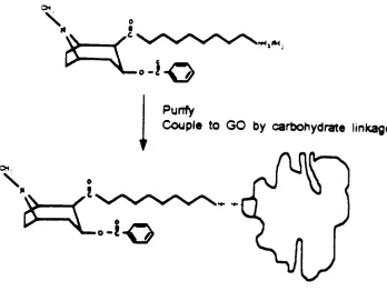
The
BEC
-peptide can also
be
conjugatedto the
carbohydrate portion ofglucose oxidase
using
peptidescontaining
carboxy
terminal hydrazides
(
method2,
seefig. 5
).
BEC
-peptide
hydrazides
canbe
preparedaccording
to
the
methods ofRamage
et al.(3). The
site -directed
immobilization
of antibodieson
hydrazide
-containing
solid supportsfollowing
oxidation oftheir
N
-linked
and
O
-linked
carbohydrate moieties
is
welldocumented
(16)
The resulting
vicinal
dialdehydes
will react withthe
BEC
- peptidehydrazides
to
form
stablehydrazone linkages.
Many
factors
needto
be
consideredin the development
of animmunoassay.
Major factors
are:sensitivity, accuracy, specificity,
et al..The
enzyme-analyteconjugates we
develop
willbe
characterizedimmunochemically
to
determine
the
effect of
the
linker
arm on conjugate performancein
animmunoassay. We
willfocus mainly
onthe
following
three factors in
assay
performance which relatespecifically
to
enzyme-analyte conjugates:
1.
Binding
ofthe
conjugateto the
antibody in
the
absence ofbenzoylecgonine.
antibody.
To be
effectivein
animmunoassay,
the
conjugate mustbind
withsufficient
affinity
to the
immobilized antibody
to
generate a colorimetric signalof
approximately
1.5
to
2.0
absorbance unitsin the
absence ofthe
free
analyte.
2.
Displacement
ofthe
bound
enzyme-analyte conjugate
by
the
analyte.In
acompetitive
assay, the
labeled
analyte will compete withthe
free
analytein
physiological
fluid. It is
necessary
to
develop
conjugatesthat
canbind
tightly
enough
to the
antibody
to
generate adetectable
signal.On
the
otherhand,
it
should also
be displaced
by
the
analytein
a specified concentration range.3.
Non
-specific
binding (
NSB
)
refersto the
conjugatesthat
are non-specifically bound
to the tube
in
the
absence of antibody.The
immunoaffinity
purification procedures
described below
should reduceNSB.
CHAPTER
II
EXPERIMENTAL DESIGN
A.
MATERIALS
1. Reagents
Glucose
oxidase(Aspergillus niger)
andhorseradish
peroxidase werefrom
Sigma Chemical Co.
(St.
Louis,
MO). Resins
andFmoc-amino
acids werefrom
Peptide
International
Inc.
Benzoylecgonine
wasgenerously
providedby
the
Research
Technology
Branch
ofthe
National Institute
onDrug
Abuse (Research
Triangle
Park,
NC). All
other reagents were ofthe
highest
quality
commercially
available:
Phenol,
liquefied
Ethanol,
absolutePotassium
cyanidePyridine
Ninhydrin
Fmoc-Gly
Fmoc-Phe
Fmoc-Gly-Wang
resinBoc-Gly-O-resin
Dichloromethane(DCM)
Dimethylformamide(DMF)
Isopropanol(iPrOH)
Piperidine
J.T. Baker Inc.
Aaper
Alcohol
andChemical
Co.
Sigma Chemical
Co.
J.T. Baker Inc.
Aldrich Chemical Inc.
Peptides
International
Inc.
Peptides
International
Inc.
Peptides International Inc.
Peptides
International
Inc.
J.T. Baker Inc.
Fisher Chemical Co.
J.T. Baker Inc.
Sigma Chemical
Co.
Diisopropylethylamine(DIEA)
Trifluoroacetic acid(TFA)
Dicyclohexylcarbodiimide(DCC)
Ethyl
etherHydrazine
N-hydroxybenzotriazole(HOBt)
Aldrich
Chemical
Inc.
Sigma
Chemical
Co.
Sigma
Chemical
Co.
E.M.
Company
Sigma Chemical
Co.
Peptides International
Inc.
2. Instruments
Syn
-Thor2000
HPLC
Peptides International
Inc.
Autochrom Co.
B.
METHODS
1.
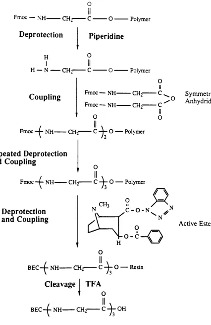
Synthesis
ofthe
Benzoylecgonine
-Peptide
Conjugate
Method 1 We
usedthe
Fmoc
solid phase peptide synthesisstrategy described
by
Stewart
andYoung
(1)
with some modifications(see figure 6). The first
glycine residue was
already
attachedto
resinby
its
carboxyl end whenpurchased and subsequent amino acid residues were attached at
the
aminoterminus
ofthe
growing
peptide.At
eachcycle,
the Fmoc-amino
group
wasdeprotected
by
a20%
piperidine/DCM
treatment followed
by
severalwashing
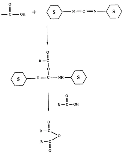
steps.Pre-formed
symmetricalanhydrides
(see figure
7)
in DCM
were mixed withthe
resin untilmonitoring
showed complete reaction.
Symmetrical
anhydrides were chosenbecause
oftheir
high
reactivities(2).
They
were preparedusing
4
equivalents of protectedamino acid and
2
equivalents ofDCC
in 50% DMF. The
urea produced wasremoved
by
filtration.
DIEA,
atertiary
amine,
was added(equivalent
to the
amount of peptide on
the
resin) 15
minutes afterthe
coupling
reactionhad
begun
to
acceleratethe
reaction.The coupling efficiency
was checkedduring
each cycle
using
the
Kaiser
test
(17).
The Kaiser
test
involves
the
following
reagents:5 g
of ninhydrinin 100
mlethanol,
80
g
ofliquefied
phenolin 20
mlethanol,
and2
ml of0.001
M
aqueouspotassium cyanide
in 98
ml of pyridine.A
few
resinbeads
were removedfrom
the
mixing
chamber,
thoroughly
washed withethanol,
and sampled with2 drops
of each reagent above
for
5
minutes at120C.
Yellow
solution and whitebeads
indicated
a complete coupling.Blue
beads indicated
anincomplete
coupling.The
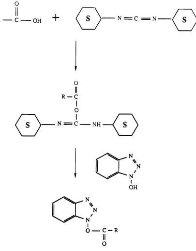
active ester method(see figure
8)
was chosento
attachthe
BEC
to the
peptide - resin.
BEC
was preactivated withDCC
andN
-
HOBt in
high
quality
DMF
to
form
the
HOBt
active ester.For HOBt
activeesters,
5
equiv. ofBEC
,DCC
andHOBt
were used.The filtrate
was reacted withthe
peptide-resin untilthe
Kaiser
test
gave a negative result.At
the
end ofthe synthesis,
the
resin was shrunkby
methanol orisopropanol
treatment.
The BEC-peptide-resin
wasdried
under vacuumfor
atleast 4 hours
before
cleavage.82.5%
trifluoroacetic
acid aqueous solution was usedto
release
the
conjugatefrom
the
resin.The
filtrate
wasbrought to dryness
and precipitatedfrom
cold ether.(For detailed
synthesis protocol seeAppendix
A)
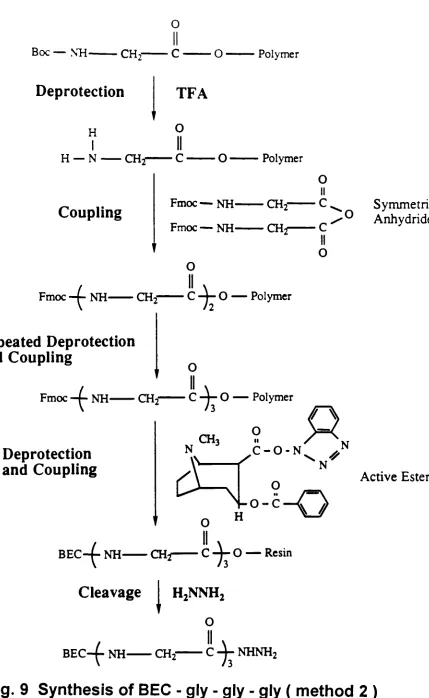
Method 2
(Peptides
Containing
Carboxy
-Terminal
Hydrazides)
BEC
-peptidehydrazides (see figure
6)
were preparedby
using
Boc
-Gly
O
-Resin.
50%
TFA/DCM
was used asdeprotection
reagent.The
peptide -0 - resin wassynthesized
following
the
Fmoc
peptide synthesisstrategy
described
above.The
active estercoupling
method was again usedto
attachBEC
to
peptide -resin.The
peptide resin wasthen
suspendedin
purifiedDMF (5
ml perg
ofresin)
and anhydroushydrazine (30
equiv. per equiv. of peptide).The
mixture was stirredfor
2 days
at room temperature.Resin
was removedby
filtration
and washedwith
DMF.
The
combinedfiltrate
and washings were evaporatedin
vacuum andthen
were purified.(For detailed
protocol seeAppendix
B)
2.
Purification
ofBEC
- peptideOur
initial
attemptsto
purify
the
BEC
-peptide
involved
cation exchangechromatography
.The
use of volatile pyridine acetatebuffers
(18)
would enableus
to
lyophilize
the
isolated
fractions
to
obtain salt-freeBEC-peptide
in
anhydrous condition.
Two
buffers
wereprepared, the
pH3.1 0.2N
pyridine acetatebuffer
andthe
pH5.0 2. ON
pyridine acetatebuffer. All
reagents weredegassed. The Dowex-50X2
resin was swollen
in
pH3.1 buffer
at a ratio of1
volume of resinto
2
volumes ofbuffer. An
HPLC
pump,
low
pressuremixing
valve and control modulefrom
the
Autochrom
Co.
were connectedto the
columnto
controlthe
flow
rate(1
ml/min)
and
to
run a gradient ofthe two
buffers. The
column was washed with3
volumesof
the
pH3.1 buffer before
the
sample was applied.The
separation wasinitiated
with pH3.1 buffer.
Elution
conditionswere:Initial
1 00%
pH3. 1 buffer
1.0
ml/min10
min100%
pH3.1 buffer
1.0
ml/min30
min100%
pH5.0 buffer
1.0
ml/min40
min100%
pH5.0 buffer
1
.0 ml/min50
min1 00%
pH3. 1 buffer
1.0
ml/minEffluent
fractions
were collectedevery
30
seconds.linear
gradientto
linear
gradientto
We
attempedto
analyzethe
purity
ofthe
crude sampleby
HPLC. Solution A
(0.1% TFA in water)
and solutionB (0.1% TFA in
acetonitrile)
were prepared.Elution
conditions were:Initial
100% A
1.0
ml/min10
min100% A
1.0
ml/minlinear
gradientto
30
min100%
B
1.0
ml/min40
min100%
B
1.0
ml/minlinear
gradientto
50
min100% A
1.0
ml/min55
min100% A
1.0
ml/minCHAPTER
III
RESULTS & DISCUSSION
A.
Synthesis
ofthe
Benzoylecgonine
-Peptide Conjugate
The
mostcommonly
usedtest to
monitor solid phase peptide synthesisis
the
Kaiser test. It is
simple and rapid and applicableto
allN-terminal
residues(1).
Any
free
primary
aminogroup
willbe indicated
by
anintense
blue
color.Hence,
it
canbe
usedto
monitorboth
the
deprotection
and coupling.It is
expectedthat
after each
attachment, the
Kaiser
test
will give a negative result.The washing
step
in the Kaiser
test
is
criticalto
avoidproducing
afalse
positive result(1). We
performed
the
Kaiser
test
to
monitorthe
coupling
reaction.The
result showed usthat
wesuccessfully
synthesizedthe
gly
-gly
-gly
- resin and attachedBEC
to
the
gly
-gly
-gly
-resin.
The
crudeyield of method1 is
90%,
method2 is 300%.
When
we usedthe
standard methodto
prepare symmetrical anhydrideusing 2
equivalents of protected amino acid and
1
equivalent ofDCC
for
eachrun,
coupling
results were checked at3
hours,
4
hours,
5
hours,
6 hours
, and8
hours
afterthe reaction,
respectively.Unfortunately,
coupling
wasincomplete
after
8 hours.
However,
30
minutes after we addedthe
second equivalent ofpreformed anhydride
to the
reactionchamber,
the Kaiser
test
gave us a negativeresult.
To
get asatisfactory
coupling,
wedecided to
use excess amino acid.The
anhydride was prepared
using 4
equivalents ofamino acid with2
equivalents ofDCC. Within
anhour
ofthe reaction,
the
Kaiser
test
indicated
complete coupling.Conjugation
ofthe
carboxylic acidgroup
ofthe
BEC
withthe
aminoterminus
ofthe
growing
peptide was attempted.Initially,
wetried
to
attachthe
BEC
symmetrical anhydride
molecules,
which arethe
actualintermediates
in
coupling
reactions,
directly
to the
peptide - resin.This technique has been found
to
givethe
best
results of all methodstried
in
many
difficult
sequences(1).
Unfortunately,
it failed.
The
reaction was monitoredby
the
Kaiser
test
for 12
hours. Blue beads
anddark
blue
solutionsindicated
an unsuccessful coupling.This
couldbe
attributedto the
fact
that
BEC
, whichhas
atertiary
aminogroup,
is
abig
molecule relativeto
glycine.The
N
-terminal
of glycine couldbe
sterically
hindered from attacking
the
BEC
symmetrical anhydride molecule.The
second approach was an active ester
method,
wherein anHOBt
ester ofBEC
was prepared
using
DCC
coupling.Two hours
aftercoupling, the
Kaiser
test
indicated
a successful coupling.B. Purification
ofBEC
- peptideWe
tried
to
purify
the
BEC
-gly
-gly
-gly
by
cation exchange chromatography.The
results wereinconclusive. The
effluentfractions
were examinedby
the
ninhydrin
identification
spray.We did
not expectto
seeany
color changefor
the
fractions
whichhad
only BEC
-.gly
-gly
-gly
residues.Unfortunately,
allthe
fractions turned
to purple,
indicating
the
presence of somefree
amino groups.Further
refinements of purification and analysis ofthe
BEC
- peptides aredescribed below.
CHAPTER
IV
Plans
/
Thoughts
A. Solid
Phase
Synthesis
ofthe
Benzoylecgonine
-Peptide Conjugate
withan enzyme cleavable
linker
armThe
BEC
-peptide could also
be
cleavedfrom
the
resinby
a protease.We
willsynthesize a peptide
in
whichthe
first
two
residues represent a specific sitefor
the
enzyme cleavage.For
example,
a peptide whichhas
a glycine andphenylalanine at
its
carboxy
terminal
end canbe
cleavedby
chymotrypsin.The
BEC
-peptide would
then
be
releasedinto
an aqueoussolution,
wheredirect
coupling
to
GO
couldbe
attemptedusing
a water solublecoupling
reagent suchas
diisopropyl
carbodiimide.B. Conjugation
ofthe Benzylecgonine
-Peptide to the
Enzyme
The
purifiedBEC-peptide
willbe
dissolved
in
an anhydrous organicsolvent,
activated with
isobutyl
chloroformate,
then
addedto the
enzyme solutionin
sodium
phosphate,
10
mmol/L,
pH7.4
(see fig. 10). The
BEC
-peptide
is
notsoluble
in
tetrahydrofuran,
therefore,
NMP is
chosenusing
these
criteria:(a)
it
must
dissolve
BEC
-peptide;
(b)
it
must notdenature
the enzyme;
(c)
it
musthave
afreezing
pointbelow
-25C,the temperature
for
the
activation reaction.C.
Purification
ofBEC
- peptide -GO
by
Immunoaffinity Chromatography
An
antibody towards BEC
willbe
immobilized
onAffi
-Gel
Hz (Bio
-Rad
Laboratories)
according
to the
manufacturer's specification.BEC
-peptide
-GO
will
be loaded
ontothe
resincontaining immobilized
anti -BEC.
Following
ahigh
salt washstep, the
conjugate willbe
eluted with citratebuffer
(10
mmol/L,
pH
3.0).
D.
Characterization
ofEnzyme
-Analyte
Conjugate
We
willfocus mainly
onthe
following
three
factors in assay
performance whichrelate
specifically
to
enzyme-analyte conjugates:
1.
Binding
ofthe
conjugateto the
antibody
in
the
absence ofbenzoylecgonine.
First,
the
antibody
willbe immobilized
on microtiter wellsusing
publishedmethods
(19).
The
wells willbe
coated withpolylysine,
whichbinds
irreversibly
to their
surface.The
carbohydrate portion ofthe
antibody
willbe
oxidized with sodium periodate.
The resulting
aldehyde groups will react withthe
e-amino groups ofthe lysine
side-chains ofthe polylysine,
thereby
immobilizing
the
antibody.Then
the
conjugate willbe incubated for 30
min.with
immobilized
antibody, the
plates washedto
remove unboundconjugate,
substrate added
for
anhour
andthen the
reaction willbe
quenched after1
hour
with1N
sulfuric acid.Absorbance
at405
nm of1.5
to
2.0
unitsis
acceptable.2.
Displacement
ofthe
bound
enzyme-analyte conjugate
by
the
analyte.We
willthen incubate
the
conjugate plus a series ofBEC
concentrationsin
a solid-phase
immunoassay
format. We
will evaluatethe
data
to
determine
the
midpoint ofthe
calibration curve andthe
level
of non-specificbinding.
3.
Non
- specificbinding.
A,05
values atBEC
concentrationstwo
orders of magnitude greaterthan the
mid point ofthe
BEC
calibration curve(
Aios
vs.logfBEC] )
shouldbe less
than
1%
ofthe
signalin
the
absence ofBEC.
Reference
1
.Stewart J
M,
Young
JD.
In:
Solid
phasepeptide synthesis.Pierce
Chemical
company,
Rockford,IL1984
2.
Brown
etal.; J.C.S. Perkin Trans.
I,
1983.
3.
Ramage
R,
Irving
SL,
Mclnnes
C. Tetrahedron Letters
1993;
34:
6599-602
4.
Kaplan
L A
,Pesce A. J.
Clinical chemistry
1989;
The C. V.
Mosby
Company,
St.
Louis,
MS.
5.
Guan
K,
Cecchini
DJ,
Giese
RW.
CarbohydrRes
1993;
246:
205-17
6.
Bille
V,
Plainshamp
D,
Lavielle
S,
Chassaing
G,
Remade
J,
Eur
J
Biochem
1989;
180:41-7
7
Studer
M,
Kroger
LA,
DeNardo
SJ,
Kukis
DL,
Meares
CF.
Bioconj
Chem
1992;
3:424-9
8.
Kerneur
C,
Homebeck
W,
Robert
L,
Moczar E. Biochem Pharmacol
1993;
45:1889-95
9.
Eidelman
0,
Yani
P,
Englert
HC,
Lang
HG,
Greger
R,
Cabantchik ZL. Am
J
Physiol 1 991
;
260:
d 094-1 03
10.
Reitz
AB,
Sonveaux
RP,
Verlander
MS,
Meimon
KL,
Hoffman
BB,
Akita
Y,
Castagnoli
N,
Goodman M. J Med Chem
1985;
28:
634-42
1
1
.Ishii
JK,
Ghosh
SS.
Bioconj
Chem
1993;
4: 34-41
12. Wilson
R,
Turner APF. Biosensors & Bioelectronics
1992;
7:165-8
13.Hosoda
H,
Tsukamoto
R,
Shishidoo
M,
Takasaki
W,
Nambara
T. Chem
Pharm Bull
1987;
35:
2856-61
14. Kellin
D,
Hartree
EF. Biochem
J
1948;42:221-9
15. Frederick
KR,
Tung J,
Emerick
RS,
Masiarz
FR,
Chamberlain
SH,
Chamberlain
SH,
et al.J Biol Chem
1990;265:3793-802
16.
O'Shannessy
DJ,
Hoffman
WL. Biotech
App
Biochem
1
987;
9: 488-96
1 7. Kaiser ET.
Natural
1
985;
31
3:788-98
18.0hno,
M. And
Anfinsen,
CB. J.
Am.
Chem. Soc.
1967,89:5994-95
19. Schramm
WS,
Paek
S-H,
Kuo
H-H,
Yang
T. Anal Chim Acta 1991
;
248:517-528
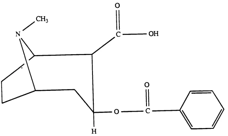
Fig.
1
Structure
of
BEC
[image:31.522.66.461.177.414.2]CQ
IO
o
a
o
w
(D
o.
m
o
a
o
o
o
3
C
ca
D
a
0
3
CO
>
3
?o n
x
9
o
o
o
a
8
lysine
side chains
o
benzoylecgonine
Fig.
3 Enzyme
-Analyte Conjugate
[image:33.522.87.438.167.370.2]Linker
Arm
CH,
Fig. 4
Attaching
BEC to Enzyme
by
Linker
Arm
[image:34.522.63.441.205.415.2]-o
Purify
Couple
to
GO
by
carbohydrate
linkage
-!0
Fig.
5
Attaching
BEC
-peptide
Hydrazide
to
enzyme
[image:35.522.62.410.187.449.2]o
Fmoc
NH
CH,
Deprotection
O
Polymer
Piperidine
H
I
H
N
CH,
O
II
C'O
Polymer
Coupling
Fmoc
NH-Fmoc
NH-O
CH2-O
II
C.
C
II
O
:o
Fmoc
-+- NH- CH2~C-j-0
Polymer
Repeated Deprotection
and
Coupling
-f
Fmoc
-f-NH
CH-j
O
II
c
V-
Polymer
Deprotection
and
Coupling
CH3
11 f XTNVN
^C-O-N^P
O
Symmetrical
Ajihydride
Active Ester
i0-*-O
1
BEC
-4-NH
CH2"Cleavage
0
II
-CTFA
V-
Resin
t
BEC-t
NH
CH2
C
4-
OH
Fig.
6
ynthesis
of
BEC
-gly
gly
-gly
(
method
1
)
[image:36.522.51.477.46.700.2]o
R
C
OH
+
N
=C
N-O
R-C
I
O
=
C
NH
(
S
R
O
II
C
OH
O
II
R
C
R
/
Fig. 7
Synthesis
of
Symmetrical Anhydride
[image:37.522.61.468.106.630.2]O
R
C
OH
+
N
N'O
R-C
O
N
NH
Fig. 8
Synthesis
of
HOBt Active
Esters
[image:38.522.60.465.54.572.2]O
Boc
NH-CH,
O-Polymer
Deprotection
H
I
H
N
CHi
TFA
o
II
c
O-Polymer
Coupling
Fmoc
NH-Fmoc
NH-CH2-CH-r
O
II
C,
c
II
o
:o
Fmoc
-+ NH-CHo
O
11
___C
-4-O
Polymer
Repeated Deprotection
and
Coupling
Fmoc
-+NH
Co
polymer
Deprotection
and
Coupling
BEC-f-
NH
CH
N
N
Symmetrical
Anhydride
Active Ester
i-z-0
i
-Cleavage
I
H2NNH2
o
BEC4-
NH
CH2
C
-j-NHNH2
Fig. 9
Synthesis
of
BEC
-gly
-gly
[image:39.522.51.480.33.731.2]CH,
N
CH-,
N
O
II
C
OH
O
II
o
c
H
\
/
t
1) DCC,
HOBt, DMF,
0C,
30
min.2)
Gly-Gly-Gly-Resin
3)
TFA
(95%)
O
II
c
/
II
\
-(-NH
C
Ct-(
\
I
/n=l
OH
10
O
H
1)
Isobutyl
chlorofonnate,NMP,
-20C,lh
2)
Glucose
oxidasein
sodium phosphatebuffer,
pH7,
lh
,CH3
N
II
C-I-NH
-4-NHC
C-)
NH
\
I
/n=l-10
Gl
coseoxidaseO
[image:40.522.57.416.56.690.2]r-'-0
Fig. 10
Synthesis
of
BEC
-GO
Conjugate
Appendix
A
Schedule
A
For
BEC
-Peptide
Synthesis
(
method
1
:using
Fmoc
-gly
- resin)
Wash 1
DCM
wash,
2
x30
ml x2
min.DMF
wash,
3
x30
ml x2
min.Deprotect
30%
(v/v)
piperidinein
DCM,
1
x30
ml x3
min.,
1
x30
ml x7
min.Wash 2
DMF
wash,
3
x30
ml x1
min.DCM
wash,
3
x30
ml x1
min.Couple
Symmetrical
anhydride ofFmoc
- amino acidin
50%
(v/v)
DCM/DMF,
20
mix15
min.DIEA,
1
equivalentto the resin,
30
min. or untilmonitoring
shows
coupling
completeRecouple
Repeat
steps aboveBEC
HOBt
active esterin
DMF,
30
min. or untilmonitoring
shows
coupling
completeWash
3
DCM
wash,
3
x30
ml x2
min.Methanol wash,
2
x30
ml x7
min.Dry
underhigh
vacuumfor
5
ormorehours
Cleavage
50
%
or82.5%
(v/v)
TFA/water,
20
ml x120
min.Ether
precipitationFilter
the
cleavage mixtureusing
a sintered glassfunnel.
Wash
the
resin severaltimes
withthe
cleavage mixture.Combine filtrates
andbring
them
to
dryness
by
rotary
evaporation.
Add
cold etherto
precipitatethe
crude sample.Appendix
B
Schedule
B For BEC
-Peptide
Synthesis
(
method
2:
using
Boc
-gly
-resin
)
Wash 1
DCM
wash,
2
x30
ml x2
min.Deprotect
50%
(v/v)
TFA/DCM,
20
ml x1 5
min.Wash
2
DCM
wash,
2
x30
ml x2
min.DMF
wash,
3
x30
ml x2
min.Couple
Symmetrical
anhydride ofFmoc
-amino acid
in 50%
(v/v)
DCM/DMF,
20
mix15
min.DIEA,
1
equivalentto the resin,
30
min. or untilmonitoring
shows
coupling
completeWash 3
DCM
wash,
3
x30
ml x2
min.DMF
wash,
3
x30
ml x2
min.Deprotect
30%
(v/v)
piperidinein
DCM,
1
x30
ml x3
min.,
1
x30
ml x7
min.Wash 4
DMF
wash,
3
x30
ml x1
min.DCM
wash,
3
x30
ml x1
min.Recouple
Repeat
stepsWash 1
to
Recouple
in Schedule A
Wash
Cleavage
DCM
wash,
3
x30
ml x2
min.DMF
wash,
3
x30
ml x2
min.High
quality
DMF
wash,
3
x30
ml x2
min.Suspend
resinin
high quality DMF (5
ml perg
ofresin)
withanhydrous
hydrazine
(30
equiv. ofresin)
for
2
days.
Bring
the filtrate to dryness
by
rotary
evaporation.