“
EVALUATION OF CENTRAL NEUROPATHY IN TYPE
2 DIABETES CASE – CONTROL STUDY
”
DISSERTATION SUBMITTED IN PARTIAL FULFILLMENT OF THE REGULATIONS FOR THE
AWARD OF DM IN NEUROLOGY
DEPARTMENT OF NEUROLOGY
PSG INSTITIUTE OF MEDICAL SCIENCES AND RESEASRCH
THE TAMILNADU Dr. M.G.R MEDICAL UNIVERSITY, CHENNAI
TAMILNADU, INDIA
CERTIFICATE
PSG Institute of Medical Sciences & Research
Coimbatore
This is to certify that Dr. E. PRASANNA VENKATESAN has prepared this dissertation entitled “EVALUATION OF CENTRAL NEUROPATHY IN TYPE 2 DIABETES CASE – CONTROL STUDY”, under my overall supervision and guidance in PSG Institute of Medical Science and Research, Coimbatore in partial fulfillment of the regulations of The TamilNadu Dr. M.G.R Medical University for the award of DM Neurology.
DR.K.RAMADOSS MD, DM., DR.S. RAMALINGAM
MD.,
Professor and Head of the Department Principal
Department of Neurology PSG IMS & R
PSG IMS & R
DECLARATION
I hereby declare that dissertation entitled “EVALUATION OF
CENTRAL NEUROPATHY IN TYPE 2 DIABETES CASE –
CONTROL STUDY” was prepared by me under the guidance and
supervision of Dr. K. Ramadoss MD, DM, PSG
IMS&R, Coimbatore. The dissertation is submitted to The TamilNadu Dr.
M.G.R. Medical University, Chennai in partial fulfillment of the
University Regulations for the award of DM degree in Neurology. This
dissertation has not been submitted for the award of any Degree or
ACKNOWLEDGEMENT
With deep sense of gratitude, I sincerely express my thanks to
Dr. K. RAMADOSS, Professor and Head, Department of Neurology,
PSG Institute of Medical Sciences & Research, Coimbatore, for his
valuable guidance and encouragement given at every stage of this project.
I would also express my sincere thanks to Dr.M.B.Pranesh,
Dr.B.Prakash, Dr.G.Lakshminarayanan Dr.R.Balakrishnan and
Dr.G.Gnana Shanmugham.
I am very much obliged and grateful to Dr. RAMALINGAM, Principal,
PSG Institute of Medical Sciences & Research, Coimbatore, for
providing facilities in carrying out this project.
I am extremely thankful to all staff who have spent their time for
collection of data and have also helped me in successful completion of
this project.
I thank Mrs. SHERLY and Mr. SARAVANAN, who helped me in
recording the data.
1 thanks my beloved parents for the confidence and encouragement given
I thank my friends for all the help they extended to me during my work
for the project. Lastly, I thank God for the wonderful team who helped
me in completion of my project.
CONTENTS
S.NO CONTENTS PAGE NO
1 INTRODUCTION 1
2 AIMS AND OBJECTIVES 4
3 REVIEW OF LITERATURE 5
4 MATERIALS AND METHODS 27
5 OBSERVATION AND RESULTS 34
6 DISCUSSION 47
7 CONCLUSION 51
8 SUMMARY 52
9 LIMITATIONS 53
EVALUATION OF CENTRAL NEUROPATHY IN TYPE 2 DIABETES:
CASE–CONTROL STUDY
ABSTRACT
Introduction: Diabetes mellitus (DM) is a global pandemic affecting almost every
organ in the body. Peripheral nervous system involvement in diabetes is well known
but there are not much studies on central nervous system involvement. Visual evoked
potential(VEP) is a sensitive, non invasive test to detect central demyelination of optic
nerve.
Aims: To compare the visual evoked potentials in type-2 DM patients with that of
healthy controls and to find out if any correlation is there with the duration or
glycemic control of the disease .
Materials and methods: We included 50 DM patients and 50 age and sex matched
controls. Patients with previous stroke, demyelination, diabetic retinopathy and other
opthalmological disorders were excluded. VEP was recorded using pattern reversal
stimulation with EMG RMS MARK II machine and p100 latency was measured.
Results: P100 latencies (ms) was significantly prolonged in diabetics with mean ± SD
of (111.24 ±5.28 ms) as compared to controls (101.30 ± 1.66 ms) with p value
<0.003. Also there was significant correlation between duration of DM and P100
latency prolongation but no significant correlation was present between glycemic
control of patients and P100 latency.
Conclusion: Abnormal VEP may be due to structural damage to myelinated optic
nerve fibres or retinal ganglion cells and it occurs even before development of
retinopathy. Hence VEP can be a used as an early marker for central neuropathy and
INTRODUCTION
Diabetes mellitus (DM) is a global pandemic affecting almost
every organ in the body. It causes serious challenge to healthcare system.
Nearly 150 million people throughout the world are affected and the
incidence increases with time as sedentary lifestyle and obesity is on the
rise. Major complications of DM are due to atherosclerosis and it can
affect any organ in body especially eyes, peripheral nerves, kidney and
heart. These are categorized into microvascular and macrovascular
complications.
Diabetic peripheral neuropathy is a major public health burden. It
is characterized by burning sensation of feet, distal weakness and absent
deep tendon reflexes especially ankle jerk. Only 15% of DM have
peripheral neuropathy clinically but upto 50% have peripheral neuropathy
by nerve conduction studies. Similarly only 10% have peripheral
neuropathy at time of diagnosis of DM but nearly 50% have neuropathy
after 25 years duration. Duration of DM and glycemic control of DM are
important factor for development of peripheral neuropathy.(1)
Various forms of peripheral neuropathy are known to occur in
DM. The most common type is distal symmetric sensory polyneuropathy.
known to occur. Rarely asymmetrical, painful proximal muscle weakness
due to diabetic amyotrophy can occur. Only 0.6% of diabetic patients
have optic nerve involvement resulting in optic atrophy.(2)
The peripheral nervous system involvement in DM has been
studied extensively in various studies but central nervous system
involvement in DM has not been studied in detail. The term “central
neuropathy” has been unknown until recently. Only after few western
studies described subclinical optic nerve involvement in DM by
electrophysiological studies the term central neuropathy was recognized.
Just like subclinical peripheral neuropathy, asymptomatic optic
neuropathy or central neuropathy can occur and it is evaluated by visual
evoked potentials. Although in diabetics the most common cause for
blindness is diabetic retinopathy asymptomatic optic nerve dysfunction
can occur as proved in various studies.
Visual evoked potential (VEP) is a non invasive, sensitive tool
which measures the impulse conducted along the central nervous
pathway. VEP measures the P100 latency which reflects the functional
abnormalities of optic pathway even in early stages. We decided to
evaluate the central neuropathy in DM patients and compare with
controls. Although there were few similar studies in past most of them
larger sample size of 100 and we also compared the latency prolongation
AIMS AND OBJECTIVES
1. To compare the visual evoked potentials in type-2 Diabetes
mellitus patients with that of healthy controls.
2. To find out if there is any correlation with duration of DM or
REVIEW OF LITERATURE
Diabetes mellitus (DM) is an metabolic disorder due to decreased
insulin secretion or action or both resulting in hyperglycemia. It is one of
the leading cause for blindness in world. It accounts for 30% of
preventable blindness. The global prevalence of DM is 6.6% in 2010. As
per international diabetes federation of 285 million diabetic subjects in
world, 70 % live in low income countries like India. India is the diabetic
capital of the world with 57 million people suffering as per 2010 data. If
no drastic steps are taken to stop this epidemic it is expected to further
increase in prevalence.
RISK FACTORS
1. Familial aggregation
2. Age
3. Adiposity
4. Body fat percentage
5. Insulin resistance
6. Life style changes due to urbanization
CHRONIC COMPLICATIONS OF DIABETES
Generally the injurious effects of DM are classified into
microvascular and macrovascular complications.(3)
MICROVASCULAR COMPLICATIONS 1. Diabetic retinopathy
2. Diabetic nephropathy
3. Diabetic neuropathy
MACROVASCULAR COMPLICATIONS 1. Coronary artery disease
2. Cerebrovascular disease
3. Peripheral vascular disease
PATHOPHYSIOLOGY OF COMPLICATIONS
Although the precise mechanism for microvascular complications
are not known it is generally believed that there are 3 pathways which are
involved in development of these complications. It is related to both
duration of DM and poor glycemic control of disease.
The central pathological mechanism in macrovascular disease is
atherosclerosis. Atherosclerosis occurs in response to oxidization of LDL
cholesterol resulting in endothelial injury and inflammation. Diabetes
enhances the effect of other co-morbid conditions like hypertension,
adhesion, plasminogen activator inhibitor and increased free radical
generation. All these factors collectively produce a state of
hypercoagulability.
PATHWAYS INVOLVED:
1. Polyol pathway 2. AGE Pathway 3. Protein kinase C
POLYOL PATHWAY
In DM the excess glucose is shunted to aldose reductase pathway
which results in sorbitol. Sorbitol is further metabolized to fructose.
Neither sorbitol nor fructose can move out of the cell and it can result in
cellular swelling. There is also depletion of myoinositol, loss of Na/K
ATPase activity and NADPH co-factors. Hence the metabolically
compromised axons are susceptible to injury and ischemia. The small
thinly myelinated fibers are more affected than large fibers and hence
ADVANCED GLYCATION END PRODUCTS
Glycation of macromolecules in diabetes results in advance
glycosylated end products. AGE are large aggregates and cannot be
cleared by normal metabolism. They are susceptible to oxidation and
resulting in oxidative damage. There is a very strong association between
AGE and development of diabetic nephropathy. AGE is a complex series
of poorly understood reactions in DM which results in endothelial
dysfunction.(4)
PROTEIN KINASE C PATHWAY:
Chronic hyperglycemia in DM can stimulate protein kinase c
pathway which mainly functions to alter vascular permeability, cellular
proliferation and blood flow. Activation of this pathway leads to increase
in VEGF and increased angiogenesis. This pathway has strong
DIABETIC RETINOPATHY:
It is the most common microvascular complication of diabetes. It
can even precede diagnosis of diabetes mellitus.(6)It is related to duration
of diabetes and degree of hyperglycemia like most of the other
microvascular complications. It is classified as proliferative and non
proliferative diabetic retinopathy. The aldose reductase pathway and
accumulation of AGE have been implicated in development of diabetic
retinopathy.(7) In addition to blindness, diabetic retinopathy indicated end
organ damage in a patient. Blindness in DM can be due to
1. Diabetic maculopathy due to ischemia or vitreo-macular traction
2. Proliferative diabetic retinopathy leading to vitreous hemorrhage or
retinal detachment
3. Neovascular glaucoma
4. CRAO/CRVO ( Central retinal artery/vein occlusion)
5. Ischaemic optic neuropathy
Strict diabetic control, regular ophthalmological evaluation and laser
CLASSIFICATION OF DIABETIC RETINOPATHY:
1. Nonproliferative (background) retinopathy
a. Simple background retinopathy
b. Dot and blot hemorrhages
c. Hard exudates
d. Microaneurysms
e. Macular edema
2. Prepoliferative retinopathy
a. Soft exudates
b. Intraretinal microvascular abnormalities (IRMA)
3. Proliferative retinopathy
a. Neovascularization of the disc
b. Neovascularization elsewhere in the retina
c. Fibrovascular proliferation
DIABETIC NEUROPATHY:
Diabetic neuropathy is a common microvascular complication
occurring in genetically predisposed individuals in addition to longer
duration of DM and poor glycemic control.(8) Recently there is association
between sensory neuropathy and impaired fasting glucose without overt
diabetes and persistent hyperglycemia with elevated HbA1c.
Both cranial and peripheral mono neuropathies which are of acute
onset are mainly due to vasculopathy of ischemic origin. Pathologically
there is ischemia of vasovasorum. The symmetrical distal polyneuropathy
do not have evidence of vasculopathy. Hence alternative theory by Dyck
proposed inflammation as possible cause. They found severe perivascular
VISUAL EVOKED POTENTIAL (VEP):
VEPS are recorded from scalp as potential differences like EEG
(electroencephalogram) in response to some visual stimuli. It checks the
entire visual pathway and any lesion along the visual path can produce
abnormal VEP. Its role in localization of lesion along visual pathway is
only limited.(9) But it is very sensitive and reproducible test which can
detect even subtle conduction defects in anterior visual pathway.
ANATOMICAL BASIS FOR VEP
The two optic nerves extends from retina to optic chiasm. Each
optic nerve is about 5cm in length. At chiasm the temporal fibers remain
uncrossed whereas the nasal fibers cross over and extend further as optic
tract. They relay in lateral geniculate body of thalamus and from which
arises optic radiation. The optic radiations terminates in striate occipital
cortex (area 17).
Following activation of striate visual cortex, P100 waveform in
VEP is generated. It primarily reflects the central field that is relayed to
area 17. Peripheral retinal stimulation does not generate P100 waveform.
The macular fibers which is responsible for central vision occupies large
area in occipital cortex.
Patient should be explained about the test and asked to sit
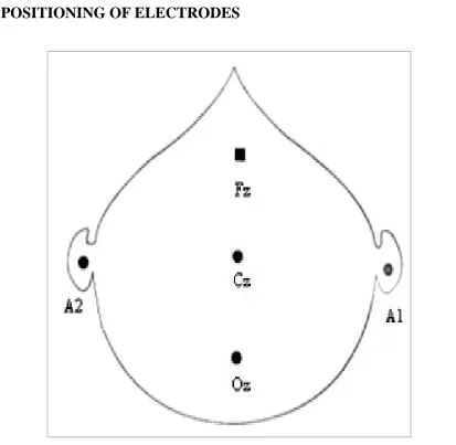
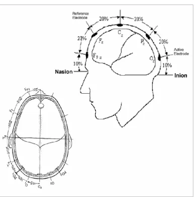
comfortably in front of PC. Standard EEG electrodes are used for
recording after degreasing the scalp. Electrodes Cz, Fz and Oz electrodes
are placed as per 10-20 international system. Oz is active, Fz is reference
and Cz is ground electrode.
1. Pattern shift VEP
2. LED goggles
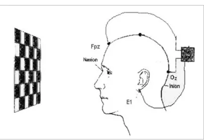
In PSVEP black and white checks are displayed in PC and patient is
instructed to look at center of checkerboard. Patient sits 100 cm from
screen. Impedance is kept less than 5 kΩ. Average of about 100 epochs
are taken so that VEP results can be reproduced.
VEP ABNORMALITIES
There are 3 types of abnormalities in VEP
1. Latency prolongation
2. Amplitude reduction
RECORDING VEPs:
Fig 1: The patient is asked to fix his eye at the centre of the
checkerboard which is flashed on front in a PC screen at a distance of
[image:24.595.103.510.96.374.2]The commonest cause for P100 prolongation is demyelination of
optic nerve. Unilateral P100 prolongation is likely prechiasmal lesion
whereas bilateral P100 prolongation cannot be localized.
VARIABLES INFLUENCING VEP
1. AGE: As the age increases P100 latency prolongs due to age
related changes. It was found that approximately 2.5ms
prolongation occurs for every decade.
2. GENDER: Males have longer latency compared to females
probably because of larger head and hormonal differences.
3. EYE MOVEMENT: Nystagmus, eye movements alter amplitude
but not latency of P100.
4. EYE DOMINANCE: P100 latency is prolonged for non dominant
eye compared to dominant eye.
5. VISUAL ACUITY: Only amplitude of P100 is affected with poor
visual acuity and not latency.
6. DRUGS: When miotics are used for pupillary constriction they
decrease the area of retinal stimulation and cause P100 latency
7. MENTAL ACTIVITY: Arithmetic calculation can increase
amplitude of P100 and decrease latency.(10)
CLINICAL APPLICATIONS OF VEP
1. DEMYELINATING DISEASES:
VEP is useful investigation in evaluation of multiple sclerosis. In
patients with history of optic neuritis more than 90% have abnormal P100
latency prolongation. It is more sensitive than MRI in detecting
abnormalities in optic pathway. Only 84% of symptomatic patients with
MS show abnormalities in MRI. It can detect subclinical demyelinating
plaque.(11)
2. OPTIC NEURITIS:
Typical optic neuritis is characterized by painful monocular vision
loss usually occurring between 20 to 50 years of age. It is difficult to
predict which of these patients with typical optic neuritis will develop MS
later. Those with recurrent episodes and with typical MRI abnormalities
3. ISCHAEMIC OPTIC NEUROPATHY:
It is characterized by painless loss of vision usually occurring in
elderly patients with vascular risk factors like DM and hypertension. It
can also occur in vasculitis and giant cell arteritis. There may be
altitudinal field defects. VEP study shows prolongation of P100 and
decrease in amplitude.(13)
4.TOXIC OPTIC NEUROPATHY:
Some of toxins which can produce optic neuropathy and blindness
are as follows
Tobacco
Alcohol
Ethambutol
Vigabatrin
Amiodarone
can cause prolongation of P100 and decrease in amplitude in both
eyes.(14,15)
5. NUTRITIONAL OPTIC NEUROPATHY:
Vitamin B12, vitamin E and thiamine deficiency can cause
6. HEREDITARY AND DEGENERATIVE DISEASES:
Following neurodegenerative conditions can produce VEP
abnormalities
Friedrich ataxia
Charcot Marie-Tooth disease
Lebers hereditary optic atrophy
Mitochondrial disease
Bilateral P100 prolongation is seen with normal amplitude. P100
prolongation usually correlates well with temporal pallor of disc.(17)
7. COMPRESSIVE LESIONS IN VISUAL PATHWAY:
Following lesions can compress the optic pathway
Meningioma
Tuberculoma
Glioma
Pituitary macroadenoma
Craniopharyngioma
The extrinsic compression of optic pathway leads to P100
8. CORTICAL BLINDNESS:
Cortical blindness due to bilateral lesion in primary visual cortex
can produce P100 prolongation whereas bilateral lesion in visual
association area with preserved primary visual cortex does not produce
abnormality in VEP.(18)
9. MALINGERING:
VEP is very helpful in detecting hysterical blindness. A normal
VEP in a patient complaining of blindness gives clue to diagnosis of
malingering. But some patients can suppress VEP and cause P100
prolongation voluntarily.
10. INTRAOPERATIVE MONITORING:
VEP can be used intraoperatively while resecting tumors of optic
pathway but has only limited role because of technical difficulties to
VEP RESULTS SHOWING P100:
Ziegler et al in their study included 12 diabetic patients both type 1
and type 2. They subjected all patients to VEP and found that diabetic
patients had P100 prolongation more than that of controls. The mean
increase in P100 latency was 116.8+/- 4.5 with a p value <0.01. They
treated the patients with continuous insulin infusion for a short period of
3 days. After 3 days of intensive blood sugar control VEP was repeated
and they found that although P100 latency was slightly prolonged
compared to controls there was significant reduction in latency compared
to previous value. They concluded that P100 prolongation in diabetic
patients were probably due to impaired glucose metabolism and is
Dolu H et al studied electrophysiological characteristics of 51
patients with type 2 DM and compared with 30 age and sex matched
controls. They did VEP, BAEP (brainstem auditory evoked potentials)
and SEP (somatosensory evoked potentials) for all patients. The
multimodal evoked potential which included VEP, BAEP and SEP were
useful in evaluating central neuropathy. They concluded that there was
significant latency prolongation suggestive of central neuropathy in
diabetic patients compared to controls. They did subgroup analysis and
found that latency prolongation in SEP, VEP, BAEP correlated well with
duration of diabetes and not with glycemic control of disease.(20)
Comi G et al also studied multimodal evoked potentials in type 2
diabetes patients using VEP, BAEP and SEP. They found that central
neuropathy due to cortical latency prolongation was more common in
diabetic patients with peripheral neuropathy. Isolated abnormalities in
VEP or BAEP or SEP was more common than all three getting affected
together. They concluded that central neuropathy may occur due to
hyperglycemia or hypoglycemia but exact cause is not known.(21)
Algan et al studied VEP in 50 type 1 diabetes and 19 type 2
diabetes. They found significant prolongation of P100 in diabetic patients
with p value less than 0.001. But on further analysis they concluded that
control of disease. Their findings were contradictory to previous
studies.(22)
Szabela DA et al studied 41 patients with type 2 diabetes. They
recorded VEP in all patients and found 22% had abnormal P100
prolongation. They further analysed age, duration of DM and metabolic
control of DM with P100 latency prolongation and concluded that there
was no correlation with either of them.(23)
Again Szabela DA et al studied 50 patients with type 1 diabetes.
They recorded VEP in all patients and found 26% had abnormal P100
prolongation. They further analysed age, duration of DM and metabolic
control of DM with P100 latency prolongation and concluded that there
was no correlation with either of them.(24)
Azal O et al studied 20 diabetic patients of which 6 were type1 and
remaining 14 were type 2. They recorded VEP in all cases and found
significant increase in P100 latency in diabetic patients with p value
<0.001. 45% of cases had P100 prolongation. They did not find any
correlation with metabolic control of DM or peripheral neuropathy. They
concluded that P100 prolongation correlated well with duration of DM.(25)
Mariani E et al conducted a case control study which included 35
and controls. They found significant prolongation of P100 latency in
cases compared to controls. They also concluded that P100 latency
prolongation correlated well with duration of DM, HbA1c and presence
of peripheral neuropathy.(26)
K Puvanendran et al studied 16 diabetic patients with VEP. They
found 81% of cases had prolonged P100 latency compared to controls.
They further analysed P100 latency with duration of DM and glycemic
control of DM and found no significant correlation existed between them.
They concluded that P100 latency prolongation correlated well with
presence of diabetic sensory neuropathy.(27)
Yaltkaya K et al studied 25 cases of DM and controls. VEP was
done to measure P100, N90 and N140. Sural nerve conduction studies
were done to detect peripheral neuropathy. They found significant P100
and N90-140 interpeak latency prolongation. The latency prolongation
correlated well with duration of DM but not with sural nerve conduction
MATERIALS AND METHODS
We conducted a prospective case control study in department of
neurology PSG institute of medical science and research from October
2011 to October 2013. Patients were chosen from neurology OPD.
INCLUSION CRITERIA
Newly diagnosed type 2 Diabetes mellitus and known case of DM were
included
WHO criteria was used for diagnosing DM;
1. Random plasma glucose of > 11.1 mmol/l
or
2. Fasting plasma glucose > 7.0 mmol/l
or
3. Two hour plasma glucose concentration > 11.1 mmol/l two hours
after 75g anhydrous glucose in an oral glucose tolerance test
EXCLUSION CRITERIA
1. Patients with long standing history of hypertension and with the
past history of cerebrovascular accident.
2. Evidence of optic atrophy
3. Past history of optic neuritis
4. Visual acuity less than 6/18
5. Patients consuming > 100 ml of alcohol daily.
6. Patients with peripheral nervous system disease unrelated to
diabetes mellitus.
7. Patients with diabetic retinopathy, cataract, glaucoma and vitreous
hemorrhage.
8. Patients with type 1 diabetes mellitus.
CONSENT: Informed consent was obtained from patients who were
willing to take part in the study. Ethical committee clearance was
METHODOLOGY:
50 diabetic patients who fulfilled the inclusion criteria were chosen
and 50 age and sex matched controls were also included. They were
subjected to detailed history to rule out stroke, past history of optic
neuritis and other ophthalmological conditions. Detailed clinical
examination, peripheral nervous system examination and
ophthalmological evaluation including visual acuity, fundus examination
was performed in all subjects. Later all patients were subjected to visual
evoked potential test.
RECORDING TECHNIQUE:
VEPS were recorded using RMS EMG EP mark 2 machine with 2
channel and routine silver chloride disc electrodes. The PC based RMS
machine was used and pattern reversal method was followed to record
P100 latency. Before undergoing VEP, patient were instructed not to
apply oil to head and take a shampoo bath. This is to decrease the
impedance to less than 5Ω. They were advised not to use any mydriatics
or meiotic 12 hours prior to VEP. If they use spectacles for refractory
error they must continue to wear it during test. VEP is recorded in dark
and quiet room. Patient sits comfortably in front of PC screen. Gentle
cleaning of scalp is done before applying electrodes using spirit. Cz, Fz
ground electrode. Fz is 12 cm above inion in frontal region, Cz in cetral
area and Oz in posterior head region as per international 10-20 system.
The patient is asked to fix his eye at the centre of the checkerboard
with checker of size 8*8cm which is flashed on front in a PC screen. The
distance between the PC screen and the subject was kept at a constant
distance of 100 cms. The aim was to achieve maximum stimulation of
foveal and parafoveal fibers at 75% contrast and a reversal rate of 1.2 Hz.
Uniocular stimulation was given separately for both eyes with white and
black checks and the potential is recorded in wave form in a computer.
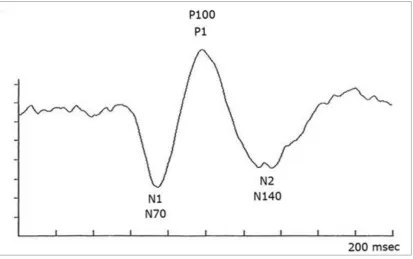
VEP measurement normally produces a series of waveforms in PC which
have negative and a positive component. The negative is N wave and
positive is takes as P wave. The parameters usually recorded are P100,
N70 and N155. Of these P100 is most important and it indicates latency
of positive wave. They were measured in microvolts. Statistical analysis
was done and p value was determined.
1. p > 0.05 (non significant)
2. p < 0.05 (significant)
WAVES IN VEP:
FIG 2: The time taken in milliseconds is marked in x-axis and the evoked
potentials in microvolt are marked in y-axis. A graph is obtained with a
positive peak PI00 and two negative peaks N70 and N155. The latency of
POSITIONING OF ELECTRODES
POSITIONING OF ELECTRODES
OBSERVATION AND RESULTS
AGE DISTRIBUTION
CHART-1: Shows 24 patients between 50 to 60 yrs, 7 patients between
40 to 50 years and 19 more than 60 yrs
7
24 19
SEX DISTRIBUTION
CHART-2: Shows 21 females and 29 males in study group
29 21
AGE VS P100
2 5 7
28.6% 71.4% 100.0%
22.2% 12.2% 14.0%
6 18 24
25.0% 75.0% 100.0%
66.7% 43.9% 48.0%
1 18 19
5.3% 94.7% 100.0%
11.1% 43.9% 38.0%
9 41 50
18.0% 82.0% 100.0%
100.0 100.0% 100.0%
Count
% within Age (Case) % within Case Count
% within Age (Case) % within Case Count
% within Age (Case) % within Case Count
% within Age (Case) % within Case Between 40 - 50 years
Between 50 - 60 years
AGE VS P100
CHART-3: Among 7 cases in 40 -50 years group 5 had prolonged P100.
In 50 -60 years group 18 had abnormal P100 and 6 had normal value.
Above 60 years all except one had abnormal P100
0 2 4 6 8 10 12 14 16 18
SEX VS P100
4 25 29
13.8% 86.2% 100.0%
44.4% 61.0% 58.0%
5 16 21
23.8% 76.2% 100.0%
55.6% 39.0% 42.0%
9 41 50
18.0% 82.0% 100.0%
100.0% 100.0% 100.0%
Count
% within Sex (Case)
% within Case P100 Count
% within Sex (Case) % within Case P100 Count
% within Sex (Case)
SEX VS P100
CHART-4: Among 29 males 25 had prolonged P100 and 4 had normal
value. In females 16 had P100 prolongation and 5 had normal P100.
0 5 10 15 20 25
Male Female
4 5
25
16
CASE VS CONTROL
CHART-5: Among 50 cases 41 had prolonged P100 and 9 had normal
P100 whereas in controls only one had prolonged P100 and rest 49 had
P100 VS HbA1c
CHART-6: Among 20 cases with well controlled DM 15 had P100
prolongation and 5 had normal P100, whereas among those with
P100 VS DURATION
CHART-7: Among those with DM of less than 5 years 8 had normal
P100 and 12 had abnormal value, whereas cases with DM more than 10
years all 20 patients had abnormal prolonged P100
0 2 4 6 8 10 12 14 16 18 20
<5 YRS 5 TO 10 >10 YRS
P100 VS PNP
CHART-8: Among cases with PNP 23 had prolonged P100 and 3 had
normal P100 whereas those without PNP 18 had prolonged P100 and 6
DISCUSSION
We had 50 cases of diabetes patients who fulfilled the inclusion
criteria after vigorously excluding many patients by history, clinical and
ophthalmological examination. VEP was done in these 50 patients as
well as 50 age and sex matched controls. In VEP there were one positive
peak (P100) and two negative peaks (N 70 AND N155). P100 is
produced by occipital striate cortex in response to stimulation of visual
cortex. P100 is the most prominent wave among all three and is easily
reproducible without much variation in an individual. The most ideal
parameter in VEP is latency as amplitude has greater variability and is
less reliable. Hence we measured P100 latency for all patients.
The mean age our population was 58.44. There were 29 males and
21 females. 7 cases were 40 to 50 years old, 24 between 50 to 60 years
and 19 more than 60 years. In our study P100 latencies (ms) was
significantly prolonged in diabetics with mean ± SD of (111.24 ± 5.28
ms) as compared to controls (101.30 ± 1.66 ms) with p value <0.003.
Among 50 cases 41 cases had prolonged P100 latency when compared to
controls only one had P100 prolongation which was statistically very
significant. Hence 81% of diabetic patients in our cases had central
neuropathy. We also noted that mean prolongation of P100 in cases was
We further divided the cases into two groups. Those with
uncontrolled DM with HbA1c > 7 and those with well controlled DM
with HbA1c < 7. Among the 30 cases in uncontrolled group 26 had P100
prolongation and in 20 cases in well controlled group 15 had prolonged
P100 latency. 75% of well controlled group and 86% of uncontrolled
group had P100 prolongation. There was no statistically significant
correlation between the two groups as p value was 0.293. Similarly we
looked into duration of DM and classified the cases into 3 groups as <5
years, 5 to 10 years and > 10 years. Among the 20 cases in <5 years
group 12 had abnormal P100 and in 5 to 10 year group 9 out of 10 had
abnormal P100. In > 10 year group all 20 had prolongation of P100
which was statistically significant (p value <0.03). Hence we noted
100% of cases with >10 year DM had abnormal VEP whereas only 60 %
had prolonged P100 in <5 year group.
We also analysed age of patients with P100 latency. We found that
5 out of 7 cases in 40 to 50 year group had abnormal P100. Likewise 18
out of 24 cases in 50 to 60 year group and 18 out of 19 cases in more
than 60 year group had prolongation of P100. 71 % of cases between 40
to 50 years, 75 % of cases in 50 to 60 year group and 94 % in more than
60 years group had central neuropathy. There was no statistical
peripheral neuropathy (PNP) with central neuropathy and classified the
cases into those with PNP and without PNP. Among 24 cases without
PNP 18 had prolonged P100 and 23 out of 26 had abnormal P100 in PNP
group. 75 % and 94 % of cases had prolonged P100 in the above groups.
There was no statistical significance and we found central neuropathy
occurring in almost equal percentage in patients with or without PNP.
Similar to our study Dolu H et al, Azal O et al, Szabela D et al, Li
P et al, Algan et al, and Comi G et al also concluded prolongation of
P100 in diabetic population in their studies. (20,25,23) But Szabela D et al
and Algan et al concluded there was no correlation between duration of
DM and P100 prolongation.(22) Ziegler et al and Li P et al summarized
that P100 prolongation correlated well with glycemic control of DM and
even improved with short term glycemic control. (19) We believe that
since the sample size was small in most studies and they also included
both type 1 and type 2 DM it produced varying results. Moreover in our
study 81% of cases had prolonged P100 whereas only 58%, 28 % and
33% of cases had P100 latency abnormality in above studies. In spite of
our strict exclusion criteria we produced the maximum percentage of
P100 prolongation. We also believe that inclusion and exclusion
criteria varied significantly between each studies resulting in varying
conclude that central neuropathy in DM correlates well with duration of
DM and not glycemic control.
At present the significance of P100 prolongation in diabetic
patients is not known. It may be due to functional disturbance in visual
conduction pathway rather than demyelination or axonal loss. It is also
possible that early diabetic preretinopathy due to retinal ganglion cell
loss may also contribute to P100 prolongation. Exact pathophysiology
for central neuropathy is not known. We suggest it may be multifactorial
like PNP both metabolic and vascular factors playing a role.
Accumulation of neuropoietic cytokines like TNF-alpha, TGF-beta in
visual conduction pathway probably causes delay in P100 latency. As
duration of DM increases further accumulation of mediators cause
SUMMARY
The mean age our population was 58.44
The P100 latencies (ms) was significantly prolonged in diabetics
with mean ± SD of (111.24 ± 5.28 ms) as compared to controls
(101.30 ± 1.66 ms) with p value <0.003.
In our study 81% of diabetic patients in our cases had central
neuropathy as evidenced by prolonged P100 latency.
In cases with DM > 10 years had prolongation of P100 which was
statistically significant (p value <0.03).
There was no statistically significant correlation between P100 and
HbA1c or age of patients.
75 % of cases without PNP had prolonged P100 and 94 % of cases
with PNP had prolonged P100. Although there was no statistical
LIMITATIONS
Although our sample size is largest when compared to other similar
studies we suggest still larger samples are required to validate the
findings in our study.
Many of type 2 DM patients who were above 60 years may have
CONCLUSION
Central neuropathy as measured by P100 latency is very common
in type 2 DM.
Similar to subclinical sensory neuropathy which is detected in
majority of DM by nerve conduction studies, subclinical central
neuropathy in DM can be detected by VEP.
It is related to duration of DM and not HbA1c unlike PNP which is
related to both.
Central neuropathy occurs even prior to development of
retinopathy or PNP.
VEP is a non invasive and sensitive screening tool to detect early
neurological involvement in DM.
Since there is a very high incidence of P100 prolongation in DM
patients its usefulness in evaluation of multiple sclerosis in a
BIBLIOGRAPHY
1. Adams R, Victor M. Brown R. et al. Special Techniques for
Neurologic Diagnosis, Adams and Victor's Principles of Neurology
- 8th Ed. The McGraw-Hill Companies, Inc.; 2005.
2. Simeon Locke. Nervous System in Diabetes. In Joslin’s Diabetes.
Lea and Febiger, Philadephia, 1971; 562-4.
3. Michael J.Fowler. Microvasular and macrovascular complications
of diabetes.2008;26:1-2.
4. Singh R, Barden A, Mori T, Beilin L. Advanced glycation end
products: a review. Diabetologia.2001; 44:129-146.
5. Meier M, King GL. Protein kinase C activation and its
pharmacological inhibition in vascular disease. Vasc Med.2000;
5:173-185.
6. Fong DS, Aiello LP, Ferris FL 3RD, Klein R: Diabetic retinopathy.
Diabetes care.2004; 27:2540-2553.
7. Gabbay KH. Hyperglycemia, polyol metabolism and complications
of diabetes mellitus. Annu Rev Med.1975; 26:521-536.
8. Boulton AJ, Vinik AI, Bril V, Freeman R et al: Diabetic
neuropathies: a statement by ADA. Diabetes Care. 2005;
9. Odom JV, Bach M, Barber C, et al. Visual evoked potentials
standard. Documenta Ophthalmologica. 2004;108(2):115–123.
10. Chiappa KH, Yiannikas C. Voluntary alteration of evoked
potentials. Ann Neurol 1982; 12:496.
11. Asselman P, Chadwick DW, Marsden CD. Visual evoked
responses in diagnosis and management of patients suspected of
multiple sclerosis. Brain 1975; 98: 261.
12. Bradley WG, Whitty WM. Acute optic neuritis: its clinical
features and their relation to prognosis for recovery of vision. J
Neurol Neurosurg Psychiat 1967; 30: 531.
13. Boghen DR, Glaser JS. Ischaemic optic neuropathy. Brain 1975;
98:689.
14. Carroll F. Etiology and treatment of tobacco alcohol amblyopia.
Am J Ophthalmol 1974; 47:713.
15. Daneshvar H, Racette L, Coupland SG, et al. Syptomatic and
asyptomatic visual loss in patients taking vigabatrin. Ophthalmol
1999; 106:1792.
16. Hennerici M. Dissociated foveal and parafoveal reponses in
17. Carroll WM, Kriss A, Baraitser M, et al. The incidence and nature
of visual pathway involvement in Friedrich Ataxia. Brain 1980;
103: 413.
18. Bodis-Wollner I. Recovery from cerebral blindness: evoked
potential and psychophysical measurements. Clin Neuro-physiol
1977; 42:178.
19. Ziegler O, Guerci B, Algan M, Lonchamp P, Weber M, Drouin P.
Improved visual evoked potential latencies in poorly control
diabetic patients after short-term strict metabolic control. Diabetes
Care 1994. 17:1 1 4 1 -7.
20. Dolu H, Ulas UH, Bolu E, Ozkardes A, Odabasi Z, Ozata M, et al.
Evaluation of central neuropathy in type II diabetes mellitus by
multimodal evoked potentials. Acta Neurol Belg.2003; 103:206-11.
21. Comi G. Evoked potentials in diabetes mellitus. Clinical
Neuroscience. 1997; 4:374.
22. Algan M, Ziegler O et al. Visual evoked potentials in diabetic
patients. Diabetes Care 12:227-9, 1989.
23. Szabela DA, Loba J, Pałenga-Pydyn D, Tybor K, Ruxer J, Split
W. Klin Oczna. 2005; 107(7-9):492-7.
24. Szabela DA, Loba J, Pałenga-Pydyn D, Tybor K, Ruxer J, Split
25. Azal O, Ozkardes A, Onde ME, Ozata M, Ozisik G, Corakc A, et
al. Visual Evoked Potentials in Diabetic Patients. Tr. J. of Medical
Sciences 1998; 28:139-42.
26. Mariani E, Moreo G, Colucci GB. Study of visual evoked
potentials in diabetics without retinopathy: correlations with
clinical findings and polyneuropathy. Acta Neurologica
Scandinavica. 1990;81(4):337–340.
27. Puvanendran K, Devathasan G, Wong PK. Visual evoked
responses in diabetes. J. Neurol Neurosurg Psychiatry. 1983;
46:643-7.
28. Yaltkaya K, Balkan S, Baysal AI. Visual evoked potentials in
diabetes mellitus. Acta Neurologica Scandinavica. 1988;
77(3):239–241.
29. World Health Organisation Department of Noncommunicable
Disease Surveillance (1999). "Definition, Diagnosis and
PROFORMA
NAME :
AGE :
SEX :
OCCUPATION :
IP NO/OP NO:
1. Do you have Diabetes? If yes, how long and treatment details?
2. Do you have Hypertension? If yes, how long and treatment details?
3. Do you smoke? If yes, how much per day?
4. Do you drink alcohol? If yes, how much per week?
5. Did you have previous attack of stroke?
6. Did you have previous eye problems? If yes, details of it?
7. Clinical peripheral neuropathy – yes or no
8. HbA1c –
9. Fundus –
PSG INSTITIUTE OF MEDICAL SCIENCES AND RESEARCH, Coimbatore
Institutional Human Ethics Committee
INFORMED CONSENT
I, Dr. E. PRASANNA VENKATESAN, postgraduate from the department of
Neurology of PSG Institiute Of Medical Sciences And Research (PSG IMS&R), am
carrying out a study on topic, “EVALUATION OF CENTRAL NEUROPATHY IN
TYPE 2 DIABETES CASE –CONTROL STUDY” to under the aegis of the
Department of Neurology, PSG IMS&R.
The objectives of this study are:
To compare the visual evoked potentials in type-2 Diabetes mellitus patients
with that of healthy controls.
To find out if there is any correlation with duration of DM or glycemic
control of Diabetes with P100 latency.
Sample size: 100
Respondents are patients with type 2 diabetes attending neurology OPD.
Consent: The above information regarding the study, has been read by me/read to me,
and has been explained to me by the investigator/s. Having understood the same, I
hereby give my consent to them to interview me and undergo VEP. I am affixing my
signature/left thumb impression to indicate my consent and willingness to participate
in this study.
Signature/left thumb impression of the study volunteer/legal representative:
MASTER CHART
CASES
S.NO PATIENTS
NAME AGE SEX HBA1C DURATION
RIGHT
P100
LEFT
P100 PNP
1 Si 58 M 9.2 20 115 114 YES
2 Ra 60 M 12.2 7 104 105 YES
3 Amu 50 F 7.3 3 102 101 NO
4 Ra 51 F 9.3 1 105 104 NO
5 Me 56 M 6.7 7 118 117 NO
6 Sh 55 F 8.3 1 106 104 NO
7 Vi 62 M 6.6 3 104 105 NO
8 Ku 64 M 7.5 1 110 111 NO
9 Na 51 M 6.9 3 112 111 YES
10 Ra 42 M 6.6 1 103 104 NO
11 Sh 53 M 10.1 2 112 111 YES
12 Dh 60 F 9.6 7 115 114 NO
13 Ku 53 F 7.1 2 106 107 NO
14 Po 57 M 7.8 10 118 117 YES
15 Am 65 F 10.1 5 111 110 YES
16 Fa 61 F 11.1 10 120 119 YES
17 Ka 50 F 7.6 10 116 117 NO
18 Ve 60 F 6.1 2 105 104 YES
19 Sg 64 M 10.1 10 116 116 YES
20 Je 60 M 6.2 11 120 119 NO
21 Su 70 M 7 12 116 115 YES
22 Sm 70 F 7.1 5 110 109 NO
23 Vl 52 F 11.3 13 112 113 NO
25 Rd 50 F 9.9 1 108 109 YES
26 Rg 60 M 14.4 1 106 107 YES
27 Ka 66 F 8.6 11 120 119 YES
28 Jl 57 M 9.9 15 119 120 YES
29 Sd 60 M 8.4 3 100 99 YES
30 Ar 66 M 10.1 30 118 117 YES
31 Ml 53 F 8.5 11 119 119 YES
32 Mk 61 M 7.5 15 117 117 YES
33 Rm 43 F 6.2 5 109 109 NO
34 PK 61 F 8.8 8 108 109 YES
35 Ch 66 M 8.6 12 117 118 YES
36 Py 63 M 9.9 6 110 110 NO
37 Mo 53 M 8.9 20 115 116 YES
38 Pl 62 M 10.8 7 110 109 NO
39 Rg 68 M 12.9 11 114 114 NO
40 Ra 60 M 11.1 11 116 116 YES
41 Sa 55 F 8.8 7 110 111 YES
42 Ar 49 M 9.7 9 111 111 NO
43 Ba 67 M 10.2 6 109 108 NO
44 Ch 59 M 12.2 2 106 107 YES
45 Ra 60 F 11.5 4 107 108 NO
46 Vl 57 F 7.6 1 103 104 NO
47 Ma 66 M 6.9 12 112 112 YES
48 Ng 50 M 7.9 16 113 114 NO
49 Gv 66 F 6.5 10 112 111 NO
CONTROLS
S.NO PATIENTS
NAME AGE SEX RIGHTP100 LEFT P100
1 Is 58 M 100 101
2 Ra 60 M 99 99
3 Um 50 F 100 100
4 Ka 51 F 102 101
5 Em 56 M 100 101
6 Ka 55 F 101 101
7 Gv 62 M 103 102
8 Ng 64 M 102 102
9 Ra 51 M 104 103
10 Ch 42 M 98 99
11 Pl 53 M 101 101
12 mo 60 F 103 102
13 Ph 53 F 98 99
14 Su 57 M 103 102
15 Sm 65 F 102 102
16 Vl 61 F 104 103
17 Gv 50 F 100 101
18 Rd 60 F 103 103
19 Rg 64 M 104 103
20 Ka 60 M 102 103
21 Jl 70 M 101 102
22 Sd 70 F 99 100
23 Ar 52 F 106 105
25 am 50 F 98 99
26 fa 60 M 99 100
27 ve 66 F 103 103
28 ch 57 M 104 104
29 py 60 M 100 101
30 mo 66 M 102 103
31 pl 53 F 100 101
32 rg 61 M 103 102
33 ra 43 F 99 100
34 sa 61 F 101 101
35 ar 66 M 104 103
36 ba 63 M 102 101
37 ch 53 M 100 99
38 ra 62 M 101 101
39 vl 68 M 99 100
40 ma 60 M 103 102
41 ng 55 F 99 98
42 gv 49 M 100 99
43 dh 67 M 101 100
44 ku 59 M 102 100
45 po 60 F 99 100
46 am 57 F 101 101
47 fa 66 M 102 102
48 ka 50 M 101 101
49 ve 66 F 103 102