Copyright © 2004, American Society for Microbiology. All Rights Reserved.
Complete DNA Sequence Analyses of the First Two Varicella-Zoster
Virus Glycoprotein E (D150N) Mutant Viruses Found in
North America: Evolution of Genotypes with an
Accelerated Cell Spread Phenotype
Charles Grose,
1* Shaun Tyler,
2Geoff Peters,
2Joanne Hiebert,
2Gwen M. Stephens,
3William T. Ruyechan,
4Wallen Jackson,
1Johnathan Storlie,
1and Graham A. Tipples
2National Microbiology Laboratory, Canadian Science Centre for Human and Animal Health, Winnipeg, Manitoba,2and University of British Columbia Virology and Reference Laboratory, St. Pauls Hospital, Vancouver, British Columbia,3
Canada; Department of Microbiology, University at Buffalo, Buffalo, New York4; and Department of Pediatrics, University of Iowa, Iowa City, Iowa1
Received 16 January 2004/Accepted 30 March 2004
Varicella-zoster virus (VZV) is considered to be one of the most genetically stable of all the herpesviruses. Yet two VZV strains with a D150N missense mutation within the gE glycoprotein were isolated in North America in 1998 and 2002. The mutant strains have an accelerated cell spread phenotype, which distinguishes them from all wild-type and laboratory viruses. Since the VZV genome contains 70 additional open reading frames (ORFs), the possibility existed that the phenotypic change was actually due to an as-yet-undiscovered mutation or deletion elsewhere in the genome. To exclude this hypothesis, the entire genomes of the two mutant viruses were sequenced and found to contain 124,883 (VZV-MSP) and 125,459 (VZV-BC) nucleotides. Coding single-nucleotide polymorphisms (SNPs) were identified in 14 ORFs. One missense mutation was discovered in gH, but none was found in gB, gI, gL, or gK. There were no coding SNPs in the major regulatory protein ORF 62. One polymorphism was discovered which could never have been anticipated based on current knowledge of herpesvirus genomics, namely, the origins of replication differed from those in the prototype strain but not in a manner expected to affect cell spread. When the two complete mutant VZV sequences were surveyed in their entirety, the most reasonable conclusion was that the increased cell spread phenotype was dependent sub-stantially or solely on the single D150N polymorphism in glycoprotein gE. The genomic results also expanded the evolutionary database by identifying which VZV ORFs were more likely to mutate over time.
The primordial varicella-zoster virus (VZV) arose around 65 million years before the present (39). Thereafter, VZV cospe-ciated first in primates and more recently in humans (40). Indeed, the two manifestations of VZV infection, namely chicken pox and zoster (shingles), may have evolved based on the exigencies of survival in small human populations through the millennia (19). The primary infection chicken pox would allow spread through a small community, after which the virus would enter a latent state in the dorsal root ganglia of all inhabitants for several decades. When one of the elderly de-veloped zoster, the virus would infect all the more-recently born nonimmune people, who in turn would contract chicken pox followed by a latent infection, lasting for several more decades.
The general characteristics of these two diseases are so sim-ilar around the world that the assumption emerged that VZV itself was genetically stable around the world. So widespread was this assumption that VZV virologists generally accepted the complete VZV DNA sequence, as published by Davison and Scott in 1986 (11), as the actual sequence for all VZV laboratory strains in common use in the 1980s and 1990s. Of
note, very few laboratories used the only sequenced strain, VZV Dumas, because of the assumption of genetic similarity among all strains. Even though different laboratory strains were used, there was in general a concordance in published data about the characterization of major VZV gene products. It was unexpected, therefore, to discover a variant VZV strain in 1998 in a child with chicken pox living in the state of Minnesota (61). This strain, called VZV-MSP, contains a mis-sense mutation in the predominant VZV glycoprotein gE (for-merly designated gp98 or gpI). In cell culture, this strain ex-hibits both an accelerated cell spread phenotype as well as a different egress pattern distinguishable from all previously ex-amined wild-type or laboratory strains (60). In human genetics, a genetic polymorphism that leads to a new phenotype in the affected human is called a mutation, as opposed to a variant. Therefore, VZV-MSP has the attributes of a mutant VZV strain. Perhaps even more surprising than the discovery of the first mutant VZV strain was the subsequent discovery of a second mutant VZV strain in Vancouver, British Columbia, Canada (68). Since the same gE mutation was observed in the VZV-BC strain, the D150N substitution can be considered an authentic single-nucleotide polymorphism (SNP). Now that two mutant VZV strains have been isolated in North America, the fundamental question immediately arose, namely, are the two viruses representative of a previously unknown VZV
sub-* Corresponding author. Mailing address: University Hospital/2501 JCP, 200 Hawkins Dr., Iowa City, IA 52242. Phone: (319) 356-2270. Fax: (319) 356-4855. E-mail: [email protected].
6799
on November 8, 2019 by guest
http://jvi.asm.org/
group in circulation among small human populations in North America? If so, how aberrant are the genomes of these mutant viruses, viz., are there other major mutations elsewhere in the genome that may contribute to the altered phenotype and, if so, are the two viruses similarly mutated?
For all the above reasons, the decision was made to se-quence the complete genomes of both MSP and VZV-BC. This undertaking led to several interesting observations about VZV genomics. Importantly, this report shows that ge-netic diversity occurs in sites that could not have been pre-dicted without sequencing the entire VZV genome and there-fore would have remained undiscovered.
MATERIALS AND METHODS
Viruses and their titration.VZV-MSP was recovered from a child in Minne-sota in late 1995; he had chicken pox (61). VZV-BC was isolated from a 75-year-old adult in Vancouver, British Columbia, Canada, in 2000; he had shingles (68). Both were compared with VZV-Dumas; this virus was isolated from a chicken pox patient in The Netherlands in the late 1970s by Anna Maria Dumas (13). She stored passage four aliquots of this virus for all subsequent studies. Since this virus was the first to be sequenced, VZV-Dumas is considered to be the prototype for European and North American strains of VZV (11). The Asian reference strain is VZV-Oka, which was isolated from a child with chicken pox in Japan in the 1970s and attenuated by multiple passage in cultured cells by Michiaki Takahashi (17). Plaque titrations to measure infectious centers in this study were performed by methods described elsewhere (20).
Virus propagation and genome purification.The VZV-MSP and VZV-BC isolates were propagated for six to eight passages in cultured human melanoma cells (Mewo strain) in Dulbecco’s modified Eagle’s medium (Gibco, Burlington, Ontario, Canada) supplemented with 5% fetal bovine serum, 1% nonessential amino acids, and 1% sodium pyruvate (20). Viral nucleocapsids were isolated from infected Mewo cells exhibiting advanced cytopathic effect, and VZV genomic DNA was extracted by previously described methods (59).
DNA sequencing strategies.Instrumentation included the ABI 377 and ABI 3100 DNA sequencers. Sequencing of VZV-MSP was carried out using a shotgun approach. The purified viral DNA was sheared to produce a random distribution of smaller fragments (generally 1 to 3 kb in length), which were then cloned into a plasmid vector. A recombinant library was generated which contained repre-sentative fragments spanning the entire genome. Random library clones were end sequenced using standard primers flanking the cloning site. Sequencing was continued until three to five genome equivalents of sequence data had been generated. The data were then assessed using Staden sequencing software to determine which plasmid clones would likely close gaps if more data were available (64). Selected clones were then resequenced using both additional primers to further extend the data from specific clones as well as PCR primers to span gaps to generate hybridization probes for screening the library for clones likely to yield missing data. Sequencing of VZV-BC was carried out using the custom primer approach. An EcoRI-restricted VZV genomic library was gener-ated. PCR primers were designed to produce hybridization probes specific for the different VZV EcoRI fragments and were used to screen the library. In addition, a panel of custom sequencing primers were designed based on the known sequence of VZV-Dumas (10) (accession number NC001348). These primers were appropriately spaced (500 bp) to obtain complete coverage of the various fragments. PCR primers were also designed to generate amplicons span-ning the EcoRI junction sites and the junction of the genome during its repli-cative phase.
Sequence analysis.The complete DNA sequences of Dumas, VZV-MSP, and VZV-BC were aligned and compared using MegAlign (clustal V algorithm; DNAStar, Madison, Wis.) (64). Any sequence variations that were detected were confirmed by resequencing the region with custom primers.
Surface plasmon resonance.Kinetic binding analyses between mouse mono-clonal antibody (MAb) 3B3 and its epitope were performed on the Biacore 3000 apparatus with the CM5 carboxy methyl dextran chip (6, 62). The respective 20-mer peptides bearing the wild-type and mutated epitopes were synthesized: IVNVDQRQYGDVFKGDLNPK and IVNVNQRQYGDVFKGDLNPK. Each was immobilized using standard amine coupling methods on separate flow cells of the same chip. With Biaevaluation 3.1 software, sensorgram data were
gen-erated from five separate concentrations of MAb: 60, 30, 15, 7.5, and 3.7g/ml.
Each concentration was run simultaneously over wild-type, mutant, and two
nonspecific control flow cells on a single CM5 chip. Total binding at each concentration was subtracted from nonspecific binding to obtain specific binding curves for each concentration. Specific binding curves for each of five concen-trations of MAb were merged onto a single timeline using Biaevaluation soft-ware. The on and off rates of antibody for peptide were determined by the bivalent analyte model, and the equilibrium constants were calculated from the on and off rates.
Nucleotide sequence accession numbers.GenBank accession numbers for the two sequences are the following: VZV-MSP, AY548170; and VZV-BC, AY548171.
RESULTS
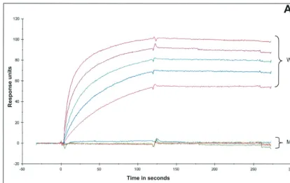
The VZV gE glycoprotein.The genetic diversity of the VZV gE gene has been a subject of speculation since the publication of the first D150N mutation in 1998 (61). The likelihood of finding a second identical mutation was considered remote. Therefore, the isolation 3 years later of VZV-BC with the same D150N coding SNP in VZV gE came as a surprise (68). As noted previously, the D residue is a critical component of the MAb 3B3 epitope. The substitution of D with an N sub-stantially reduced the amount of immunostaining, as gauged by inspection under laser scanning confocal microscopy (60). In order to quantify with precision the loss of reactivity by the murine MAb, the affinity of MAb 3B3 to its epitope with either a D or an N residue was measured by surface plasmon reso-nance with a Biacore apparatus. For comparison, the binding curves for wild-type and mutant peptides at five different MAb concentrations were merged onto a single timeline (Fig. 1A and B). The data showed a marked reduction in affinity of MAb 3B3 for the mutated epitope. For example, the associa-tion rate was 152 times greater for the wild-type peptide, while the dissociation rate was nearly 10 times greater for the mutant peptide. The equilibrium association constant, therefore, was 1,400 times greater for the wild-type peptide.
Viruses containing the gE(D150N) glycoprotein have other attributes. Generally, under the usual conditions of VZV in-fection of cultured cells, the mutant viruses exhibited a pro-gression of cytopathic effect about 30% faster than that of the typical wild-type virus (Fig. 1C). Based on data in herpes sim-plex virus type 1 (HSV-1) studies (12), the presumption was that the gE mutation was likely responsible for the altered VZV phenotype. Yet there was a reasonable alternative hy-pothesis, namely, that an as-yet-undiscovered alteration in the VZV-MSP genome (deletion, duplication, or addition) was responsible for the faster cell spread phenotype. At the present time, there is insufficient information about wild-type VZV genomics upon which to base any conclusion. To explore this hypothesis, the entire genomes of VZV-MSP and VZV-BC were sequenced and annotated. The mutant genomes con-tained 124,883 and 125,459 nucleotides, respectively.
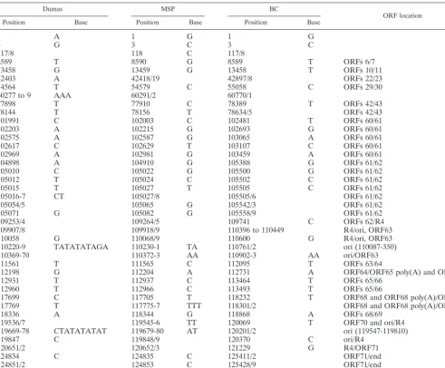
Other VZV glycoproteins.The gE glycoprotein forms a com-plex with gI (ORF 67) (18). There were no polymorphisms noted in gI (Table 1). There was a coding SNP, however, in the ectodomain of the gH protein (ORF 37), namely, a proline-to-leucine change in residue 269 (Table 1). The P-to-L substi-tution has been observed in numerous European strains and also in Asian strains (70). One of the strains not having this SNP is VZV-VSD, a North American strain extremely closely related to VZV-Dumas (14). However, it is again important to note that none of the other strains with this gH SNP has a concomitant gE mutation.
on November 8, 2019 by guest
http://jvi.asm.org/
FIG. 1. Characterization of the mutated epitope and the mutant virus. (A) Biacore specific binding curves. Using BIAevaluation 3.1 software, sensorgram data from five separate concentrations of 3B3 MAb injected over bound wild-type (WT) epitope synthetic peptide (top five curves) or bound mutant (MT) synthetic peptide (bottom five curves) were merged onto a single timeline. (B) Concentrations of 3B3 MAb used in the experiment shown in panel A. To achieve specific binding, the binding from a control chip was subtracted from total binding shown by either the WT or MT synthetic peptide. The kinetic and equilibrium constants were determined using the Biacore technology as described in Materials and Methods. (C) Infectious center assay of WT and MT virus. Infectious center assays were carried out by previously described methods (8, 20). The numbers of infectious centers present at 24 and 48 h postinfection were determined for WT and MT viruses. The average increase of infectious centers at each interval was tabulated.
on November 8, 2019 by guest
http://jvi.asm.org/
No polymorphism was observed in the gH chaperone pro-tein gL (ORF 60) (Table 1). A second potential start site has been found in gL in several Asian strains, but in none of the European strains (70). Likewise, there were no polymorphisms in gK (ORF 5) or gB (ORF 31) (42). However, polymorphisms were observed in gC (ORF 14). VZV gC is important for viral tropism for skin cells (43). This glycoprotein is unusual in that it contains an in-frame repeating structure of 14 amino acids near the amino terminus. The entire VZV genome contains five sets of these repeating elements, and the set within gC is R2 (11). The R2 repeats consist of tandem arrays of 42-bp sequences, followed by a final 32-bp sequence. The number of R2 repeating sequences can vary from 3 to 14, but most strains have between 6 and 9. VZV-Dumas and VZV-BC contain seven repeats, ABABAAA and ABBBBAA, respectively, while VZV-MSP contains eight repeats, AAABBBBB (Tables 2 and 3).
Five VZV repeating elements.In addition to the R2 in ORF 14, there are four additional repeating sequences in the VZV genome (11, 26). R1 and R3 occur within genes 11 and 22, respectively. R4 is located within the repeated regions of the unique short portion of the genome, while R5 is found within the right-hand portion of the unique long segment, as previ-ously noted (26). The R1 repeating sequences consisted of varying arrangements of 18-bp elements and 15-bp elements followed by 3-bp elements; the R1 regions were different in all three strains (Table 3). The R3 region included multiple copies of 9-bp elements, in frame within ORF 22. As with R2, differ-ences were observed in all three strains (Table 3). Moreover, these differences in R3 sequences largely accounted for the difference in genome size between VZV-MSP and VZV-BC. The R4 region is made up of variable numbers of a 27-bp element.
In contrast to results with R1 through R3, VZV-Dumas and VZV-MSP shared the same R4 elements, while VZV-BC con-tained an additional two 27-bp inserts. R5 contains tandem
arrays of an 88-bp element followed by a 24-bp element. Among the three strains, VZV-MSP and VZV-BC had an identical R5 structure, but that of VZV-Dumas was different. Prior investigators compared the stability of the R1 and R5 repeats from viruses after passages 12 to 14 and 41 to 43. They observed little or no difference and concluded that R1 and R5 are very stable during multiple passages (26, 32). In addition, others have noted that R2 and R3 may change after 85 pas-sages in cell culture, but no changes were detected after only 10 passages (21, 22).
VZV enzymes.Like all alphaherpesviruses, VZV encodes a thymidine kinase gene (ORF 36). Thymidine kinase catalyzes the phosphorylation of thymidine to thymidylate (67). Similar to its HSV counterpart, this enzyme is dispensable in VZV grown in cultured cells. However, the enzyme is essential for effective antiviral therapy, because the thymidine kinase is re-sponsible for the first phosphorylation step of the nucleoside analog acyclovir. In patients receiving acyclovir treatment for severe VZV infection, acyclovir-resistant mutant viruses have arisen, although less frequently than HSV resistant viruses. Because both patients with gE(D150N) viruses had received antiviral treatment, there was a possibility of mutation in the thymidine kinase gene. Both gE mutant viruses had an amino acid substitution of serine to leucine at position 288 of ORF 36 (Table 1). This polymorphism has been observed in several VZV strains and did not alter the ability of the VZV thymidine kinase to function in phosphorylation (45, 49). The ORF 36 protein, therefore, did not have a mutation known to be asso-ciated with acyclovir resistance.
[image:4.603.47.539.80.306.2]A coding polymorphism was observed in the large subunit of the ribonucleotide reductase (ORF 19), which catalyzes the reduction of ribonucleotides to deoxyribonucleotides (Table 1) (25). The polymorphism led to a K-to-E substitution. There is insufficient mutational analysis of ORF 19 to determine the effect of the SNP. The small subunit of the ribonucleotide reductase is encoded by ORF 18. No polymorphisms were
TABLE 1. Coding polymorphisms within VZV ORFs
Dumas MSP BC
ORF no. Amino acidchange
Position Base Position Base Position Base
14117 to 14149 14118 to 14120 14117 to 14120 11 Deletion
20753 T 20709 A 20708 T 14, R2 T to S
20795 A 20751 A 20750 T 14 S to T
20879 A 20835 T 20834 T 14, R2 T to S
20921 A 20877 T 20876 A 14 S to T
20963 A 20919 T 20918 A 14 S to T
21017/8 20974 to 21015 20972/3 14 Insertion
28836 T 28834 T 28791 C 19 K to E
33732 A 33730 A 33687 G 21 K to E
40660 T 40658 T 40615 C 22 V to A
41485 C 41483 T 41440 C 22 A to V
41486 G 41484 C 41441 G 22 A to V
63448 T 63460 T 63939 C 34 T to A
65669 C 65681 T 66160 T 36 S to L
66879 C 66891 T 67370 T 37 P to L
69756 T 69768 T 70247 C 38 S to G
78698 C 78710 T 79188 C 43 R to C
90202 G 90214 T 90692 T 51 H to Q
99186 T 99198 T 99676 C 56 Y to H
99789 T 99801 T 100279 C 58 I to V
116255 G 116261 A 116788 A 68 D to N
on November 8, 2019 by guest
http://jvi.asm.org/
observed in this ORF. The sequences of the two VZV serine/ threonine protein kinases, ORF 47 and ORF 66, were also examined because of their importance to the virus life cycle (44, 48). However, no coding SNPs were detected.
The IE62 major regulatory protein. As with all herpesvi-ruses, VZV genes are grouped into three temporal classes: immediate early (IE), early, and late. The first set of genes to
[image:5.603.48.540.81.487.2]be expressed are the IE or alpha genes, which are predomi-nantly involved in regulation of viral transcription. An under-lying hypothesis of this project was that the gE mutation ac-counted for the altered phenotype. An alternative explanation is that a regulatory event enhanced viral replication. VZV IE62 is the major viral transcriptional regulator, an even-more-dom-inant player in viral gene regulation than its HSV-1 homolog,
TABLE 2. Polymorphisms within VZV intergenic regions
Dumas MSP BC
ORF location
Position Base Position Base Position Base
1 A 1 G 1 G
3 G 3 C 3 C
117/8 118 C 117/8
8589 T 8590 G 8589 T ORFs 6/7
13458 G 13459 G 13458 T ORFs 10/11
42403 A 42418/19 42897/8 ORFs 22/23
54564 T 54579 C 55058 C ORFs 29/30
60277 to 9 AAA 60291/2 60770/1
77898 T 77910 C 78389 T ORFs 42/43
78144 T 78156 T 78634/5 ORFs 42/43
101991 C 102003 C 102481 T ORFs 60/61
102203 A 102215 G 102693 G ORFs 60/61
102575 A 102587 G 103065 A ORFs 60/61
102617 C 102629 T 103107 C ORFs 60/61
102969 A 102981 G 103459 A ORFs 60/61
104898 A 104910 G 105388 G ORFs 61/62
105010 C 105022 G 105500 G ORFs 61/62
105012 T 105024 C 105502 C ORFs 61/62
105015 T 105027 T 105505 C ORFs 61/62
105016-7 CT 105027/8 105505/6 ORFs 61/62
105054/5 105065 G 105542/3 ORFs 61/62
105071 G 105082 G 105558/9 ORFs 61/62
109253/4 109264/5 109741 C ORFs 62/R4
109907/8 109918/9 110396 to 110449 R4/ori, ORF63
110058 G 110068/9 110600 G R4/ori, ORF63
110220-9 TATATATAGA 110230-1 TA 110761/2 ori (110087-350)
110369-70 110372-3 AA 110902-3 AA ori/ORF63
111561 T 111565 C 112095 T ORFs 63/64
112198 G 112204 A 112731 A ORF64/ORF65 poly(A) and ORF65
112931 T 112937 C 113464 T ORFs 65/66
112960 T 112966 C 113493 T ORFs 65/66
117699 C 117705 T 118232 T ORF68 and ORF68 poly(A)/ORF69
117769 T 117775-7 TTT 118301/2 ORF68 and ORF68 poly(A)/ORF69
118336 A 118344 G 118868 A ORFs 68/69
119536/7 119545-6 TT 120069 T ORF70 and ori/R4
119669-78 CTATATATAT 119679-80 AT 120201/2 ori (119547-119810)
119847 C 119848/9 120370 C ori/R4
120651/2 120652/3 121229 G R4/ORF71
124834 C 124835 C 125411/2 ORF71/end
124851/2 124853 C 125428/9 ORF71/end
124880-2 AGA 124882-4 AGG 125457 G ORF71/end
TABLE 3. VZV reiterations R1 through R5
Region Repeating sequence
VZV-Dumas VZV-MSP VZV-BC
R1 ABBABBCBBCBBBBABDAB ABBABBABBABBABAB ABBABBABBABDABAB
R2 ABABAAAx AAABBBBBx ABBBBAAx
R3 AAAAABAx ABABABABAx AAAABAABAAAAABAx
R4 AAAAAx AAAAAx AAAAAAAx
R5a ABA(4T, 61G, 72A) ABA(4T, 61G) ABA(4T, 61G)
aFor R5, A⫽88 bases (4G, 61A, and 72G) and B⫽24 bases. Davison and Scott (11) defined R5 as an 88-bp repeat (which they termed A), then a 24-bp repeat
(no name), followed by another 88-bp repeat which they again termed A, although it differed from the first A in the three positions noted above.
on November 8, 2019 by guest
http://jvi.asm.org/
[image:5.603.45.542.625.709.2]ICP4 (7, 52, 53). In HSV-1, VP16 is a potent alpha gene transducing factor. In the VZV system, the VP16 homolog called ORF 10 lacks the acidic domain required for much of its transactivating activity. VZV IE62, unlike HSV ICP4, pos-sesses a potent N-terminal acidic activation domain and can activate its own promoter, thereby playing a similar role to VP16 regarding its own expression. Furthermore, VZV IE62 can transactivate promoters within all three temporal classes. In this sequence analysis, two IE62 polymorphisms were un-covered but neither led to an amino acid change. The first was a C-to-G change in both VZV-MSP and VZV-BC. This change has been observed in all VZV IE62 genes sequenced to date by our laboratory (14, 70). The second was a G-to-C change in VZV-BC only. Otherwise stated, at an amino acid level the IE62 proteins of all three viruses were identical. Since ORF 62 is located in the short repeat sequences, it has a duplicate gene, ORF 71. ORF 62 and ORF 71 were identical in each of the mutant VZV strains.
The promoter region of IE62.As part of the same
hypoth-esis, we also examined thecis-acting sequences that control the
expression of IE62 (Table 2) (46, 57). If the expression of IE62 were up regulated, one consequence would be an altered growth phenotype. The promoter region of IE62 has been investigated in several studies (41, 54). The TATA box in VZV-Dumas consists of the sequence T TTT AA located 97 bp from the first methionine. The same TATA box was found in the same region of both the VZV-MSP and VZV-BC ge-nomes. A string of eight G nucleotides was present at 111 bp, and further upstream two octamer motifs, ATG TAAA TGAAAT and TTTTG CAT, were identified. Again, these motifs and their positions in the sequences upstream of the IE62 coding region in the two mutant viruses were identical to those found in VZV-Dumas (29).
VZV proteins involved in viral replication.The VZV genes involved in viral replication include ORF 6 (primase; HSV UL52), ORF 16 (polymerase processivity factor; HSV UL42), ORF 28 (polymerase catalytic subunit; HSV UL30), ORF 29 (DNA binding; HSV UL29), ORF 51 (origin binding; HSV UL9), ORF 52 (helicase; HSV UL8), and ORF 55 (helicase; HSV UL5). When these sequences were examined in the two mutant viruses, all were identical to those in VZV-Dumas, with the exception of the origin binding protein ORF 51 (31, 58). The latter protein contained a missense mutation at VZV-Dumas nucleotide 90,202, resulting in an H-to-Q substitution (Table 1). In general, however, the viral replication proteins were not a site of genetic divergence within the two VZV gE(D150N) mutant viruses.
Short genome segment and viral origins of replication.The VZV unique short region of the genome and its inverted re-peat sequences contain genes 62 to 71 (10). The diploid genes within the inverted sequences include genes 62, 63, and 64, as well as their duplicates 71, 70, and 69, respectively. When all regions of the sequenced VZV-MSP and VZV-BC genomes were examined and compared to that of VZV-Dumas, a pro-pensity for intergenic polymorphisms was observed in the unique short segment plus the adjacent ORF 61-ORF 62 in-tergenic region in the UL segment (Tables 1 and 2). With regard to the ORF 61 promoter region, all four intergenic nucleotide changes in VZV-MSP and VZV-BC occurred out-side the previously described TATA and CATT elements. In short, when compared to the VZV-Dumas strain, there were mainly single nucleotide substitutions, additions, and deletions within the short segment of the genome (Table 2). However, with regard to the signature coding SNP in the two mutant viruses, no changes were found in the promoter regions pre-ceding either ORF 67 (gI) or ORF 68 (gE) (28, 37).
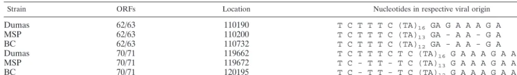
The VZV genome contains two origins of DNA replication, both of which are resident in the repeat regions flanking the short region. They are located in the intergenic regions be-tween ORFs 62 and 63 and bebe-tween ORFs 70 and 71 (65, 66). Unlike HSV-1, VZV does not have a third origin of replication in the long segment of the genome. The VZV ori sequences contain a core element consisting of a palindrome with a cen-tral TA dinucleotide repeat. Based on the overall high level of relatedness between the DNA sequences of Dumas and the mutant strains already described in Results, it was of consid-erable surprise and interest that there were obvious nucleotide differences between the origins of replication for the three viruses (Table 4). Whereas VZV-Dumas contains 16 TA dinucleotides, in VZV-MSP this stretch was shortened to 13 and in VZV-BC it was just 12. Within each origin palindrome, the nucleotides upstream and downstream of the dinucleotide sequence also were variable between the North American strains and VZV-Dumas, including identical nucleotide dele-tions just downstream of the TA repeat in the ori flanked by ORFs 62 and 63 and a different pair of deletions occurring upstream of the TA repeat in the ori flanked by ORFs 70 and 71 (Table 4).
DISCUSSION
[image:6.603.41.549.82.156.2]The VZV gE(D150N) mutant VZV strains have the at-tributes of increased cell-to-cell spread in cultured cells, as well as rapid destruction of an infected human skin explant in the
TABLE 4. VZV strain DNA replication origin polymorphismsa
Strain ORFs Location Nucleotides in respective viral origin
Dumas 62/63 110190 T C T T T C (TA)16 GA G A A A G A
MSP 62/63 110200 T C T T T C (TA)13 GA - A A - G A
BC 62/63 110732 T C T T T C (TA)12 GA - A A - G A
Dumas 70/71 119662 T C T T T C T C (TA)16 G A A A G A A A
MSP 70/71 119672 T C - T T - T C (TA)13 G A A A G A A A
BC 70/71 120195 T C - T T - T C (TA)12 G A A A G A A A
aThe VZV ori lies between the designated ORFs; the location indicates the first nucleotide in each sequence.
on November 8, 2019 by guest
http://jvi.asm.org/
SCID-hu mouse model and a novel egress pattern observed with scanning electron microscopy (60, 61). VZ virions typi-cally emerge onto the cell surface in a pattern called viral highways, whereas the mutant viruses egress uniformly over the surface in a pattern similar to that of HSV-1 and HSV-2. When taken together, these properties allow the gE mutant viruses to be distinguished from all other VZV strains. The effects of temperature and cell type were investigated further in several cell-to-cell spread experiments (8). At both 32 and 37°C, VZV-MSP spread faster than wild-type VZV. This effect was markedly greater in epidermal cells than in fibroblast cells. When the combined effects of cell spread, incubation temper-ature, and virus strain (wild type or gE mutant) were subjected to an analysis of variance, a statistically significant two-way
interaction was evident (P⬍0.001). Namely, the magnitude of
the effects of both temperature and virus strain varied signifi-cantly as a function of cell type. Otherwise stated, the gE mutant virus spread faster and the magnitude of the cell spread phenotype was dependent on both temperature and cell type. The question as to why this gE mutation occurred was dif-ficult to address when only a single mutant virus had been found. Because the likelihood of a VZV mutation historically has been considered rare, the possibility was often mentioned that the mutation may have occurred by chance in a single patient and that we discovered the mutant virus by serendipity. The isolation of the second mutant virus at a geographically distant site lessens the likelihood of chance; in other words, there is no known connection between the two individuals in Minnesota and British Columbia, other than northern Euro-pean ancestry (68). Rather, we raise the possibility that the gE mutation represents positive selection. The mutation has oc-curred in a surface glycoprotein exposed to the humoral im-mune response of any human infected with chicken pox or zoster. Furthermore, in humans with VZV infection, the gE glycoprotein is one of the earliest and strongest immunogens (4, 18, 35). As proven by the Biacore binding data in this
report, the missense mutation almost completely (⬃90%)
eliminates an epitope defined by murine MAb 3B3; this epitope is involved in both complement-dependent neutraliza-tion and antibody-dependent cytotoxicity (50). Under this sce-nario, the SNP may represent an antibody escape mutant virus. Similar mutations occur commonly in surface glycoproteins of RNA viruses, such as influenza A virus and measles virus (72). Although T-cell immunity appears pivotal for ultimately recov-ering from a herpesvirus infection, B-cell immunity may mod-ulate the ability of virus to attach to and subsequently infect new cells, for example, as virus exits the viremic phase and enters the epidermis late in the incubation period (1). Thus, VZV-MSP and VZV-BC may represent gE escape mutants which arose independently within isolated human populations in North America. To date, gE sequences from more than 150 additional strains have been published, and no additional D150N polymorphisms have been uncovered (55, 63, 70).
There is a second possible scenario based on the glycopro-tein sequence data. Rather than solely involving gE, the changed virus phenotype may be due to concomitant mutations in two VZV surface proteins, namely, gH along with gE. The mutation in gH is a proline-to-leucine substitution at residue 269 in the ectodomain. It has long been known that any proline mutation in a protein may be important, because of the
fre-quent and often major changes to secondary structure. How-ever, consequences of a proline-to-leucine substitution (or vice versa) have been especially well documented in numerous pro-teins over the past 20 years. A computer-assisted search of this mutation uncovered over 500 published papers, ranging from diverse genetic disorders in humans to numerous examples of variant microorganisms, including the prion protein (2, 3, 5, 15, 27, 33, 69). In all of these cases, the mutation was associated with a change in phenotype or biologic activity, such as altered receptor function. Of parenthetical interest, even one of the VZV thymidine kinase mutations leading to acyclovir resis-tance is an L92P substitution (34). If a leucine or proline mutation involves a receptor, the binding affinity of ligand to receptor is usually reduced. Although we have previously doc-umented the same P269L substitution in the gH glycoprotein in several VZV strains, in addition to MSP and VZV-BC, we have not observed an increased cell spread phenotype in any gH variant strain containing the prototype gE glycopro-tein (60). In the VZV system, gE (or the gE/gI complex) appears to be primarily responsible for cell-to-cell spread, while gH (or the gH/gL complex) is the major fusogen. What is postulated, therefore, is a linkage between the gH P269L mutation and the gE D150N mutation, leading to a subsequent accelerated cell spread phenotype in the mutant viruses. This hypothesis seems even more reasonable now that a functional interaction has been documented between the two VZV sur-face glycoproteins (51).
Identification of the obvious differences in the DNA repli-cation of origin sequences among the Dumas and MSP and BC strains was a striking finding which could not have been antic-ipated based on limited available genomic data about the sta-bility of the herpesvirus origin sequence. The VZV minimal origin contains a palindromic sequence in which a variable-length TA repeat is flanked upstream and downstream by TC-rich and GA-rich sequences, respectively. The second ele-ment making up the minimal VZV ori sequence is a binding site for the VZV origin-specific binding protein encoded by ORF 51, located just upstream of the palindrome (65, 66). It is believed, by analogy with HSV-1 (23, 24, 36), that initiation of DNA replication involves site-specific binding by the ORF 51 gene product and possibly binding of other viral or cellular factors downstream of the palindrome, followed by unwinding and denaturation of the TA repeat with the aid of the ORF 29 product and subsequent recruitment of the remainder of the replication proteins (58). Based on current understanding, therefore, the two nucleotide deletions observed in the se-quences flanking the TA repeats would likely not have a major effect on the positioning of the transacting factors involved in initiation of replication and thus not affect efficiency of that process in either a positive or negative manner. Therefore, a rapid cell-to-cell spread phenotype is unlikely to result even in part from these ori polymorphisms.
Finally, with regard to the broader picture of VZV evolu-tion, it is now clear that VZV genomes can be split into major geographic clades on the basis of SNP analysis: they include Asian and European-North American strains (47, 55, 71). These clades presumably represent viruses cospeciating with humankind during the great migrations out of Africa into Asia and Europe. For example, when VZV-MSP and VZV-BC are placed in a phylogenetic tree, it is immediately apparent that
on November 8, 2019 by guest
http://jvi.asm.org/
they are very closely related to VZV-Dumas, but they are not closely related to VZV-Oka or other Asian strains located on a different branch (17, 70). In the future, based on DNA analyses conducted by different laboratories, it should be pos-sible to predict which of the 71 VZV genes will be subject to more genetic variation over time (9, 14, 16, 38, 63). Presum-ably, other VZV mutant viruses will be found. Certainly what is most remarkable of all is that these extensive VZV genomic data emphasize again that relatively few SNPs within structural proteins of evolutionarily closely related VZV strains can lead to major phenotypic changes in the social behavior of the mutated viruses (30, 56).
ACKNOWLEDGMENTS
We acknowledge the pivotal contributions of Andrew Davison in his report of the first complete VZV DNA sequence. We thank Sytske Welling-Wester for comments about VZV strains and Michael Gray for technical assistance.
This research was supported in part by NIH grants AI22795 (C.G.) and AI18449 (W.T.R.), as well as a grant from the Office of the Chief Scientists, Health Canada (G.A.T.).
REFERENCES
1. Abendroth, A., and A. M. Arvin.2000. Host response to primary infection, p.
142–156.InA. M. Arvin and A. A. Gershon (ed.), Varicella zoster virus.
Cambridge University Press, Cambridge, England.
2. Adriouch, S., C. Dox, V. Welge, M. Seman, F. Koch-Nolte, and F. Haag.2002. Cutting edge: a natural P451L mutation in the cytoplasmic domain impairs
the function of the mouse P2X7 receptor. J. Immunol.169:4108–4112.
3. Anwar, R., L. Gallivan, C. Trinh, F. Hill, and A. Markham.2001. Identifi-cation of a new Leu354Pro mutation responsible for factor XIII deficiency.
Eur. J. Haematol.66:133–136.
4. Arvin, A. M., and A. A. Gershon.1996. Live attenuated varicella vaccine.
Annu. Rev. Microbiol.50:59–100.
5. Barron, R. M., V. Thomson, E. Jamieson, D. W. Melton, J. Ironside, R. Will, and J. C. Manson.2001. Changing a single amino acid in the N-terminus of murine PrP alters TSE incubation time across three species barriers. EMBO
J.20:5070–5078.
6. Choulier, L., G. Orfanoudakis, P. Robinson, D. Laune, M. Ben Khalifa, C. Granier, E. Weiss, and D. Altschuh.2002. Comparative properties of two peptide-antibody interactions as deduced from epitope delineation. J.
Im-munol. Methods259:77–86.
7. Cohen, J. I., D. Heffel, and K. Seidel.1993. The transcriptional activation domain of varicella-zoster virus open reading frame 62 protein is not
con-served with its herpes simplex virus homolog. J. Virol.67:4246–4251.
8. Cole, N. L., and C. Grose.2003. Membrane fusion mediated by herpesvirus
glycoproteins: the paradigm of varicella-zoster virus. Rev. Med. Virol.13:
207–222.
9. Davison, A. J.2000. Molecular evolution of alphaherpesviruses, p. 25–50.In
A. M. Arvin and A. A. Gershon (ed.), Varicella zoster virus. Cambridge University Press, Cambridge, England.
10. Davison, A. J., C. M. Edson, R. W. Ellis, B. Forghani, D. Gilden, C. Grose, P. M. Keller, A. Vafai, Z. Wroblewska, and K. Yamanishi.1986. New com-mon nomenclature for glycoprotein genes of varicella-zoster virus and their
glycosylated products. J. Virol.57:1195–1197.
11. Davison, A. J., and J. E. Scott.1986. The complete DNA sequence of
varicella-zoster virus. J. Gen. Virol.67:1759–1816.
12. Dingwell, K. S., and D. C. Johnson.1998. The herpes simplex virus gE-gI complex facilitates cell-to-cell spread and binds to components of cell
junc-tions. J. Virol.72:8933–8942.
13. Dumas, A. M., J. L. Geelen, M. W. Weststrate, P. Wertheim, and J. van der Noordaa.1981. XbaI, PstI, and BglII restriction enzyme maps of the two
orientations of the varicella-zoster virus genome. J. Virol.39:390–400.
14. Faga, B., W. Maury, D. A. Bruckner, and C. Grose.2001. Identification and mapping of single nucleotide polymorphisms in the varicella-zoster virus
genome. Virology280:1–6.
15. Golding, G. B., and C. Strobeck.1982. Expected frequencies of codon use as
a function of mutation rates and codon fitnesses. J. Mol. Evol.18:379–386.
16. Gomi, Y., T. Imagawa, M. Takahashi, and K. Yamanishi.2000. Oka varicella vaccine is distinguishable from its parental virus in DNA sequence of open
reading frame 62 and its transactivation activity. J. Med. Virol.61:497–503.
17. Gomi, Y., H. Sunamachi, Y. Mori, K. Nagaike, M. Takahashi, and K. Ya-manishi.2002. Comparison of the complete DNA sequences of the Oka
varicella vaccine and its parental virus. J. Virol.76:11447–11459.
18. Grose, C.2002. The predominant varicella-zoster virus gE and gI
glycopro-tein complex, p. 183–211.InE. Bogner and A. Holzenburg (ed.), Structure
function relationships of human pathogenic viruses. Kluwer Academic Press, London, England.
19. Grose, C.1999. Varicella-zoster virus: less immutable than once thought.
Pediatrics103:1027–1028.
20. Grose, C., and P. A. Brunell.1978. Varicella-zoster virus: isolation and propagation in human melanoma cells at 36 and 32 degrees C. Infect.
Immun.19:199–203.
21. Hawrami, K., D. Harper, and J. Breuer.1996. Typing of varicella zoster virus
by amplification of DNA polymorphisms. J. Virol. Methods57:169–174.
22. Hayakawa, Y., T. Yamamoto, K. Yamanishi, and M. Takahashi.1986. Anal-ysis of varicella-zoster virus DNAs of clinical isolates by endonuclease HpaI.
J. Gen. Virol.67:1817–1829.
23. He, X., and I. R. Lehman.2001. An initial ATP-independent step in the unwinding of a herpes simplex virus type I origin of replication by a complex of the viral origin-binding protein and single-strand DNA-binding protein.
Proc. Natl. Acad. Sci. USA98:3024–3028.
24. He, X., and I. R. Lehman.2000. Unwinding of a herpes simplex virus type 1
origin of replication (OriS) by a complex of the viral origin binding protein
and the single-stranded DNA binding protein. J. Virol.74:5726–5728.
25. Heineman, T. C., and J. I. Cohen.1994. Deletion of the varicella-zoster virus large subunit of ribonucleotide reductase impairs growth of virus in vitro.
J. Virol.68:3317–3323.
26. Hondo, R., and Y. Yogo.1988. Strain variation of R5 direct repeats in the right-hand portion of the long unique segment of varicella-zoster virus DNA.
J. Virol.62:2916–2921.
27. Hsiao, K., H. F. Baker, T. J. Crow, M. Poulter, F. Owen, J. D. Terwilliger, D. Westaway, J. Ott, and S. B. Prusiner.1989. Linkage of a prion protein
missense variant to Gerstmann-Straussler syndrome. Nature338:342–345.
28. Ito, H., M. H. Sommer, L. Zerboni, H. He, D. Boucaud, J. Hay, W. Ruyechan, and A. M. Arvin.2003. Promoter sequences of varicella-zoster virus glyco-protein I targeted by cellular transactivating factors Sp1 and USF determine
virulence in skin and T cells in SCIDhu mice in vivo. J. Virol.77:489–498.
29. Kantakamalakul, W., W. T. Ruyechan, and J. Hay.1995. Analysis of
vari-cella-zoster virus promoter sequences. Neurology45:S28–S29.
30. Keller, J. M., P. G. Spear, and B. Roizman.1970. Proteins specified by herpes simplex virus. 3. Viruses differing in their effects on the social behav-ior of infected cells specify different membrane glycoproteins. Proc. Natl.
Acad. Sci. USA65:865–871.
31. Kinchington, P. R. and J. I. Cohen.2000. Viral proteins, p. 74–104.InA. M. Arvin and A. A. Gershon (ed.), Varicella zoster virus. Cambridge University Press, Cambridge, England.
32. Kinoshita, H., R. Hondo, F. Taguchi, and Y. Yogo.1988. Variation of R1 repeated sequence present in open reading frame 11 of varicella-zoster virus
strains. J. Virol.62:1097–1100.
33. Klinkhamer, M. P., N. A. Groen, G. C. van der Zon, D. Lindhout, L. A. Sandkuyl, H. M. Krans, W. Moller, and J. A. Maassen.1989. A leucine-to-proline mutation in the insulin receptor in a family with insulin resistance.
EMBO J.8:2503–2507.
34. Lacey, S. F., T. Suzutani, K. L. Powell, D. J. Purifoy, and R. W. Honess.1991. Analysis of mutations in the thymidine kinase genes of drug-resistant vari-cella-zoster virus populations using the polymerase chain reaction. J. Gen.
Virol.72:623–630.
35. LaRussa, P. S., A. A. Gershon, S. P. Steinberg, and S. A. Chartrand.1990. Antibodies to varicella-zoster virus glycoproteins I, II, and III in leukemic
and healthy children. J. Infect. Dis.162:627–633.
36. Lehman, I. R., and P. E. Boehmer.1999. Replication of herpes simplex virus
DNA. J. Biol. Chem.274:28059–28062.
37. Ling, P., P. R. Kinchington, M. Sadeghi-Zadeh, W. T. Ruyechan, and J. Hay.
1992. Transcription from varicella-zoster virus gene 67 (glycoprotein IV).
J. Virol.66:3690–3698.
38. Loparev, V. N., T. Argaw, P. R. Krause, M. Takayama, and D. S. Schmid.
2000. Improved identification and differentiation of varicella-zoster virus (VZV) wild-type strains and an attenuated varicella vaccine strain using a
VZV open reading frame 62-based PCR. J. Clin. Microbiol.38:3156–3160.
39. McGeoch, D. J., and A. J. Davison.1999. The molecular evolutionary history
of the herpesviruses, p. 441–466.InE. Domingo, R. G. Webster, and J. J.
Holland (ed.), Origin and evolution of viruses. Academic Press, London, England.
40. McGeoch, D. J., A. Dolan, and A. C. Ralph.2000. Toward a comprehensive
phylogeny for mammalian and avian herpesviruses. J. Virol.74:10401–10406.
41. McKee, T. A., G. H. Disney, R. D. Everett, and C. M. Preston.1990. Control of expression of the varicella-zoster virus major immediate early gene.
J. Gen. Virol.71:897–906.
42. Mo, C., J. Suen, M. Sommer, and A. Arvin.1999. Characterization of vari-cella-zoster virus glycoprotein K (open reading frame 5) and its role in virus
growth. J. Virol.73:4197–4207.
43. Moffat, J. F., L. Zerboni, P. R. Kinchington, C. Grose, H. Kaneshima, and A. M. Arvin.1998. Attenuation of the vaccine Oka strain of varicella-zoster virus and role of glycoprotein C in alphaherpesvirus virulence demonstrated
in the SCID-hu mouse. J. Virol.72:965–974.
44. Moffat, J. F., L. Zerboni, M. H. Sommer, T. C. Heineman, J. I. Cohen, H.
on November 8, 2019 by guest
http://jvi.asm.org/
Kaneshima, and A. M. Arvin.1998. The ORF47 and ORF66 putative protein kinases of varicella-zoster virus determine tropism for human T cells and
skin in the SCID-hu mouse. Proc. Natl. Acad. Sci. USA95:11969–11974.
45. Morfin, F., D. Thouvenot, M. De Turenne-Tessier, B. Lina, M. Aymard, and T. Ooka.1999. Phenotypic and genetic characterization of thymidine kinase from clinical strains of varicella-zoster virus resistant to acyclovir.
Antimi-crob. Agents Chemother.43:2412–2416.
46. Moriuchi, H., M. Moriuchi, and J. I. Cohen.1995. Proteins andcis-acting elements associated with transactivation of the varicella-zoster virus (VZV) immediate-early gene 62 promoter by VZV open reading frame 10 protein.
J. Virol.69:4693–4701.
47. Muir, W. B., R. Nichols, and J. Breuer.2002. Phylogenetic analysis of varicella-zoster virus: evidence of intercontinental spread of genotypes and
recombination. J. Virol.76:1971–1979.
48. Ng, T. I., and C. Grose.1992. Serine protein kinase associated with
varicella-zoster virus ORF 47. Virology191:9–18.
49. Ng, T. I., Y. Shi, H. J. Huffaker, W. Kati, Y. Liu, C. M. Chen, Z. Lin, C. Maring, W. E. Kohlbrenner, and A. Molla.2001. Selection and character-ization of varicella-zoster virus variants resistant to
(R)-9-[4-hydroxy-2-(hy-droxymethy)butyl]guanine. Antimicrob. Agents Chemother.45:1629–1636.
50. Padilla, J. A., and C. Grose.2001. Varicella-zoster virus with a lost gE epitope: evidence for immunological pressure by the human antibody
re-sponse. Arch. Virol. Suppl.17:27–39.
51. Pasieka, T., L. Maresova, K. Shiraki, and C. Grose.2004. Regulation of varicella-zoster virus induced cell-to-cell fusion by the endocytosis
compe-tent glycoproteins gH and gE. J. Virol.78:2884–2896.
52. Peng, H., H. He, J. Hay, and W. T. Ruyechan.2003. Interaction between the varicella zoster virus IE62 major transactivator and cellular transcription
factor Sp1. J. Biol. Chem.278:38068–38075.
53. Perera, L. P., J. D. Mosca, W. T. Ruyechan, G. S. Hayward, S. E. Straus, and J. Hay.1993. A major transactivator of varicella-zoster virus, the immediate-early protein IE62, contains a potent N-terminal activation domain. J. Virol.
67:4474–4483.
54. Perera, L. P., J. D. Mosca, M. Sadeghi-Zadeh, W. T. Ruyechan, and J. Hay.
1992. The varicella-zoster virus immediate early protein, IE62, can positively
regulate its cognate promoter. Virology191:346–354.
55. Quinlivan, M., K. Hawrami, W. Barrett-Muir, P. Aaby, A. Arvin, V. T. Chow, T. J. John, P. Matondo, M. Peiris, A. Poulsen, M. Siqueira, M. Takahashi, Y. Talukder, K. Yamanishi, M. Leedham-Green, F. T. Scott, S. L. Thomas, and J. Breuer.2002. The molecular epidemiology of varicella-zoster virus:
evidence for geographic segregation. J. Infect. Dis.186:888–894.
56. Ruyechan, W. T., L. S. Morse, D. M. Knipe, and B. Roizman.1979. Molec-ular genetics of herpes simplex virus. II. Mapping of the major viral glyco-proteins and of the genetic loci specifying the social behavior of infected
cells. J. Virol.29:677–697.
57. Ruyechan, W. T., H. Peng, M. Yang, and J. Hay.2003. Cellular factors and
IE62 activation of VZV promoters. J. Med. Virol.70(Suppl. 1):S90–S94.
58. Ruyechan, W. T., and J. Hay.2000. DNA replication, p. 51–73.InA. M.
Arvin and A. A. Gershon (ed.), Varicella zoster virus. Cambridge University Press, Cambridge, England.
59. Safronetz, D., A. Humar, and G. A. Tipples.2003. Differentiation and quan-titation of human herpesviruses 6A, 6B and 7 by real-time PCR. J. Virol.
Methods112:99–105.
60. Santos, R. A., C. C. Hatfield, N. L. Cole, J. A. Padilla, J. F. Moffat, A. M. Arvin, W. T. Ruyechan, J. Hay, and C. Grose.2000. Varicella-zoster virus gE escape mutant VZV-MSP exhibits an accelerated cell-to-cell spread
pheno-type in both infected cell cultures and SCID-hu mice. Virology275:306–317.
61. Santos, R. A., J. A. Padilla, C. Hatfield, and C. Grose.1998. Antigenic variation of varicella zoster virus Fc receptor gE: loss of a major B cell
epitope in the ectodomain. Virology249:21–31.
62. Saunal, H., and M. H. Van Regenmortel.1995. Mapping of viral
conforma-tional epitopes using biosensor measurements. J. Immunol. Methods183:
33–41.
63. Shankar, V., S. Fisher, B. Forghani, and A. Vafai.2001. Nucleotide sequence analysis of varicella-zoster virus glycoprotein E epitope coding regions.
Vac-cine19:3830–3833.
64. Staden, R., K. F. Beal, and J. K. Bonfield.1998. Computer methods in
molecular biology, p. 115–130.InS. Misener and S. A. Krawetz (ed.),
Bioin-formatics methods and protocols. Humana Press Inc., Totowa, N.J. 65. Stow, N. D., and A. J. Davison.1986. Identification of a varicella-zoster virus
origin of DNA replication and its activation by herpes simplex virus type 1
gene products. J. Gen. Virol.67:1613–1623.
66. Stow, N. D., H. M. Weir, and E. C. Stow.1990. Analysis of the binding sites for the varicella-zoster virus gene 51 product within the viral origin of DNA
replication. Virology177:570–577.
67. Suzutani, T., L. C. Davies, and R. W. Honess.1993. Kinetic studies of the predicted substrate-binding site of varicella-zoster virus thymidine kinase.
J. Gen. Virol.74:1011–1016.
68. Tipples, G. A., G. Stephens, C. Sherlock, M. Bowler, B. Hoy, D. Cook, and C. Grose.2002. New variant of varicella-zoster virus. Emerg. Infect. Dis.
8:1504–1505.
69. Tsuji, S., P. V. Choudary, B. M. Martin, B. K. Stubblefield, J. A. Mayor, J. A. Barranger, and E. I. Ginns.1987. A mutation in the human
glucocerebro-sidase gene in neuronopathic Gaucher’s disease. N. Engl. J. Med.316:570–
575.
70. Wagenaar, T. R., V. T. Chow, C. Buranathai, P. Thawatsupha, and C. Grose.
2003. The out of Africa model of varicella-zoster virus evolution: single nucleotide polymorphisms and private alleles distinguish Asian clades from
European/North American clades. Vaccine21:1072–1081.
71. Wagenaar, T. R., C. Grose, V. N. Loparev, D. S. Schmid, and J. Breuer.2003. Genomic analysis of varicella-zoster virus: primers for individual open
read-ing frames. J. Clin. Virol.28:104–110.
72. Woelk, C. H., O. G. Pybus, L. Jin, D. W. Brown, and E. C. Holmes.2002. Increased positive selection pressure in persistent (SSPE) versus acute
mea-sles virus infections. J. Gen. Virol.83:1419–1430.