1
STUDY OF CLINICAL, SONOLOGICAL
AND HISTOPATHOLOGICAL
CORRELATION OF OVARIAN TUMOR
DISSERTATION SUBMITTED FOR
M.D (BRANCH – II)
(OBSTETRICS & GYNAECOLOGY)
APRIL 2013
THE TAMILNADU
DR.M.G.R. MEDICAL UNIVERSITY
2
CERTIFICATE
This is to certify that this dissertation titled ―STUDY OF CLINICAL,
SONOLOGICAL AND HISTOPATHOLOGICAL CORRELATION OF
OVARIAN TUMOR‖ submitted by DR. AISHWARYA JAGAN to the faculty of
Obstetrics and Gynecology, The TamilNadu Dr. M.G.R. Medical University,
Chennai in partial fulfillment of the requirement for the award of MD degree
Branch II Obstetrics and Gynecology, is a bonafide research work carried out by
her under our direct supervision and guidance from September 2011 to August
2012.
Dr.UMA, M.D.,D.G.O., Dr.P. ANGAYARKANNI M.D.(O.G), DCH,
Professor of Obstetrics and Professor and Head of the Department,
Gynecology, Department of Obstetrics and
Department of Obstetrics and Gynecology,
Gynecology, Madurai Medical College,
Madurai Medical College, Madurai.
3
DECLARATION
I, Dr. AISHWARYA JAGAN solemnly declare that the dissertation titled
―STUDY OF CLINICAL, SONOLOGICAL AND HISTOPATHOLOGICAL
CORRELATION OF OVARIAN TUMOR‖ has been prepared by me. This is
submitted to The Tamilnadu Dr. M.G.R. Medical University, Chennai, in
partial fulfillment of the regulations for the award of MD degree (Branch II)
Obstetrics & Gynaecology. I also declare that this bonafide work has not been
submitted in part or full by me or any others for any award, degree or diploma to
any other university or board either in India or abroad.
.
Place: Madurai Dr. AISHWARYA JAGAN.
4
ACKNOWLEDGEMENT
I owe my thanks to The Dean Dr. N. MOHAN M.S., Madurai Medical
College for allowing me to avail the facilities needed for my dissertation.
I am deeply indebted to DR. P. ANGAYARKANNI, M.D. (O.G.), D.C.H.,
Professor and Head of the Department of Obstetrics and Gynaecology, Madurai
Medical College, Madurai, for her able guidance, inspiration and the
encouragement she rendered at every stage of this study.
I am very grateful to my Former Professor and Head of Department
Dr. S. Dilshath M.D., D.G.O., and Prof. Dr. Uma M.D., D.G.O., for their
valuable guidance in conducting and completing the study. I express my gratitude
to other Professors, Dr. Ambigaimeena, M.D., D.G.O, Dr. S. Geetha, M.D.,
D.G.O., Dr. T. Uma Devi, M.D., D.G.O. and Dr. Revwathy Kailairajan, M.D.,
D.G.O, Department of Obstetrics and Gynaecology for allowing me and helping
me in conducting my study in their respective units.
I extend my heartful thanks to Dr. Usha Ravikumar, M.D., Professor and
Head of the Department of Pathology, Dr. N. Sundari M.D.R.D., Professor and
Head of Department of Radiology and Dr. Jayaraman D.M.R.D., for their
5
I thank all my Assistant Professors for their kind co-operation in helping
me to do this study.
Last but not the least I gratefully acknowledge my thanks to all my
Colleagues and the co-operation of the patients without whom this study would not
6
CONTENTS
S.NO. TITLE PAGE
1. INTRODUCTION 1
2. AIM AND OBJECTIVES 3
3 REVIEW OF LITERATURE 4
HISTORICAL ASPECTS 9
ANATOMY OF OVARY 11
PATHOLOGY OF OVARIAN TUMORS 19
4. MATERIALS AND METHODS 51
5. RESULTS 54
6. DISCUSSION 68
7. SUMMARY 80
8. CONCLUSION 83
ANNEXURES
Bibliography
Proforma
Master Chart
Ethical Committee Approval Certificate
7
LIST OF CHARTS
NO. TITLE
1 Incidence of Benign, Borderline and Malignant tumors as per HPE
2 Mean age distribution
3 Parity distribution
4 Per abdomen consistency of Ovarian Tumor
5 Laterality
6 USG Volume Score
7 USG Structural Score
8 Morphological score
9 Results as per USG total morphological score and HPE
10 Comparison of Sensitivity and Specificity with other
8
LIST OF TABLES
Table No. Title Page No.
1 Incidence of Benign, Borderline and Malignant
tumors as per HPE
54
2 Mean age distribution 55
3 Parity distribution 56
4 Mode Of Presentation 57
5 Per abdomen consistency of Ovarian Tumor 58
6 Laterality 59
7 USG Volume Score 60
8 USG Structural Score 61
9 Total Morphological Score 62
10 Results as per USG Total Morphological Score and HPE
63
11 Efficacy of Morphological Score with HPE as Gold Standard
63
12 Morphological Score and Malignancy 65
13 Types of tumor 66
14 Comparison of incidence of Ovarian Tumors 68
15 Comparison of age distribution of benign Ovarian Tumors
69
16 Comparison of age distribution of malignant Ovarian Tumors
70
17 Comparison Of Sensitivity And Specificity With Other Studies
9
LIST OF PHOTOGRAPHS
Figure No. Title
1 Cells of Ovary
2 Histiogenesis of Ovarian tumors
3 Serous Cyst Adenoma
4 Papillary Serous Cyst Adenocarcinoma
5 Mucinous Cystadenoma
6 Papillary Mucinous Cyst Adenocarcinoma
7 Endometrioid Tumor
8 Malignant Brenner Tumor
9 Fibrothecoma
10 Granulosa Cell tumor
11 Benign Cystic Teratoma
12 Immature Teratoma
13 Krukenbergs Tumor
14 Ovarian Tumor complicating pregnancy
15 Morphological scoring System
16 Borderline Mucinous Ovarian Tumor
17 Leiomyosarcoma of ovary
18 Structural score
18a Smooth wall, Sonolucent
18b Smooth wall, Diffuse echogenicity
18c Wall thickening, <3mm fine septa
18d Papillary projections, septa> 3mm
18e Complex, predominantly solid
18f Complex, solid and cystic areas
10
INTRODUCTION
Ovary is a very important organ as it is concerned with reproduction.The
ovary consists of mesenchymal cells which are multipotential and sex cells which
are totipotent.So their stimulation may result in any type of tumor.1, 2
Of all gynaecological cancers, ovarian malignancies representsthe greatest
clinical challenge because of greater range and variety of tumors with uncertain
origin, with no known premalignant lesion and variability in the rate of disease
progression.1 Around 70% of patients with ovarian tumors are diagnosed only at
advanced stages due to unavailability of effective screening method and lack of
specific clinical presentations at early stage of the disease.
Ovarian cancer is the eighth leading cause of cancer and fifth leading cause
of cancer related death in females. Every year 204,000 women are diagnosed to
have ovarian cancer and almost 125,000 women die due to ovarian cancer
worldwide.1
The histiogenesis of ovarian tumor revolves around four main components,
mainly surface epithelium, germ cells, sex cord and specialized ovarian stroma.2
Ovarian tumors can occur in all ages, but there are differences in the
11
during younger age group arethe germ cell tumors. Sex cord stromal tumours
occurs in women of all ages.
In premenopausal women 7% of tumors are frankly malignant and 30 % of
themin postmenopausal women are malignant.
90 to 95% of malignant tumorsof the ovary are Surface Epithelial Ovarian
cancers and the remaining 5 to 10% constitute the Sex Cord Stromal tumors and
the Germ cell tumors of the ovary.
Ultrasonography is used extensively to differentiate benign and malignant
tumors of ovary.
This study is on the OVARIAN TUMOR MORPHOLOGICAL
INDEXING – a quantitative analysis relating tumor morphology from
12
AIM AND OBJECTIVES
1. To analyse the sensitivity and specificity of a morphological
scoring system in differentiating benign and malignant tumors of
the ovary.
13
REVIEW OF LITERATURE
Ovarian cancer is the second most common of all gynaecological cancers
and accounts for 10 – 15 % of gynaecological malignancies in developing
countries including India.
Ovarian cancer remains as the leading cause of all cancer related deaths
among gynaecological malignancies in United States. The incidence of tumors of
ovary is highest in Sweden (19.6/100,000) and the United States (15.4/100,000)
and lowest in Japan (10.1/100,000).3
Sonography is considered to be the investigation of choice for the evaluation
of ovarian tumors due of its high sensitivity, acceptability, and low cost.Campbell
et al in 1989 was the first to propose the use of sonography for ovarian cancer
screening.4He reported a high correlation coefficient of 97% between the
ultrasonographically detected ovarian volumes and the actual ovarian volume
measured after oophorectomy.
Several studies have been reported to evaluate the benefits of morphologic
scoring systems, in quantifying and standardizing the interpretation of sonographic
14
Granberg et al in 1989 concluded that the ultrasound is highly reliable in
predicting the characteristics of an ovarian cyst.5They reported that the most
predictive characteristic feature of malignancy in an ovarian cyst is the papillary
projections seen on the inner surface of the cyst wall.
Sassone et al in 1991, suggested an index with four different morphologic
features of ovarian cyst architecture, which includes wall structure, septations, cyst
wall thickness and echogenicity.6 He gave a discrete score for each specific
character and was evaluated on 143 women with ovarian tumors. The
morphological index thus obtained was 100% sensitive and 83% specific in the
distinguishing benign tumor from that of a malignant one.
DePriest et al in 1994 recommended a morphologic index system, with only
3 structural characteristics - cyst wall, ovarian volume and septae.7This was
analysed on 213 women with ovarian tumors. The sensitivity was 89% and
specificity was only 70% in predicting ovarian cancer according to his study.
Lerner etalin 1994 used the Sassone classification system and defined the
relative importance of the structural components by a multiple regression analysis.8
Lerneretal appliedan individual score to each of the morphological components by
simplifying the indices. The new scoring system was evaluated in 350 women with
15
Ferrazzi et al in 1997 designed a new scoring system that was evaluated on
330 women with ovarian masses.9According to the study the sensitivity of this
scoring system was 87% and the specificity was 67%.
In 2001 Mol et al conducted an external validation of different
morphological indices including those by Lerner et al, Granberg et al, Finkler et
al, De Priest et al, Sassone et al and Ferrazzi et al.10Mol et al conducted a study on
170 ovarian tumor to compare the predictive accuracy of these scoring systems.
They reported a lower accuracy than the original scoring indices, with a high
sensitivity ranging from 77% to 93% and specificity ranging from 21% to 89 %.
Ueland and collegues in 2003 evaluated the Morphology indexing and
concluded it to be an inexpensive and accurate method in differentiating benign
ovarian tumors and malignant ovarian tumors, it can be used as an effective tool to
plan the management of ovarian tumors.11 The useof Doppler flow studies along
with this morphological indexing have not shown to improve the diagnostic
accuracy of morphological indexing.
Persistent ovarian masses and cysts studied in the Hirosaki and Kentucky
trials showed a very low likelihood ratio for malignancy tumors - 9.4% in
Kentucky trial and 7.0% in Hirosaki trial. By applying the morphological scoring
16
the reported a high sensitivity ranging from 80% to 90% with a positive predictive
value of > 20%.
Since 1980s, the color Doppler flow sonography was considered as an
important tool for the predictor of malignancy in ovarian tumors because Doppler
flow gives indirect information on the metabolism and direct information on the
vascular anatomy of the ovaries.
Hata et al 1999, studied about the colour Doppler assessment of intratumoral
blood flow in tumors of the ovary.12
According to The National Cancer Data Base Report on Ovarian Cancer,the
risks for ovarian cancers were associated with age, nulliparity and a family history
of the ovarian tumors. They reported that the women‘s lifetime risk of developing
cancer of ovaryis 1 in 70 (1.4%). Women who have a family history of one or
more affected first-degree relatives will have an increase in risk upto 5% - 7%. It is
been reported that only 3% -9% of the women with family history will actually
manifest hereditary cancer syndromes.
Approximately 90% of malignant ovarian tumors in adult are of epithelial
17
Shruti et al in 2008 studied on the incidence and management of ovarian
tumours in 75 cases and reported that 90% of ovarian tumours were of epithelial
origin, 75% were benign serous type, 20% of them were mucinous and 2% were
endometriod type.14
In 2010, GG Swamy et al did a study on 120 cases and concluded that 71.6%
were benign, 25% were malignant and 3% were borderline tumors.15 About two
third of all benign neoplasms was seen in patient between 20 to 40 years age,
whereas two third of all malignant neoplasms were seen after the age of 40 yrs.
The youngest patient in the study conducted by Swamy et al was 12 years of age
18
HISTORICAL ASPECTS
Herophilus first described the mammalian ovaries, he called it female testis.
Soranus describes the ovary as didymus [twin]. Vesalius and De Fabrica of
1543 labelled ovary as the testis of the uterus. Vesalius, first described the ovarian
follicle and corpus luteum.
In 1666, Janswammer dam explained that ―the human female testis are
comparable to ovaria of birds‖.
Esmond long in 1761, described all the common tumours of ovary.
Leonardo Da Vinci drew accurately the anatomy of the uterus and ovaries.
Carl von Rokintansky of Vienna, his name is attached to the Rokitansky
protuberance of dermoid tumour, also called ―dermoid mamilla‖, ―dermoid
protuberance‖ and ―embryonic node‖.
F.Von Werdt proposed the term ―granulosa cell tumour‖ in 1914.
Friedrich Ernst krukenerg described a ―fibrosarcoma ovarii mucocellular
carcinomatodes‖ in 1896 under the mistaken impression that the lesion was
primary malignancy of ovary. In 1902 Schlagenhauter corrected the error and
19
Struma ovary was first described by Ludwig pick in 1903.
Fritz brenners in 1907 published paper describing 3 cases of Brenner
tumour, which now bear his name.
The term ―Adenoacanthoma‖ was coined by Malcolm Dockerty of the Mayo
clinic in 1954.
The first histological description as germ cell tumours was given by M.
Cherassu in 1906.
Robert mayor first described the mixed and less differentiated forms of
tumours which produce viriliszing effect and named as ―Adreoblastoma‖, in 1930
it was replaced by ―Arrhenoblastoma‖.
In 1936 Joe Vincetmeigs presented 7 cases of syndrome of ovarian fibroma,
ascitie and hydrothorax. In 1937, Rhoads and Terrell suggested the eponym
20
ANATOMY OF OVARY
The ovaries arenodular ovoid structures, located on eachside of the uterus. It
is related to the lateral pelvic wall and attached to the posterior layer of the broad
ligament, posteroinferior to the fallopian tubes.
They are pink in colour with a smooth surface in young adults. With
advancing age they become more greyish with a puckered and scarred surface due
to repeated ovulation.They areabout 3 cm long, 1.5 cm wide. The thickness of the
ovary is approximately 1cm. The weight of each ovary ranges from 2 - 3.5 gm.16
Each ovary has 2 ends – uterine and tubal end. There are two borders – free
border and mesoovarium. They have two surfaces – lateral and medial.
RELATIONS:
The upper end is also known as the tubal end. It gives attachment to the
fimbrial end of the tubes. It is also attached to the infundibulopelvic ligament. The
lower end is also known as the uterine end. It gives attachment to the ovarian
ligament.3.16
The free border is also called the posterior border. Upper part of free border
is related with the uterine tube while it is related to the ureter and internal iliac
21
attached to the posterior leaf of the broad ligament. A cleft in this border called the
hilum, transmits the ovarian nerves and vessels.
The medial surface of the ovary is related to the fimbrial end of the uterine
tube. The lateral surface is convex and lies on the peritoneal depression of the
lateral pelvic wall, called the ovarian fossa. The lateral surface is related to the
obturator nerves and vessels.
BLOOD SUPPLY:
The ovarian artery is the main arterial supply of the ovary. Ovarian artery
arises directly from the abdominal aorta and enters the ovary through the
infundibulo pelvic ligament, mesoovarium and mesosalphinx. The ovaries are also
supplied by uterine artery.16,25
The pampiniform plexus of veins coming out of the hilum form a single vein
called the ovarian vein. The left ovarian vein drains into left renal vein and right
ovarian vein drains into inferior venacava.
LYMPHATIC DRAINAGE:
22
NERVE SUPPLY:
Twigs from the aortic plexus pass around the ovarian artery forms the
ovarian plexus. Ovarian plexus consists of sympathetic fibres from T10- T11 and
parasympathetic fibres from the vagus.16,25
1. The ovarian surface epithelium ( germinal epithelium) forms the outermost
layer
2. The cortex is covered by the tunica albuginea, which is lined by germinal
epithelium of waldeyer that consist of a layer of cuboidal cells.
3. The ovarian cortex consists of stroma and ovarian follicles.2,25
a. Ovarian follicle : Contains within it the membrana granulosa,
cumulus oophorus, the corona radiata, primary oocyte and the zona
pellucida. The zona pellucida, antrum, theca of follicle and liquor
folliculi are also included within the follicle.
b. Stromal tissue: The oocytes are included within the stromal tissue,
they are derived from the mesenchymal cells and are made up of
connective tissue and interstitial cells. They have the ability to
respond to hormones like leutinizing hormone or human chorionic
23
4. The inner most layer is the medulla. The medulla and cortex cannot be
distinctly differentiated. The medulla is made of loose connective tissue
derived mostly from mesonephric cells.
5. The hilum (rete ovarii): The hilum is the point where the mesoovarium is
attached to the ovary. It contains blood vessels and nerves, and hilus cells
which become active in response to steroidogenesis to form tumours.
CELLS OF THE OVARY3,16 (Fig – 1)
Germ cells: At birth the oocytes represents the germ cells. Germs cellsare capable
of reproducing tissue of all germ layers and are considered to be the cells of origin
of teratomas.
Granulosa cells: The granulosa cells lie in a single layer around the oocytes. They
proliferate under the influence of FSH, forming a fluid that contains the precursor
of the zona pellucida.
The Call-Exner bodies,small round masses of dense pink materials
surrounded by a rosette of granulosa cells are a specific product of granulosa cells,
normal and neoplastic. They synthesize estrogen and various intermediates,
including dehydroepiandrosterone. They enlarge at the time of ovulation to form
24
Theca cells: As the maturing graffian follicle enlarge, the immediate surrounding
stromal cells enlarge to become rounded and plump, called luteinization. Follicle
associated theca cells when activated produce estrogen.
Corpus luteum: The graffian follicle ruptures in response to LH surge and expels
the oocyte to become a corpus luteum. The corpus luteum is the main source of
progesterone but it also synthesizes estrone and E2, and also androgen.
The corpus luteum producesrelaxin during gestation andpuerperium,
probably under the control of HCG.
Hilus cells (HilarLeydig cells): These are clusters of large cells with abundant
pink cytoplasm associated with nonmyelinated nerve fibers seen in the hilum of the
ovary. They contain crystalloid of Reinke. Their physiological significance of in
ovary has not been demonstrated.
Vestigial structure: Persistence of mesonephros as isolated small duct in the
mesovarium andaplexiform glandular structure, the rete ovarii are situated on the
25
ETIOLOGY OF OVARIAN TUMOUR1,2,3
The etiology of epithelial cancers of the ovary remains obscure.
1. Geographic factors-more common in women of European and North
American origin. Low incidence is seen in Japan.
3. Menstrual factors - early menarche and late menopause
4. Reproductive factors
Nulliparity and low parity are at high risk. There is 40% decrease in risk
following first pregnancy, and 14% decrease in risk following subsequent
pregnancy.
Elevation in circulating progestin levels during pregnancy protect from
ovarian cancer.
5. Oral contraceptives17
Long term use of combination of contraceptive reduces the risk of ovarian
cancer by 50%. The duration of protection last up to 25 years after the last use.
This is due to the inhibitory effect of the OCP on ovulation. OCP protect
against both malignant tumors of the ovaryand functional cysts of the ovarybut not
26
6. Dietary factors
There is increased risk with high fats, and low fiber, carotene and vitamin
intake.
7. Peritoneal irritants
Increased risk in women working in asbestos related industries and women
who used talc on the perineum as they reach ovaries by ascending through the
vagina and cervix.
8. Family history
Approximately 90% of inherited cancers of the ovary are due to mutations in
the genes BRCA1 or BRCA2.18
a. BRCA 1 gene – located on chromosome 17, long arm.Germlinemutation in
this gene are responsible for 80 – 90% of hereditary ovarian cancers.
b. BRCA 2 gene –located on chromosome 13, long arm .Germlinemutation in
this gene are responsible for 15% of hereditary ovarian cancers.
The lifetime risk of ovarian cancer is approximately 39% in BRCA 1 carriers
and 11% in BRCA 2 carriers.
c. Hereditary Site specific ovarian cancer (HSSOC) -this is characterized by
27
first or second degree relatives who have epithelial ovarian cancer.
Accounting for 5 – 10 % of all hereditary ovarian tumors.
d. Lynch syndrome II- Hereditary non polyposis colorectal cancer (HNPCC) -
this is characterized by early onset (<50 years of age) colon cancer (85%),
ovarian cancer (10to12%) and endometrial cancer (40to60%)t. It is caused
by mutation in mismatch repair MLH1 and MSH2 and makes up 5 – 10% of
all hereditary ovarian tumors.
9. ―Incessant ovulation theory‖19
In nulliparity ovarian surface epithelium form multiple cortical inclusion
cysts (CICs) as a result of rupture of follicles due to ovulation that occurs
cyclically. The ovarian surface epithelium, undergoes mulllerian metaplasia when
they get embedded in the cortex, which on exposure to inflammatory stimuli and
hormones results in DNA damage further leading to defined mutation and
formation of endometrioid, mucinous and serous tumors of low grade.
10. Other factors
a. Tubal ligation has decreased risk.
b. Increased incidence in women with bloodgroup ‗A‘.
c. Increased risk in women who had been treated with hormone replacement
28
PATHOLOGY OF OVARIAN TUMORS1,2,3,20.25
The ovarian tumors are categorized into 3 main types:
1. Surface epithelial-stromal tumours
2. Sex cord-stromal tumours
3. Germ cell tumor
They are classified according to the anatomic structure of origin. Further,
each category is divided into a various subtypes. A combinations of these various
subtypes found adjacent or mixed together are possibly present with different
29
WHOHISTOLOGIC CLASSIFICATION OF OVARIAN TUMORS2.3,20
I. Surface epithelial-stromal tumors
1. Serous tumors:
a. Benign
i. Cystadenoma and papillary cystadenoma
ii. Surface papilloma
iii. Adenofibroma and cystadenofibroma
b. Borderline malignant (carcinoma of low malignant potential)
i. Cystadenoma and papillary cystadenoma
ii. Surface papilloma
iii. Adenofibroma and cystadenofibroma
c. Malignant
i. Adenocarcinoma, papillary adenocarcinoma
ii. Surface papillary carcinoma
iii. Malignant adenofibroma and cystadenofibroma
2. Mucinous tumors, endocervical-like and intestinal-type:
a. Benign
i. Cystadenoma and papillary cystadenoma
ii. Surface papilloma
iii. Adenofibroma and cystadenofibroma
b. Borderline
i. Cystadenoma and papillary cystadenoma
ii. Surface papilloma
30
c. Malignant
i. Adenocarcinoma and cystadenocarcinoma
ii. Malignant adenofibroma and cystadenofibroma
3. Endometrioid tumors:
a. Benign
i. Adenoma and cystadenoma
ii. Adenofibroma and cystadenofibroma
b. Borderline malignant
i. Adenoma and cystadenoma
ii. Adenofibroma and cystadenofibroma
c. Malignant
i. Carcinoma
a. Adenocarcinoma
b. Adenoacanthoma
c. Malignant adenofibroma and cystadenofibroma
ii. Endometroid stromal sarcomas
iii. Mesodermal (Mullerian) mixed tumours, homologous
andHeterologous
4. Clear cell tumors:
a. Benign: Adenofibrom
b. Borderline malignant
c. Malignant: carcinoma and adenocarcinoma
5. Transitional cell tumors:
a. Brenner tumor
31
c. Malignant Brenner tumor
d. Transitional cell carcinoma (non-Brenner type)
6. Squamous cell tumors
7. Mixed epithelial tumors (specify components):
a. Benign
b. Borderline
c. Malignant
8. Undifferentiated carcinoma
II. Sex cord-stromal tumors
1. Granulosa-stromal cell tumors:
a. Granulosa cell tumors
b. Thecoma-fibroma group
2. Sertoli-stromal cell tumors, androblastomas:
a. Well differentiated
i. Sertoli cell tumour and tubular androblastoma
ii. Tubular androblastoma with lipid storage and Sertoli cell
tumour with lipid storage
iii. Sertoli-leydig cell tumour
iv. Leydig cell tumour and hilus cell tumour
b. Of intermediate differentiation
c. Poorly differentiated (sarcomatoid)
d. With heterogenous elements
32
4. Gynandroblastoma
5. Unclassified
6. Steroid (lipid) cell tumors:
a. Stromal luteoma
b. Leydig cell tumor
c. Unclassified
III. Germ cell tumors
a. Dysgerminoma:
a. Variant-with syncytiotrophoblast cells
b. Yolk sac tumors (endodermal sinus tumors):
a. Polyvesicular vitelline tumor
b. Hepatoid
c. Glandular
c. Embryonal carcinoma
d. Polyembryoma
e. Choriocarcinoma
f. Teratomas:
a. Immature
b. Mature
i. Solid
33
c. Monodermal and highly specialized
i. Struma ovarii
ii. Carcinoid
iii. Struma ovarii and carcinoid
IV. Gonadoblastoma
a. Pure
b. Mixed with dysgerminoma or other form of germ cell tumour
V. Germ cell sex cord-stromal tumor of non gonadoblastoma type
VI. Tumors of rete ovarii
VII. Mesothelial tumors
VIII. Tumors of uncertain origin and miscellaneous tumors
a. Small cell carcinoma
b. Tumour of probable Wolifian origin
c. Hepatoid carcinoma
IX. Gestational trophoblastic diseases
X. Soft tissue tumors not specific to ovary
XI. Malignant lymphomas, leukemias, and plasmacytomas
XII. Unclassified tumors
XIII. Secondary (metastatic) tumors
34
Surface Epithelial Tumors20,21
The surface epithelium of the ovary mimics the mesothelium, which forms
the inner epithelialliningof the abdomen and pelvis.
They are classified as
1. Benign - lack of invasive behaviour and exuberant cellular proliferation.
2. Borderline - no invasive behaviour but have exuberant cellular proliferation
3. Malignant- invasive behaviour
Approximately 60% of all tumors of ovary and nearly 90% of malignant
tumors of the ovary are Surface epithelial tumors. They usually occur in middle or
older women but very rarely can be seen in younger adults, evenbefore puberty.
Surface epithelial stromal tumorshas 5 subtypes:
a. Serous,
b. Mucinous,
c. Endometrioid,
d. Clear Cell and
35
Malignant epithelial-stromal tumors those who lack specific differentiation are
grouped as Undifferentiated and those without any specific subtype are classified
as Adenocarcinomas Not Otherwise Specified (NOS).
1. SEROUS TUMORS20,22
They are formed by the invagination of the surface ovarian epithelium and
are lined by tall columnar ciliated epithelial cells resembling those of tubal
epithelium. Psammoma bodies characterize serous tumors. They account for nearly
30% of all tumors of the ovary.
a. Benign serous tumors:
They account for 40% of tumors of the ovary. It is bilateral 40% of cases.
Occurs in 4th and 5th decade of life (Fig -3).
Gross: They may be cystic, papillary or adenofibromatous on macroscopic
appearance. Size may vary from 20 – 30 cms. Typically presents with smooth
walled cyst wall with small papillary projections.Papillary serous cystadenoma
occurs in combination with a cyst and the papillae on the inner wall or on the outer
surface (exophytic). Often associated with ascites and implantation of tumor
fragments on the peritoneal surfaces giving a false impression of malignancy.
36
b. Borderline serous tumors:
They account for 10 % – 15 % of seroustype of ovarian tumors. 30% of
tumors are bilateral, 40% of patients have small tumorlets in abdominal and pelvic
cavity.
Gross:They have an increased number of papillary projections.
Microscopy: Increased complexity of the papillae of the stromal tissue with
epithelial stratification and nuclear atypia.
c. Malignant serous tumors:
They account for 75% of epithelial tumors and 25 % of serous tumors are
malignant.60% of tumors are bilateral. Usually occurs in 6th decade (Fig -4).
Gross: They have multiple cystic loculations and solid areas. The tumor masses
are irregular with solid or papillary areas and capsular nodularity.
Microscopy: Even more complex growth with infiltration of the underlying stroma
by solid tumors. Individual tumor cells have extreme degrees of atypia.
Histologically, they may be papillary, adenopapillary or of diffuse pattern. Well
differentiated carcinoma form glands & have papillary structures. Psammoma
37
large nuclei called giant cells, the papillae are more complex with a high degree of
cellular budding.
2. MUCINOUS TUMORS20,22:
Mucinous tumors are made of cells which mimics those of the intestinal
epithelium orendocervical epithelium. They account for 20 % – 25 % of all ovarian
tumors.
a. Benign
They account for 20 – 25 % of all ovarian tumors. 10% of them are bilateral.
Usually occurs in 3rd and 5th decade (Fig -5).
Gross:They are smooth walled, lobulated with whitish or bluish white hue. Cut
section shows thick, viscid mucin. The cyst is frequently multiloculated with
papillary growth arising from septum.
Microscopy:They are lined by a single layer of tall columnar epithelium with dark
staining basal nucleus but without any cilia. The epithelial characteristics are like
those of endocervix.
b. Borderline:
They account for 10 %- 15% of all mucinous type of ovarian tumors. These
38
endocervical type of borderline tumors where else only 10% of intestinal types are
bilateral.
They are similar to benign tumors but may have cystic chambers with
papillary areas and few solid areas. Intestinal variety of tumors may rarely be seen
in association with pseudo myxoma peritonei.
c. Malignant:
They account for 5 – 10 % of all ovarian tumors. Bilaterality is seen in 8 –
10% of tumors. Usually occurs in 6th decade of life.
Gross: They have larger cystic spaces and solid areas with more papillary
projections with large areas of hemorrhage and necrosis.
Microscopy:Tumors of well differentiated type are similar to mucin secreting
adenocarcinoma of intestinal origin.
3. ENDOMETRIOID TUMOR:20,21
They constitute 6 – 8 % of all epithelial tumors. They are formed by cells
which are similar to those of the endometrium. They are associated with
39
a. Benign:
Benign tumors are unusual and they account for only 2% of epithelial
tumors. These tumors are mostly unilateral in origin.
Gross: They are large usually measuring 10 – 25 cms. Outer surface is smooth
with firm fibrous cut surfaces with variably sized cystic spaces.
Microscopy:These tumors are formed of cudoidal or columnar epithelial cells
arranged in a glandular or acinar pattern. They are adenofibromas or
cystadenofibromas. The tumor stroma resembles that of ovary rather than that of
endometrium.
b. Proliferating:
They exhibit one of following features: multilocular cystadenofibromas or
cystic tumours with solid friable nodules.
Microscopy: They are low grade proliferating tumours has glands and cystic
spaces embedded in stroma. High grade proliferating tumour shows less stroma,
more intra cystic course, papillary and glandular proliferation.
c. Malignant :
Endometrioid adenocarcinoma: Accounts for 15-20% of epithelial ovarian
40
13-28% of these tumors have a bilateral origin.
Gross: These tumors have prominent solid areas with cystic spaces. The cystic
fluid is bloody or brown.
Microscopy: Well differentiated carcinoma have glandular growth pattern with
irregular budding and have good prognosis. Moderately differentiated carcinoma
show more complex glandular / micro glandular pattern.Poorly differentiated
carcinoma have round, punched out, broader longer papilla with squamous and an
adenomatous type of growth pattern.
4. CLEAR CELL TUMORS:20,21
Clear cell tumors are uncommon, with most of tumors having malignant
potential. They account for 4-5% of all epithelial tumors of the ovary. They are
usually seen in the 5th decade of life. Approximately 15 – 20% of tumors are
bilateral in origin.
The hall mark of clear cell tumors are large polyhedral cells with eosinophilic
granular cytoplasm or peg like ‗hob nail‘ cells with little cytoplasm and bulbous
nuclei that bulge into lumen of cystic spaces.
a. Benign:
41
clear fluid. Average size is 10-12 cm.
Microscopy: Histologically, adenofibromas with tubular spaces lined by one or
two layers of hobnail cells, clear cells, or indifferent cuboidal cells. Nuclei are
regular in size and shape. Cells contain intracytoplasmic glycogen.
b. Malignant :
Gross: They are large (15-20 cm), thick walled unilocular cysts containing blood
stained fluid.
Microscopy: The common type is small to large sheets of polyhedral clear cells
separated by delicate fibrovascular septa. Second type is tubulo papillary type.
Mitosis are less frequent than in other epithelial carcinoma (2/10 HPF).
Eosinophilic hyalinised stroma and hobnail cells are the characteristic features.
5. TRANSITIONAL CELL TUMORS ( BRENNERS): 20,21
Transitional cell tumors are made up of cells that mimicsthe bladder epithelium.
They are formed from ovarian surface epithelium that transforms into urothelium
like cells.
42
a. Benign
Most brenner tumors are benign. Occur in 5th and 6th decade of life.
Gross:Most of them are very small, well circumscribed firm, solid bosselated
tumours with rubbery consistency and faintly lobulated grey white cut surface.
Microscopy: They have sharply demarcated epithelial cellnests set in fibrous
stroma (walthard nests), large cystic spaces and large blunt papillae. The epithelial
cells are round with clear margins and eosinophilic to clear cytoplasm. The oval
nuclei have obvious nucleoli and longitudinal grooving (Coffee bean appearance).
Microspaces are characteristic feature of transitional cell carcinomas.
b. Borderline :
Gross: Cut surfaces have variable multicystic component with watery fluid into
which friable papillary or polypoid nodules project.
Microscopy: Coarse papillary projections and cysts are lined by multilayered
atypical epithelium cells are similar to benign tumours.
c. Malignant :
Gross: They are partly cystic and contain watery or mucinous fluid and have
43
Microscopy: These show hetrogenous solid carcinomatous proliferation. Cysts are
lined by multilayered epithelium with hyperchromatic and pleomorphic
nucleoli.(Fig.-8)
6. MIXED EPITHELIAL TUMOURS:
TheWorld Health Organization follows the 10% rule – means that the minor
component must form at least 10% of the tumor mass to call it as a mixed tumour.
Common combinations include clear cell carcinoma and endometrioid
carcinoma as both these tumours usually originate rise endometriosis.
7. UNDIFFERENTIATED CARCINOMA:
They accounts for 5% of all cancers of the ovary and only 1% of surface
epithelial tumors. These tumors areformed by cells with high grade malignant
potentials, with high nuclear atypia and loss of cytoplasmic differentiation. Two
main types are recognized - the large cell and small cell types. About 50% are
bilateral.
II. SEX CORD TUMOURS OF THE OVARY 20,23
Sex cord-stromal tumors make up 5% of all neoplasms of the ovary. These
44
Sexcord stromal tumors are usually seen in association with certain endocrine
manifestations.
A. GRANULOSA CELL TUMOURS
These tumors account for 2 % of all tumors of the ovary. They originate
from the rests of primitive granulosa cells unused in folliculogenesis. It is the
commonest ovarian stromal tumor. Rokitansky first described these tumors.
The hallmark of these tumours is the presence of cells that resemble those of
follicular granulosa or their luteinized variants.
Around 2% of these tumor are bilateral in origin. Inhibin, secreted by
granulosa cells to regulate FSH secretion is tumour marker.
There are two majorforms:
The adult type and
The juvenile type
a. Adult granulosa cell tumors:
Constitutes 95% of GCT, usually occurs in postmenopausal women. Clinical
presentations mainly depends on the hormone production, estrogen, they may be
45
leads to development of endometrial carcinoma in about 5- 10 % of cases. These
tumors are of low grade malignant potential. (Fig.-9)
b. Juvenile granulosa cell tumors:
These tumors constitute 5% of all ovarian GCT. These tumors are usually
unilateral, and almost 50% of themoccur before puberty. Due to their estrogen
production, most tumors result in precocious puberty.
Gross: The external surface is smooth or bosselated. Cut surface appears partly
cystic andpartly solid. Solid areas are soft or rubbery in consistency and cystic
spaces usually contain some proteinaceous fluid or altered blood.
Microscopy: They exhibitinsular or trabecular, macro follicular or micro follicular
patterns. Micro follicular is common - has multiple rounded small spaces made of
cystic degeneration of granulosa cells and they contain PAS positive material.
These spaces are known as call-exner bodies (30-50%).
B. THECOMA — FIBROMA GROUP OF TUMOURS
These tumors arise from the stromal tissue of the ovary. The thecoma,
fibroma and fibrothecoma constitute this group of tumors. These tumors account
for 4% of all tumors of the ovaryand they occur in both pre as well as in post
46
Thecoma:
These tumor cells resemble the ovarian theca interna, mainly composed of
spindle cells with lipid elements contained within the cytoplasm. Thecomas are
usually seen in postmenopausal age group and are unilateral in origin. Thecomas
usually have estrogenic features, suchas postmenopausal bleeding, in some cases
may have hyperplasia of theendometrium, and even endometrial cancer. Most of
the thecomas are characteristically benign in nature.
Gross: Thecomas vary in size from a nodule to a large, firm rubbery solid tumour.
Cut section appears bright yellow to orange in colour. They are almost always
unilateral.
Microscopy: They consist of one or two patterns.
Typical thecomas consists of large ill-defined nodular masses of eosinophilic
or vacuolated cells with less fibrous connective tissue. Edema or myxoid
change is prominent.
Second pattern is ‗luteinized thecoma‘ — has the appearance of fibroma or
47
Fibroma:
Fibromas are the commonest neoplasm of the sex cord tumor group. Although
fibroma are said to arise from nonfunctioning stromal tissue with no estrogenic
activity, lipid-rich thecoma can have some estrogenic manifestations.
Fibromas usually occur in the middle aged women. Fibromas are mostly
benign and few with increase inmitotic activity are may have a malignant potential
called the fibrosarcomas.
On rare occasions these tumors may be bilateral and associated with basal
cell carcinoma of nevoid type called as the Gorlinsyndrome. They may be seen in
association with Meigs syndrome ( ascites, ovarian tumor and pleural effusion on
right side).
Gross: Fibromas are large tumours with slightly bosselated or smooth serosal
surface. They vary in consistency from rubbery to stony hard solid mass. The cut –
section appears white and whorled with few regions of cystic degeneration.5% of
cases are bilateral in origin.
Microscopy: They consist of variably cellular bundles and intersecting
48
C.ANDROBLASTOMA (Sertoli — leydig cell tumours)
(Arrhenoblastoma) 35
They account for approximately only 1 % of all sex cord — stromal
tumours. They are characterized by presence of sertoli cells, leydig cells or
fibroblastic cells. Only <2% in them are bilateral in origin.
1. Sertoli cell tumours
Sertoli cell tumors are formed by proliferation of cells similar to rete ovarii.
They are a rare group of sex cord-stromal ovarian tumors constituting<0.5% of all
tumors of the ovary.They are usually seen in the 4th decade of life. Sertoli cell
tumors are non-function neoplasms, but may occasionally be able to induce sexual
developmentprecociouslyby hormone production or rarely may cause virilization
in girls.
Gross: Sertoli cell tumors are usually <10cm they are solid, firm, encapsulated
typically, yellow or brown, lobulated masses.
Microscopy: They are formed by highly differentiated uniform tubules lined by
single layer of cells with basal nuclei and clear cytoplasm. The epithelial cells
49
d. Leydig cell Tumours (Hilus cell tumour)
Gross: These tumors are mostly unilateral and found in hilar region of ovary and
have soft or fleshy consistency.
Microscopy: They are formed of oval or polyhedral cells, which slender rod like
bodies with square or tapering ends known as Reinke crystals are present.
e. Sertoli — leydig cell tumours
Sertoli-Leydig cell tumors are made up of cells that mimic stromal and
epithelial cells of the testis. They are rare sex cord-stromal ovarian tumors make
up<0.5% of all ovarian neoplasms. They usually presents in the 3rd decade of life.
These tumors usually cause virilization in about 30% of patients, but they can also
rarely produce estrogenic manifestations in few patients.
They are further classified into 5 subtypes:PoorlyDifferentiated,
Intermediate Differentiation, Well Differentiated, Mixed and Retiform Type.
Gross: These tumors are usually solid withcompletely or partial cystic areas. The
tumors have a yellowish tinge appearance with vesicular or polypoid structures in
their inner surface. Most ofthe tumors are unilateral in origin.
Microscopy: Well differentiated tumours have tubular structures lined by cells of
50
Intermediate differentiation tumours contain cells of sertoli cells, arranged
incords, trabeculae or tubules with abundant mesenchymal stroma.
Poorly differentiated tumours are consists sheets of closely packed spindle
shaped cells.
D.GYNANDROBLASTOMA:
These tumours show intermingling of well differentiated ovarian and
testicular elements. They are usually unilateral in origin.
Gross: Gynandroblastoma are small and solid pole tumours. Cut section shows
pink to yellow fleshy nodules.
Microscopy: The ovarian elements resemble mature granulosa cells with call —
exner bodies and the testicular element resemble the tubules lined by typical leydig
and/ or sertoli cells with Reinke crystals.
E. STEROID (LIPID) CELL TUMOUR
Lipid cell tumors are classified as
Hilus cell tumor
51
Steroid cell tumors not otherwise specified - NOS
Gross : These appear as a soft, yellowish or yellowish - brown nodules.
Microscopy : The cells are polyhedral or rounded, large mimicking the lutein
cells, adrenocortical cells and the leydig cells.
Stromal luteomas:
These are situated in the stroma of ovarian tissue and lack Reinke
crystalloids. These tumors are androgenic rarely.
Hilus cell tumours:
These tumors are usually andrognic and presence of Reinke crystalloids is a
characteristic feature.
Both Hilus cell tumor and Stromal luteomsa occur usually in
postmenopausal age group. These tumors are usually benign.
Steroid cell tumours not otherwise specified:
They are seen in younger age group. Approximately 25 – 40% of these
52
III. GERM CELL TUMOURS20,24
Germ cell tumours are the second most common of ovarian tumors only after the
surface epithelial tumours of ovary.
GCT arises from totipotent germ cells which are capable of both extra
embryonic and embryonic differentiation.
A.DYSGERMINOMA
These are the most common malignant germ cell tumour of the ovary. Peak
during 2nd and 3rd decades. Bilateral in 10% of cases. Serum lactate dehydrogenate
useful in monitoring individual for disease recurrence.
Gross: Pure dysgerminoma are solid, rapidly growing tumour and with glistening
smooth capsule. They may be oval, round or lobulated in appearance. On cut
section, solid and varying from firm soft rubbery and from grey pink to yellowish
tan colour.
Microscopy:They containaggregates or strands of uniform large cells with
lymphocytes, plasma cells, eosinophils, and show a granulomatous reaction with
53
B. YOLK SAC TUMOURS
Yolk sac tumours in its pure form is the most frequent malignant germ cell
tumour only next to dysgerminoma. The average age ranges from 16-19 years.
Majority are unilateral.Alpha-fetoprotein is commonly produced by these tumours.
Gross: They resemble dysgerminoma with red and yellow areas of necrosis and
hemorrhage.
Microscopy: Microscopic features are highly variable. Glomeruloid Schiller —
Duval bodies are typical. The presence of diastase-resistant hyaline globules and
PAS positive is a characteristic feature of these tumors
C. EMBRYONAL CARCINOMA
Embryonal carcinoma is a raretumour. 60% of the tumours present with
hormonal manifestations.It typically produce HCG and 75% also secrete AFP.
Gross: They closely resembles those of yolk sac tumours. Most of the tumours are
unilateral.
Microscopy: They resembles testicular embryonal carcinoma.
D.POLYEMBRYOMA
54
embryoid bodies, closely resembling early, embryo. Serum AFP or HCG levels or
both may be elevated.
E.CHORIOCARCINOMA
Non gestational choriocarcinoma are extremely rare malignant tumours.
Choriocarcinomas secrete beta HCG, which are very useful tumour marker.
Gross: Choriocarcinoma are large, rapidly growing tumours and nearly always
hemorrhagic.
Microscopy: These tumour are composed of solid aggregates containing a
centrally located cytotrophoblast and a peripherally located syncytiotrophoblast.
F.TERATOMA
Benign cystic teratoma:
Ususally occurs in the age group of 20 years to 40 years. In adults they
account for 20% of all ovarian tumors and in children they account for 50% of all
ovarian tumors.
Usually unilocular with smooth surface. They contain hair and sebaceous
55
structures commonly seen are teeth, cartilage, bone bronchial mucous membrane
and thyroid tissue. Rarely intestinal mucous membrane, pancreas and liver tissue
can be seen in the walls of dermoid cyst(Fig – 11).
Immature teratoma (Malignant teratoma):
Among germ cell tumor immature teratoma are the third most common
tumour. Usually occurs in children and young adults with a mean age of 18 yrs.
These tumors are usually solid with a trabeculated appearance on cut section. The
solid component of the tumor consist of bone and cartilage where else the
sebaceous material and hair form the cystic component of the tumor (Fig -12).
Monodermalteratoma
Primitive neuroectodermal tumours: These primitive neuroectodermal tumours
(PENTs) are highly malignant neoplasms. Collections of small undifferentiated
cells (primitive neuroepithelium) exhibiting varing degree of nuclear
pleomorphism characterise the microscopic appearance.
Sebaceous tumours: It‘s a rare form of a monodermal teratoma. The
characteristics necrotic ―Cheesy‖ cyst contents seen in typical teratoma are present
56
Cystic struma ovarii: These lesions are seen in women at any age, the average age
is 46 years. It is a smooth external surface with small foci of fibrous adhesions. A
clear to green down fluid fills the lumen of the cysts.
G.MIXED FORM
The prognosis of these tumors depend on the presence of components of
endodermal sinus tumor, teratoma grade III or choriocarcinoma. Tumors
accounting for more than one third of these components have a bad prognosis.
Tumors with less than one third of these components or when seen in association
with teratomagarde I &II, embryonal carcinoma or dysgerminoma have a better
prognosis.
IV. GERM CELL -SEX CORD STROMAL TUMOURS
Gonadoblastoma:
These are rare tumour that arise from a dysgenetic gonad. These tumour
consist of both sex cord -stromal cells, that have sertoli or granulose cell and
immature germ cells.
V. METASTATIC TUMOURS
Metastatic ovarian tumors are very common, accounting for 10% of all
57
Krukenbergs tumour are secondary growths in the ovary. The common sites
of primary are - stomach (70%), large intestine (15%) and breast (5%)
Gross :
These tumours are usually bilateral with smooth surface and at times slightly
bossed. They are freely mobile, not adherent to the adjacent structures and do not
show any capsular infilteration. These tumors maintatin the shape of the ovary. Cut
section shows a waxy consistency with occasional areas of cystic changes due to
degeneration.
Microscopy:
Stroma appears cellular or myxomatous. With large signetring cells which
are ovoid with eccentric nuclei and granular cytoplasm.
Metastatic melanomas and renal cell carcinoma may be confused with
granulosa cell tumor and clear cell carcinoma. Leukemias and lymphomas may
involve the ovaries usually in their late stage.
VI. OVARIAN TUMOURS ASSOCIATED WITH PREGNANCY20,26,27
Benign ovarian tumors are common in pregnancy. The incidence depends on
whether the tumor was noted on ultrasound examination – 1 in 50 live births or on
58
1500 live births (Fig – 14).
The most common benign ovarian tumor that occurs during pregnancy is
benign cystic teratoma (30%) followed by cystadenomas (15%). Malignant ovarian
tumors account for only 1 to 2% of all ovarian tumors that complicate pregnancy.
The most common malignant ovarian tumor complicating pregnancy is
dysgerminoma (60%) followed by epithelial tumors and sex cord stromal tumors.
Whether benign or malignant, most of the ovarian tumors complicating
pregnancy are unilateral in origin.
The optimal time for surgical intervention is 16 weeks to 18 weeks.
Conservative surgical management for benign tumors has an excellent outcome.
The overall 5 year survival rate for malignant ovarian tumors complicating
pregnancy depends on the cell type, stage of the disease and on the trimester in
59
CLINICAL MANIFESTATIONS
Difference between benign and malignant ovarian tumours
Benign Malignant
1. Age Reproductive age Extremes of age group (Premenarchal, and post-menopausal)
2. Rapidity of growth
Slow Rapid
3. Pain and tenderness
Absent unless complicated
Present
4. Surface Smooth Irregular
5. Consistency Usually cystic Solid nodular and irregular
6. Number Unilateral
15% bilateral
75% bilateral
7. Fixation Mobile Fixty present
8. Ascites Usually absent (Except in fibroma)
Present
9. Oedema of feet
Bilateral Unilateral with vulvaloedema
10. Metastasis Absent Present
11. Capsule Intact Ruptured
12. Appearance Uniform Variegated
13. Blood vessels
No engorged vessels on the surface
Large blood vessels present on the surface
14. Features Unilocular cyst, few papillary projection, no solid areas
60
MATERALS AND METHODS
This is a prospective study conducted in Department of Obstetrics and
Gynaecology, in Govt. Rajaji Hospital, Madurai from September 2011 to August
2012.
The study group includes patients who were admitted at Government Rajaji
Hospital with an ovarian tumor confirmed by transadbominal ultrasound
examination. Totally 136 patients were evaluated during this study period. A
standard proforma was used for collection of data.
Transabdominal sonography was performed on all patients using a 3.5 –
5MHztransducer. The ovary was measured in all its three dimensions, and the
volume of the ovary was calculated with the use of the ellipsoid formula (length x
width x height x 0.523). Cystic ovarian tumor with a papillary projections and
solid areas, echogenicity, presence of septum and the presence or absence of free
fluid in the extra tumoral space were noted. Morphology indexing was performed
according to Ueland and collegues in 2001.
Two different descriptive category were evaluated ( Fig15) :
1. Volume of tumor and
61
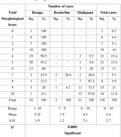
A score from 0-5 was assigned for each of the component. A total score ranging
from 0 and 10 for every tumor.
Observations of tumor septa, diffuse echogenicity and extra tumoral free
fluid were includedwithin the category of morphological features.
Following morphology indexing,eachtumor was surgically removed and
werehistologicallyclassified according to the WHO System of classifying ovarian
tumors.
Statistical Tools
The information collected regarding all the selected cases were recorded in a
Master Chart. Data analysis was done with the help of computer using
Epidemiological Information Package (EPI 2002).
Using this software, range, frequencies, percentages, means and standard
deviations were calculated.
Sensitivity, specificity, accuracy, positive predictive value and negative
62
Sensitivity = True positivex 100
True positive + False negative
Specificity = True negativex 100
False positive + True negative
Positive predictive value = True positive x 100
True positive + False positive
Negative predictive value = True negativex 100
True negative + False negative
Accuracy = True positive + True negative x 100
63
RESULTS
A total of 136 ovarian tumors were studied. Table 1 illustrates the incidence
benign, borderline and malignant tumors based on histopathological examination.
Of the 136 ovarian tumors 92 cases were benign (67.6%), 3 were borderline (2.2%)
and 41 were malignant (30.2%).
Table : 1 Incidence of benign , borderline and malignant tumors as per HPE
Type of tumor
Cases
No. %
Benign 92 67.6
Borderline 3 2.2
Malignant 41 30.2
Total 136 100
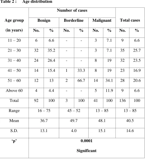
The mean age was 40.5 years (range, 13–85 years).60 patients belong to the
age group of 40 years or older and 76patients were less than 40 years of age. The
64
Table 2 : Age distribution
Age group
(in years)
Number of cases
Total cases
Benign Borderline Malignant
No. % No. % No. % No. %
11 – 20 6 6.6 - - 3 7.1 9 6.6
21 – 30 32 35.2 - - 3 7.1 35 25.7
31 – 40 24 26.4 - - 8 19 32 23.5
41 – 50 14 15.4 1 33.3 8 19 23 16.9
51 – 60 12 13 2 66.7 14 34.1 28 20.6
Above 60 4 4.4 - - 5 11.9 9 6.6
Total 92 100 3 100 41 100 136 100
Range 16 – 75 45 – 52 13 – 85 13 – 85
Mean 36.7 49.7 48.1 40.5
S.D. 13.1 4.0 15.1 14.6
‗p‘ 0.0001
65
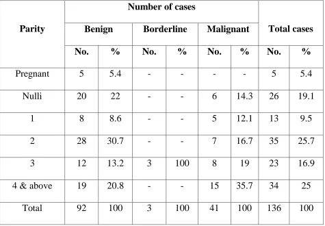
Table 3 : Parity distribution
Table 3 illustrates the parity distribution of ovarian tumours. Ovarian tumors
were common in multipara of 2 which is considered as statistically significant.
Benign tumors were common in 2nd parity women and malignant tumors were
common in women with parity 4 and above constituting 33% and 35.7%
respectively. Incidence of benign tumours in nulliparous women was 22% and
malignant tumours were 14.3%. There were 5 cases (5.4%) of ovarian tumor
complicating pregnancy. Parity
Number of cases
Total cases
Benign Borderline Malignant
No. % No. % No. % No. %
Pregnant 5 5.4 - - - - 5 5.4
Nulli 20 22 - - 6 14.3 26 19.1
1 8 8.6 - - 5 12.1 13 9.5
2 28 30.7 - - 7 16.7 35 25.7
3 12 13.2 3 100 8 19 23 16.9
4 & above 19 20.8 - - 15 35.7 34 25
66
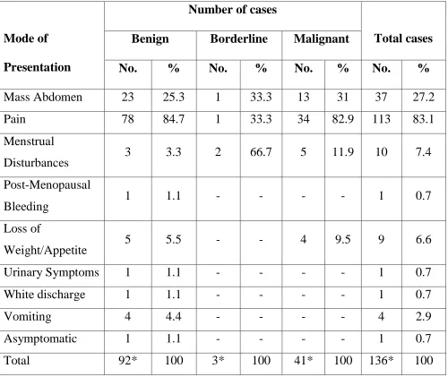
Table 4 : Mode of Presentation
* There was more than one mode of presentation in many cases.
Table 4 illustrates the mode of presentation of ovarian tumors. The most
common presenting features in both benign and malignant ovarian tumours were
pain abdomen with an incidence of 84.7% and 82.9% respectively. Mode of
Presentation
Number of cases
Total cases
Benign Borderline Malignant
No. % No. % No. % No. %
Mass Abdomen 23 25.3 1 33.3 13 31 37 27.2
Pain 78 84.7 1 33.3 34 82.9 113 83.1
Menstrual
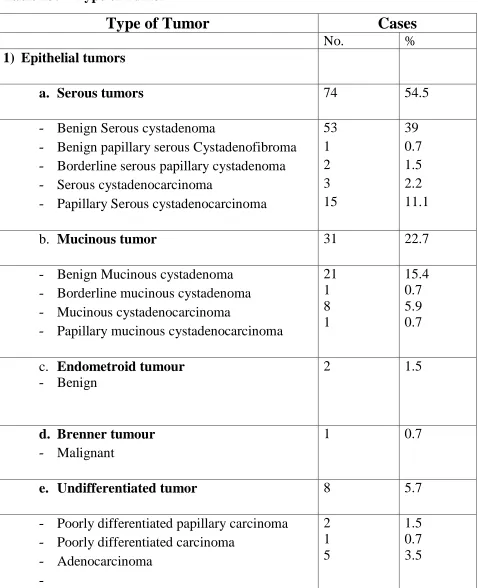
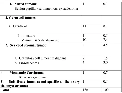
Disturbances 3 3.3 2 66.7 5 11.9 10 7.4
Post-Menopausal
Bleeding 1 1.1 - - - - 1 0.7
Loss of
Weight/Appetite 5 5.5 - - 4 9.5 9 6.6
Urinary Symptoms 1 1.1 - - - - 1 0.7
White discharge 1 1.1 - - - - 1 0.7
Vomiting 4 4.4 - - - - 4 2.9
Asymptomatic 1 1.1 - - - - 1 0.7
67
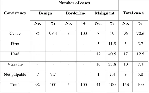
Table 5 : Per abdomen consistency of Ovarian Tumor
Consistency
Number of cases
Total cases
Benign Borderline Malignant
No. % No. % No. % No. %
Cystic 85 93.4 3 100 8 19 96 70.6
Firm - - - - 5 11.9 5 3.7
Hard - - - - 17 40.5 17 12.5
Variable - - - - 10 23.8 10 7.4
Not palpable 7 7.7 - - 1 2.4 8 5.8
Total 92 100 3 100 41 100 136 100
Table5 illustrates the consistency of ovarian tumor on clinical examination.
Most benign tumors were cystic in consistency (93.4%) and malignant tumors
were hard in consistency (40.5%) and around 23.8% of malignant tumors have a
68
Table 6 : Laterality
Laterality
Number of cases
Total cases
Benign Borderline Malignant
No. % No. % No. % No. %
Unilateral 86 92.3 2 66.7 22 53.6 110 80.9
Bilateral 6 6.6 1 33.3 19 45.2 26 19.1
Total 92 100 3 100 41 100 136 100
Table 6 illustrates the unilateral or bilateral involvement of ovaries in benign
and malignant tumors. Most ovarian tumors were unilateral (80.9%). 92.3% of
69
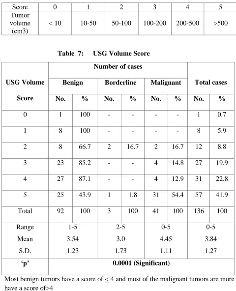
Table 7 illustrates the score for tumors based on the tumor volume by ultrasound.
Score 0 1 2 3 4 5
Tumor volume
(cm3)
[image:69.612.63.543.95.687.2]< 10 10-50 50-100 100-200 200-500 >500
Table 7: USG Volume Score
USG Volume
Score
Number of cases
Total cases
Benign Borderline Malignant
No. % No. % No. % No. %
0 1 100 - - - - 1 0.7
1 8 100 - - - - 8 5.9
2 8 66.7 2 16.7 2 16.7 12 8.8
3 23 85.2 - - 4 14.8 27 19.9
4 27 87.1 - - 4 12.9 31 22.8
5 25 43.9 1 1.8 31 54.4 57 41.9
Total 92 100 3 100 41 100 136 100
Range Mean S.D. 1-5 3.54 1.23 2-5 3.0 1.73 0-5 4.45 1.11 0-5 3.84 1.27
‗p‘ 0.0001 (Significant)
70
Table8: illustrates the score for tumors based on the tumor structures by ultrasound.
Score 0 1 2 3 4 5
Structure
Smooth wall, sonolucent
Smooth wall, diffuse echogenicity Wall thickening, <3mm fine septa Papillary projections, septa> 3mm Complex, predominantly solid Complex, solid& cystic areas,
extratumoral fluid