White Rose Research Online URL for this paper:
http://eprints.whiterose.ac.uk/97328/
Version: Accepted Version
Proceedings Paper:
Chandler, JH, Jayne, DG, Neville, A et al. (1 more author) (2015) A novel multiple electrode
direct current technique for characterisation of tissue resistance during surgery. In:
Proceedings of the Annual International Conference of the IEEE Engineering in Medicine
and Biology Society, EMBS. 37th Annual International Conference of the IEEE Engineering
in Medicine and Biology Society (EMBC), 25-29 Aug 2015, Milan, Italy. IEEE , pp.
8022-8025.
https://doi.org/10.1109/EMBC.2015.7320254
[email protected] https://eprints.whiterose.ac.uk/
Reuse
Unless indicated otherwise, fulltext items are protected by copyright with all rights reserved. The copyright exception in section 29 of the Copyright, Designs and Patents Act 1988 allows the making of a single copy solely for the purpose of non-commercial research or private study within the limits of fair dealing. The publisher or other rights-holder may allow further reproduction and re-use of this version - refer to the White Rose Research Online record for this item. Where records identify the publisher as the copyright holder, users can verify any specific terms of use on the publisher’s website.
Takedown
If you consider content in White Rose Research Online to be in breach of UK law, please notify us by
Abstract— Electrochemical and electrical characteristics have the potential to help differentiate between, and assess the health state of, different biological tissues. However, measurement and interpretation of these characteristics is non-trivial. We propose a new DC galvanostatic sensing method for application to laparoscopic cancer surgery. This presents a simple and cost-effective measurement coupled with straightforward data interpretation. This paper describes the electrochemical and electrical theory underpinning the technique. Additionally, we describe a measurement system employing this technique and present an investigation into the feasibility of using it for measuring the resistance of different tissue types. Measurements were performed on ex vivo porcine liver, colon and rectum tissues. Outputs were consistent with theory and showed a significant difference between the resistance of the different tissue types, (one-way ANOVA, F(2, 28) = 1369, p < 0.01). These findings indicate that this novel technique may be viable as a low cost method for the discrimination and health assessment of tissues in clinical scenarios.
I. INTRODUCTION
Advances in intraoperative imaging and measurement systems may facilitate a move toward personalised surgery [1, 2]. In this scenario, appropriate sensing would allow real-time staging and therefore tailoring of surgical procedures to the patient [1]. However, there is currently no prominent method for delivering relevant real-time information to the surgeon intraoperatively. A particular research focus has been on the development of intraoperative imaging systems to aid in the delineation of anatomical features and localisation of cancerous targets [3-6]. A variety of targeted and non-targeted fluorescent probes are being investigated for image-guided surgery. Although attractive for ease of application, optical imaging methods are dependent on the specificity and sensitivity of the probes and are limited by the depth of light penetration into tissues [7]. Progression of these probes to the clinic is also hampered by delays due the necessary safety assurance procedures [3, 6].
More localised measurements using the mechanical [8] or electrochemical (EC) and electrical properties of tissue may offer an alternative to imaging and can arguably deliver a faster route to surgical integration [9-11]. Electrical tissue characteristics, measured using the AC technique of Bioimpedance Spectroscopy (BIS), have shown differences between healthy and diseased tissues. [10-12]. However,
Research supported by Leeds Cancer Research UK (CRUK) Centre (CRUK grant no. 483355).
J.H. Chandler, D.G. Jayne, A. Neville and P.R. Culmer are with the University of Leeds, UK (phone: +44 (0)113 343 2186; fax: +44 (0) 113 242 461; e-mail: [email protected]).
these differences are often target tissue specific and require specific frequency range selection and complex electronic model analogies to be applied in order to make statistical distinction. A DC biogalvanic method for tissue characterisation has also been investigated as a means of determining the electrical properties of tissue [13]. This utilises a tissue-based galvanic cell as a power source and characterises an internal resistance, which has shown specificity to tissue type and damage [9, 13, 14]. Although the simplicity of this DC method and characterisation technique is suitable for surgery, it is subject to errors from the polarisation of the electrodes and the non-Ohmic behaviour of the electrochemical cell [15].
In this paper, we present a novel method for obtaining DC tissue resistance values. The technical development of a suitable measurement system has been described and an investigation into the feasibility of using the method for tissue type differentiation has also been presented. Resistance variation, measured using the described method, between ex vivo porcine tissues (liver, colon and rectum) has been reported and discussed.
II. THE MULTIPLE ELECTRODE TECHNIQUE
The proposed measurement technique is applied to a standard electrochemical cell connected through a tissue salt bridge. This consists of a current carrying electrode pair (working ( ) and counter electrodes ( )), and two stable reference (potential measurement) electrodes ( ). Application of significant current demand upon an EC cell necessitates a change in potential of the current-carrying electrodes. For an electrode under a kinetic regime of activation control, its overpotential can be calculated using the Tafel equation (1) [16]. This relates the change in electrode potential, relative to its equilibrium potential , to the current density of the cell via the Tafel parameters and
.
Assuming a suitable counter electrode is present within the cell, the potential difference between the working and any reference electrode can be determined as (2). Here the terms and represent anodic polarisation of the (+ve) and cathodic polarisation of the (-ve) respectively. A suitable reference electrode will have a stable equilibrium potential ( ) and be highly non-polarisable relative to the , leading to a negligible cathodic ovepotential ( ) . For the cell in question, represents the resistance of the tissue salt-bridge between the working and reference electrodes. When multiplied by the current density, this indicates the potential drop across the tissue. Assuming a non-polarisable and
A novel multiple electrode direct current technique for
characterisation of tissue resistance during surgery
Figure 1. Schematic of the resin mounted four electrode cell configuration; dimensions included (all in mm).
Figure 2. Four electrode control and measurement system; working, reference and counter electrodes shown (WE, RE & CE respectively).
Figure 3. Porcine rectal mucosa tissue (ex vivo) during a four-electrode test (zero strain condition).
stable reference electrode, calculation of a potential difference from comparison of the potential from two reference points yields (3). This crucially allows subtraction of the polarisation and leaves a current-potential relationship equal to a cell resistance (4). It is this resistance ( ) that is proposed as a useful comparative metric for tissue assessment and discrimination during surgery.
The theoretical prediction of (3) is that measurement of the difference between the responses, measured from two separated reference electrodes, for a controlled current flow will give a linear correlation. The gradient of the vs plot will represent the difference in resistance for the two resistive pathways from to the respective .
III. MATERIALS AND METHOD
To evaluate the feasibility of measuring cell resistance using the described method, a set of electrodes and a control and measurement system were developed. The simplest configuration of 4 electrodes was employed for initial testing. Extruded zinc wire (99.9 % pure) with a 1 mm diameter was used for the counter and reference electrodes. The working electrode was extruded copper wire (99.9 % pure) with a 1 mm diameter. The electrode size was selected to allow for significant electrode separation without inducing a large cell size. Copper wire was soldered onto the back of each electrode to form connection to the external control and DAQ devices. The electrode set was configured to the desired separation and set into non-conductive resin (VaridurTM,
Buehler, USA), leaving the end face of the electrode wires exposed. The electrode faces were wet ground to 1200 grit and rinsed with distilled water prior to each test. Fig. 1 shows the configuration of the manufactured electrode set.
To allow control of the current between the working and counter electrodes a commercial potentiostat was employed (CompactStat, Ivium Technologies). This was configured to operate galvanostatically, thereby inducing electrode
polarisation through control of the cell current. The potential differences between working and reference electrodes were recorded using a standard data acquisition device (NI myDAQ, National Instruments). Custom software (LabVIEW, National Instruments) was used to allow synchronisation, measurement and control of the system. Fig. 2 shows the developed system in relation to the electrode configuration of Fig. 1. Current and voltage data from the system were recorded at 10 Hz.
An investigation into the feasibility of using the method to measure the resistance of different tissue types was performed. Measurements were taken on ex vivo porcine liver, colon and rectum. Animals used were bred and sacrificed in accordance with UK Home Office regulations (Animals [Scientific Procedures] Act 1986). Each specimen was tested using the same electrode geometry over ten repeats. Initial testing indicated that excessive pressure can lead to tissue damage, with an associated drop in measured resistance. Tissue-electrode contact was therefore controlled to a minimal strain condition as shown in Fig. 3.
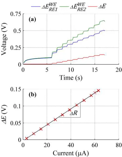
[image:3.612.327.548.545.704.2]Figure 4. (a) polarisation response for reference electrodes, and (b) corresponding voltage vs current resistance characterisation.
Figure 5. Results for ex vivo porcine liver, colon and rectum tissues showing (a) mean normalised measured voltage difference vs current (shaded region indicates range from repeats), and (b) mean calculated
resistance ± SD (n = 10).
current step of 1 µA. Prior to current stepping the cell was held at the starting current value for 5 s to allow for electrode stabilisation. Each tissue type was tested over 10 repeats. The voltage-current response for the electrodes was characterised using a linear regression fit (LabVIEW, National Instruments) to define the tissue resistance. A statistical comparison of the tissue types was performed (one-way ANOVA) in order to establish if resistance is specific to the tissue types tested.
IV. RESULTS
Typical tissue measurements showed a separation in measured potential differences as a function of time (current) for the two reference electrodes, Fig. 4(a). It can be seen that the reference closest to the working electrode ( ) shows a lower potential than the more distant electrode ( ). The difference between these two voltages was proposed as being proportional to the difference in resistance of the current pathways from the working electrode to the respective reference. Fig. 4(b) shows that this resistive difference is approximately constant, delivering a linear relationship between the voltage differences and cell current. Linear regression analysis showed a high quality fit ( > 0.99) across tissue types.
Repeat resistance measurements measured on each tissue type are shown within Fig. 5(a). Linearity is shown with low variability induced across repeat measurements (shaded region). The gradients of each tissue type are shown to be distinct. Fig. 5(b) summarises the mean ± SD (n=10) for each tissue type. A one-way ANOVA shows significant difference between the resistance of the different tissue types, F(2, 28) = 1369, p < .01.
V. DISCUSSION
Testing across different ex vivo porcine tissue types has demonstrated the proposed resistance characterisation methodology. Through control of the cell current and comparison of the response of the working electrode as viewed from two electrical reference points, a tissue resistance has been determined. The stepwise response of the reference voltages during measurement (Fig. 4(a)) shows that the current steps induce a significant and measureable alteration in the working electrodes potential. The theoretical prediction of this response being linearly proportional to cell current is apparently upheld.
The extension of the methodology to different tissue types has shown resistance variation. In order of increasing resistance, rectal, liver and colon tissues showed a range from 550-2300 . A particularly distinct difference is shown between the colon and rectum tissues which may not be expected as they are anatomically connected. These tissues do however, differ in muscle wall structure. In addition, it is possible that the relatively thin colon tissue may alter the current pathway under the electrode configuration tested, leading to an increased resistance. Detailed correlation of this resistance to the cellular structure of the tissues is beyond the scope of this paper. However, the resistance values recorded are likely to be influenced by the fat content and salt concentration of the respective tissues.
[image:4.612.70.266.450.702.2]stable potential. No reference electrode can meet these criteria, including the zinc metal material used in this study. Upon closer inspection however, this assumption may be relaxed to a point where the reference electrode potentials only need to be stable and only during the measurement process. Any relative separation of the reference potentials would shift the measured voltage differences in the ordinate axis but leave the gradient, and therefore the resistance, unchanged. As the copper working electrode is more readily polarised relative to zinc [15], and current flow is directed through a separate counter electrode, minimal alteration of the reference electrodes is expected. Any relative shifts in reference potential would propagate as deviation from a linear voltage-current trace, which has not been seen.
A second consideration of the theoretical assumption is that of the working electrode kinetic behaviour. The overpotential relationship proposed within (2) employs the Tafel equation, modelling the reduction process as activation controlled. Cathodic polarisation of copper in a aqueous or tissue salt bridge system shows mass-transport limitation at relatively small overpotentials (~100 mV), [15]. This therefore invalidates (2) over much of the potential range induced within this study. However, it is only the (Tafel) model of the working electrode overpotential that is invalid. Moving from (2) to (3) requires cancellation of the overpotential. Ignoring the actual electrode overpotential relationship, and assuming it is the same from both reference electrodes, its value may still be cancelled leading to an equally valid (4) and therefore characterised resistance. In addition to kinetic considerations, this cancellation is also applicable in the case of time-dependent effects suggesting measurement rate independence of the technique.
The results presented within Fig. (5) show specificity for tissue type based on resistance and not resistivity. A simple comparison using volume conductor assumptions shows AC liver tissue resistance to be approximately 100 times greater than found within this study [17]. However, the volume conductor assumption is not appropriate for the electrode configuration utilised, and BIS tissue resistivity values are typically frequency dependent [10]. The comparative metric for this study was therefore left as tissue resistance. Investigation into the influence of cell geometry on the determined resistance is required and may be coupled with numerical modelling to allow for appropriate conversion to resistivity.
Testing indicated temporal sensitivity in measurement of < 1 . When applied to tissues in vivo, with a continuous blood supply and improved hydration, lower resistance values would be expected. It is therefore necessary to determine if this sensitivity will be sufficient for in vivo health discrimination. It is proposed that relative testing may form the most appropriate protocol, where diseased tissue is assessed relative to an identified and measured healthy subject-specific baseline. Application to ex vivo tissues has however shown specificity to tissue type, and the stability and potential scalability of the technique make it a suitable candidate for integration into surgical tools for real-time tissue assessment.
VI. CONCLUSION
The novel technique presented allows for characterisation of a DC based tissue resistance. The arrangement of the electrodes delivers mitigation of a number of electrochemical phenomena that would otherwise skew the determined resistance. Application of the technique to tissues has shown specificity to tissue type across liver, colon and rectum. These findings indicate that the presented technique may offer a viable low cost method for tissue assessment within clinical scenarios such as laparoscopic surgery.
REFERENCES
[1] J. P. Tiernan, I. Ansari, N. A. Hirst et al., “Intra-operative tumour detection and staging in colorectal cancer surgery,” Colorectal Disease, vol. 14, no. 9, pp. e510-e520, 2012.
[2] R. P. Tufano, and E. Kandil, “Considerations for personalized surgery in patients with papillary thyroid cancer,” Thyroid, vol. 20, no. 7, pp. 771-6, Jul, 2010.
[3] A. Taruttis, and V. Ntziachristos, “Translational Optical Imaging,” American Journal of Roentgenology, vol. 199, no. 2, pp. 263-271, 2012/08/01/, 2012.
[4] B. E. Schaafsma, J. S. D. Mieog, M. Hutteman et al., “The clinical use of indocyanine green as a near-infrared fluorescent contrast agent for image-guided oncologic surgery,” Journal of Surgical Oncology, vol. 104, no. 3, pp. 323-332, 2011.
[5] Y. Kondo, Y. Murayama, H. Konishi et al., “Fluorescent detection of peritoneal metastasis in human colorectal cancer using 5-aminolevulinic acid,” International Journal of Oncology, 2014/05/06/, 2014.
[6] M. Mehrmohammadi, S. J. Yoon, D. Yeager et al., “Photoacoustic Imaging for Cancer Detection and Staging,” Current molecular imaging, vol. 2, no. 1, pp. 89-105, 2013/03//, 2013.
[7] E. de Boer, N. J. Harlaar, A. Taruttis et al., “Optical innovations in surgery,” British Journal of Surgery, vol. 102, no. 2, pp. e56-e72, 2015.
[8] A. S ftoiu, P. Vilmann, T. Ciurea et al., “Dynamic analysis of EUS used for the differentiation of benign and malignant lymph nodes,” Gastrointestinal Endoscopy, vol. 66, no. 2, pp. 291-300, 8//, 2007. [9] J. H. Chandler, A. Hood, P. R. Culmer et al., “Technological
assessment of the biogalvanic method for tissue characterization,” Physiological Measurement, vol. 35, no. 2, pp. 297, 2014.
[10] B. R. Lee, W. W. Roberts, D. G. Smith et al., “Bioimpedance: Novel use of a minimally invasive technique for cancer localization in the intact prostate,” The Prostate, vol. 39, no. 3, pp. 213-218, 1999. [11] R. J. Halter, A. Schned, J. Heaney et al., “Electrical Properties of
Prostatic Tissues: I. Single Frequency Admittivity Properties,” The Journal of Urology, vol. 182, no. 4, pp. 1600-7, 10//, 2009.
[12] N. Chauveau, L. Hamzaoui, P. Rochaix et al., “Ex Vivo Discrimination between Normal and Pathological Tissues in Human Breast Surgical Biopsies Using Bioimpedance Spectroscopy,” Annals of the New York Academy of Sciences, vol. 873, no. 1, pp. 42-50, 1999.
[13] A. Golberg, H. D. Rabinowitch, and B. Rubinsky, “Galvanic apparent internal impedance: An intrinsic tissue property,” Biochemical and Biophysical Research Communications, vol. 389, no. 1, pp. 168-171, 2009.
[14] A. Golberg, S. Laufer, H. D. Rabinowitch et al., “In vivo non-thermal irreversible electroporation impact on rat liver galvanic apparent internal resistance,” Physics in Medicine and Biology, vol. 56, no. 4, pp. 951-63, 2011.
[15] J. H. Chandler, P. R. Culmer, D. G. Jayne et al., “Assessment of electrochemical properties of a biogalvanic system for tissue characterisation,” Bioelectrochemistry, vol. 101, pp. 138-145, 2015/02//, 2015.
[16] E. Gileadi, Physical Electrochemistry, Weinheim: Wiley-VCH Verlag, 2011.