This is a repository copy of
Diverse roles and interactions of RNA structures during the
replication of positive-stranded RNA viruses of humans and animals
.
White Rose Research Online URL for this paper:
http://eprints.whiterose.ac.uk/87704/
Version: Accepted Version
Article:
Tuplin, AK orcid.org/0000-0003-2524-707X (2015) Diverse roles and interactions of RNA
structures during the replication of positive-stranded RNA viruses of humans and animals.
Journal of General Virology, 96. pp. 1497-1503. ISSN 1465-2099
https://doi.org/10.1099/vir.0.000066
© 2015, The Author. Published by the Microbiology Society. This is an author produced
version of a paper published in Journal of General Virology. Uploaded in accordance with
the publisher's self-archiving policy.
[email protected] https://eprints.whiterose.ac.uk/ Reuse
Unless indicated otherwise, fulltext items are protected by copyright with all rights reserved. The copyright exception in section 29 of the Copyright, Designs and Patents Act 1988 allows the making of a single copy solely for the purpose of non-commercial research or private study within the limits of fair dealing. The publisher or other rights-holder may allow further reproduction and re-use of this version - refer to the White Rose Research Online record for this item. Where records identify the publisher as the copyright holder, users can verify any specific terms of use on the publisher’s website.
Takedown
If you consider content in White Rose Research Online to be in breach of UK law, please notify us by
Working Title: Diverse roles and interactions of RNA structures during
the replication of positive-stranded RNA viruses of humans and animals
Contents category: Positive-strand RNA viruses
Author: Andrew Tuplin
Affiliations:School of Molecular and Cellular Biology, Faculty of Biological Sciences, University of Leeds, Leeds, UK. LS2 9JT.
10
E. Mail: [email protected] Tel: +44 (0)113 3435582
Word count: Summary: 117 words. Main text including legends and citations: 3116 words.
Abstract
Positive-stranded RNA viruses include important human, animal and
plant pathogens. Their genomes are able to fold into complex structures
20
stabilised by base pairing between individual nucleotides, many of which
are highly conserved and have essential functions during virus replication.
With new studies and technological advances the diversity of roles,
mechanisms and interactions in which such structured viral RNA functions
is becoming increasingly clear. It is also evident that many RNA structures
do not function as discrete elements but through mechanisms involving
multiple, long-range and often dynamic RNA-RNA interactions. Through a
range of examples and recent advances, this review illustrates the diverse
roles and mechanisms of structured viral RNA during the replication of
positive-stranded RNA viruses infecting humans and animals.
30
Introduction
Positive-stranded RNA viruses are a phylogenetically diverse
grouping including families such as the Flaviviridae, Coronaviridae, and Manuscript Including References (Word document)
Picornaviridae. They possess single-stranded RNA genomes (~3-32 Kb)
that function as both genetic material and mRNA template. With selection
pressure to maximise their coding potential, their genomes have evolved
a range of non-template functions involved in processes including virus
translation, replication, sub-genomic mRNA transcript production,
40
encapsidation and modulation of host antiviral responses. Such
non-template functions can be mediated through nucleotide sequence
composition (Atkinson et al., 2014), specific motifs or more commonly via
RNA structures interacting with host or viral trans-activating factors. RNA
structures within virus genomes are stabilised by Watson/Crick base
pairing and may comprise a simple stem-loop with duplexed stem and
single-stranded terminal-loop, or complex higher-order structures such as
pseudoknots and dynamic long-range RNA-RNA interactions.
While their host range, structure and life cycle are extremely
50
diverse the requirement for positive-strand RNA virus genomes to act as
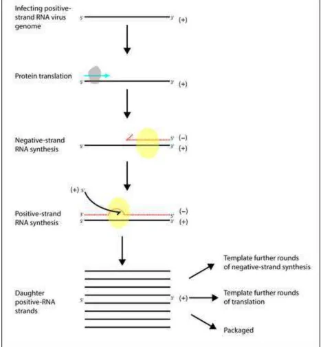
both genetic material and mRNA remain in common, Fig. 1. Following
release into the cytoplasm of a permissive cell, infecting genomes initially
template translation of viral proteins including RNA-dependent RNA
polymerase (RdRp), which catalyses synthesis of RNA negative strand
intermediates that in turn template synthesis of positive-strand RNA
molecules. Daughter positive-strand RNA is multifunctional and templates
further rounds of genome replication and translation or can be packaged
into new virion particles. In many instances mechanisms for temporal and
spatial regulation of these key events depend on structured RNA. Such
60
functional RNA elements act through an extremely diverse range of
mechanism involving interaction with host/viral trans-activating factors
and complex, often dynamic, RNA-RNA interactions. Our understanding of
how such mechanisms function has until recently been limited, however
with new structural mapping methods such as SHAPE (Merino et al.,
2005; Wilkinson et al., 2006) and powerful in silico prediction algorithms,
we are starting to develop a clearer picture of the interactions involved.
Structured RNA and its dynamic interactions play an essential role in the
replication cycle of positive-stranded RNA viruses infecting the full range
and the range of mechanisms in which they are involved, this review
cannot be an exhaustive discussion of all functional RNA elements (for
reviews of structured RNA in positive-strand RNA viruses of plants and
prokaryotes see (Pathak et al., 2011) and (Harvey et al., 2013;
Olsthoorn, 2011) respectively). Consequently, using a range of examples
and recent advances, this review illustrates the mechanistic and structural
diversity of viral RNA elements involved in the replication of
positive-strand RNA viruses that infect animal and human hosts.
Translation
80
Positive-strand RNA viruses compete with endogenous cellular
mRNAs for host translational machinery. In many instances these viruses
have evolved functional RNA structures that impart a competitive
advantage in recruiting translation initiation factors. Alternatively, such
structures may provide temporal control to translation events by
modulating the efficiency of initiation or reprograming translational
extensions.
Initiation
90
Cellular translation is generally initiated by recognition of an m7G cap by eukaryotic initiation factor 4F (eIF4F) - a complex of eIF4A, eIF4E
and eIF4G - that recruits the 40S ribosome pre-initiation complex to the
mRNA. Many viruses inhibit host cell translation through cleavage or
modification of cap-dependent factors and may directly recruit canonical
and non-canonical factors in a cap-independent process, via structured
RNA designated internal ribosome entry sites (IRES). Based on structure,
mechanism and initiation factor requirements, IRES elements can be
grouped into four categories. Type I elements include poliovirus, type II
100
foot and mouth disease virus (FMDV), type III hepatitis C virus (HCV) and
type IV the intragenic region (IGR) cricket paralysis virus (CrPV) IRES.
The 40S preinitiation complex either binds at an upstream position and
subunit binding is initiated through different combinations of
canonical/non-canonical translation factors. However, type IV elements
such as the CrPv IGR-IRES, interact directly with the 40S subunit,
independent of cellular initiation factors (Jan & Sarnow, 2002).
110
Modulation
IRES elements are generally located within 5’ non-coding regions
(NCRs), however their initiation function may be modulated by RNA
structures or long-range interactions within other regions of the virus
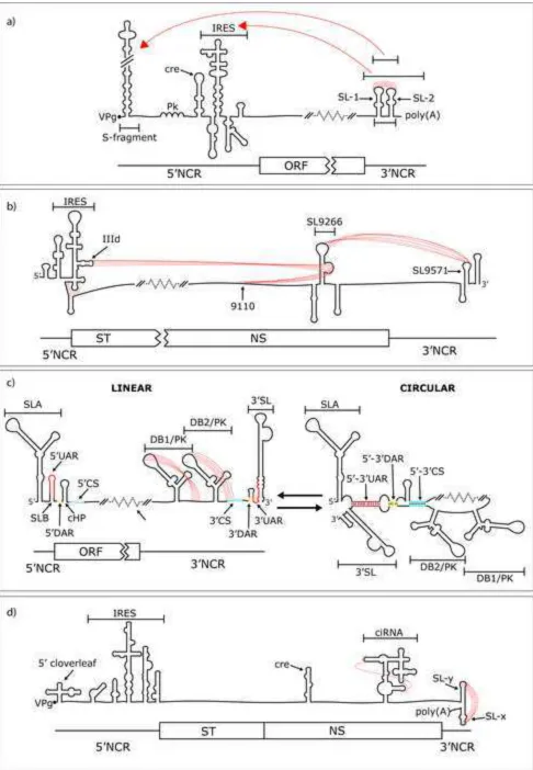
genome. FMDV translation is up regulated by a long-range interaction between its IRES and a region of the 3’NCR containing RNA structures
SL1, SL2 and a poly(A) tract, Fig. 2(a) (Lopez de Quinto et al., 2002;
Serrano et al., 2006). Rather than specific stem-loops, enhancement is
dependent on the overall higher-order RNA folding structure adopted by
120
the entire IRES and 3’NCR (Serrano et al., 2006). Interestingly, this
structure is competitive with an alternative interaction between SL1-SL2 and the S-fragment stem-loop in the 5’NCR. It has been postulated that
the S-fragment is involved in genome replication and that the alternative
mutually exclusive long-range interactions represent a mechanism
modulating FMDV translation and replication (Serrano et al., 2006).
Similarly, regulation of HCV translation involves various complex
(and potentially competing) higher order RNA interactions, Fig. 2(b).
Stem-loop SL9266 (alternatively 5BSL3.2 (You et al., 2004) or SLV (Lee
130
et al., 2004)) in the RdRp encoding region of the open reading frame
(ORF), is the core of a dynamic pseudoknot (SL9266/PK), undergoing
multiple RNA-RNA interactions involved in regulation of virus translation
and replication. The sub-terminal bulge of SL9266 forms RNA-RNA
interactions with both the IIId loop of the IRES (Romero-Lopez &
Berzal-Herranz, 2012) and an upstream region of the RdRp encoding region
centred on nucleotide 9110 (Diviney et al., 2008). The two potentially
competing interactions are mutually exclusive, with the IIId loop
interaction inhibiting HCV translation (Romero-Lopez & Berzal-Herranz,
(Diviney et al., 2008). The terminal-loop of SL9266 forms a ‘kissing-loop’ interaction with stem-loop SL9571 (alternatively SL2) in the 3’NCR (Friebe et al., 2005). The ‘kissing-loop’ is a dynamic structure forming open and
closed conformations, both of which are essential for efficient virus
replication (Tuplin et al., 2012). We have shown that the closed
conformation stimulates HCV IRES initiated translation (Tuplin et al.,
unpublished results) and the open structure preferentially binds cellular
protein EWSR1 up-regulating genome replication (Oakland et al., 2013). It
has been suggested that SL9266/PK functions in the temporal control of
early translation and replication events (Diviney et al., 2008; Oakland et
150
al., 2013). However, much remains to be understood regarding this
complex mechanism of dynamic RNA-RNA and RNA-protein interactions, in
particular how alternative conformations are stabilised and interact with
different trans-activating factors.
Many positive-strand RNA viruses that use capped, rather than
IRES initiated translation, also contain RNA structures that confer
regulatory control and a competitive advantage over cellular mRNA
initiation. The 3’NCR of flaviviruses such as Dengue virus (DENV) contain
two dumbbell-like stem-loops (DB1 and DB2) that form local pseudoknots
160
(DB/PK) necessary for optimal translation, Fig. 2(c) (Manzano et al.,
2011). Eukaryotic m7G cap dependent translation is regulated by
interaction between the mRNA 3’ poly(A) tract and poly(A) binding protein
(PABP) that recruits eIF4G, triggering initiation complex formation and
cyclisation. DENV lacks a poly(A) tract, however the DB/PKs
independently bind PABP, enabling efficient initiation; even under
conditions of reduced eiF4G (Polacek et al., 2009).
Reprogramming
170
Many positive-strand RNA viruses increase the coding capacity of
their genome through translation reprograming mechanisms such as stop
codon read-through or programmed -1 ribosomal frameshifting.
Reprograming enables translation of C-terminal extended polypeptides
proteins are translated as a polyprotein from a single ORF but in many
instances (for example Sindbis virus) an in frame UGA stop codon is
located upstream of the RdRp coding region (Strauss et al., 1983). In a
proportion of ribosomes the stop signal is read through in a mechanism stimulated by (but not dependent on) a large 3’ RNA structural element
180
(Firth et al., 2011). Alternatively, ribosomal frameshifting generally
depends on signals incorporating structured RNA and ‘slippery’ nucleotide
motifs. Coronaviruses (CoV) contain a signal in ORF1a that enables a
proportion of elongating ribosomes to switch into the -1 reading frame
(ORF1b), bypassing a stop codon and translating c-terminal extended
ORF1a/b products (Giedroc & Cornish, 2009; Plant & Dinman, 2008). In
SARS-CoV the signal incorporates a pseudoknot containing three internal
stems at which a proportion of ribosomes pause and stutter, shifting into the -1 reading frame at an upstream ‘slippery’ motif.
190
Genome replication
Temporal and spatial regulation of negative-strand synthesis is
frequently provided by structured RNA elements in the positive-strand 3’NCR, whilst growing evidence suggests that RNA structures in the negative strand 3’NCR influence positive-strand synthesis. However, it is
becoming increasingly clear that regulation also often involves RNA interactions and structure in both the 5’NCR and ORF. Such mechanisms
can function via direct RNA-RNA complementarity or RNA-binding protein intermediates and frequently initiate genome cyclisation between the 5’
200
and 3’ NCRs.
Flaviviruses
Flaviviruses possess structurally dynamic genomes that switch
between linear and circular conformations, both of which are essential for
virus replication. Genome cyclisation is stabilised by direct RNA-RNA
interactions between inverted complementary repeats (UAR, DAR and CS
motifs) at either end of the genome, Fig. 2(c) (Gebhard et al., 2011;
strand synthesis (Alvarez et al., 2005a; Khromykh et al., 2001; Lo et al.,
2003) and a balance between both conformations is critical for virus
replication (Villordo et al., 2010). DENV genome replication is dependent on activation of viral RdRp via binding 5’NCR stem-loop SLA before
genome cyclisation repositions the activated replication complex at the
3’NCR transcription initiation site (Filomatori et al., 2011; Filomatori et al.,
2006). Although SLA and genome cyclisation are essential for DENV
negative-strand synthesis they are not sufficient. Evidence from various
laboratories indicates the involvement of further RNA rearrangements and
structures in both NCRs and the ORF. For example, the 3’NCR DB/PKs
220
enhance transcription and the base of stem-loop 3’SL represses it due to overlapping with the 3’UAR cyclisation signal (Filomatori et al., 2011; Manzano et al., 2011); consequently the base of 3’SL must open to allow 5’-3’UAR binding and subsequent negative strand synthesis.
Flavivirus cyclisation is dependent on direct RNA-RNA
complementarity rather than protein factors (Alvarez et al., 2005b).
However, various cellular and viral proteins, essential to efficient virus
replication, bind in structured regions of the genome and have been
postulated to control or stabilise conformational rearrangement. In
230
arboviruses such as DENV and alphaviruses, stem-loop mutants have
been observed with different phenotypes in mammalian and mosquito
cells (Fayzulin & Frolov, 2004; Gorchakov et al., 2004; Groat-Carmona et
al., 2012; Villordo & Gamarnik, 2013). Such differences most likely reflect
differences in trans-activating protein factor availabilities, although
differences in intracellular environments such as temperature and cation
concentrations may also play a role.
Picornaviruses
240
The genome of poliovirus contains various functional RNA
structures such as cis replicating elements (cre) responsible for
form a ‘kissing-loop’, Fig. 2(d) (Ogram & Flanegan, 2011). Cellular
poly(rC) binding protein 2 (PCBP2) interacts with the poliovirus IRES during translation initiation and with stem-loop 5’CL, as part of a
ribonucleoprotein complex with viral protein 3CDpro (5’CL-RNP), during genome replication. Formation of the 5’CL-RNP is essential for initiation of
250
negative strand synthesis, with genome cyclisation occurring through interaction between PCBP2 in the 5’CL-RNP and PABP bound to the 3’NCR.
PCBP2 is cleaved by poliovirus proteinase 3CD rendering it unable to bind the IRES and preventing translation initiation, while binding to 5’CL and
thus replication is unaffected. Indeed, cleavage of PCBP2 by 3CD is
necessary for poliovirus replication and may constitute a switching
mechanism between translation and RNA replication (Chase et al., 2014). The 3’NCR ‘kissing-interaction’ is also required for efficient replication and
surprisingly its disruption has a much greater inhibitory effect than deleting the entire 3’NCR, possibly due to faulty positioning of the
5’CL-260
RNP in ‘kissing-loop’ mutants blocking successful initiation. Structures
within the poliovirus genome are relatively well documented. However,
the dynamics of their interactions within host cells and the role of other
regions of structured RNA remain to be fully characterised. For example,
two recent studies revealed a number of novel structures within the ORF (Burrill et al., 2013; Song et al., 2012), one of which - element 3D-7000 –
is implicated in RNA replication through an as yet unknown mechanism
(Burrill et al., 2013).
Evasion of innate immune responses
270
Antiviral innate responses generally depend on recognition of viral
RNA through pathogen-associated molecular patterns (PAMPs) by pattern
recognition receptors (PRRs). Recognition of viral RNA PAMPs (such as
double-stranded molecules, length, sequence, structure, and location),
trigger a range of antiviral responses. Consequently, viruses have evolved
numerous countermeasures, often involving structured RNA, to block
recognition by PRRs or overcome innate responses. For example, an RNA
2012). Many positive-stranded RNA viruses, such as HCV, exhibit
extensive RNA structure designated genome-scale ordered RNA structure
(GORS) (Simmonds et al., 2004). Distinct from discrete structural
elements, GORS inhibits detection by cellular PRRs with its presence
correlating to persistent viruses (Witteveldt et al., 2014). A number of
PRR interferon induced proteins bind 5’ caps lacking 2’-O methylation, in alphaviruses secondary structures in their 5’NCR alter this binding and
block their antiviral effect (Hyde et al., 2014). Alternatively, highly
structured sub-genomic flavivirus RNAs (sfRNA) - produced during
flavivirus infection as the product of host Xrn1 endonuclease activity
290
terminating at a pseudoknot resistant to Xrn1 helicase activity (Chapman
et al., 2014) - increase viral pathogenicity in mice, play a role in inhibition
of a type I interferon responses and are involved in silencing RNAi in
insect cells (Schnettler et al., 2012). Such varied mechanisms by which
structured RNA interacts with or manipulates antiviral responses,
illustrates its key role in the evolutionary arms race between viruses and
host innate immunity.
Summary and perspectives
300
Using a range of examples and recent advances this review
illustrates the diversity of mechanisms by which structured RNA influences
different stages of positive-RNA virus replication. As our understanding of
these mechanisms develops, in part through new methodologies enabling
structural mapping of longer RNA molecules at higher resolutions, the importance and diversity of – often long-range and dynamic - higher order
RNA structures is becoming increasingly clear. For example, various
studies have clearly demonstrated that flavivirus genomes switch between
alternative RNA conformations and that a balance between the two is
critical to virus replication. However, our understanding of the
310
mechanisms and trans-activating factors involved in controlling or sensing
such complex and often dynamic interactions is limited. Indeed,
elucidation of transient RNA-RNA and RNA-protein interactions remains
challenging and we have little comprehension of how essential RNA
intracellular environments and compartments. In summary, recent
advances have made a significant impact on our understanding of viral
RNA structure and its diverse roles across a range of mechanisms
essential to positive-strand RNA virus replication. With future studies
refining and extending our understanding, it can be expected that the
320
potential of structured RNA as an antiviral target will be further
investigated and that a clearer understanding of their dynamics during
virus replication will inform the study of RNA structures in other systems,
such as eukaryotic mRNA and long non-coding RNA function.
Bibliography
Alvarez, D. E., De Lella Ezcurra, A. L., Fucito, S. & Gamarnik, A. V. (2005a).
Role of RNA structures present at the 3'UTR of dengue virus on
translation, RNA synthesis, and viral replication.
Virology
339,
200-330
212.
Alvarez, D. E., Lodeiro, M. F., Luduena, S. J., Pietrasanta, L. I. & Gamarnik, A.
V. (2005b). Long-range RNA-RNA interactions circularize the dengue
virus genome.
Journal of virology
79, 6631-6643.
Atkinson, N. J., Witteveldt, J., Evans, D. J. & Simmonds, P. (2014). The
influence of CpG and UpA dinucleotide frequencies on RNA virus
replication and characterization of the innate cellular pathways
underlying virus attenuation and enhanced replication.
Nucleic acids
research
42, 4527-4545.
Burrill, C. P., Westesson, O., Schulte, M. B., Strings, V. R., Segal, M. & Andino,
340
R. (2013). Global RNA structure analysis of poliovirus identifies a
conserved RNA structure involved in viral replication and infectivity.
Journal of virology
87, 11670-11683.
Chapman, E. G., Costantino, D. A., Rabe, J. L., Moon, S. L., Wilusz, J., Nix, J. C. &
Kieft, J. S. (2014). The structural basis of pathogenic subgenomic
flavivirus RNA (sfRNA) production.
Science
344, 307-310.
Chase, A. J., Daijogo, S. & Semler, B. L. (2014). Inhibition of
poliovirus-induced cleavage of cellular protein PCBP2 reduces the levels of
viral RNA replication.
Journal of virology
88, 3192-3201.
Diviney, S., Tuplin, A., Struthers, M., Armstrong, V., Elliott, R. M., Simmonds,
350
P. & Evans, D. J. (2008). A hepatitis C virus cis-acting replication
element forms a long-range RNA-RNA interaction with upstream
RNA sequences in NS5B.
Journal of virology
82, 9008-9022.
Fayzulin, R. & Frolov, I. (2004). Changes of the secondary structure of the 5'
end of the Sindbis virus genome inhibit virus growth in mosquito
cells and lead to accumulation of adaptive mutations.
Journal of
virology
78, 4953-4964.
recruitment and activity of the dengue virus polymerase.
J Biol Chem
360
286, 6929-6939.
Filomatori, C. V., Lodeiro, M. F., Alvarez, D. E., Samsa, M. M., Pietrasanta, L. &
Gamarnik, A. V. (2006). A 5' RNA element promotes dengue virus
RNA synthesis on a circular genome.
Genes & development
20,
2238-2249.
Firth, A. E., Wills, N. M., Gesteland, R. F. & Atkins, J. F. (2011). Stimulation of
stop codon readthrough: frequent presence of an extended 3' RNA
structural element.
Nucleic acids research
39, 6679-6691.
Friebe, P., Boudet, J., Simorre, J. P. & Bartenschlager, R. (2005). Kissing-loop
interaction in the 3' end of the hepatitis C virus genome essential for
370
RNA replication.
Journal of virology
79, 380-392.
Gebhard, L. G., Filomatori, C. V. & Gamarnik, A. V. (2011). Functional RNA
elements in the dengue virus genome.
Viruses
3, 1739-1756.
Giedroc, D. P. & Cornish, P. V. (2009). Frameshifting RNA pseudoknots:
structure and mechanism.
Virus Res
139, 193-208.
Gorchakov, R., Hardy, R., Rice, C. M. & Frolov, I. (2004). Selection of
functional 5' cis-acting elements promoting efficient sindbis virus
genome replication.
Journal of virology
78, 61-75.
Groat-Carmona, A. M., Orozco, S., Friebe, P., Payne, A., Kramer, L. & Harris, E.
(2012). A novel coding-region RNA element modulates infectious
380
dengue virus particle production in both mammalian and mosquito
cells and regulates viral replication in Aedes aegypti mosquitoes.
Virology
432, 511-526.
Han, J. Q., Townsend, H. L., Jha, B. K., Paranjape, J. M., Silverman, R. H. &
Barton, D. J. (2007). A phylogenetically conserved RNA structure in
the poliovirus open reading frame inhibits the antiviral
endoribonuclease RNase L.
Journal of virology
81, 5561-5572.
Harvey, S. C., Zeng, Y. & Heitsch, C. E. (2013). The icosahedral RNA virus as a
grotto: organizing the genome into stalagmites and stalactites.
Journal of biological physics
39, 163-172.
390
Hyde, J. L., Gardner, C. L., Kimura, T., White, J. P., Liu, G., Trobaugh, D. W.,
Huang, C., Tonelli, M., Paessler, S., Takeda, K., Klimstra, W. B.,
Amarasinghe, G. K. & Diamond, M. S. (2014). A viral RNA structural
element alters host recognition of nonself RNA.
Science
343,
783-787.
Jan, E. & Sarnow, P. (2002). Factorless ribosome assembly on the internal
ribosome entry site of cricket paralysis virus.
Journal of molecular
biology
324, 889-902.
Keel, A. Y., Jha, B. K. & Kieft, J. S. (2012). Structural architecture of an RNA
that competitively inhibits RNase L.
Rna
18, 88-99.
400
Khromykh, A. A., Meka, H., Guyatt, K. J. & Westaway, E. G. (2001). Essential
role of cyclization sequences in flavivirus RNA replication.
Journal of
virology
75, 6719-6728.
Lee, H., Shin, H., Wimmer, E. & Paul, A. V. (2004). cis-acting RNA signals in
the NS5B C-terminal coding sequence of the hepatitis C virus
genome.
Journal of virology
78, 10865-10877.
untranslated region of West Nile virus by use of a reporting replicon
that differentiates between viral translation and RNA replication.
410
Journal of virology
77, 10004-10014.
Lopez de Quinto, S., Saiz, M., de la Morena, D., Sobrino, F. & Martinez-Salas,
E. (2002). IRES-driven translation is stimulated separately by the
FMDV 3'-NCR and poly(A) sequences.
Nucleic acids research
30,
4398-4405.
Manzano, M., Reichert, E. D., Polo, S., Falgout, B., Kasprzak, W., Shapiro, B. A.
& Padmanabhan, R. (2011). Identification of cis-acting elements in
the 3'-untranslated region of the dengue virus type 2 RNA that
modulate translation and replication.
J Biol Chem
286, 22521-22534.
Merino, E. J., Wilkinson, K. A., Coughlan, J. L. & Weeks, K. M. (2005). RNA
420
structure analysis at single nucleotide resolution by selective
2'-hydroxyl acylation and primer extension (SHAPE).
J Am Chem Soc
127, 4223-4231.
Oakland, T. E., Haselton, K. J. & Randall, G. (2013). EWSR1 binds the
hepatitis C virus cis-acting replication element and is required for
efficient viral replication.
Journal of virology
87, 6625-6634.
Ogram, S. A. & Flanegan, J. B. (2011). Non-template functions of viral RNA in
picornavirus replication.
Current opinion in virology
1, 339-346.
Olsthoorn, R. a. V. D., J. (2011).
Bacteriophages with ssRNA
: eLS.
Pathak, K. B., Pogany, J. & Nagy, P. D. (2011). Non-template functions of the
430
viral RNA in plant RNA virus replication.
Current opinion in virology
1, 332-338.
Plant, E. P. & Dinman, J. D. (2008). The role of programmed-1 ribosomal
frameshifting in coronavirus propagation.
Frontiers in bioscience : a
journal and virtual library
13, 4873-4881.
Polacek, C., Friebe, P. & Harris, E. (2009). Poly(A)-binding protein binds to
the non-polyadenylated 3' untranslated region of dengue virus and
modulates translation efficiency.
The Journal of general virology
90,
687-692.
Romero-Lopez, C. & Berzal-Herranz, A. (2012). The functional RNA domain
440
5BSL3.2 within the NS5B coding sequence influences hepatitis C
virus IRES-mediated translation.
Cellular and molecular life sciences :
CMLS
69, 103-113.
Schnettler, E., Sterken, M. G., Leung, J. Y., Metz, S. W., Geertsema, C.,
Goldbach, R. W., Vlak, J. M., Kohl, A., Khromykh, A. A. & Pijlman, G. P.
(2012). Noncoding flavivirus RNA displays RNA interference
suppressor activity in insect and Mammalian cells.
Journal of
virology
86, 13486-13500.
Serrano, P., Pulido, M. R., Saiz, M. & Martinez-Salas, E. (2006). The 3' end of
the foot-and-mouth disease virus genome establishes two distinct
450
long-range RNA-RNA interactions with the 5' end region.
The Journal
of general virology
87, 3013-3022.
Simmonds, P., Tuplin, A. & Evans, D. J. (2004). Detection of genome-scale
ordered RNA structure (GORS) in genomes of positive-stranded RNA
viruses: Implications for virus evolution and host persistence.
Rna
Song, Y., Liu, Y., Ward, C. B., Mueller, S., Futcher, B., Skiena, S., Paul, A. V. &
Wimmer, E. (2012). Identification of two functionally redundant RNA
elements in the coding sequence of poliovirus using
computer-generated design.
Proc Natl Acad Sci U S A
109, 14301-14307.
460
Strauss, E. G., Rice, C. M. & Strauss, J. H. (1983). Sequence coding for the
alphavirus nonstructural proteins is interrupted by an opal
termination codon.
Proc Natl Acad Sci U S A
80, 5271-5275.
Tuplin, A., Struthers, M., Simmonds, P. & Evans, D. J. (2012). A twist in the
tail: SHAPE mapping of long-range interactions and structural
rearrangements of RNA elements involved in HCV replication.
Nucleic acids research
40, 6908-6921.
Villordo, S. M., Alvarez, D. E. & Gamarnik, A. V. (2010). A balance between
circular and linear forms of the dengue virus genome is crucial for
viral replication.
Rna
16, 2325-2335.
470
Villordo, S. M. & Gamarnik, A. V. (2009). Genome cyclization as strategy for
flavivirus RNA replication.
Virus Res
139, 230-239.
Villordo, S. M. & Gamarnik, A. V. (2013). Differential RNA sequence
requirement for dengue virus replication in mosquito and
mammalian cells.
Journal of virology
87, 9365-9372.
Wilkinson, K. A., Merino, E. J. & Weeks, K. M. (2006). Selective 2'-hydroxyl
acylation analyzed by primer extension (SHAPE): quantitative RNA
structure analysis at single nucleotide resolution.
Nat Protoc
1,
1610-1616.
Witteveldt, J., Blundell, R., Maarleveld, J. J., McFadden, N., Evans, D. J. &
480
Simmonds, P. (2014). The influence of viral RNA secondary structure
on interactions with innate host cell defences.
Nucleic acids research
42, 3314-3329.
You, S., Stump, D. D., Branch, A. D. & Rice, C. M. (2004). A cis-acting
replication element in the sequence encoding the NS5B
RNA-dependent RNA polymerase is required for hepatitis C virus RNA
replication.
Journal of virology
78, 1352-1366.
490
Figure legends
Figure 1
Replication of positive-strand RNA virus genomes
Generalised schematic representation of single ORF, positive-strand RNA virus, replication. After release into permissive cell cytoplasm the infecting positive-strand RNA molecule acts as an mRNA and is translated by host
510
cell machinery. Translation initiates at the 5’ of the ORF, producing a polyprotein (blue arrow) that is cleaved into mature viral proteins. As viral protein levels build up synthesis of negative-strand RNA intermediates by viral RdRp is initiated from the 3’ end of the viral genome. Negative-strand RNA acts as a template for synthesis of daughter positive-Negative-strand RNA molecules, which in turn can act as template for further rounds of translation and replication or are packaged with viral structural proteins into new virion particles. As translation and negative strand synthesis initiate at opposing ends of the genome they are mutually exclusive processes that are tightly regulated, both specially and temporally, within
520
the cell. Where known, regulation mechanisms often involve dynamic interactions between host and/or virally encoded proteins and viral RNA structures.
Figure 2
RNA secondary structures and higher-order interactions within positive-strand RNA virus genomes
530
Simplified cartoon representations of viral genomes showing functional local RNA structures and higher-order RNA-RNA interactions between genome regions as dashed red lines. Breaks in genomes and regions not shown are represented by diagonal lines (-//-) and black-dashed lines respectively. Relative genome positions are indicated below each RNA molecule by schematic genome maps showing NCRs and the ORF. A) FMDV showing ‘kissing-loop’ interactions between the terminal loops of SL-1 and SL-2 and alternative long-distance interactions between different regions of the 3’NCR and either the S-fragment or IRES. Pk represents a region of predicted pseudoknots. B) HCV showing a ‘kissing-loop’
540
interaction between the terminal loops of SL9266 and SL9571 and alternative long-range interactions between the sub-terminal bulge of SL9266 and either the IRES or upstream to a position centred on
nucleotide position 9110. C) DENV showing alternative linear and circular conformations and DB pseudoknot interactions. Unbroken red, yellow and blue lines represent complementary UAR (upstream AUG region), DAR (downstream AUG region) and CS (cyclisation sequence) motifs
respectively. D) Poliovirus showing pseudoknot interactions between regions of the crRNA element and between the terminal loops of SL-y and SL-x.
Figure 1 post review
Figure 2 post review