MULTIPLE ROLES OF FLAVONOIDS IN
PLANT-RHIZOBIA SYMBIOSES AND
PLANT-PATHOGEN INTERACTIONS
S a m i r a Hassan
J a n u a r y 2 0 1 5
Australian
National
University
D E C L A R A T I O N
This thesis contains my original work and has not been submitted for any other award. The specific contributions by others are referred to in the Acknowledgements.
Samira Hassan
"The brick walls are therefor a reason. The brick walls are not there to keep us out The brick walls are there to give us a chance to show how badly we want something."
ACKNOWLEDGEMENTS
First and foremost, I would like to thank my supervisor Dr Uirike Mathesius for her continual support during my PhD candidature. I would like to also extend my gratitude towards my advisors Dr Peter Solomon and Dr Adrienne Hardham for their valuable insights. I am grateful for the ANU Postgraduate Scholarship and Australian Government's Research Training Scheme for their financial support to complete this work.
I'm particularly thankful to Dr Anton Wasson who initially worked on the roles of flavonoids in plant-rhizobia symbioses as part of his thesis. The constructs for silencing chalcone synthase in the vector pHellsgateS was generated by Anton Wasson and Dr Flavia Pellerone. Uirike Mathesius and Anton Wasson collected the plant material for transcriptome analysis using microarrays. I contributed with data analysis and inference. The Nod factor overexpressing rhizobia strain E65 was kindly provided by Dr Sharon Long and Dr Melanie Barnett. Thanks to Dr. Naresh Verma for brainstorming ideas on how best to integrate GPP into the E65 strain.
I am also thankful to fellow PhD student; Jason Ng for optimizing a method for lAA quantification using LC MS/MS. Dr Thy Thruong of the RSB Mass Spectrometry Facility also assisted me in running mass spectrometry and provided excellent support even on weekends. I am indebted to Professor Lloyd Sumner of the Noble Foundation, USA for training me in the fundamental concepts of mass spectrometry. My thanks also extend to his colleagues Drs David Huhman, Dongsik Yang, Bonnie Watson, Zhentian Lee and Mohamed Bedair for demonstrating the methods for quantification of plant metabolites including flavonoids so that 1 could set up similar protocols at the ANU.
The staffs at the Research School of Biology have provided excellent support services to allow me to complete my research. 1 am particularly thankful to Stephanie McCaffery for helping me optimise plant-growth rooms during the stress of building moves. Sharon Seltabanis showed tremendous patience when autoclaving my constant influx of plant-growth media. And Roseanne Holm who was never tired of assisting me in chasing up product suppliers.
ABSTRACT
Flavonoids are secreted by plant roots to activate and attract nitrogen-fixing
bacteria known as rhizobia. Rhizobia perceive flavonoids and reciprocate with
secretion of Nod factors required to initiate nodule organogenesis. In this thesis I
have explored the roles flavonoids play in the roots during early nodulation of
Medicago truncatula. To achieve this goal, I silenced or overexpressed genes
encoding key enzymes in the flavonoid biosynthesis pathway in hairy root
cultures. I used RNA interference as a mechanism to silence multiple copies of the
genes chalcone synthase, flavonol synthase, isoflavone synthase,
dihydroflavonol-4-reducase and flavone synthase II. 1 also used a 35S promoter to overexpress the
genes chalcone synthase, flavonol synthase, isoflavone synthase and
dihydroflavonol-4-reducase, although only isoflavone synthase overexpression showed quantifiable
increase in the targeted metabolites using LC-MS/MS.
Flavonoids have been described to affect the spatio-temporal balance of the plant
hormone auxin through the inhibition of polar auxin transport and its breakdown.
Using constitutive Nod factor overexpressing rhizobia to inoculate
flavonoid-silenced [CHSi) roots, 1 have demonstrated that flavonoids regulate nodule
organogenesis in part through the control of auxin synthesis in the root, by
regulating gene expression of YUCCAl, a central enzyme in auxin biosynthesis. In
contrast, flavonoids did not alter the expression of genes encoding PIN and LAX
auxin efflux and influx transporters, even though these flavonoids were required
for the inhibition of acropetal auxin transport by rhizobia. The inhibition of polar
auxin transport was regulated most strongly by root flavonols and isoflavonoids,
however, only flavonols were found to be important for nodule formation. The
synthetic auxin transport inhibitor, TIBA, failed to complement CHSi roots,
suggested that flavonoids play additional roles in nodule organogenesis apart from
controlling auxin transport.
To explore further functions for flavonoids in nodulation, I studied the
transcriptomic changes 6 and 24 hours post inoculation of CHSi roots with
constitutive Nod factor expressing rhizobia. Inoculated CHSi roots
accumulated many defense related genes compared to control roots, but failed to express structural enzymes that could be involved in infection thread formation.
These roots also differentially expressed genes encoding proteins involved in the
synthesis and response factors of hormones gibberelins, ethylene, cytokinin and
auxin. GFP-labelled, constitutively Nod factor expressing rhizobia were shown to
form infection threads in control but not CHSi roots, suggesting that flavonoids are
involved in infection of rhizobia. CHSi roots also did not trigger reactive oxygen species accumulation, suggested to be required for cross-linking of glycoproteins
in the matrix of infection threads.
Flavonoids have previously also been demonstrated to be important as
plant-defense compounds. Using the transgenic hairy roots with altered flavonoid metabolite profile, I suggest a link between auxin-mediated plant-defense and the
presence of flavonoids in the roots. This was investigated by challenging roots with the pathogens Rhizoctonia solani (AGS) and Aphanomyces euteiches. One
mechanism by which roots show tolerance to pathogens is through secondary root formation to outgrow and evade pathogens. Roots showing reduced expression of
flavonol synthase showed a reduction in the ability to form secondary roots and
showed increased susceptibility to the fungal and oomycete pathogens.
Conversely, isoflavonoids are precursors to a number of protective phytoalexins. Increasing the expression of isoflavone synthase led to an increase in tolerance to
the root pathogens. This could have important implications in increasing productivity of agricultural crops exposed to environmental stresses.
T A B L E OF C O N T E N T S
CHAPTER 1. THE ROLE OF FLAVONOIDS IN ROOT-RHIZOSPHERE SIGNALING - OPPORTUNITIES AND
CHALLENGES FOR IMPROVING PLANT-MICROBE INTERACTIONS 1
1.1 INTRODUCTION 3
1 . 2 FLAVONOIDSINTHE RHIZOSPHERE 7
1 . 3 MULTIPLE ROLES FOR FLAVONOIDS IN NODULATION 11 1.4 EFFECTS OF FLAVONOIDS ON QUORUM SENSING-REGULATED BEHAVIOURS 1 4
1.5 MYCORRHIZAL SYMBIOSES 1 6 1.6 FLAVONOIDS ARE INVOLVED IN DEFENCE AGAINST ROOT PATHOGENS 1 8
1.7 FLAVONOIDS IN NEMATODE INTERACTIONS 2 1
1.8 FLAVONOIDS CAN CAUSE ALLELOPATHY 2 1
1 . 9 FLAVONOID METABOLIC FLUX ENGINEERING 2 2 1 . 1 0 CHALLENGES IN MANIPULATING THE FLAVONOID PATHWAY 2 4
1 . 1 1 AIMS OF THIS STUDY 2 7
CHAPTER 2. GENERATION OF RNAI AND OVEREXPRESSION CONSTRUCTS FOR INDUCING FLAVONOID
METABOLIC CHANGES 29
2 . 1 INTRODUCTION 3 0
2 . 2 METHODS 3 7 2.2.1 Construction of RNAi vectors 39
2.2.2 Construction of Overexpression vectors 42
2.2.3 Gene amplification 46 2.2.4 Transformation of bacterial cells 48
2.2.5 Agrobacterium-mediated gene silencing in M. truncatula roots 50
2.2.6 Media composition 51 2.2.7 Confirmation of RNA silencing or overexpression through quantitative real-time PCR 52
2.2.8 Confirmation of metabolite changes through LC-MS/MS 54
2 . 3 RESULTS 5 7 2.3.1 Transformation efficiencies in M. truncatula roots expressing RNAi or overexpression
constructs 57 2.3.2 Quantification of transcript levels of genes encoding flavonoid biosynthesis enzymes in
silenced and overexpressing hairy roots 57 2.3.3 LC MS/MS Quantification for flavonoid aglycones and glucosides 60
2 . 4 DISCUSSION 6 5
CHAPTER 3. FLAVONOLS AND ISOFLAVONOIDS CONTROL AUXIN TRANSPORT AND ACCUMULATION
DURING NODULE DEVELOPMENT IN M. TRUNCATULA 71
3 . 1 INTRODUCTION 7 2 3.1.1 Auxin metabolism and homeostasis during plant-development 72
3 . 2 METHODS 8 0 Statistical analysis 89
3 . 3 RESULTS 9 0 3.3.1 Silencing of flavonoids reduces the number of nodules in M. truncatula roots 90
3.3.2 Flavonoid silenced roots foil to inhibit auxin transport in response to rhizobia inoculation 90
3.3.3 Silencing of flavonoids alters the accumulation of auxin in roots inoculated with rhizobia 93
3.3.4 Silencing of flavonoids does not alter the PIN or LAX gene expression in M. truncatula ....95 3.3.5 Expression of genes encoding auxin biosynthesis proteins increases in flavonoid silenced
roots during early stages post rhizobia inoculations 100 3.3.6 Silencing of different branches of the flavonoid biosynthesis alters nodulation in M.
truncatula 101 3.3.7 Acropetal auxin transport is not inhibited by rhizobia in flavonol and isoflavonoid silenced
roots 102
3.3.8 Inhibition of auxin transport is insufficient to rescue nodulation in flavonoid deficient roots 106
3 . 4 DISCUSSION 1 0 9 3.4.1 Flavonoids regulate polar auxin transport in response to rhizobia 109
3.4.2 Local abundance oflAA and conjugates reduces at 24 hpi with rhizobia in M. truncatula
hairy roots and this is regulated by flavonoids no 3.4.3 Root flavonoids do not alter the expression of PIN or LAX genes in response to rhizobia 111
3.4.4 Flavonoids regulate YUCCAl synthesis during early stages of rhizobial infection 112 3.4.5 Isoflavonoid regulated auxin-transport inhibition is not essential for indeterminate nodule
formation in M. truncatula roots 114 3.4.6 Auxin transport inhibition by TIBA was insufficient to rescue nodulation in CHSi roots ..115
3 . 5 CONCLUSION! 1 1 7
CHAPTER 4. ADDITIONAL ROLES OF FLAVONOIDS IN SYMBIOTIC NODULE ORGANOGENESIS 119
4 . 1 INTRODUCTION 12O 4.1.1 Early symbiotic interactions between legumes and rhizobia 12O
4.3.1 Verification of gene expression levels using gRT-PCR 136 4.3.2 Silencing offlavonoids leads to extensive changes in transcript levels 137
4.3.3 Transcriptional changes observed in plant-hormone responsive genes 143 4.3.4 GO enrichment analysis shows a significant over-representation of genes involved in an
oxidoreductive response in flavonoid silenced roots 151
4.4 CONCLUSIONS 163
CHAPTER 5. THE ROLES OF FLAVONOIDS IN PLANT-PATHOGEN INTERACTIONS 167
5.1 INTRODUCTION 168
5.2 METHODS 173
5.3 RESULTS 175
5.3.1 The silencing of FLS reduced the root growth following an infection with A. euteiches ..175 5.3.2 The overexpression ofDFR gene increased the roots' tolerance to A. euteiches 181 5.3.3 The silencing of FLS and DFR genes significantly reduces roots' tolerance to R. solani.... 187
5.3.4 The overexpression oflFS increased the roots' tolerance to R. solani 191
5.4 DISCUSSION 210
5.4.1 Effect of auxin accumulation on defence 211 5.4.2 Flavonols and isoflavonoids may be important for lateral root formation when evading a
pathogenic infection 212 5.4.3 Overall metabolic changes in flavonoid silenced roots indicate a difference in response to
a nectrophic and a biotrophic pathogen 214
5.5 CONCLUSION 215
CHAPTER 6. GENERAL DISCUSSION 217
6.1 SUMMARY OF THE MAIN RESULTS 217
6.2 MANIPULATION OF THE FLAVONOID BIOSYNTHESIS PATHWAY 218
6.3 ROLE OF FLAVONOIDSIN REGULATION OF AUXIN TRANSPORT AND AUXIN ACCUMULATION DURING NODULATION221
6.4 ADDITIONAL ROLES OF FLAVONOIDS IN SYMBIOTIC INTERACTIONS WITH RHIZOBIA 227
6.5 ROLE OF FLAVONOIDS IN RESPONSE TO ROOT-PATHOGENS 231
6.6 CONCLUSIONS 234
APPENDIX 237
List of Figures
FIGURE 1 . 1 - MA)OR BRANCHES OF THE FLAVONOID BIOSYNTHESIS PATHWAY 5 FIGURE 1 . 2 - SPATIAL DIFFERENCES IN FLAVONOID ACCUMULATION WITHIN AND BETWEEN CELLS. 6
FIGURE 1 . 3 - SCHEMATIC OVERVIEW OF FLAVONOID FUNCTIONS IN THE RHIZOSPHERE 10
FIGURE 2 . 1 - INDUCTION OF R N A INTERFERENCE IN PLANTS 3 5 FIGURE 2 . 2 - SCHEMATIC OVERVIEW OF THE FLAVONOID BIOSYNTHESIS PATHWAY IN M. TRUNCATULA 3 6
FIGURE 2 . 3 - AN OVERVIEW OF THE METHODS USED TO CREATE TRANSGENIC HAIRY ROOTS THAT WERE 3 8 EXPRESSING SILENCING OR OVEREXPRESSING FLAVONOID CONSTRUCTS
F I G U R E 2 . 4 - THE EFFICIENCY OF TRANSFORMATION FOR THE M. TRUNCATULA ROOTS TRANSFORMED 5 8 WITH A RHIZOGENESCARmmC THE RNAI CONSTRUCTS
FIGURE 2 . 5 - THE EFFICIENCY OF TRANSFORMATION FOR THE M. TRUNCATULA ROOTS TRANSFORMED 5 8 WITH A RHKOCFNGS CARRYING THE OVEREXPRESSION CONSTRUCTS
FIGURE2.6 - T H E RELATIVE TRANSCRIPT ABUNDANCE OF SILENCED GENES IN RESPECTIVE 5 9 TRANSFORMED ROOTS
FIGURE 2 . 7 - THE RELATIVE TRANSCRIPT ABUNDANCE OF OVEREXPRESSED GENES IN RESPECTIVE 5 9 TRANSFORMED ROOTS
FIGURE 2 . 8 - SELECTED FLAVONOID METABOLITES CONCENTRATIONS IN ROOTS EXPRESSING RNAI 6 3 CONSTRUCTS
FIGURE 2 . 9 - SELECTED FLAVONOID METABOLITES CONCENTRATIONS IN ROOTS EXPRESSING 6 4 OVEREXPRESSION CONSTRUCTS
FIGURE 3 . 1 - AUXIN FATE IN PLANT ROOT CELLS AND INTERACTIONS WITH RHIZOBIA 7 6
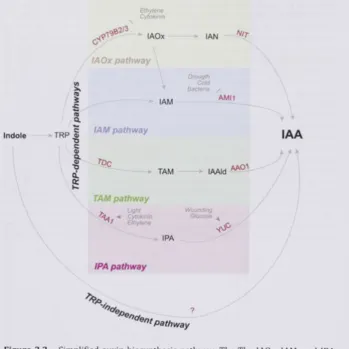
FIGURE3.2 - SIMPLIFIED AUXIN BIOSYNTHESIS PATHWAY 7 8 FIGURE 3 . 3 - SCHEMATIC DIAGRAM SHOWING THE PLACEMENT OF ^H-IAA BLOCK 8 4
FIGURE 3 . 4 - AVERAGE NODULE NUMBERS IN CONTROL AND CHSI ROOTS 9 0 FIGURE 3 . 5 - EXPRESSION OF THE AUXIN RESPONSIVE PROMOTER G H 3 FUSED TO B- GLUCURONIDASE 9 2
( G U S ] REPORTER SYSTEM IN CONTROL AND CHSI TRANSFORMED HAIRY ROOTS
FIGURE 3 . 6 - AUXIN TRANSPORT MEASUREMENTS IN CONTROL AND CHSI ROOTS 9 2 FIGURE 3 . 7 - THE CONCENTRATIONS OF I A A AND ITS CONJUGATES AS DETECTED IN CONTROL AND CHS\ 9 4
ROOTS
FIGURE 3 . 8 - THE RELATIVE GENE EXPRESSIONS OF PIN 1-10 9 7 FIGURE 3 . 9 - THE RELATIVE GENE EXPRESSION OF LAXL-4 9 9 FIGURE 3 . 1 0 - THE RELATIVE EXPRESSION OF YUCCAL, 2 AND 3 1 0 0 FIGURE 3 . 1 1 - AVERAGE NODULE NUMBERS IN CONTROL AND DIFFERENTIALLY SILENCED ROOTS 1 0 1
FIGURE 3 . 1 2 - AVERAGE NUMBER OF NODULES IN CONTROL AND FLS\ 1 0 2 FIGURE 3 . 1 3 - ACROPETAL AUXIN TRANSPORT MEASUREMENTS IN DIFFERENTIALLY SILENCED ROOTS 1 0 4
FIGURE 3 . 1 4 - ACROPETAL AUXIN TRANSPORT MEASUREMENTS SHOWING THE RESCUE OF AUXIN 1 0 5 TRANSPORT INHIBITON PHENOTYPE IN FLSL AND IFS\ ROOTS
FIGURE 3 . 1 5 - OPTIMIZATION OF TIBA-INDUCED PSEUDONODULE FORMATION IN M. TRUNCATULA 1 0 7 HAIRY ROOTS
FIGURE 3 . 1 6 - STRUCTURE OF A NODULE [ A ) AND A PSEUDO-NODULE ( B ) . 1 0 8 FIGURE 3 . 1 7 - NODULES AND PSEUDO-NODULES IN CONTROL AND CHSI ROOTS 1 0 8 F I G U R E 3 . 1 8 - MEDICAGO GENE EXPRESSION ATLAS DETAILS THE EXPRESSION LEVELS OF YUCCAL 1 1 3
FIGURE 4 . 1 - MAJOR BIOSYNTHESIS PATHWAYS OF GIBBERELLIC ACID. ETHYLENE, CYTOKININ AND 1 2 4 AUXIN
FIGURE 4 . 2 - THE MOLECULAR EVENTS IN EARLY NODULE ORGANOGENESIS IN LEGUMES 1 2 7
FIGURE 4 . 4 - V E N N DIAGRAM SHOWING T W O - W A Y A N O V A INTERACTION [GENOTYPE*INOCULATION) 1 3 7 FIGURE 4 . 5 - V E N N DIAGRAMS SHOWING 2-FOLD INCREASE/DECREASE IN TRANSCRIPT ABUNDANCES 1 4 2 FIGURE 4 . 6 - C H A N G E S IN ROOT-HAIRS IN CONTROL AND CHSI ROOTS AFTER RHIZOBIA INOCULATION 1 5 4
FIGURE 4 . 7 - RELATIVE EXPRESSION OF RIPL TRANSCRIPT 1 5 8
FIGURE 4 . 8 - N U M B E R OF NODULES IN R O S INHIBITOR TREATED CONTROL AND CHS] ROOTS 1 5 9
FIGURE 4 . 9 - ACCUMULATION OF R O S IN THE CONTROL AND CHS] ROOTS 1 6 1 FIGURE 5 . 1 - TRANSGENIC HAIRY ROOTS OF M. RRUNC/IRT/M INFECTED WITH A EURE/CHK HYPHAL 1 7 6
PLUG
FIGURE 5 . 2 - CHANGES IN ROOT LENGTH 7 AND 1 4 DPI WITH A EUTEICHES 1 7 7 FIGURE 5 . 3 - CATEGORIZED CHANGES IN ROOT LENGTHS 7 AND 1 4 DPI WITH A EUTEICHES 1 7 8
FIGURE 5 . 4 - SHOOT WEIGHTS OF R N A I EXPRESSING COMPOSITE PLANTS 1 4 DPI WITH A EUTEICHES 1 8 0 FIGURE 5 . 5 - SECONDARY ROOTS THAT EMERGED IN FLAVONOID SILENCED PLANTS INOCULATED WITH A 1 8 0 EUTEICHES
FIGURE 5 . 6 - TRANSGENIC HAIRY ROOTS OF M. TRUNCATULA INFECTED WITH A. EUTEICHES' HYPHAL 1 8 2 PLUG
FIGURE 5.7 - ROOT LENGTH CHANGES 7 AND 1 4 DPI WITH A EUTEICHES ]N ROOTS EXPRESSING OX 1 8 3 CONSTRUCTS
FIGURE 5 . 8 - CATEGORICAL ROOT LENGTH CHANGES IN ROOT LENGTHS POST INOCULATION WITH 1 8 4
A. EUTEICHES
FIGURE 5 . 9 - NUMBER OF SECONDARY ROOTS EMERGING IN R N A I EXPRESSING ROOTS 1 4 DPI WITH 1 8 5
A. EUTEICHES
FIGURE 5 . 1 0 - SECONDARY ROOTS EMERGENCE DISTRIBUTION IN ROOTS 1 4 DPI WITH A EUTEICHES 1 8 6 FIGURE 5 . 1 1 - SHOOT FRESH WEIGHTS OF THE COMPOSITE PLANTS WITH R N A I EXPRESSING ROOTS 1 8 6 1 4 DPI WITH A EUTEICHES
FIGURE 5 . 1 2 - TRANSGENIC HAIRY ROOTS OF M. TRUNCATULA INFECTED WITH R. SOLANI MYCELIAL PLUG 1 8 8 FIGURE 5 . 1 3 - CHANGES IN ROOT LENGTH 7 AND 1 4 DPI WITH R. SOLANI IN HAIRY ROOTS EXPRESSING 1 8 9 DIFFERENT R N A I CONSTRUCTS
FIGURE 5 . 1 4 - SHOOT FRESH WEIGHTS OF THE COMPOSITE PLANTS WITH R N A I EXPRESSING ROOTS 1 4 1 8 9 DPI WITH R. SOLANI
FIGURE 5 . 1 5 - NUMBER OF SECONDARY ROOTS THAT EMERGED BELOW THE SITE OF INFECTION IN OF 1 9 0 ALL THE COMPOSITE PLANTS WITH R N A I EXPRESSING ROOTS 1 4 DPI WITH R. SOLANI
FIGURE 5 . 1 6 - CHANGES IN ROOT LENGTH 7 AND 1 4 DPI WITH R. SOLANI OF ROOTS EXPRESSING 1 9 2 DIFFERENT FLAVONOID OVEREXPRESSION CONSTRUCTS
FIGURE 5 . 1 7 - CHANGES IN SHOOT BIOMASS COMPARED 1 4 DPI WITH BETWEEN R. SOLANI INOCULATED 1 9 2 AND MOCK INOCULATED PLANTS
FIGURE 5 . 1 8 - TRANSGENIC HAIRY ROOTS OF M. TRUNCATULA INFECTED WITH R. SOLANI MYCELIAL PLUG 1 9 3 FIGURE 5 . 1 9 - CHANGES IN ROOT LENGTH 7 AND 1 4 DPI WITH R. SOLANI OF ROOTS EXPRESSING 1 9 4
DIFFERENT FLAVONOID OVEREXPRESSION CONSTRUCTS
FIGURE 5 . 2 0 - CHANGES IN SHOOT BIOMASS COMPARED 1 4 DPI WITH BETWEEN R. SOLANI INOCULATED 1 9 6 AND UNINOCULATED PLANTS
FIGURE 5 . 2 1 - NUMBER OF SECONDARY ROOTS THAT EMERGED BELOW THE SITE OF INFECTION IN R N A I 1 9 6 EXPRESSING ROOTS 1 4 DPI WITH R. SOLANI
FIGURE 5 . 2 2 - CATEGORICAL CHANGE IN ROOT LENGTHS 7 AND 1 4 DPI WITH R. SOLANI 1 9 7
FIGURE 5 . 2 3 - FOLD CHANGE DIFFERENCES IN FLAVONOID CONCENTRATIONS BETWEEN CONTROL 2 0 1 ROOTS, SET AT 1 (DASHED LINE) AND UNINFECTED, R. SOLANI INFECTED AND A EUTEICHES INFECTED
ROOTS EXPRESSING R N A I CONSTRUCTS
FIGURE 5 . 2 4 - FOLD CHANGE DIFFERENCES IN FLAVONOID CONCENTRATIONS BETWEEN CONTROL 2 0 2 ROOTS, SET AT 1 [DASHED LINE] AND UNINFECTED, R. SOLANI INFECTED AND A EUTEICHES INFECTED
FIGURE 5 . 2 5 - FOLD CHANGE DIFFERENCES IN FLAVONOID CONCENTRATIONS BETWEEN CONTROL 2 0 3 ROOTS, SET AT 1 (DASHED LINE) AND UNINFECTED, R, SOLAN! INFECTED AND/I. EUTEICHES WFECTED
ROOTS EXPRESSING RNAL CONSTRUCTS
FIGURE 5 . 2 6 - F O L D CHANGE DIFFERENCES IN FLAVONOID CONCENTRATIONS BETWEEN CONTROL ROOTS 2 0 7 AND UNINFECTED, R. SOLAN! INFECTED AND /I, EUTEiCHES INFECTED ROOTS EXPRESSING
OVEREXPRESSION CONSTRUCTS
FIGURE 5 . 2 7 - FOLD CHANGE DIFFERENCES IN FLAVONOID CONCENTRATIONS BETWEEN CONTROL 2 0 8 ROOTS AND UNINFECTED, R. SOLAN! INFECTED AND A. EUTE!CHES
FIGURE 6.1 - A RECTANGULAR CLADOGRAM OFTHE ORDERS OF GREEN PLANTS 2 2 5 FIGURE 6.2 - THE CHANGES IN TRANSCRIPT ABUNDANCES OF GENES INVOLVED IN THE EARLY RHIZOBIA 2 2 9
INTERACTION IN CHS] ROOTS
FIGURE 6.3 - MODEL FOR HOW THE FLAVONOIDS MAY AFFECT RHIZOBIA-INDUCED INFECTION THREAD 230 FORMATION IN ROOT-HAIRS.
List of Tables
T A B L E 2 . 1 - KEY FLAVONOID BIOSYNTHESIS ENZYMES, DETECTED METABOLITES AND THE PUTATIVE 3 3 FUNCTIONS OF DIFFERENT CLASSES OF FLAVONOIDS
T A B L E 2 . 2 - COMPONENTS OF A ROUTINE P C R AMPLIFICATION REACTION USING ECONOTAQ 4 6 T A B L E 2 . 3 - COMPONENTS OF P C R AMPLIFICATION REACTION USING PLATINUM PFX POLYMERASE 4 7 T A B L E 2 . 4 - COMPONENTS OF THE TRANSFORMATION BUFFERS FOR R B C L COMPETENT E. COLI CELLS 4 9
T A B L E 2 . 5 - COMPOSITION OF FAHREUS AND MODIFIED-FAHREUS GROWTH MEDIA 5 1
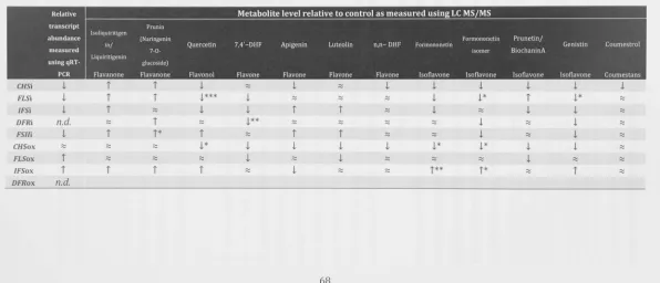
T A B L E 2 . 6 - COMPOSITION OF BERGENSEN'S MODIFIED MEDIUM 5 2 T A B L E 2 . 7 COMPONENTS OF A QUANTITATIVE REAL-TIME P C R REACTION 5 3 T A B L E 2 . 8 - P R I M E R PAIRS USED FOR QUANTITATIVE REAL-TIME P C R REACTIONS 5 4 T A B L E 2 . 9 - S U M M A R Y OF THE CHANGES IN TRANSCRIPT AND METABOLITE LEVELS IN UNINFECTED M. 6 8
TRUNCATULA ROOTS TRANSFORMED WITH R N A SILENCING CONSTRUCTS
T A B L E 3 . 1 - T H E PRIMERS USED TO AMPLIFY THE P/W AND M X G E N E S 8 2 T A B L E 3 . 2 - T H E PRIMERS USED TO AMPLIFY THE YUCCA GENES 8 3 T A B L E 3 . 3 - RESPONSE FACTORS AND REGRESSION COEFFICIENT FOR THE FOUR AUXIN ANALYTES 8 7
T A B L E 3 . 4 - T H E MEAN CONCENTRATIONS OF THE LAA AND ITS CON|UGATES IN CONTROL AND C H S I 9 4
T A B L E 3 . 5 - T H E RELATIVE EXPRESSION OF MEDICAGO PIN GENES 9 8 T A B L E 3 . 6 - T H E RELATIVE EXPRESSION OF MEDICAGO LAX GENES 9 9 T A B L E 4 . 1 - S U M M A R Y OF ACTIVITIES OF ANTIOXIDANT SYSTEMS IN RHIZOBIA-INFECTED NODULES 1 2 9
T A B L E 4 . 2 - T H E PRIMERS USED TO AMPLIFY GENES FOR CONFIRMATION OF MICROARRAY DATA 1 3 4 T A B L E 4 . 3 - VERIFICATION OF EXPRESSION PATTERNS OF SEVEN GENES THROUGH Q R T - P C R 1 3 6 T A B L E 4 . 4 - SUBSET OF GENES SHOWING INCREASED EXPRESSIONS DUE TO INOCULATION WITH RHIZOBIA 1 4 0
T A B L E 4 . 5 - CHANGES IN EXPRESSION OF SELECTED HORMONE RELATED GENES IN CW5I AND CONTROL 1 4 9 T A B L E 4 . 6 - SIGNIFICANT FOLD-CHANGES IN EXPRESSION OF REDOX RELATED GENES IN CHSI AND 1 5 5 CONTROL ROOTS
T A B L E 5 . 1 - A SUMMARY OF THE FLAVONOID CONTENT CHANGES IN ROOTS EXPRESSING DIFFERENT 2 0 4 R N A I CONSTRUCTS
T A B L E 5 . 2 - A SUMMARY OF THE FLAVONOID CONTENT CHANGES IN ROOTS EXPRESSING DIFFERENT 2 0 9 OVEREXPRESSION CONSTRUCTS
T A B L E 6 . 1 - A SURVEY SHOWING THE PRESENCE OF FLAVONOID BIOSYNTHESIS ENZYMES AND AUXIN 2 2 6 SYNTHESIS ENZYMES ACROSS VARIOUS PLANT ORDERS
List of Abbreviations
Ae - Aphanomyces euteiches ANOVA - Analysis of Variance
ATI - Auxin transport inhibitor/inhibition BMM - Bergersen's modified m e d i u m CHS - Chalcone synthase
DFR - Dihydroflavonol-4-reductase dpi - Days post inoculation
dsRNA - double stranded RNA
E65 - Sinorhizobium meliloti A2102 overexpressing nodD3 EV - Empty vector
FLS - Flavonol synthase FSII - Flavone synthase II GFP - Green fluorescent protein GUS - P" glucuronidase
hpi - Hours post inoculation HR - Hypersensitive response lAA - Indole-3-acetic acid IFS - Isoflavone synthase IT - Infection thread
LC MS/MS - Liquid chromatography tandem mass spectrometry LOD - Limit of detection
LOQ - Limit of quantitation NF - Nod factor
NPA - 1-N-naphthylphthalamic acid Ox - Overexpression
PAT - Polar Auxin Transport PGR - Polymerase chain reaction QG - Quiescent Centre
qRT-PCR - Quantitative Real-time PGR RAM - Root Apical Meristem
Chapter 1. The role of flavonoids in rootrhizosphere signaling
-opportunities and challenges for improving plant-microbe
interactions
The chapter presented here has been published in Journal of Experimental Botany in 2012. Two updates of the same chapter have been published as book chapters.
HASSAN, S. & MATHESIUS, U. 2012. The role of flavonoids in root-rhizosphere signalling: opportunities and challenges for improving plant-microbe interactions. Journal of Experimental Botany, 63, 3429-44.
HASSAN, S. & MATHESIUS, U. 2013. Roles of Flavonoids in Symbiotic Root-Rhizosphere Interactions. In: BRUIJN, F. ]. D. (ed.) Molecular Microbial Ecology of the Rhizosphere. John Wiley & Sons, Inc. doi:10.1002/9781118297674.ch51
HASSAN, S. & MATHESIUS, U. in press. Flavonoids play multiple roles in symbiotic root-rhizosphere interaction. In: BRUIJN, F. J. D. (ed.) Biological Nitrogen Fixation. Wiley/Blackwell.
Summary
flavonoid exudation to the rhizosphere. Possible challenges in that endeavour
include limited knowledge of the mechanisms that regulate flavonoid transport
and exudation, unforseen effects of altering parts of the flavonoid synthesis
pathway on fluxes elsewhere in the pathway, spatial heterogeneity of flavonoid
exudation along the root as well as alteration of flavonoid products by
microorganisms in the soil. In addition, the overlapping functions of many
flavonoids as stimulators of functions in one organism and inhibitor of another
1.1 Introduction
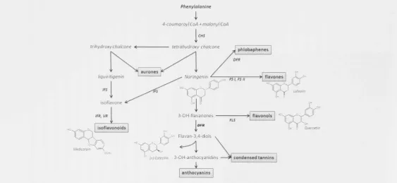
The flavonoid pathway is one of the best-studied biosynthetic pathways of specialized metabolites. Flavonoids are phenylpropanoid metabolites, most of which are synthesized from p-coumaroyl-CoA and malonyl-CoA and share their precursors with the biosynthetic pathway for lignin biosynthesis [Stafford, 1990]. However, some rare flavonoids are synthesized from CoA esters of substrates like cinnamic acid or dihydro-coumaric acid, e.g. (Friederich et al., 1999], To date, more than 1 0 , 0 0 0 flavonoids have been identified in plants and their synthesis appears to be ubiquitous in plants (Ferrer et al., 2008]. Their diversity stems from the generation of a number of basal flavonoid structures that include flavones, flavonols, flavan-3-als, flavanones, isoflavonoids, isoflavans and pterocarpans (Figure 1.1]. The flavonoid skeleton can be modified by glycosylation, malonylation, methylation, hydroxylation, acylation, prenylation or polymerisation, leading to the diversity of end products (Winkel-Shirley, 2 0 0 1 ] . These substitutions have important effects on flavonoid function, solubility, mobility and degradation.
al., 1998, Erlejman et al., 2004}. Flavonoid localisation and synthesis in different
cell types and in response to environmental stimuli can be regulated by a number
of transcription factors, in particular of the MYB and bHLH families (Koes et al.,
2005, Quattrocchio et al., 2006). In many cases, the regulation of cell specificity is
unknown.
Flavonoids can also be transported within and between cells and tissues. Within
the cell, flavonoids are likely to move via vesicle-mediated transport or through
membrane bound transporters of the ABC (ATP Binding Cassette) or MATE
[Multidrug And Toxic Extrusion compound) families [Zhao and Dixon, 2009).
Flavonoids transport into vacuoles can be achieved by conjugation of glutathione
with flavonoids in the cytoplasm, followed by ATP-driven transport via
glutathione-S-transferase pumps [Marrs et al., 1995, Goodman et al., 2004, Mueller
et al., 2000). Long distance transport of flavonoids is less well understood but has
been demonstrated in Arabidopsis, where application of flavonoids to the root or the shoot led to their transport towards distal tissues [Buer et al., 2007).
Application of transporter inhibitors showed that the long distance transport is
likely to be mediated by members of the ABC transporter families and is also
altered by glutathione, which is likely to act as a transport vehicle for flavonoids
after binding. To date, the exact mechanisms of flavonoid transport out of cellular
organelles and out of the cell, as well as long distance transport remain poorly
Phenylalanine
i
[image:25.612.28.592.35.294.2]4-coumaroyl CoA + malonyl CoA CHS
Figure 1.1 - Major branches of the flavonoid biosynthesis pathway. Some of the critical enzymes are highlighted in bold and are abbreviated as follows - CHS, Chalcone
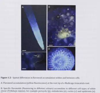
F i g u r e 1 . 2 - Spatial d i f f e r e n c e s in f l a v o n o i d accumulation w i t h i n and b e t w e e n cells.
A: F l a v o n o i d accumulation ( y e l l o w f l u o r e s c e n c e ) at t h e r o o t tip o f a M e d i c a g o truncatula root.
B: Specific f l a v o n o i d s Cfluorescing in d i f f e r e n t c o l o u r s ) a c c u m u l a t e in d i f f e r e n t cell t y p e s o f w h i t e c l o v e r ( T r i f o l i u m r e p e n s ) , f o r e x a m p l e p e r i c y c l e ( p ) , e n d o d e r m i s ( e ) , c o r t e x ( c ) and e p i d e r m i s ( e ) . F l a v o n o i d s are located in v a c u o l e s ( v ) of cortical cells.
C: F l a v o n o i d s ( f l u o r e s c i n g o r a n g e ) in the cell w a l l a n d / o r m e m b r a n e o f a c u r l e d r o o t hair o f M e d i c a g o truncatula in r e s p o n s e t o S i n o r h i z o b i u m m e l i l o t i infection.
D; N u c l e a r localisation of f l a v o n o i d s in a cross section t h r o u g h the r o o t tip o f w h i t e c l o v e r .
[image:26.408.22.371.16.340.2]1.2 Flavonoids in the rhizosphere
Flavonoids are not only found within the plant but constitute a large part of root
exudates (Cesco et al., 2 0 1 0 ] . Flavonoid exudation into the rhizosphere is not well
understood although some progress has been made towards the identification of
transporters. Flavonoids are likely to be actively exuded from roots, often in
response to elicitors (Schmidt et al., 1 9 9 4 , Armero et al., 2 0 0 1 ) . ABC transporter
mutants
of Arabidopsis
were shown to have altered root exudate profiles, although they likely affect multiple compounds [Badri et al., 2 0 0 8 ) . Exudation of theisoflavonoid genistein from soybean root plasma membrane vesicles was
ATP-dependent and most likely catalysed by an ABC-type transporter [Sugiyama et al.,
2 0 0 7 ) . Several phenylpropanoid exudates were affected in the ABC transporter
mutant
abcgSO,
although it was not shown whether this transporter directly transports the altered phenolics (Badri et al., 2 0 0 9 ) . Altogether, most of thetransporters responsible for flavonoid exudation into the rhizosphere, their
location or regulation are so far unknown. Flavonoids can also be released
passively from decomposing root cap and border cells (Shaw et al., 2 0 0 6 , Hawes et
al., 1 9 9 8 ] . Apoplastic p-glucosidases have been found to release isoflavones from
their conjugates in soybean roots, and this could be an important mechanism for
releasing active flavonoid aglycones during root-microbe interactions (Suzuki et
a l . , 2 0 0 6 ] .
Many studies have determined types and concentrations of flavonoids in root
exudates (summarized in Cesco et al., 2 0 1 0 , see also Leon-Barrios et al., 1 9 9 3 ] ,
although most of these were from plants grown in solution. Both aglycones and
glycosides of flavonoid can be found in root exudates. Their concentrations vary
widely and depend on plant growth condition, sampling techniques, nutrient
supply and plant species (Cesco et al., 2 0 1 0 ] . In general there is only little
information of actual flavonoid concentrations in soil and how these
concentrations change in space and time. In addition, there are large differences in
exudation of flavonoids along the root, with larger amounts being reported to be
exuded from the root tip (Graham, 1991, Hawes et al., 1 9 9 8 ; see also Figure 1.2a].
citrate release, is spatially and temporally regulated to coincide with maturation of
the cluster roots (Weisskopf et al., 2006, Tomasi et al., 2008). Solid phase root
zone extraction with the use of micro-tubes that can be placed along the root could
be used in future studies to determine spatial and temporal changes in flavonoid
exudation along roots grown in soil (Mohney et al., 2009, Weidenhamer et al.,
2009}
Once in the rhizosphere, the fate of flavonoids depends on various conditions in
the soil. Flavonoids can be absorbed to the cell wall and to soil particles with
cationic binding sites, thus becoming unavailable (Shaw and Hooker, 2008].
Depending on their modifications, flavonoid solubility and mobility in the soil
varies. While glycosylation improves their solubility in water, it is likely that
flavonoid glycosides are quickly deglycosylated by microorganisms and plant
exoenzymes, leaving the more hydrophobic aglycone (Hartwig and Phillips, 1991).
Flavonoid persistence in the soil varies and can be less than 72 h, depending on
the structure [Shaw and Hooker, 2008). Persistence in non-sterile soil can be
much shorter than in sterile soil, suggesting degradation by microorganisms.
Some bacteria metabolize flavonoids as a carbon source, while others specifically
modify flavonoids. For example, rhizobia can modify
nod
gene inducing flavonoids
by partial breakdown to produce flavonoids more or less active as
nod
gene
inducers (Rao and Cooper, 1995).
Flavonoids also alter the soil by acting as antioxidants and metal chelators.
Chelation and reduction of metals can alter nutrient concentration in the soil, and
this might have importance especially for the availability of phosphorus and iron.
For example, an isoflavonoid identified in
Medicago sativa(alfalfa) root exudates
was able to dissolve ferric phosphate, thus making both phosphate and iron
available to the plant (Masaoka et al., 1993). Flavonoids, including genistein,
quercetin and kaempferol can also alter iron availability by reducing Fe(lll) to
Fe(II) and by chelating iron otherwise unavailable in iron oxides (Cesco et al.,
2010).
Flavonoids can be synthesized and released specifically in response to abiotic and
biotic signals in the rhizosphere (Dixon and Paiva, 1995). For example, flavonoid
quercetin
1.3 Multiple roles for flavonoids in nodulation
Most legumes have the ability to form root nodules that house symbiotic nitrogen fixing bacteria or rhizobia. In addition, members of several families of non-legumes, so-called actinorhizal plants, form symbioses with nitrogen-fixing actinomycetes, in particular Frankia species. The rhizobia fix atmospheric
nitrogen into a form that the plant can use to fulfill its nitrogen requirement while the plant provides the bacteria with a source of carbon. Nodules are root organelles that are developed through signal exchange between the plant roots and the bacteria. In this symbiosis, flavonoids act as chemoattractants, inducers of nodulation [nod] and other genes, determinants of host specificity, developmental
regulators and regulators of phytoalexin resistance in rhizobia (Cooper, 2004).
One well-studied role of root-exuded flavonoids is their action as regulators of nod genes in rhizobia. A number of nod gene products are necessary to synthesize
species-specific Nod factors, lipochitin oligosaccharides required for nodule formation in the host [Spaink, 1995). The nod gene transcription is regulated by
NodD, a transcription factor of the LysR family of transcriptional regulators. Binding of an appropriate flavonoid to NodD is thought to enhance the access of RNA polymerase and improve transcriptional ability of the nod genes at the site in the promoter where NodD is localized (Li et al., 2008, Peck et al., 2006). The perception of flavonoids by rhizobia is also linked to elevation in concentrations of intracellular calcium in rhizobia that subsequently induces NodD proteins for Nod factor expression (Moscatiello et al., 2010]. The first flavonoids to be discovered to act as nod gene inducers were luteolin, isolated from M. sativa and 7,4' dihydroxyflavone from Trifolium repens (white clover] (Peters et al., 1986,
Redmond et al., 1986]. Since then many other flavonoids have been discovered to have nod gene inducing roles (summarized by Cooper, 2004). Most of these flavonoids are active as nod gene inducers at nano- to low micromolar
concentrations. It has been suggested that a mixture of flavonoids is more effective in inducing nod genes as opposed to a single compound (Bolanos-Vasquez and
proteins of different rhizobia is partially responsible for the host specificity of the
symbiosis.
Interestingly, some flavonoids also show nod gene repressing activity for certain rhizobia. For example, the isoflavonoids medicarpin and coumestrol have been
shown to negatively control Nod factor production in Sinorhizobium meliloti
[Zuanazzi et al., 1998). The nod gene activators and repressors together are thought to maintain an optimal level of Nod factor production and prevent
elicitation of defense responses by the plant [Savoure et al., 1997, Zuanazzi et al.,
1998).
Flavonoid exudation from the root changes during the symbiosis. Altered
flavonoid exudates have been found in rhizobia-inoculated roots of several
legumes. This alteration in flavonoid profiles could fine-tune Nod factor synthesis
during different stages of symbiosis [Dakora et al., 1993, Schmidt et al., 1994).
Altered flavonoid exudate profiles could also be the result of flavonoid metabolism
by rhizobia themselves, causing changes in the activity of flavonoids as nod gene inducers or repressors [Rao and Cooper, 1995).
Some flavones and flavonones that induce nod genes, such as luteolin and apigenin, have also been shown to evoke a strong chemoattractant response from
the rhizobia, with different flavonoids attracting different Rhizobium species [Aguilar et al., 1988, Dharmatilake and Bauer, 1992). These responses occur at
flavonoid concentrations of 10 to as little as lO-i" M, a much lower minimum
concentration that reported for flavonoid activity as nod gene inducers.
In actinorhizal symbioses, flavonoids might also play a role in host specificity and
selection of rhizobia, even though no canonical nod genes have been found in
Frankia (Normand et al., 2007). Flavonoids were found to accumulate inside actinorhizal nodules [Laplaze et al., 1999). Flavonoids from seeds of actinorhizal
plants were shown to enhance or inhibit symbiosis, although the flavonoids were
not identified in this study (Benoit and Berry, 1997). Flavonoids extracted from
fruits of the host Myrica gale had positive effects on growth and nitrogen fixation in compatible, but negative effects in incompatible Frankia strains (Popovici et al.,
2010), suggesting that flavonoids could play a role in selection of compatible
rhizobia by the host. This is similar to the situation in legumes, w h e r e it has been
s h o w n that the phytoalexin medicarpin produced by clover and medic species
inhibits the g r o w t h of incompatible but not that of compatible strains (Pankhurst
and Biggs, 1980). This effects might be due to the fact that certain ( i s o j f l a v o n o i d s
can induce resistance to phytoalexins in rhizobia at micromolar concentrations
[Parniske et al., 1991], thus enabling host plants to simultaneously exude
phytoalexins to inhibit pathogens while still allowing rhizobial infection.
Flavonoids have also been shown to regulate a number of other Rhizobium genes,
including those for exopolysaccharide synthesis, which is important for regulating
defense responses in the host. For example, genistein at 1 ^iM concentration
altered exopolysaccharide concentration and composition in Rhizobium fredii
cultures [Dunn et al., 1992). In addition, type III secretion systems, which play a
role in nodulation in some rhizobia, as well as the production of exported proteins,
can be induced by flavonoid exudates (Krishnan et al., 2003]. P r o t e o m e analysis
also found a number of other proteins in response to host flavonoids, many of
which await characterization (Guerreiro et al., 1997]
Nod factors are important in inducing the initial response of root cortical cell
division and root hair curling. Certain flavonoids act as negative regulators of
auxin transport and could thus cause accumulation of auxin at the nodule
initiation site to stimulate cell division and nodule organogenesis [Mathesius et al.,
1998b, Wasson et al., 2006, Boot et al., 1999]. Exactly h o w flavonoids redirect
auxin transport during nodule initiation is not known, but the perception of Nod
factors by the plant is thought to induce endogenous flavonoids that could locally
inhibit auxin transport [Mathesius et al., 1998a]. In M. truncatula, silencing of
different branches of the flavonoid pathway s h o w e d that flavonols like kaempferol
are the most likely candidates for auxin transport inhibitors [Zhang et al., 2009].
W h e t h e r auxin transport regulation occurs during nodulation of legumes forming
determinate nodules, e.g. soybean, is still unclear, but it is likely that other
Changes in auxin accumulation could also be due to auxin breakdown by
peroxidases, which can be modulated by flavonoids. The isoflavonoid
formononetin, which accumulates in the nodule primordia of white clover,
accelerated auxin breakdown. In contrast, a derivative of 7,4'-dihydroxyflavone
[DHF] and free DHF, which accumulate in the vacuoles of the cortical cells that
later form the nodule primordia, were shown to inhibit auxin breakdown
(Mathesius, 2001). The local changes in auxin may be critical in regulating cell
divisions during nodule development.
One of the opportunities in nodulation research could be the expression of
appropriate flavonoid synthesis pathways in non-legumes in future efforts to
extend nodulation beyond legumes. In addition, altered branches of the flavonoid
pathway could be engineered to extend legume host ranges to non-specific
rhizobia. This is demonstrated in Chapter 2 where 1 have engineered
M. truncatula
roots to express silencing or overexpression constructs targeting known genes
encoding flavonoid biosynthesis enzymes. I have further investigated if auxin
accumulation and response in flavonoid-silenced roots are altered in response to
rhizobia inoculation (Chapter 3).
1.4 Effects of flavonoids on quorum sensing-regulated behaviours
In the past years, a n u m b e r of molecules have been identified that interfere with q u o r u m sensing, including halogenated furanones produced by red algae (Manefield et al., 1999]. A number of land plants have been shown to synthesize quorum-sensing mimics, which can both inhibit and stimulate AHL-dependent genes in various reporter strains, although most of these compounds remain unidentified (Teplitski et al., 2000, Gao et al., 2003). The first mimic signal identified from plants was lumichrome, a riboflavine derivative (Rajamani et al., 2008). Another potential AHL mimic is the phenolic compound p-coumaric acid, a lignin precursor that can be exuded by roots into the soil (Bodini et al., 2009). P-coumaric acid can also be produced by breaking down flavonoids from root exudates (Rao and Cooper, 1995). In addition, coumaric acid can form p-coumaroyl-homoserine lactone, a distinct quorum-sensing signal used by some bacteria (Schaefer et al., 2008). Thus, p-coumaroyl-HSL could have two functions, to sense the presence of a host plant and to control density-dependent bacterial behaviors. A flavonoid identified from the medicinal tree Combretum albiflorum, catechin, which also occurs in many other plant species, also showed activity as a quorum-sensing mimic, although at rather high concentrations of between 0.125 and 4 m M [Vandeputte et al., 2010). Catechin can also be present in the rhizosphere of plants, for example as an exudate of spotted knapweed, where it acts as a potent allelochemical (Weir et al., 2003). Another flavonoid with inhibitory effects on quorum sensing regulated reporters is naringenin, which was shown to inhibit quorum sensing in E. coli and Vibryo fischeri at concentrations of 20 -360 |.iM (Vikram et al., 2010) as well as in P. aeruginosa at 4 mM concentration (Vandeputte et al. 2011). Naringenin is exuded by some legume roots and also acts as a nod gene inducer in rhizobia (Novak et al., 2002). Therefore, it would be interesting to test the effect of naringenin on quorum sensing-regulated genes in rhizobia.
Interestingly, the flavonoid pathway is activated in legumes by exposure to q u o r u m sensing signals from rhizobia, and it has also been shown that bacterial AHLs (at 50 nM concentrations) can stimulate production of AHL mimics by M. truncatula (Mathesius et al., 2003). These results strongly suggest a link between AHL perception by plants, activation of the flavonoid pathway and possible
feedback on bacteria by production of possible AHL mimics. However, effective
concentrations of potential flavonoid mimics in the rhizosphere will have to be
established. A recent study found that nod gene inducing flavonoids increased AHL
synthesis in three species of rhizobia at low micromolar concentrations,
concomitant with enhanced expression of AHL synthesis genes (Perez-Montano et
al., 2 0 1 1 ) . This suggests coordination between nod gene induction and quorum
sensing, possibly to enhance symbiotic behaviours of rhizobia. If flavonoids indeed
act as quorum sensing mimics and activators in plant-associated bacteria at
relevant concentrations this could be explored as an avenue to alter the ability of
bacteria to colonize and infect host plants.
1.5 Mycorrhizal symbioses
Mycorrhizal fungi are important symbionts of the majority of land plants that
contribute primarily to plant phosphorus nutrition. Mycorrhizal symbioses are
stimulated under phosphorus deficiency in the soil. Mycorrhizal fungi germinate
from spores and form hyphae in the soil which branch in response to root
exudates, which attract the hyphae to a host root. Hyphae then penetrate the host
root tissue and form ecto- or endomycorrhizal invasion structures (Harrison,
2 0 0 5 ] . Some of the host exudates that stimulate spore germination, hyphal
branching in the soil and root colonization, often in a symbionts-specific manner,
have been identified as flavonoids [Steinkellner et al., 2007, Siqueira et al., 1991,
Scervino et al., 2007, Scervino et al., 2005, Kikuchi et al., 2 0 0 7 ] . Most of these
studies reported active flavonoid concentrations of between 0.5 and 20 ^iM. Not
surprisingly, some of the flavonoids enhancing mycorrhizal infection are induced
under phosphorus stress (Akiyama et al., 2 0 0 2 ] . The isoflavonoid coumestrol has
been identified as a particularly active stimulator of hyphal growth (Morandi et al.,
1 9 8 4 ] and an M. truncatula mutant hyper-accumulating coumestrol was also
found to be hyperinfected by its mycorrhizal symbiont (Morandi et al., 2 0 0 9 ] .
Flavonoids are also likely to play a role during fungal invasion and arbuscule
formation inside the root. Infection of roots with vesicular arbuscular mycorrhizal
fungi was shown to induce the flavonoid pathway in a number of host species, in
particular in infected cells (Harrison and Dixon, 1994].
Flavonoid accumulation starts before the onset of infection and was shown to vary with different stages of infection and in response to different symbionts (Harrison and Dixon, 1993, Larose et a!., 2002], One of the roles of flavonoids inside the root could be to regulate defense reactions, and it has been hypothesized that mycorrhizal invasion triggers a temporary defense response in the root that involves induction of flavonoid phytoalexins (Harrison and Dixon, 1993). However, compared to the induction of phytoalexins in response to pathogens, mycorrhizal induction of these flavonoids is relatively low (Morandi, 1996). Flavonoids may also be responsible for an autoregulation of mycorrhization at later stages of the symbiosis (Larose et al., 2002). Split-root studies have demonstrated that formononetin and its glycoside are down-regulated systemically by either rhizobia or mycorrhizae, concomitant with autoregulation of both symbioses, and that their external application restored the symbioses (Catford et al., 2006).
Interestingly, pyranoisoflavones produced by white lupin, which is not a host for mycorrhizal fungi, inhibited hyphal branching of mycorrhizal fungi, suggesting that flavonoids could play both stimulating and inhibitory roles on fungal symbionts in the soil (Akiyama et al., 2010). However, inhibitory activity of flavonoids on hyphal branching was also reported from host plants (Tsai and Phillips, 1991), and therefore, it is likely that host and non-host plants can modulate the establishment of symbiosis by altering the profile of flavonoid exudates. While flavonoids clearly appear to enhance mycorrhization through stimulation of spore germination, hyphal branching and host infection, their presence in the host is not essential for the symbiosis. Experiments in flavonoid-deficient carrot and maize plants have shown that mycorrhizal infection was not abolished (Becard et al., 1995).
other hand, coumestrol is induced by mycorrhizal symbionts in M. truncatula
(Harrison and Dixon, 1 9 9 4 ) , where it could act as a nod gene inhibitor in S. meliloti
(Zuanazzi et al., 1 9 9 8 ) . It is possible that combinations of flavonoids, rather than
single compounds, need to be tested in more detail for their combined effects on
multiple symbionts. The tripartite symbiosis between soybean and its mycorrhizal
and rhizobial symbionts was shown to enhance nodulation compared to
inoculation of plants only with rhizobia. In the co-inoculated plants, flavonoid
profiles changed specifically in response to both symbionts, although interestingly,
flavonoid accumulation was largely inhibited by the symbionts, including the
flavonoids with activities as nod gene and hyphal branching inducers (Antunes et
al., 2 0 0 6 ] . Therefore, enhanced symbiosis in tri-partite interactions might be due
to enhanced nutrient uptake rather than, or in addition to, stimulation of
flavonoids.
The protection of the host plant from pathogens by mycorrhizal fungi has also
been partially attributed to the enhanced synthesis of flavonoid (phytoalexins) in
response to the mycorrhizal symbiont [Morandi, 1 9 9 6 ) , although data are still
scarce and the protective effect could have multiple causes [Borowicz, 2 0 0 1 ) .
While some studies have shown increased phenolic content of roots co-inoculated
with mycorrhizal fungi and pathogens (Cordier et al., 1 9 9 8 , Dehne and Schonbeck,
1 9 7 9 ) , others found decreased phytoalexin accumulation in co-inoculated plants
compared to plants only inoculated with mycorrhizal symbionts (Carlsen et al.,
2 0 0 8 ) .
1.6 Flavonoids are involved in defence against root pathogens
Flavonoids and other phenolics have been found to inhibit a range of root
pathogens and pests ranging from bacteria to fungi and insects (Makoi and
Ndakidemi, 2 0 0 7 ) . This has been attributed to their role as antimicrobial toxins
(Cushnie and Lamb, 2 0 1 1 ) and anti- or pro-oxidants (Jia et al., 2 0 1 0 ) . Their role
within the plant as antioxidants is suspected to be protective, although clear
evidence is lacking (Hernandez et al., 2 0 0 9 ) . Some of the major gaps in knowledge
pertain to fact that because of their highly oxidative nature, the transport and
storage of flavonoids is tightly regulated. Hence, the mechanism in which these
compounds may become available to prevent oxidative stress in case of a pathogen attack is unknown.
The challenge from a pathogen can lead to de novo synthesis of flavonoid phytoalexins that exhibit antifungal and antibacterial activities. These molecules can also be stored in an inactive form to function as broad-spectrum phytoanticipins to mount a quick defense against future attacks (Lattanzio et al., 2006).
Isoflavonoids represent a major class of phytoalexins in legumes. Using a promotenGUS fusion, it was shown that the expression of isoflavone synthase [Figure 1.1] was elevated when elicited with salicylic acid and cyst nematodes (Subramanian et al., 2004} suggesting their direct involvement in plant defense. Derivatives of isoflavonoids called pterocarpans such as medicarpin, pisatin and maackiain are known to have antimicrobial properties [Naoumkina et al., 2010). Maackiain also inhibited the oomycete Pythium graminicola at a concentration of 20 |ag/L (Jimenez-Gonzalez et al., 2008). Medicarpin from alfalfa and pea protects the plants from the pathogenic fungus Rhizoctonia solani (Pueppke and Vanetten, 1974, Kapulnik et al., 2001) possibly by having inhibitory activities on the elongation of fungal germ tube and mycelial growth (Blount et al., 1992, Higgins, 1978). There is also genetic evidence that pisatin from pea contributes to resistance against the fungus Nectria haematococca (VanEtten and Wu, 2004), as a knockdown of enzymes responsible for pisatin biosynthesis reduced its concentration from 28 ng/g dry weight by one third and increased the susceptibility of pea roots towards the fungal infection.
death. Alternatively, it is also suggested that some pterocarpans target the
m e m b r a n e A T P a s e and mitochondrial electron transport for degradation, leading
to cell death [Graham et al., 2007).
It is also interesting to note that in some plant-microbe interactions the
pterocarpans may not accumulate within the roots but be secreted instead. W h e n
chickpea seedlings w e r e challenged by the endogenous elicitor glutathione, an
increase in pterocarpan biosynthesis w a s observed ( A r m e r o et al., 2001]. It was
also shown that these compounds w e r e released by the roots to the surrounding,
possibly to cause damage to the pathogens before they can infect the roots.
Flavonols also contribute to resistance against pathogens. One of the most w i d e l y
distributed flavonol, quercetin has strong antimicrobial properties. Quercetin
binds to GyrB subunit of Escherichia coli DNA gyrase and inhibits the A T P a s e
activity. H o w e v e r , the promotion of DNA cleavage was induced only at
concentrations above 80 |iM (Naoumkina et al., 2010, Plaper et al., 2003].
Quercetin also inhibited the growth of the fungus Neurospora crassa ( P a r v e z et al.,
2004]. The plant carnation {Dianthus caryophyllus) mounts a defense against
Fusarium attacks through increasing the concentration of the fungitoxic flavonol triglycoside of kaemferide at concentrations as l o w as 50 nM (Curir et al., 2005].
Global gene expression studies have also shown that elevation of flavonoid
synthesis occurs w h e n M. truncatula plants w e r e challenged by Phymatotrichopsis
omnivora, cause of cotton root rot disease [Uppalapati et al., 2009]. In order to successfully colonize the plant, virulent strains of pathogens such as P. omnivore,
N. haematococca and R. solani have 'learned' to evade many of these flavonoids [Denny and Vanetten, 1981, Denny and Vanetten, 1982, Padmavati et al., 1997,
Pedras and Ahiahonu, 2005].
An appealing opportunity to enhance plant protection would be to engineer plants
with increased, or inducible expression of effective flavonoid phytoalexins and
phytoanticipins, however, it might be necessary for the plant to synthesize a range
of active antimicrobial flavonoids to avoid emergence of resistance by pathogens.
Using the constructs generated in this study, 1 have investigated if overexpression
of isoflavone synthase led to increased tolerance against root pathogens such as R. solani and Aphanomyces euteiches. In future, it would be imperative to test whether ectopic expression of these flavonoids would cause any harm to beneficial rhizosphere organisms.
1.7 Flavonoids in nematode interactions
Plant parasitic nematodes, including root knot, cyst and root lesion nematodes, constitute some of the major root pathogens. Many of these pathogens exhibit a wide host range, often of thousands of plant species. Sedentary endoparasitic nematodes cause the formation of feeding structures that are characterized by multiple cell divisions and endoreduplication in root tissues, leading to the formation of galls or cyst (Goverse et al., 2000, Gheysen and Mitchum, 2011). Invasion of roots with root knot and cyst nematodes induces the flavonoid pathway in infection structures (Jones et al., 2007, Hutangura et al., 1999), and it has been hypothesized that the flavonoids could act as regulators of auxin transport and accumulation during gall formation [Hutangura et al., 1999, Grunewald et al., 2009). In M. truncaCula plants deficient in flavonoids, gall formation still occurred, although galls were smaller and showed less cell divisions (Wasson et al., 2009). In flavonoid-deficient Arabidopsis and tobacco mutants, reproduction of several species of nematodes was not affected (Wuyts et al., 2006a, Jones et al., 2007). However, flavonoids did have an effect on nematode behaviour, for example certain flavonoids acted as repellents for specific nematode species and inhibited their motility and hatching at mM concentrations (Wuyts et al., 2006b). Therefore, while flavonoids do not seem to be essential for feeding site development in the host plant, flavonoids exuded into the rhizosphere could alter nematode attraction to the roots. This could be exploited for designing nematode trap plants that could be intercropped to reduce the infection of crop roots.
1.8 Flavonoids can cause allelopathy
ecological consequences. In some cases, flavonoids have been implicated as allelochemicals in the rhizosphere. The parasitic weed Striga constitutes one of the major problems in African agriculture with yield losses up to 1 0 0 % in large parts of sub Saharan Africa, thus inhibiting its germination would be an important achievement (Gressel et al., 2004). In a search for intercropping plants, the forage legume Desmodium uncinatum was found to significantly inhibit post-germination and attachment of Striga, and this inhibition was mimicked by several (iso)flavonoids identified from its root exudates [Khan et al., 2010, Hooper et al., 2010]. In addition, at least one of the isoflavones stimulated germination, which could be used to cause 'suicidal' germination of the weed. Therefore, the use of Desmodium as a 'push-pull' intercrop plant has been a cheap and successful strategy for smallholder farmers to control Striga infestations (Hooper et al., 2009, Khan et al., 2006).
The success of some invasive weeds has also been attributed to flavonoid allelochemicals. For example, spotted knapweed [Centaurea maculosa), which has been invading large parts of North America, exudes [-)-catechin, which can induce reactive oxygen species in susceptible species that lead to cell death and demise of the root system (Bais et al., 2003). A racemic mixture of catechin can also have detrimental effects on legume nodulation at high (~3 mM) concentrations [Alford et al., 2009). (-)-catechin has also been suggested to inhibit germination and growth of native species, but it has been questioned whether soil concentrations of (-)-catechin would be high enough to be effective [Blair et al., 2005, Duke et al., 2009).
1.9 Flavonoid metabolic flux engineering
The biosynthesis of flavonoids involves several branches of pathways to which multiple strategies could be applied to alter the metabolic flux. The production of secondary metabolites requires the plant to invest its energy and therefore, the pathways are tightly regulated. The initial committed step for flavonoid biosynthesis is catalyzed by chalcone synthase (CHS). Silencing of CHS transcripts has been demonstrated to cause dramatic decrease in flavonoid accumulation within the plant (Wasson et al., 2006, Zhang et al., 2009).
Many strategies can be adopted for engineering the flux of metabolites through
the biosynthesis pathways. Some of these include
-1 - Changes in enzyme specificity through changes in the active site configuration.
By studying the crystal structure of the enzymes' active sites, novel methods to
manipulate it may be developed to affect substrate specificity. |ez et al. (2002)
demonstrated that the substrate specificity could be altered by inducing single
point mutations at the active site of CHS.
2 - Since flavonoid biosynthesis is an energy consuming process, the plants utilize
control points for each branch. Knockouts or knockdowns of these critical
enzymes would direct the flux into alternative branches. Some of the enzymes that
may be directed with this strategy include isoflavone synthase, flavone synthase II
and vestitone reductase. Zhang et al. (2009) confirmed that knockdown of
isoflavone synthase and flavone synthase led to the generation of roots deficient in
isoflavonoids and flavones, respectively. The authors were then able to
demonstrate that plants can nodulate even in the absence of isoflavonoids that are
thought to play an essential role in the process.
3 - Transporters. Flavonoids are synthesized in the cytoplasm but often have to
be transported for exudation into the rhizosphere or storage. Similar to enzymes,
the expression of transporters would also have an impact in the direction of the
flux. This was recently demonstrated when a MATE type transporter was knocked
down to generate proanthocyanidin-rich plants [Zhao et al., 2011, Zhao and Dixon,
2009).
4 - Modification enzymes. Transport, activity and storage of flavonoids often
require modifications of their structure. Changes in abundance or activity of
enzymes responsible for these modification may increase or decrease generation
of the end product. An example of such an enzyme is 0-methyl transferase (OMT)
that is responsible for 4'-0-methylation and 3'-0'-methylation of isoflavonoids in
the generation of phytoalexins. Silencing of OMTs in pea affects the flux in the
biosynthetic pathway leading to the reduction in the phytoalexin pisatin [Liu et al.,
2006). A similar strategy may be applied to other modification enzymes although
it may be challenging to find enzymes that are not catalytically promiscuous and allowing control of a single pathway.
5 - Transcription factors. Altered expression of transcription factors regulating different parts of the flavonoid pathway could be an approach to altering activity of more than one enzyme. Members of the MYB, bHLH and WD40 transcription factors play an important role in regulating the flavonoid pathway (Koes et al., 2005, Du et al., 2010). For example, it has been shown that ectopic expression of MYB transcription factors can significantly increase anthocyanin synthesis in tomato (Butelii et al., 2008). In soybean, isoflavonid synthesis was increased by a combination of ectopic expression of maize transcription factors and inhibition of the competing anthocyanin pathway [Yu et al., 2003).
1.10 Challenges in manipulating the flavonoid pathway
While it appears opportunistic to modify the flavonoid pathway in order to manipulate root-rhizosphere interactions, the many interactions between plants, flavonoids and microorganisms demand a cautionary approach. First, while most studies on the function of flavonoids have been done under laboratory conditions, their demonstration in real rhizosphere conditions remains to be carried out to determine how effective some of the flavonoids functions remain under conditions of breakdown, adsorption, metabolism and altered solubility in the soil. Second, there are likely to be unforseen effects of flavonoids on non-target organisms. Whereas certain flavonoids could enhance nodulation or mycorrhization of host plants, they could also indirectly affect bacterial quorum sensing, plant-plant interactions and soil biochemistry. For example, catechin could inhibit quorum sensing in host related soil bacteria, while it may also suppress plant growth as a potent allelopathic signal. Similarly naringenin, which induced nod gene expression in several rhizobia, could have an effect on quorum sensing regulation in non-target bacteria. Exudation of isoflavonoids from soybean can stimulate attraction of its symbionts Bradyrhizobium japonicum as well as the devastating pathogen Phytophthora sojae (Morris et al., 1998). In addition, metabolism of flavonoids by rhizosphere bacteria could alter their activity and availability in the soil and could affect other bacteria (Shaw et al., 2006). Third, altering
concentrations or forms of flavonoids inside the plant tissue is lii<ely to have effects on plant development, which could be either beneficial or detrimental for the plant host. Flavonoids, via their effect on auxin transport (Brown et al., 2001] have been shown to alter cell morphology, root growth, gravitropism, and light responses (Buerand Djordjevic, 2009, Buerand Muday, 2004, Ringli etal., 2008],
Flavonoid exudation is also likely have effects on microbial community structure in the soil because it could increase species that use flavonoids as a carbon source while inhibiting the growth of others as phytoalexins (Walker et al., 2003, Rao and Cooper, 1994). Increased phenolics exudation in the Arabidopsis abcg30 mutant was shown to have wide ranging effects on bacterial and fungal community structure, although this mutation also affected other exudates (Badri et al., 2009).
Alteration of flavonoid synthesis is feasible and has been demonstrated in many studies in various plants, and both overexpression and inhibition of the flavonoid pathway using RNA interference have been successful (Wang et al., 2011). In some cases, the transfer of single gene encoding flavonoid enzymes might be sufficient to have an effect. For example, transfer of stilbene synthase from grapevine to tobacco resulted in increased resistance to Botrytis cinerea (Main et al., 1993). Altering glycosylation or targeting vacuolar transporters are other options that would allow the release of stored flavonoids from the vacuole for subsequent export (Weisshaar and Jenkins, 1998, Zhao et al., 2011b). One aspect of the manipulation of flavonoid synthesis or glycosylation is that it would be most efficient if it was targeted in the appropriate tissues (Figure 1.2), for example the root epidermis for subsequent exudation. This would require the use of epidermal-specific promoters, which are currently not available for most crop plants.
An important drawback in the manipulation of specific branches of the flavonoid biosynthesis pathway could be that it alters fluxes through other branches of the flavonoid pathway or related pathways (Wang et al., 2011, Liu et al., 2002). This has been cited as one reason why sufficient flavonoid accumulation for large scale production in engineered plants has not been successfully achieved (Fowler and Koffas, 2009). For example, mutants that show changes in the flavonoid pathway