0022-538X/84/030873-08$02.00/0
CopyrightC 1984, American SocietyforMicrobiology
Protein Processing Map
of Poliovirus
MARK A. PALLANSCH,1t OLEN M. KEW,'t BERTL. SEMLER,2§ DANIEL R. OMILIANOWSKI,' CARLW.
ANDERSON,3 ECKARD WIMMER,2 ANDROLAND R. RUECKERTl*
Biophysics Laboratory, Graduate School, andBiochemistry Department, College of AgriculturalandLife Sciences, University of Wisconsin-Madison, Madison, Wisconsin537061; Department of Microbiology, SchoolofMedicine, State University ofNew YorkatStony Brook, Stony Brook, New York117942; andDepartment of Biology, BrookhavenNational
Laboratory, Upton, New York
119733
Received 31August 1983/Accepted7November 1983
Fivepreviously unmapped proteins (Sa, 7d, 8, 9b, and 10)werelocatedontheproteolytic processingmap
of thepolyprotein. One of the proteins, 9b,appearstobe the sisterfragment ofacleavage reaction (P3-9->
P3-9b+VPg). Twoof the other newly mapped proteins, 8 and 10, have been identifiedassisterfragments of
X-related proteins 3b and 5b; thus, P2-3b -* P2-8 + P2-5b and P2-5b -* P2-10 + P2-X. The remaining proteins,5aand7d,mappedin the lb proteinandappear toresult from thecleavagesP3-lb-* P3-5a + P3-6b andP3-4b-* P3-7d +P3-6b. Theseassignmentsaccountforover95% of the totalpolioviralproteinsand
complete the mapping of the major processing pathways.
Synthesis of polioviral polyprotein (molecular weight,
247,000) is accompanied by extensive
processing,
which usually begins while still inthe nascent state to formthreemajor precursors called P1-la, P2-3b, and P3-lb. These precursors are then cleaved into a
variety
ofsmallerprod-ucts(Fig. 1A andB)that have beenmapped
by
avariety
ofapproaches including kinetic studies on the flowof radiola-beled amino acidsthrough proteins synthesized in the pres-ence and absence ofthe drug pactamycin (5, 23, 32, 33), tryptic analysis (13, 21, 25, 26, 37),
immunoprecipitation
(35), and radiochemical sequencing (14, 16, 26-28), which became feasible with the availability of the RNA sequence (14, 20, 22).
As a result of these studies, 19 of 26 electrophoretic
components found in poliovirus-infected cells, after poly-acrylamide gel electrophoresis ofthe radiolabeledproteins,
havebeen correlatedwithpositions intheprotein processing
map (Fig. 1C). We now report the identity and location of proteins representing the last three majorgaps in this map
(dotted lines). We also report the location of two minor proteins that appear to originate from previously
unrecog-nizedcleavages ofproteins P3-lbandP3-4.
MATERIALSANDMETHODS
Virus and cells.Poliovirus type 1 (Mahoney) was obtained
from C. Pfau at Rensselaer Polytechnic Institute; C. Pfau
hadobtainedthevirus fromS. Penman at the Massachusetts Institute ofTechnology. A second Mahoney strain from E.
Wimmer(24) was used for the mapping of VPg and
P3-lb
andforradiochemical microsequencing. This second virusisthe
strain sequenced recently by Wimmer's group (14). Virus used for tryptic mapping was progagated in H-HeLa cells that were grown in medium B (19) containing 10% added volume of calf serum (K. C. Biologicals, Inc., Lenexa, Kans.). Theinfection of cells in suspension was with 50 to 100PFU per cell by the procedures of McGregor et al. (17), except that a 30-minute virus attachment period was used.
* Correspondingauthor.
tPresentaddress: RockefellerUniversity,NewYork, NY 10021. tPresentaddress: Divisionof Viral Diseases, Center for Infec-tiousDiseases, Centers for Disease Control, Atlanta, GA 30333.
§Present address:Department of Microbiology, College of Medi-cine, University ofCalifornia, Irvine, CA 92717.
873
Virus used formicrosequencingwaspropagatedinS3-HeLa cells as describedpreviously (27).
Preparation and purification of radiolabeled poliovirus VPg. Virions were radiolabeled by the addition of
L-[4,5-3H]lysine
(5.0 mCi to 4 X10⁷
infected cells) at 2.5 hpostinfection. Cell-associated virus was harvested at 6 h
postinfection, theviruswaspurified,and theVPgRNAwas phenol extracted as described previously (21). The
[3H]ly-sine VPg RNA was useddirectlyin tryptic peptide analysis.
Preparation of radiolabeled proteins for tryptic peptide analysis. Because processing rates differ for various
pro-teins, it was necessary to utilize three types of labeling
conditions inpreparing the proteins usedfor tryptic analysis: (i) continuous labeling for short time periods
(pulse-label-ing), (ii) short labeling followed by the addition of a large excess of unlabeled amino acid and continued incubation
(pulse-chase labeling), and (iii) continuous labeling in the presenceof ZnCl2.
The pulse-labeling protocol was used to isolate proteins P1-la,
P3-ib,
P1-3a, P2-3b, P3-5a,P2-Sb,P2-X, P2-8, P3-9a,and P2-10. This was carried out by adding radiolabeled amino acidsat3.5 hpostinfection. Incorporationwasfor15 or 30min. The cells were then lysed with sodium dodecyl sulfate asdescribed below.
The pulse-chase labeling protocol was used to isolate
proteinsP3-2, P1-3c,P3-4b, P3-6a, P3-6b, P3-7d and P3-9b.
Incorporation of radiolabeled amino acids was for 30 min
beginning at 3 h postinfection. At this point cells were sedimented (820 x g for 3 min) and then suspended in the samevolume of medium A (19) withleucine andlysineat0.1
mM, a 100-fold excess over the initial concentration of the
radiochemical. The cells were then incubated for an
addi-tional 90 minbefore harvestingby sodium dodecyl sulfate
lysis.
Thethird procedure, labeling viral proteins in thepresence ofZnCl2(6), was used toisolate rapidly cleaved precursors. This was used primarily to obtain relatively large amounts of
proteinP3-lb. Fetal calf serum (0.05%) andZnCl2 (1.0 mM
in 0.1 mMHCI,final concentration) were added 6 minbefore the addition of the radiochemical at 3.5 h postinfection.
874 PALLANSCH ET AL.
Four radiochemicals were used to label polioviral pro-teins: L-[4,5-3H]leucine, L-[U-14C]leucine, L-[4,5-3H]lysine, and L-[U-14C]lysine. Atypical lysate was prepared by label-ing 4 x
107
cells in a 10-ml culture in amino acid-deficient medium A (19) with 500 ,uCi/ml for the 3H-labeled com-pounds or 50 ,uCi/ml for the14C-labeled
compounds.Cell lysates were made by sedimenting the infected cells (820 x gfor5 min), suspending them at 8 x
107
cells per ml indistilledwater,andadding an equal volume of solubilizing solution (2% sodium dodecyl sulfate, 1 M urea, and 0.2% mercaptoethanol). The lysate was then placed in a boiling water bath for 5 min. The preparation of samples for electrophoresis by dialysis has been described previously(19).
Preparativeelectrophoresis for peptide mapping. Prepara-tive electrophoresis of cell lysates was carried out with cylindrical gels (25 by 1.2 cm) of 10% polyacrylamide in phosphate bufferasdescribed previously (30). Five percent polyacrylamide was used for preparative electrophoresis of protein P3-lb (13, 21). After electrophoresis, gels were
Mobility
C
non-ba 3b lb
3o30 5b Bck
- ~~... 5b. 1- I _
3c X 9 2
VPO VP3 VPI 7t 4
VP4 VP2 VP, 60 6b
2000-1000
-FIG. 1. Electrophoreticprofiles and processingmapofpolioviral proteins found inlysatesofvirus-infected HeLa cells (A) aftera 10-min labeling period with[35S]methionineand (B)as inA, followed bya90-min chase. Electrophoresis wascarriedoutasdescribed in the text. The processing map of the polioviral polyprotein (C) is from reference 25 modified to reflect recent mapping of VPg-containingprecursor9(4,26) and the tentative (in brackets)location
ofprotein lc. Most cleavages occuratglutamine-glycine pairs (14, 26-28).
-3000
)00- b219 L,,
woo
VP_
lb-~
)0 50
FRACTION NUMBER
FIG. 2. Tryptic peptide analysis showing relationship of proteins lb, 2, 9a, 9b, and VPg. Proteins were radiolabeled with [3H]- or
[14C]lysineasdescribed in thetext.Protein lbwasradiolabeledwith[3H]lysine (AandB)or['4C]lysine (D)inthepresenceof 1mMZnCl2. Proteins 2 and 9awerelabeled with[14C]lysine; protein 9bwaslabeledwith[3H]lysine.Protein lbwasisolatedona5%polyacrylamide gel;the otherswerelabeledon a10%gel(13). A mixture ofdifferentially labeled proteinswas reduced andalkylatedbefore trypsin treatment;the digestwaschromatographedon acolumn of Chromobeadstype Pandassayedforradioactivity asdescribedpreviously (13, 31).
A
B
.0
[image:2.612.65.544.89.679.2]PROTEIN PROCESSING MAP OF POLIOVIRUS 875
6GI
T NY I E S L GA A F G S G F TQ Q I S DKITE fractionated in 1-mm segments with an automatic gel frac-6GPLQY
KDLKIDIKTSPP
PECINDLLQA
tionator(10).
Theproteins
were recoveredby
leaching
fromthe gel (30), and samples of each fraction were counted for
A radioactivity. The resulting gel profile was used to identify
proteins, and the fractions corresponding to the various
4 proteins were
pooled by centrifugation
through
filters asdescribed
previously
(13).
If any doubt existed as to aprotein
peak's identity,
thatprotein
was not used withoutfurther direct electrophoretic comparisons to a reference
lysate.
Thecriteria forpeak identity
were(i)
reference tothe2 electrophoreticprofilesof Butterworth(5),(ii) kinetic
behav-I? &
Uior
of the protein duringpulse-chase
analysis, and (iii)o relative amount of protein (e.g., tracings in Fig. 1).
x g 0 X
Tryptic
peptide
mapping.
Proteinsdifferentially
labeled with[H]-
or[14C]lysine
were mixed, reduced,alkylated,
c::
7§
acid2 ,
precipitated,
,
,
digested with tolylsulfonylphenylalanyl
E
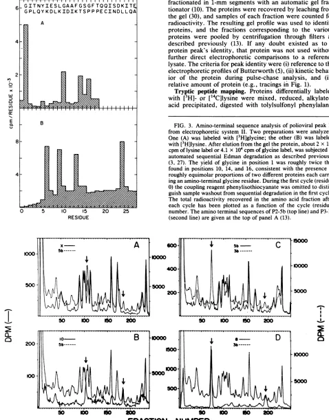
QL _ B FIG. 3. Amino-terminal sequence analysis of polioviral peak 10
from electrophoretic systemII. Two preparations were analyzed. One (A) was labeled with [3H]glycine; the other (B) was labeled 8_ with
[3H]lysine.
After elution from thegel theprotein, about 2x10⁵
cpmof lysine labelor 4.1 x10⁴cpmofglycine label,wassubjectedto_ & m automated
sequential
Edmandegradation
as describedpreviously
(3, 27). The yield ofglycine in position 1 was roughly twice that found in
positions
10, 14, and16,
consistent with the presence of 4 W Xroughly
equimolar proportions
oftwodifferent proteinseachcarry-ing
anamino-terminalglycine
residue.During
thefirstcycle
(residue0)
thecoupling
reagentphenylisothiocyanate
was omittedto distin-guishsamplewashoutfromsequential degradationin thefirstcycle. The total radioactivity recovered in the amino acid fraction after each cycle has been plotted as a function of the cycle (residue)0 5 10 15 20 25 number. The amino terminal sequences of P2-Sb (top line) and P3-lb RESIDUE (second line) are given at the top of panel A(13).
ooo
t
A
X-
5bC
100
500SbX>
0
0
S
0
4~~~~~W
C]
-00
N°
j
j@
so
1
200
o
o
o
Bo
o
200
500~~~5
F
NUBE
o I0- 0~~~~~~~oN
FIG. 4. Trypticpeptide analysisofleucine-labeledproteins 3b, 5b,X,8,and 10. Arrows inpanelsAand Bindicate differencesbetween
proteins5bandX, whichappeartobe accountedforbypeptidesfrom 10. Arrows inpanelsC and Dindicatetwopeptidesfromprotein8that arefoundin 3b,butnot in5b.
[image:3.612.73.551.77.688.2] [image:3.612.80.548.381.699.2]876 PALLANSCH ET AL.
ND
0E
5 10 15 20
RESI DUE
FIG. S. Amino-terminal sequence analysis of protein 8 radiola-beled with [3H]glycine (A) or [3H]lysine (B). The amount of radioac-tivity applied to the sequencer was 15cpmfor glycine and 4.2 x 10⁴ cpm for lysine. A mechanical mnalfunction resulted in the loss of residue 3 from the lysine-labeledprotein. The predictedamino acid sequence for the aminoterminuisof protein P2-3b is indicated at the top ofpanel A.
chloromethyl ketone (TPCK)-trypsin, and analyzed by cat-ion-exchange chromatography exactly as previously
de-scribed (13, 30). If protein sample volumes exceeded 2.2 ml
they were reduced in volume by lyophilization before tryptic
analysis.
Preparationiof radiolabeled proteins for radiochemical mi-crosequencing. Suspension cultures of S3 HeLa cells (2.5 ml
at x 106 cells per ml) were infected at a multiplicity of
PFU per cell. Infected cell cultures were labeled from 3 to h postinfection (400 ,uCiof [3HJglycine or[3H]lysineper ml) and were harvested immediately after the labeling period.
Infected cell lysates were made, the lysates were run on preparative gels, and the proteins were electrophoretically
eluted from gel strips as described previously (27).
Radiochemical microsequencing. Radioactively labeled proteins that had been polyacrylamide gel purified, were
subjected to automated Edman degradation in a Beckman 890C sequencer as described previously (2, 27, 28).
RESULTS
Isolation and identification of polioviral proteins for tryptic peptide analysis. Twodifferent sodium dodecyl sulfate-poly-acrylamide gel systems were used in this study, reflecting the differentlaboratories in which the work was carried out. TheLaemmli system (discontinuous Tris-glyine) was used to isolate proteins used for microchemical sequencing, whereas
a modified Maizel system (continuous, phosphate buffered) was usedfor proteins analyzed by tryptic comparisons. The
electrophoretic patterns obtained with these two systems, with one important exception to be discussed below, were generally very similar (compare Fig. 2 in reference 11 with
Fig. 1in thiswork).With theexceptionof proteins 7a and lc, all of the numbered peaks shown in Fig. 1 were character-ized by tryptic mapping by procedures described previously (13, 25). Thesetryptic maps confirmed all of the previously reported relationships (Fig. IC), and the results will there-fore not be presented exceptasthey relate to the identifica-tion ofpreviously unmapped proteins.
Characterization ofproteins9a and9b.Earlierstudies have shown thatproteins
lb,
2,and4arerelatedby the cleavagepathway
lb -* 2 4(5, 32, 33)via thepathways
2->7c +4(25, 27) and lb 9 + 2 (4, 26). Protein P3-9, also called preVPg-3 (4),contains at its carboxy-terminal end sequences
ofthe genome-linked protein VPg (2, 26). One of the points
clarified by the present study is the relationship of protein P3-9 to peaks 9a and 9b in Fig. 1.
As shown inFig. 2A, the tryptic profile of protein 2 (solid
line) lacks four peaks (arrows) found in protein lb (dotted
line). These four missing peaks were present in digests of
peak9a(solid linein Fig. 2B). The profiles in Fig. 2C show that protein 9b (solid line) is closely related to 9a (dotted
line),but lacks two peaks found in digests of VPg (Fig. 2D). The presenceofVPg sequencesinprotein9aindicatesthat the latterprotein corresponds to protein P3-9, whereas the absence of VPg sequences inprotein9b suggests that it is the
cleavage complement produced by removalofVPg; thus 9a
->9b + VPg.This conclusionwasreinforcedby microchem-ical sequencingstudies. These studiesalsorevealed that the reason protein 9b had not previously been detected in the Laemmli gel system is because it migrates in that system
withband 10. Thus the pattern of glycine andlysineresidues
obtained from sequence analysis of band 10 could not be matched to anysinglestretchof amino acidspredicted bythe
polioviral RNA sequence (Fig. 3). However the sequencing profiles of peak 10 matched perfectly the amino-terminal
sequencespredictedforanequimolarmixture of
proteins
P2-5b and P3-lb. Thus in the first 25residues,glycine appeared at positions 1, 10, 14, and 16 exactly as predicted by theRNAsequence(14) for the amino terminus ofproteinP2-Sb
(first line at the top of Fig. 3); in the corresponding 25 residues ofproteinP3-lbglycine appeared onlyatposition 1
(secondline in Fig. 3). Similarly
lysine
occurredonly once,atposition24, in the first 25 residuesofprotein P2-Sb,but it occurred three times inprotein P3-lb, at
positions
6,9,and 13. We conclude that protein peak 10 on Laemlli gels contained an equimolar mixture of two proteins. Becausepeak 10is distinctfromthepreviouslyidentified
protein
9itmustcontain the proteincorrespondingto9b ingelsystemI (Fig. 1). We show below that the other component probably correspondsto protein 10.
Mapping of protein 10. The presence in
peak
10 ofse-quences from the amino-terminal end of
protein
P2-Sb [image:4.612.84.275.65.479.2]---
,~~~~
50 o 50 00
*RACTIO
NUMBER0
[image:5.612.70.548.75.353.2]NUMBER
FIG. 6. Tryptic peptide analysis ofproteins P3-lb, P3-2, P3-4, 5a, and 7d. Proteins P3-lb, P3-6a, P3-6b, and 7d were labeled with
[14C]leucine; the others, P3-2, P3-4,andSa,werelabeled with[3H]leucine.Filledarrowsinpanels A, C,and Dindicatepeptidesfrom7dthat
arefound inproteins P3-4andP3-6a,butnotinproteinP3-6b.OpenarrowsinpanelsB and D indicatepeptidesofP3-6b andP3-lbthatarenot
found inprotein5a.
gested that it corresponded to the heretofore unidentified
sisterfragment ofproteinP2-Xin thecleavage P2-Sb--
P2-10 + P2-X. The electrophoretically determined molecular weight of protein 10 is alsoconsistent with thishypothesis. Attempts to confirm this identity by using tryptic peptide analysiswereinconclusive, but consistent with this
possibili-ty.Asshownin Fig. 4A, the tryptic profileofproteinP2-5b
(dotted line)containedanumberof minorpeaksnotfound in
proteinP2-X(solid line).Atleasttwoof thesemissingpeaks (arrows) appearedtobe presentin the profilesofprotein10
(FIg. 4B).Thus thecumulativeevidence,basedonsequence
analysis, apparent molecular weight, and tryptic analysis provides consistent evidence in favor of identification of
protein10asthe sisterfragment producedwhenprotein P2-Sb iscleavedtoformprotein P2-X.
Mappingofprotein8. As shown inFig. 4C, proteinP2-3b
(dotted line) containsatleasttwotryptic peptides lackingin
protein 5b(solid line). These twopeaks, shown by arrows, arefound inprotein8 (compare Fig. 4C andD), suggesting
thatprotein 8 is the sister ofprotein P2-5b in the cleavage
reaction P2-3b-* P2-8 + P2-5b. This assignment was
con-firmedby microchemical sequencingwith protein 8
radiola-beled with [3H]glycine or [3H]lysine (Fig. 5). Thus glycine
occurredatpositions 1, 3, and 13,whereaslysine appeared
at positions 7 and 1S, exactly as predicted for the unique
amino-terminal sequence ofprotein P2-3b (Fig. 5A).
Identification of protein7das acleavageproductof protein
P3-2. As showninFig. 6A, tryptic digests of protein 7d(solid line) contain five major peaks (filled arrows) that coincide
with peaks in protein P3-4b (dotted line). These same five peaksalsooccurinprotein P3-6a (Fig. 6C), butnotinprotein
P3-6b (filled arrows, Fig. 6D). This result indicates that
protein 7d is the product of a previously unrecognized cleavageofprotein P3-4b orP3-6a.
Originofprotein 5a. Figure 6B shows that all five major
peaksinthe trypticprofile ofprotein5a(solid line) are also present in the tryptic profile of protein lb (dotted line). In
addition the profile of5aalso contains a humber oftryptic peaks (filled arrows) common toprotein 7d (compare solid
arrowsinFig.6A andB). Finally,anumber of theremaining lbpeptides, i.e.,thosenotpresentin the5aprofile,coincide withpeaks observedindigestsfromprotein P3-6b (compare Fig. 6B and D, open arrows). These results indicate that
peakSaoriginatesfromcleavageofproteinP3-1b;thusP3-lb
-*P3-5a+P3-6b. The kinetic behavior ofpeakSaina
pulse-chaseexperiment (Fig. 1)iscompatiblewiththishypothesis; sois itselectrophoretic mobility,whichis about that expect-ed for the49,000-daltonmasscomputedfor thisproteinfrom
the RNAsequence (Table 1).
DISCUSSION
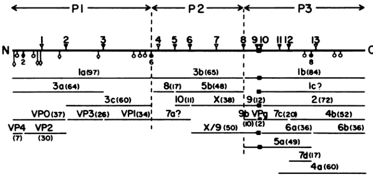
Arevised processing mapof the polioviral polyprotein is
illustrated in Fig. 7. We have divided the polyprotein into
three regions, P1, P2, and P3 (14), allowing for easier
recognition of the numerous cleavage products. For exam-ple, theproteinsare designatedPl-la, P2-3b, P3-7c, etc.
All 27 proteins in this map can be accounted forby 12 cleavage sites(Table 1). Nineof thesecleavages,whichare probably carried out by virus-coded protein P3-7c (11),
occurin glutamine-glycine (QG) pairs, of which thereare a
total of 13 (14). One of these sites, QG-11, is cleaved only rarely, yielding a protein, designated P3-4a (29), which is associated with the viral RNA replication complex (9). In addition to the nine known QG cleavage sites, three other
I
Q-4000
2000
878 PALLANSCH ET AL.
TABLE 1. Proteins of poliovirus
C terminus NH,terminus or orCOOH Protein L434namea Molwtb NH, terminal terminal
cleavagesitec cleavage
site'
P1-la 1 97,247 Blocked G YG1
P3-lb 3 84,234 QG-8 P
3b/9 2-3AB 77,000 YG-1 QG-10
P3-1c 3BCD 76,000d QG-9? P?
P3-2 3CD 72,132 QG-10 P
P1-3a 1ABC 63,786 Blocked G QG-3
P2-3b 2 64,953 YG-1 QG-8
P1-3c 1CD 59,930 QG-2 YG-1
P3-4a 60,000 QG-11 P
P3-4b 3D 52,481 QG-12 P
X/9 2C-3AB 50,000 GQ-6 GQ-10
P3-5ae 48,550 QG-8 YG-2
P2-Sb 2BC 48,273 QG-5 QG-8
VPO 1AB 37,352 Blocked G QG-2
P3-6a 3C' 36,450 QG-10 YG-2
P2-X 2C 37,555 QG-6 QG-8
VP1 1D 33,521 QG-3 YG-1
P3-6b 3D' 35,700 YG-2 P?
VP2 1B 29,985 NS-2 QG-2
VP3 iC 26,410 QG-2 QG-3
P2-7a 2AB 25,500d YG-1? QG-6?
P3-7c 3C 19,669 QG-10 QG-12
P3-7de 16,780 QG-12 YG-2
P2-8 2A 16,680 YG-1 QG-5
P3-9 3AB 12,100 QG-8 QG-10
P3-9b 3B 9,750 QG-8 QG-9
P2-10 2B 10,720 QG-5 QG-6
VP4 10 7,385 Blocked G NS-2
VPg 3B 2,354 QG-9 QG-10
a From R. R. Rueckertand E. Wimmer, submitted for
publica-tion.
bThemolecular weight given for each protein is that predicted
from theRNAsequence (14)using theindicatedamino andcarboxy termini.
Sites are as shown in Fig.7.Glycine(G) is the amino terminus of the polyprotein; phenylalanine (P) is the carboxy terminus. Other abbreviations: N, asparagine; Q, glutamine; S, serine; Y,tyrosine; and ?, uncertain.
dApparent rholecularweight wasdeterminedby electrophoretic mobility in the continuous phosphate-buffered gel system.
eThe molecular weight estimate of P3-7d is basedonthe differ-encein molecularweights betweenP3-6aandP3-7c. The molecular weight estimate of protein Sais basedon the sumofthe molecular weights of proteins 6aand 9a (seethetext).
cleavage sites distinct fromQG cleavageshavebeen
identi-fied. One is an asparagine-serine pairin VPO (16), which is
cleaved by an unidentified protease to form VP4 and VP2 duringthevirion maturation step(12). Theothertwo cleav-age sites are
tyrosine-glycine
(YG) pairs,
one of which is locatedbetweenproteins
laand3b.(14);
theother is locatedbetween6a
and
6b(11).Inhibition
ofviralprocessing
in vitrobyantibodies
against protein
7cfailtoinhibit YGcleavages
(11),
indicating involvementof another protease,possibly
acellular protease with a
chymotrypsin-like
specificity
(15).
Forall threetypesof
cleavages
(QG,YG,
asparagine-serine)
proteolytic processing occurs by a
single
cleavage
eventwithout furthertrimmingof thenewly
generated
amino acid termini(2, 8).In the interest of standardization we have
adopted
the nomenclature of Kitamura et al. (14). Thus theprotein
earlier called 9a in this report is hereafter
designated protein
9.Similarly protein 4 is hereafter designated 4b to distinguish it from 4a(9).
P2familyofproteins.Theidentification ofprotein P2-8as
thecleavage complement ofP2-Sb and of P2-10 as the sister fragment ofX(Fig. 7) completes thelasttwomajor gaps in
the P2region.These assignments do not however
necessari-ly impnecessari-lythatprotein P2-X isderivedonly through cleavage
of precursors P2-3b and P2-Sb, since kineticstudies (M. A.
Pallansch and R. R. Rueckert, manuscript in preparation) indicate thatthepolyproteincanalsobecleavedatsites
QG-5 andQG-6whilestill in thenascent state torelease
proteins
P2-8,P2-10, and P2-X directly as primary cleavageproducts.
Moreover, two otherminor cleavage products, 3b/9 and X/9, which are unusual in that they span the P2-P3 cleavage site
(Fig. 7), have recently been identifiedby immune
precipita-tionandmicrosequence analysis (34). ProteinX/9(molecular weight, 50,000) is similar in size to proteinP3-5a(Table 1), but is probably produced in substantially smaller amounts (34). It may correspond to the unidentified precursor-like proteinthat migrates between bands 4 and Sa (Fig. 1A).
Theassignment of protein P2-7a as an uncleaved formof
proteins (P2-8 andP2-10) is based upon its
electrophoretical-ly determined molecular weight, about 27,400, and is
there-fore stillhighlytentative.Attemptstoconfirm itsidentityby tryptic analysis were hindered by difficulty in completely
freeing P2-7a from the large peak of VP3 with which it comigrates (Fig. 1).
Multiple cleavagepathwaysforP3proteins. The complex-ity of the P3 region of the processing map is due tomultiple
cleavages, a phenomenon first observed in thehomologous
P3 proteins ofhuman rhinovirus 1A (18). The same kind of complexcleavages are observed in the P3 proteins of aphtho-viruses (7),butappear to be absent inthecardioviruses(5). Radiochemical sequencing studies have shown that pro-teinsP3-7c and P3-4b are produced by cleaving protein P3-2
at QG-12 (27), whereas proteins P3-6a and P3-6b are
pro-duced byanalternative cleavage atYG-8 (11); thus (Fig.7) P3-2-*P3-6a +P3-6b. Theidentification of protein P3-7das afragment from the left end ofproteinP3-4 suggests that this protein may arise from acleavage at YG-8 in P3-4b. If P3-7d isproduced by such acleavage, one must conclude that the
cleavages of sites QG-12 and YG-8are notmutually
exclu-sive; in other wordsprotein P3-4b may not bea stable end
productbut could be further cleaved atthe YG-8 site,and thus4b-- 7d +6b;oratsite
QG-12,
and thus 6a-- 7c +7d.In view of the multiple potential cleavage sites in P3-2, however, theprecise mappingofP3-7d mustawait terminal amino acid sequence analysis. Similarlyitappears that the same YG-8 site can be cleaved inprotein lbtoproducethe
cleavage
pathway
lb S-> a + 6b.Finally P3-lbmay be cleavedat QG-11 toyield P3-4a (9,
29), but the corresponding N-terminal fragment of this
cleavage has notbeen detected and may be
rapidly
degrad-ed.
VPg andpotential precursors. ProteinP3-Sa,likeproteins P3-9 and P3-lb, contains VPg sequences (Fig. 7) and is therefore of potential interest as a VPg
donor,
which is believed to be a requirementfor initiating synthesis of newpolioviralRNAmolecules(38).Itis of interest that 5a and lb werenotimmunoprecipitatedfrom labeled
poliovirus-infect-ed HeLa cell extracts by anti-VPg serum made against a
syntheticpeptidecorrespondingtotheseven
carboxy-termi-nalamino acids of VPg (27). As
previously suggested
(26), [image:6.612.61.299.80.402.2]P3
,
4 4
6666
6
la0(97)a
I
I 1
14 5 6 7 8 910
IlI1
I3
ITT T Y T VT
3b(65
I, u8n7)
5b4e
I3c(60)
'a10(11)
X(3s)
'19(12)
VPO(37)
VP3(26)
VPh3
74a?
VP4
VP2
I(7) (30)
V c(20* 4b(52)
X/9(SO)
(-)(2)
60(36) 6b(36)1 .
5o(49)
I 7d('7)
4a
(60)FIG. 7. Processing map of thepolioviral polyprotein. The polyprotein (heavy line) is divided into three regions (P1, P2, and P3) for convenience in classifying cleavage products. Amino acid pairs (sites) known to be cleaved are indicatedby filled symbols; apparently uncleaved sitesareindicatedbyopensymbols: (V, V)glutamine-glycine (QG); (0, 0) tyrosine-glycine (YG); (O, *) asparagine-serine (NS).
Theglutamine-glycine sitesareall believedtobe cleavedbyP3-7c,avirus-encoded coded protease. Theagentsresponsibleforcleavageof
sitesNS-2, YG-6,andYG-8 havenotyetbeen identified. Proteins P3-4a(9,29), X/9 (34),and 3b/9(datanotshown; 34)areproducedinonly trace amounts.Thisimpliesthat site QG-11is cleavedonly rarelyand that siteQG-8israrelyleft uncleaved.Assignmentsforproteins7aand
lcaretentative (see the text). (v)VPg.
otherhand, protein lbandaproteinthatmaycorrespondto
5a(pre-VPg 2)wereimmunoprecipitated byantiseradirected
against larger synthetic peptides (corresponding to all 22or
the 14amino-terminal amino acids ofVPg) (4).Theincreased
efficacyofimmunoprecipitationin thelattercaseisevidently due to the different and increased number of epitopes
present in the longer peptides.
Anothermolecule of interest as apotential VPg donoris protein lc, which has a proteolytic map similar to that of protein P3-2 (37). Thus P3-ic appears to be generated by cleavageofQG-9 justtothe left ofQG-10andwould contain theVPgsequences at itsamino terminus. It isworth noting
inthis regard that therelatively minoramountof these VPg-containing molecules is no argument against their potential importance as VPgdonors since the number of RNA
mole-culesrequiredforsynthesisof the 60-subunitvirionsisonly aminorfractionof the total number of translations.
Finally identification of protein P3-9b as the cleavage complement of VPg suggests that it is generated by the
reaction,9-k 9b +VPg. Proteins P3-9b and P2-10comigrate
intheLaemmligel electrophoresissystem,and this mixture was earlierdesignated P3-10 (26, 29, 34). The nomenclature
used in Fig. 7 will be followed hereafter. The amount of protein P3-9b seen aftera 90-min chase is substantial (Fig. 1B), yet attempts tolocate VPg in infected cells have been
unsuccessful (4, 14, 26) evenwith radioactive amino acids
other than methionine, which is absent from VPg. The
reasonfor this is still unclear.
Otherminorproteins. The map illustrated in Fig. 7
repre-sents over 95% of the total methionine incorporated into poliovirus-infected cells andthereforeencompassesthe ma-jor cleavage reactions. Nevertheless the map does not exhaust the complete repertoire of proteins produced in poliovirus-infected cells(1). Inspection of the
electrophoret-ic profiles inFig. 1reveals several unidentified minorpeaks in the region between la and 2 and between 3a and Sa.
Additional unidentified spots have been detected with two-dimensional electrophoresis, although none is present in large amounts(36). This suggests that someof the cleavage
sites mapped as inactive are in fact cleaved on rare
occa-sions. The possible functional significance of such minor
cleavages is currently unclear.
ACKNOWLEDGMENTS
This work was supported by grant MV-331 from the American CancerSociety, by Public Health Service GrantsAl 15122 andCA 28146 from the National Institutes of Health, and by the U.S. Department of Energy. M.A.P.wasaNationalScienceFoundation predoctoral Fellow, O.M.K. was the recipient ofa Public Health Service Postdoctoral Fellowship from the National Institute of Allergy and Infectious Diseases, and B.L.S. was supported by Public Health Service Postdoctoral Fellowship Al 05935 from the National Institutes ofHealth.
LITERATURE CITED
1. Abraham,G., and P. D.Cooper. 1975. Polioviruspolypeptides examined inmoredetail. J. Gen. Virol. 29:199-213.
2. Adler,C.J.,M. Elzinga,and E. Wimmer. 1983.The genomic-linked protein of picornavirus. VIII. Complete amino acid
sequenceofpoliovirusVPgandcarboxyterminal analysisofits
precursor,P3-9. J. Gen. Virol.64:349-355.
3. Anderson,C. W. 1982. Partialsequencedetermination of meta-bolicallylabeled radioactiveproteinsandpeptides,p. 147-167. In J. Setlow and A. Hollaender(ed.),Geneticengineering, vol. 4.PlenumPublishing Corp.,New York.
4. Baron, M. H., and D. Baltimore. 1982. Antibodies against the chemically synthesized genome-linked protein of poliovirus reactwith native virus-specific proteins.Cell 28:395-404. 5. Butterworth, B. E. 1973. A comparison of the virus-specific
polypeptidesofencephalomyocarditis virus,human rhinovirus-1A,andpoliovirus.Virology56:439-453.
6. Butterworth,B.E., and B. D. Korant. 1974. Characterization of thelarge picornaviral polypeptides producedin thepresenceof zinc ion.J. Virol. 14:282-291.
7. Doel,T.R., D. V.Sangar,D.J. Rowlands, andF.Brown.1978. A re-appraisal of the biochemical map of foot-and-mouth dis-easevirus RNA. J. Gen. Virol. 41:395-404.
8. Emini, E. A., M. Elzinga, and E. Wimmer. 1982. Carboxy-terminal analysis of poliovirus proteins: the termination of poliovirusRNAtranslation and the location ofuniquepoliovirus polyproteincleavage sites.J. Virol. 42:194-199.
9. Etchison,D., and E. Ehrenfeld. 1980. Viralpolypeptides
associ-ated with the RNA replication complex in poliovirus-infected cells. Virology 107:135-143.
.4
Pi
NF
30(64)
C
Ib(84)
Ic?
;-I [image:7.612.125.493.71.246.2]880 PALLANSCH ET AL.
10. Gilson, W., R. Gilson, and R. R.Rueckert. 1972. An automatic high precision acrylamide gel fractionator. Anal. Biochem. 47:321-328.
11. Hanecak, R., B. L. Semler, C. W. Anderson, and E. Wimmer. 1982. Proteolytic processing of poliovirus polypeptides: anti-bodies to polypeptide P3-7c inhibit cleavage at glutamine-glycine pairs. Proc. Natl. Acad. Sci. U.S.A. 79:3973-3977. 12. Jacobson, M. F., J. Asso, and D. Baltimore. 1970. Further
evidence on the formation of poliovirus proteins. J. Mol. Biol. 49:657-669.
13. Kew, 0. M., M. A. Pallansch, D. R. Omilianowski, and R. R. Rueckert. 1980. Changes in three of the four coat proteins of oral poliovaccine strain derived from type A poliovirus. J. Virol. 33:256-263.
14. Kitamura, N., B. L. Semler, P. G. Rothberg, G. R. Larsen, C. J. Adler, A. J. Dorner, E. A. Emini, R. Hanecak, J. J. Lee, S.van der Werf, C. W. Anderson, and E. Wimmer. 1981. Primary structure, gene organization and polypeptide expression of poliovirus RNA. Nature (London) 291:547-553.
15. Korant, B. D. 1972. Cleavage of viral precursorproteins in vivo and in vitro. J. Virol. 10:751-759.
16. Larsen, G. R., C. W. Anderson, A. J. Dorner,B. L.Semler,and E. Wimmer. 1982. Cleavage sites within thepolioviruscapsid protein precursors. J. Virol. 41:340-344.
17. McGregor, S., L. Hall, and R. R. Rueckert. 1975. Evidence for theexistence of protomers in the assembly of encephalomyocar-ditis virus. J. Virol. 15:1107-1120.
18. McLean, C., T. J. Matthews, and R. R. Rueckert. 1976. Evi-dence ofambiguous processing and selectivedegradation inthe noncapsidproteins of rhinovirus 1A.J. Virol. 19:903-914. 19. Medappa, K. C., C. McLean, andR. R. Rueckert. 1971. On the
structure of rhinovirus 1A.Virology 44:259-270.
20. Nomoto, A., T. Omata, H. Toyoda, S. Kuge, H. Horie, Y. Kataoke, Y.Genba, Y.Nakano,and N. Imura. 1982. Complete nucleotide sequence of the attenuatedpoliovirus Sabin1strain genome.Proc. Natl.Acad. Sci. U.S.A. 79:5793-5797. 21. Pallansch, M. A., 0.M. Kew, A.C. Palmenberg, F.Golini,E.
Wimmer,and R. R. Rueckert.1980.PicornaviralVPg sequences are contained in thereplicase precursor. J. Virol.35:414-419. 22. Racaniello,V.R.,and D. Baltimore.1981. Molecularcloningof
polioviruscDNAanddetermination of thecomplete nucleotide sequence of the viral genome. Proc. Natl. Acad. Sci. U.S.A. 78:4887-4891.
23. Rekosh,D.1972.Gene order of thepolioviruscapsid proteins.J. Virol. 9:479-487.
24. Rothberg,P.G., T.J.R. Harris,A. Nomoto,and E.Wimmer. 1978.
04-(5'-Uridylyl)tyrosine
is the bond between the genome-linkedproteinandtheRNAofpoliovirus.Proc. Natl. Acad. Sci. U.S.A. 75:4868-4872.25. Rueckert, R.R.,T.J. Matthews,0.M.Kew,M.Pallansch,C. McLean,and D.Omilianowski. 1979. Synthesis andprocessing
of picornaviral polyprotein, p. 113-125. In R. Perez-Bercoff
(ed.),Themolecularbiologyofpicornaviruses. Plenum Publish-ing Corp., New York.
26. Semler,B.L., C.W.Anderson,R.Hanecak,L. F.Dorner,and E.Wimmer. 1982.Amembrane-associated precursor to poliovi-rus VPg identified by immunoprecipitation with antibodies directedagainst asynthetic heptapeptide. Cell28:405-412. 27. Semler, B.L., C.W.Anderson,N.Kitamura,P. G.Rothberg,
W. L. Wishart, and E. Wimmer. 1981. Poliovirus replication
proteins: RNA sequence encoding P3-lb and the sitesof proteo-lytic processing. Proc. Natl. Acad. Sci. U.S.A.78:3464-3468. 28. Semler, B.L., R. Hanecak,C. W. Anderson, and E. Wimmer.
1981. Cleavage sites in the polypeptide precursors ofpoliovirus protein P2-X. Virology 114:589-594.
29. Semler, B. L., R. Hanecak, L. F. Dorner, C. W.Anderson,and E.Wimmer. 1983. Poliovirus RNA synthesis in vitro: structural elements andantibody inhibition. Virology126:624-635. 30. Shealy, D. J., and R. R. Rueckert. 1978. Proteins of
Rous-associatedvirus61,anavian retrovirus:commonprecursor for glycoproteins gp85 and gp35 and use of pactamycin to map translational order of proteins in the gag,pol, and env genes.J. Virol. 26:380-388.
31. Shih, D. S., C.T.Shih,0. Kew, M. Pallansch, R. Rueckert,and P. Kaesberg. 1978. Cell-free synthesis and processing of the proteins of poliovirus. Proc. Natl. Acad.Sci. U.S.A. 75:5807-5811.
32. Summers,D. F., and J. V.Maizel, Jr.1971.Determination of the gene sequence ofpoliovirus with pactamycin.Proc.Natl. Acad. Sci. U.S.A.68:2852-2856.
33. Taber, R.,D.Rekosh,and D.Baltimore. 1971. Effect of pacta-mycin onsynthesis of poliovirus proteins:amethod forgenetic mapping. J. Virol. 8:395-401.
34. Takegami,T., B. L.Semler,C. W.Anderson,andE.Wimmer. 1983. Membranefractions active in poliovirus RNAreplication containVPg precursor polypeptides. Virology 128:33-47. 35. Vrijsen, R., P. Brioen, and A. Boeye. 1980. Identification of
poliovirus precursor proteinsbyimmunoprecipitation.Virology 107:567-569.
36. Wiegers,K.J., and R. Dernick. 1981. Poliovirus-specific poly-peptides in infected HeLa cells analyzed by isoelectricfocusing and2D-analysis. J. Gen. Virol. 52:61-69.
37. Wiegers,K. J.,and R. Dernick. 1981. Peptide maps of labeled poliovirus proteins after two-dimensional analysis by limited proteolysis and electrophoresis in sodiumdodecyl sulfate. Elec-trophoresis 2:98-103.
38. Wimmer, E. 1982. Genome-linked proteins of viruses. Cell 28:199-201.
![FIG.1.proteinstheminbyfromcontainingof protein Electrophoretic profiles and processing map of polioviral found in lysates of virus-infected HeLa cells (A) after a 10- labeling period with [35S]methionine and (B) as in A, followed a 90-min chase](https://thumb-us.123doks.com/thumbv2/123dok_us/1434608.95972/2.612.65.544.89.679/proteinstheminbyfromcontainingof-electrophoretic-profiles-processing-polioviral-infected-labeling-methionine.webp)
![FIG.S.tivitybeledresiduetopcpmsequenceAmino-terminalsequenceanalysis of protein8 radiola- with [3H]glycine (A) or [3H]lysine (B)](https://thumb-us.123doks.com/thumbv2/123dok_us/1434608.95972/4.612.84.275.65.479/fig-s-tivitybeledresiduetopcpmsequenceamino-terminalsequenceanalysis-protein-radiola-glycine-lysine.webp)
![FIG. 6.foundare[14C]leucine; Tryptic peptide analysis of proteins P3-lb, P3-2, P3-4, 5a, and 7d](https://thumb-us.123doks.com/thumbv2/123dok_us/1434608.95972/5.612.70.548.75.353/fig-foundare-c-leucine-tryptic-peptide-analysis-proteins.webp)