RESEARCH NOTE
Optimized fixation of actin filaments
for improved indirect immunofluorescence
staining of rickettsiae
Monika Danchenko
1, Lucia Csaderova
1, Pierre Edouard Fournier
2and Zuzana Sekeyova
1*Abstract
Objective: The objective was to investigate fixative solutions: 3.7% formaldehyde, 4% paraformaldehyde, 4% para-formaldehyde in the cytoskeletal buffer and 4% parapara-formaldehyde in PHEM buffer (containing PIPES, HEPES, EGTA and MgCl2), applicable for immunofluorescence assay.
Results: Herein we optimized this serological technique, testing four fixative solutions, for the sensitive detection of rickettsial antigens, and preservation of intracellular structures of the host cells, particularly filamentous actin. Rickett-sial antigens were presented equally well both with formaldehyde and all paraformaldehyde-based fixations, but only protocol with 4% paraformaldehyde in PHEM buffer allowed accurate imaging of actin filaments, and simultaneously allows monitoring of rickettsiae using actin-based motility during infection inside the host cells.
Keywords: Immunofluorescence assay, Rickettsiae, Cytoskeleton fixation, Cell culture, Actin-based motility
© The Author(s) 2019. This article is distributed under the terms of the Creative Commons Attribution 4.0 International License (http://creat iveco mmons .org/licen ses/by/4.0/), which permits unrestricted use, distribution, and reproduction in any medium, provided you give appropriate credit to the original author(s) and the source, provide a link to the Creative Commons license, and indicate if changes were made. The Creative Commons Public Domain Dedication waiver (http://creativecommons.org/ publicdomain/zero/1.0/) applies to the data made available in this article, unless otherwise stated.
Introduction
Members of the genus Rickettsia are Gram-negative strictly intracellular Alphaproteobacteria, multiplying by binary fission in the cytoplasm and nuclei of infected host cells [13, 19]. Rickettsiae are arthropod-vectored pathogens, causing from mild to very severe diseases in animals and humans, which are widely distributed throughout the world [2, 20]. Serology using indirect immunofluorescence assay (IFA) is considered as the “gold standard” for diagnostic confirmation [16]. Molec-ular detection using polymerase chain reaction (PCR) demonstrates the presence of the bacterium itself. How-ever, a positive PCR does not predict the viability of bac-teria and does not provide an accurate indication of their localization in tissues.
According to phylogenomic studies, Rickettsia spp. are divided into spotted fever group (SFG), typhus group (TG), transitional group (TRG) and ancestral group (AG)
[8, 18]. SFG rickettsiae and one TG species, R. typhi, can polymerize actin of eukaryotic host cells and use actin-based motility for intra- and intercellular spread. Assem-bly of the filamentous actin “tail” transports the pathogen through the host cytosol and to cell membranes [9, 11]. Therefore, actin “tails” appear as adequate target to be visualized during active rickettsial infection in the eukar-yotic cell.
We would like to investigate four fixative solutions: 3.7% formaldehyde (FA), 4% paraformaldehyde (PFA), 4% paraformaldehyde in cytoskeletal buffer (PFA + CB) [24] and 4% paraformaldehyde in PHEM buffer (PFA-PHEM) [25], with the aim to optimize preservation of rickettsial antigens, the morphology of the host cell constituents, and to visualize dispersal of actin “tails”.
Main text
Methods Reagents
Paraformaldehyde, MES hydrate, EGTA, HEPES, Triton X-100 and Tween 20 were from Sigma-Aldrich (USA). Blotting-grade blocker non-fat dry milk was from Bio-Rad (USA). Potassium chloride, magnesium dichloride,
Open Access
*Correspondence: [email protected]
1 Biomedical Research Center, Slovak Academy of Sciences, Dúbravská
cesta 9, 845 05 Bratislava, Slovakia
potassium dihydrogen phosphate, potassium hydrogen phosphate, sodium hydrogen phosphate, sucrose and formaldehyde (FA) were from Slavus (Slovakia). Sodium chloride was from CentralChem (Slovakia). Potassium hydroxide and sodium hydroxide were from Lachema (Czech Republic). PIPES was from AppliChem (Ger-many). Glutamic acid was from Duchefa Biochemie (Netherlands). Dulbecco’s Modified Eagle’s Medium, trypsin 10x with versene and fetal bovine serum were from Lonza (Switzerland). Vectashield Mounting Medium with DAPI was from Vector Laboratories (UK). We handled all chemical compounds according to the recommendations in their safety data sheets.
Antibodies
Rhodamine Red-X goat anti-rabbit IgG (H + L) polyclonal secondary antibody was from Invitrogen (reference num-ber R6394, lot: 1402199, made in the USA). Alexa Fluor 488 phalloidin was from Invitrogen (reference number A12379, lot: 1378369, made in the USA). We prepared primary anti-Rickettsia akari and anti-Rickettsia slovaca rabbit antibodies (in serum) in our experimental animal facility at Biomedical Research Center.
Bacterial species and host cell line
The bacterial species used in the study were Rickettsia akari, strain MK (Kaplan, ATCC VR-148) and Rickett-sia slovaca, strain 13-B (ATCC VR-1639). Bacteria were propagated in confluent monolayers of Vero cell line (African green monkey kidney cells, ATCC CCL-81), maintained in RPMI 1640 medium supplemented with 2 mM l-glutamine, 25 mM HEPES and heat-inactivated fetal bovine serum at 34 °C in a humidified 5% CO2 incu-bator. Rickettsiae were purified by isopycnic density gra-dient centrifugation [1, 29]. Infections of the host cells were initiated from frozen rickettsial stocks stored in SPG buffer (0.218 M sucrose, 3.76 mM KH2PO4, 7.1 mM K2HPO4 and 4.9 mM potassium glutamate). The concen-tration of the rickettsiae was determined by Real-Time quantitative PCR as described earlier [5].
Immunofluorescence assay
One day prior to the experiment, host cells were trypsi-nized and seeded onto 12-well plates appropriate for tissue culture, containing round glass coverslips and supplemented RPMI medium. Cultured to semi-conflu-ence, Vero cells were infected with rickettsiae (multiplic-ity of infection, MOI = 25). After 48 h, the medium was aspirated, and infected cells were gently washed twice with pre-warmed PBS (0.13 M NaCl, 0.01 M Na2HPO4, 1.5 mM KH2PO4, pH 7.2).
Next, four different fixatives were added for com-parison: 3.7% formaldehyde in PBS pH 7.2 (FA), 4%
paraformaldehyde in PBS pH 7.2 (4% PFA), 4% para-formaldehyde in cytoskeleton buffer (4% PFA + CB) con-taining 10 mM MES hydrate pH 6.1, 1 mM KCl, 3 mM MgCl2, 2 mM EGTA and 0.32 M sucrose [24] and 4% paraformaldehyde in PHEM buffer (PFA-PHEM) con-taining 60 mM PIPES, 25 mM HEPES, 10 mM EGTA, 2 mM MgCl2, pH 6.9 [25]. We allowed fixation at 37 °C for 15 min in the case of 3.7% FA and 4% PFA, or at room temperature for 10 min in the case of 4% PFA-PHEM and 4% PFA + CB.
After removing the fixatives, cells on the coverslips were washed three times with PBS, moderately agitat-ing for 2 min on a shaker. For permeabilization, we added 0.1% Triton X-100 in PBS to each well and cells were incubated at room temperature for 7 min. This step was followed by washing with PBS as described above. Block-ing was performed at 37 °C for 1 h with 5% milk solution in PBS.
After aspiration of the blocking solution, cells on the coverslips were washed, and intracellular bacteria were subsequently probed with a primary antibody. Rabbit serums containing polyclonal anti-Rickettsia antibod-ies were diluted 1:100 with 5% milk in PBS and samples were incubated at 37 °C for 1 h. Each coverslip was then washed three times to remove unbound antibodies by adding 0.1% Tween 20 in PBS, moderately agitating for 5 min on a shaker. For visualization of rickettsiae, we applied fluorophore-conjugated goat anti-rabbit second-ary antibody diluted 1:1000 into 5% milk in PBS. Sub-sequently, cell cultures were incubated at 37 °C for 1 h in the dark. Afterward, the coverslips were thoroughly washed with 0.1% Tween 20 in PBS.
To localize filamentous actin, the cells were probed with fluorescently tagged phalloidin, diluted into 5% milk in PBS according to the manufacturer’s instruction. Plates were incubated at room temperature for 30 min in the dark. After three further prolonged washing with 0.1% Tween 20 in PBS, coverslips were air-dried and counterstain with mounting medium containing DAPI. Specimens were stored at ‒ 20 °C.
Controls
Slides with infected cells treated only with 5% milk in PBS and conjugated secondary antibody mixture were included to ensure the absence of nonspecific reactivity with the sample. Uninfected cells treated with both pri-mary and secondary antibodies were used as negative controls to show the specificity of the primary antibody used for detection of rickettsiae.
Fluorescent microscopy
Germany) in multi-track scanning mode using Plan Apochromat 100×/1.40 oil immersion objective. The microscope settings were as follows: excitation of 420 nm, band pass filter 420–480 nm to visualize nuclei, excitation 488 nm, band pass filter 505–550 nm for actin (fluorophore Alexa Fluor 488), excitation 543 nm and long pass filter over 560 nm for rickettsia (fluoro-phore Rhodamine Red-X). Brightness and contrast were adjusted, if necessary, by LSM Image Browser software from Zeiss.
Results
We used a range of available fluorophores which facili-tate the simultaneous localization of as many as three
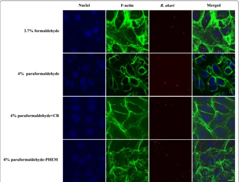
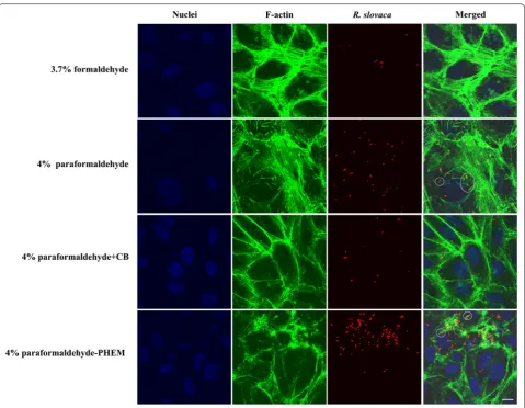
structures/epitopes of interest in a single cell: Rickettsia sp. (Rhodamine Red), filamentous actin (Alexa Fluor 488) and host cell nuclei (DAPI). Representative results from the comparison of the suitability of aldehyde fixatives for infected Vero cells in vitro with two different species of the genus Rickettsia, namely R. akari and R. slovaca, are shown in Figs. 1 and 2, respectively.
A rickettsial antigen is preserved equally well
with formaldehyde and paraformaldehyde‑based fixation We detected selective and robust staining for rick-ettsial antigens with both tested species. We did not observe relevant differences in the quality of the micro-graphs, which would be attributed to the use of either
Fig. 1 Indirect immunofluorescent staining of Vero cells infected with R. akari and comparison of four fixative solutions. We applied an anti-R. akari
[image:3.595.57.540.290.657.2]formaldehyde or paraformaldehyde. Co-localized signal (yellow reaction product—equals a red signal of rickett-siae and green signal of filamentous actin) was demon-strated in almost all captured merge images, providing proof of intracellular localization of bacteria.
Additionally, we also prepared appropriate controls. Unin-fected Vero cells probed with both primary and conjugated secondary antibodies gave no rhodamine red response. Like-wise, slides with infected cells but without primary antibodies against R. akari or R. slovaca displayed no specific red signal.
Fixation with PFA‑PHEM buffer allows accurate imaging of actin filaments
Phalloidin (a fungal toxin of Amanita phalloides that binds specifically to filamentous actin), conjugated to a
fluorophore, showed strong signal intensity with all four tested fixatives in infected (Figs. 1, 2) and control cells.
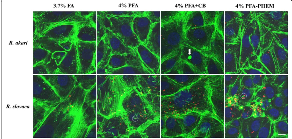
Formaldehyde fixation retained cytoskeletal structure the most poorly compared to other tested fixatives; fila-mentous actin appeared as beads on long fibers, giving it a broken appearance. Paraformaldehyde allowed reveal-ing actin more faithfully as continuous filaments showreveal-ing the overall shape of host cells. We were able to visualize “actin-tail” formation of R. akari and R. slovaca in both, 4% PFA- and PFA-PHEM-fixed infected host cells (high-lighted in white circles in Figs. 1, 2, and 3, respectively).
Vero cell line, mock or infected, treated with 4% PFA + CB gave nonspecific signals more often than samples fixed with 4% PFA or PFA-PHEM (highlighted with a white arrow in Fig. 3) thus, we do not recommend this procedure.
Fig. 2 Indirect immunofluorescent staining of Vero cells infected with R. slovaca and comparison of four fixative solutions. We employed an anti-R. slovaca rabbit serum as primary antibody and goat anti-rabbit Rhodamine Red-X secondary antibody (red fluorescence signal) to probe bacteria. To visualize “comet-tail” formation (in white circles), we applied Alexa Fluor 488 phalloidin for F-actin staining (green fluorescence signal).
[image:4.595.59.539.85.457.2]Paraformaldehyde dissolved in PHEM buffer pre-served the proper architecture of actin filaments forming a distinctive net throughout the cell. Therefore, we rec-ommend using 4% PFA-PHEM buffer as the optimized procedure for immunofluorescent localization of Rick-ettsia sp. and “actin-tail” formation by these pathogens. However, we would like to note, that 4% PFA can also be used as a viable simplified alternative.
The summary of our findings in merged images of infected Vero cells with rickettsiae fixed with different cytoskeletal preserving solutions is shown in Fig. 3.
Discussion
Here we present the effects of four aldehyde-based fixa-tives on Vero cells infected with rickettsiae (namely R. akari and R. slovaca) in vitro and provide a detailed opti-mized protocol for immune-based protein localization in three-color images (Figs. 1, 2 and 3).
The ability of rickettsiae to use actin-based motility was confirmed in several species of the SFG [3, 4, 12,
27]. Rickettsia slovaca, is commonly found in Dermacen-tor ticks in Slovakia [23, 26]. Comparing to R. rickettsii and R. conorii, which both typically exhibit a “comet tail” greater than 10 µm, R. slovaca seems to form it shorter,
about 5 µm [10, 28]. Similarly, R. akari belonging to the transitional group of rickettsiae, promotes directional actin polymerization [12, 14].
Indirect immunofluorescence is widely used for the detection of pathogens in cell cultures [6, 15]. The first step in any successful immunocytochemical localization is the choice of fixative and fixation conditions [17].
Formaldehyde (the 1st alternative of fixation) works by chemically bonding adjacent macromolecules (neigh-bouring proteins) together, locking them in place with little morphological damage, so-called cross-linking [30].
Paraformaldehyde (the 2nd) is polymeric formaldehyde dissolved in water or a buffer. The free methanediols in the resulting solution are reactive with amine groups on proteins and other cellular structures that contain nitrogen. PFA crosslinks amino groups without chang-ing the tertiary structure of proteins, therefore leavchang-ing most epitopes available for binding antibodies [7, 21]. Paraformaldehyde dissolved in the cytoskeletal buffer (PFA + CB) (the 3rd) with the inclusion of sucrose keeps the cell iso-osmotic, which also might help to preserve internal cell structures. Similarly, paraformaldehyde in PHEM buffer (the 4th) containing a high concentration of EGTA (10 mM or more) conserves structural integrity of all fibrous components of the cytoskeleton [22].
[image:5.595.55.539.419.650.2]Conclusions
We showed that an optimized fixation of actin filaments with 4% PFA-PHEM improved indirect immunofluores-cence staining of rickettsiae. This allowed to accurately visualizing the actin-based motility of R. akari and R. slovaca. We suggest this approach as possibly applicable for imaging of other intracellular bacteria in host cells as well.
Limitations
• choice of fixative
• choice of fixation conditions • sensitivity of antibodies
• specificity for an antigen–antibody interaction.
Abbreviations
AG: ancestral group; FA: formaldehyde; IFA: immunofluorescence assay; MOI: multiplicity of infection; PBS: phosphate-buffered saline; PCR: polymerase chain reaction; PFA: paraformaldehyde; PFA + CB: paraformaldehyde in cytoskeletal buffer; PFA-PHEM: paraformaldehyde in PHEM buffer; PHEM buffer: containing PIPES, HEPES, EGTA and MgCl2; SFG: spotted fever group; TG:
typhus group; TRG : transitional group.
Acknowledgements
The authors thank the Renáta Szalayová for her enthusiastic participation and professional technical assistance.
Authors’ contributions
MD designed and performed the experiments. MD and ZS wrote the manu-script. LC and PEF analysed data and revised the manumanu-script. ZS coordinated the study. All authors read and approved the final manuscript.
Funding
This study was financially supported by the projects: VEGA 2/0010/19 from the Scientific Grant Agency of Ministry of Education, Science, Research and Sport, as well as SRDA-0280-12 (the study design, data collection and analysis), and SRDA SK-FR-2017-0005 from the Slovak Research and Development Agency, and the French National Research Agency under the program “investisse-ments d’avenir”, reference ANR-10-IAHU-03 (interpretation of data, decision to publish and preparation of the manuscript).
Availability of data and materials
The datasets used and analysed during the current study are available from the corresponding author on reasonable request.
Ethics approval and consent to participate
The usage of animals in the experiment was approved by the Ethical Com-mittee of the Biomedical Research Center, Slovak Academy of Sciences and the State Veterinary and Food Administration of the Slovak Republic (Permit Number: SK U 01016 292/16-221j). The experiments were performed under standard conditions in the experimental animal facility of the Biomedical Research Center, Slovak Academy of Sciences.
Consent for publication
Not applicable.
Competing interests
The authors declare that they have no competing interests.
Author details
1 Biomedical Research Center, Slovak Academy of Sciences, Dúbravská cesta
9, 845 05 Bratislava, Slovakia. 2 UMR VITROME, AMU, IRD SSA, AP-HM, IHU
Mediterranee Infection, Marseille, France.
Received: 23 July 2019 Accepted: 3 October 2019
References
1. Ammerman NC, Beier-Sexton M, Azad AF. Laboratory mainte-nance of Rickettsia rickettsii. Curr Protoc Microbiol. 2008. https ://doi.
org/10.1002/97804 71729 259.mc03a 05s11 .
2. Azad AF, Beard CB. Rickettsial pathogens and their arthropod vectors. Emerg Infect Dis. 1998;4(2):179–86.
3. Baldridge GD, Burkhardt N, Herron MJ, Kurtti TJ, Munderloh UG. Analysis of fluorescent protein expression in transformants of Rickettsia mona-censis, an obligate intracellular tick symbiont. Appl Environ Microbiol. 2005;71(4):2095–105.
4. Balraj P, El KK, Vestris G, Espinosa L, Raoult D, Renesto P. RickA expression is not sufficient to promote actin-based motility of Rickettsia raoultii. PLoS ONE. 2008;3(7):e2582.
5. Bohacsova M, Filipcik P, Opattova A, Valarikova J, Quevedo DM, Skultety L, Novak M, Sekeyova Z. Survival of rat cerebrocortical neurons after rickettsial infection. Microbes Infect. 2015;17(11–12):845–9.
6. Fritschy JM, Härtig W. Indirect immunofluorescence of cultured cells. New York: Wiley; 2001.
7. Fujiwara K. Techniques for localizing contractile proteins with fluorescent antibodies. Curr Top Dev Biol. 1980;14(Pt 2):271–96.
8. Gillespie JJ, Beier MS, Rahman MS, Ammerman NC, Shallom JM, Pur-kayastha A, Sobral BS, Azad AF. Plasmids and rickettsial evolution: insight from Rickettsia felis. PLoS ONE. 2007;2(3):e266.
9. Goldberg MB. Actin-based motility of intracellular microbial pathogens. Microbiol Mol Biol Rev. 2001;65(4):595–626.
10. Gouin E, Gantelet H, Egile C, Lasa I, Ohayon H, Villiers V, Gounon P, Sanson-etti PJ, Cossart P. A comparative study of the actin-based motilities of the pathogenic bacteria Listeria monocytogenes, Shigella flexneri and Rickettsia conorii. J Cell Sci. 1999;112(Pt 11):1697–708.
11. Heinzen RA. Rickettsial actin-based motility: behavior and involvement of cytoskeletal regulators. Ann N Y Acad Sci. 2003;990:535–47.
12. Heinzen RA, Hayes SF, Peacock MG, Hackstadt T. Directional actin polym-erization associated with spotted fever group Rickettsia infection of vero cells. Infect Immun. 1993;61(5):1926–35.
13. Heymann WR. Rickettsial microbiology. Clin Dermatol. 1996;14(3):243–4. 14. La SB, Meconi S, Fenollar F, Rolain JM, Roux V, Raoult D, et al. Emended
description of Rickettsia felis (Bouyer et al. 2001), a temperature-depend-ent cultured bacterium. Int J Syst Evol Microbiol. 2002;52(Pt 6):2035–41. 15. Levin PA. Light microscopy techniques for bacterial cell biology. Methods
in microbiology, vol. 31. London: Academic Press Ltd.; 2002. p. 115–32. 16. Luce-Fedrow A, Mullins K, Kostik AP, St John HK, Jiang J, Richards AL.
Strat-egies for detecting rickettsiae and diagnosing rickettsial diseases. Future Microbiol. 2015;10(4):537–64.
17. McCaffery JM, Farquhar MG. Localization of GTPases by indirect immu-nofluorescence and immunoelectron microscopy. Methods Enzymol. 1995;257:259–79.
18. Merhej V, Angelakis E, Socolovschi C, Raoult D. Genotyping, evolution and epidemiological findings of Rickettsia species. Infect Genet Evol. 2014;25:122–37.
19. Olano JP. Rickettsial infections. Ann N Y Acad Sci. 2005;1063:187–96. 20. Perlman SJ, Hunter MS, Zchori-Fein E. The emerging diversity of Rickettsia.
Proc Biol Sci. 2006;273(1598):2097–106.
21. Robinson RW, Snyder JA. An innovative fixative for cytoskeletal compo-nents allows high resolution in colocalization studies using immunofluo-rescence techniques. Histochem Cell Biol. 2004;122(1):1–5.
22. Schliwa M, van Blerkom J. Structural interaction of cytoskeletal compo-nents. J Cell Biol. 1981;90(1):222–35.
•fast, convenient online submission •
thorough peer review by experienced researchers in your field • rapid publication on acceptance
• support for research data, including large and complex data types •
gold Open Access which fosters wider collaboration and increased citations maximum visibility for your research: over 100M website views per year •
At BMC, research is always in progress.
Learn more biomedcentral.com/submissions
Ready to submit your research? Choose BMC and benefit from:
24. Small JV, Rinnerthaler G, Hinssen H. Organization of actin meshworks in cultured cells: the leading edge. Cold Spring Harb Symp Quant Biol. 1982;46(Pt 2):599–611.
25. Sobue K, Fujio Y, Kanda K. Tumor promoter induces reorganization of actin filaments and calspectin (fodrin or nonerythroid spectrin) in 3T3 cells. Proc Natl Acad Sci USA. 1988;85(2):482–6.
26. Spitalska E, Stefanidesova K, Kocianova E, Boldis V. Rickettsia slovaca and Rickettsia raoultii in Dermacentor marginatus and Dermacentor reticulatus ticks from Slovak Republic. Exp Appl Acarol. 2012;57(2):189–97. 27. Teysseire N, Chiche-Portiche C, Raoult D. Intracellular movements of
Rick-ettsia conorii and R. typhi based on actin polymerization. Res Microbiol. 1992;143(9):821–9.
28. Van Kirk LS, Hayes SF, Heinzen RA. Ultrastructure of Rickettsia rickett-sii actin tails and localization of cytoskeletal proteins. Infect Immun. 2000;68(8):4706–13.
29. Weiss E, Coolbaugh JC, Williams JC. Separation of viable Rickettsia typhi from yolk sac and L cell host components by renografin density gradient centrifugation. Appl Microbiol. 1975;30(3):456–63.
30. Wheatley SP, Wang YL. Indirect immunofluorescence microscopy in cultured cells. Methods Cell Biol. 1998;57:313–32.
Publisher’s Note