Proteomic Analysis Reveals Age-related Changes in Tendon
Matrix Composition, with Age- and Injury-specific Matrix
Fragmentation
*
□SReceived for publication, March 19, 2014, and in revised form, July 16, 2014Published, JBC Papers in Press, July 30, 2014, DOI 10.1074/jbc.M114.566554 Mandy J. Peffers‡1,2, Chavaunne T. Thorpe‡§1,3, John A. Collins‡, Robin Eong‡¶, Timothy K. J. Wei‡¶ Hazel R. C. Screen§, and Peter D. Clegg‡
From the‡Department of Musculoskeletal Biology, Institute of Ageing and Chronic Disease, University of Liverpool, Leahurst
Campus, Neston CH64 7TE,§Institute of Bioengineering, Queen Mary University of London, Mile End Road, London E1 4NS, and
¶School of Life Sciences, Ngee Ann Polytechnic, Singapore 599489
Background:Alterations in tendon matrix composition with aging and injury are poorly understood.
Results:Aging and injury resulted in distinct protein profiles, with age-specific peptide fragmentation in injury.
Conclusion:Identification of protein cleavages associated with aging and injury suggest impaired maintenance and repair in aged tendon.
Significance:Novel peptide fragments identified are potential biomarkers of tendon injury and age-related degeneration.
Energy storing tendons, such as the human Achilles and equine superficial digital flexor tendon (SDFT), are highly prone to injury, the incidence of which increases with aging. The cel-lular and molecular mechanisms that result in increased injury in aged tendons are not well established but are thought to result in altered matrix turnover. However, little attempt has been made to fully characterize the tendon proteome nor determine how the abundance of specific tendon proteins changes with aging and/or injury. The aim of this study was, therefore, to assess the protein profile of normal SDFTs from young and old horses using label-free relative quantification to identify differ-entially abundant proteins and peptide fragments between age groups. The protein profile of injured SDFTs from young and old horses was also assessed. The results demonstrate distinct proteomic profiles in young and old tendon, with alterations in the levels of proteins involved in matrix organization and regu-lation of cell tension. Furthermore, we identified several new peptide fragments (neopeptides) present in aged tendons, sug-gesting that there are age-specific cleavage patterns within the SDFT. Proteomic profile also differed between young and old injured tendon, with a greater number of neopeptides identified in young injured tendon. This study has increased the knowl-edge of molecular events associated with tendon aging and injury, suggesting that maintenance and repair of tendon tissue may be reduced in aged individuals and may help to explain why the risk of injury increases with aging.
Current descriptions of tendon extracellular matrix (ECM)4 in the literature list the main components as collagen type I, proteoglycans (predominantly small leucine-rich proteogly-cans (SLRPs)) (1), minor collagens (types III, V, VI, XII) (2), elastic fibers (3), and glycoproteins (4). The most abundant pro-tein, collagen type I, aligns with the tendon long axis and aggregates in a series of hierarchical levels to form fibrils, fibers, fascicles, and finally the whole tendon (5). At the larger hierar-chical levels the collagen is interspersed with a proteoglycan-rich matrix. This multilevel fiber composite organization results in a tendon with high uniaxial strength that is able to resist the large tensional forces experiencedin vivo.
It is well established that tendon functional integrity decreases with aging, predisposing aged tendons to degenera-tion and injury (6, 7). Addidegenera-tional risk factors for tendon injury include high levels of repetitive loading (8), genetic factors (9, 10), and chronic inflammation (11). However, the cellular and molecular mechanisms underpinning this increased risk in ten-dons are not well understood. Several studies have reported alterations in matrix content as a function of aging, including increased type III collagen (12), changes in cross-link profile as a result of glycation (13, 14), and accumulation of partially degraded collagen within the matrix (14). Age-related altera-tions to the non-collagenous matrix have also been identified, with decreased glycosaminoglycan and cartilage oligomeric matrix protein (COMP) levels (15, 16). Several of these studies indicate altered matrix turnover with aging. In cartilage, ECM fragmentation patterns have demonstrated novel potential sub-strates and cleavage sites for specific enzymes (17). Although a recent study has identified stage-specific peptide fragments in tendon disease (18), there is also a need to identify age-specific *This work was supported by a project grant from the Horserace Betting Levy
Board, UK (prj/752) and the Biosciences and Biotechnology Research Council, UK (BB/K008412/1).
Author’s Choice—Final version full access.
□S This article containssupplemental Table 1– 4. 1Both authors contributed equally to this work.
2Supported by Wellcome Trust Integrated Veterinary Research Training
Fel-lowship WT088557MA.
3To whom correspondence should be addressed: Institute of
Bioengineer-ing, School of Engineering and Materials Science, Queen Mary University of London Mile End Rd., London E1 4NS, UK. Tel.: 44-207-882-5368; E-mail: [email protected].
4The abbreviations used are: ECM, extracellular matrix; ANOVA, analysis of
variance; COMP, cartilage oligomeric matrix protein; DAVID, Database for Annotation, Visualization, and Integrated Discovery; GdnHCl, guanidine hydrochloride; GO, gene ontology; SDFT, superficial digital flexor tendon; SLRP, small leucine-rich proteoglycan; STRING, Search Tool for Retrieval of Interacting Genes/Proteins; Bis-Tris, 2-[bis(2-hydroxyethyl)amino]-2-(hydroxymethyl)propane-1,3-diol.
at UNIV OF NOTTINGHAM on April 16, 2015
http://www.jbc.org/
cleavage sites in tendon as this will enable the understanding and distinction of the ECM degradative mechanisms associated with aging and disease.
It is important to further characterize the tendon ECM and identify aging changes in both health and disease, as it is likely that alterations to minor matrix components may have a pro-found influence on tendon function. However, some minor components of the tendon matrix may not yet have been iden-tified. Although proteomic analysis has been used to identify many novel proteins in other connective tissues such as carti-lage (19, 20), a review of the current literature shows few studies that have undertaken a proteomic analysis of tendon. Consid-ering the studies that have addressed tendon proteomics, some have assessed the proteins produced by tendon fibroblastsin vitro(21, 22), whereas others have investigated alterations in protein profile as a result of artificially induced injury (23, 24). Smithet al.(25) investigated changes in pericellular proteins during development, and Dakinet al.(18) studied normal and diseased tendons from horses with a wide age range but do not report any data regarding age-related alterations in protein content. To the authors’ knowledge, no studies have assessed age- and injury-associated changes in the tendon extracellular matrix protein profile.
In the current study we used equine tendon tissue to study the effect of aging and injury on tendon matrix composition. The horse is an accepted and relevant model in which to study musculoskeletal aging and injury, as it is a relatively long-lived species in which age-related musculoskeletal diseases, such as tendon injury, show a very similar epidemiology, etiology, and pathology to that seen in human age-related musculoskeletal diseases (14, 26–30). In both species the most commonly injured tendons are those that store and return energy during locomotion. In the human it is the Achilles tendon that is the major energy store and the most prone to injury (31), whereas in the horse, the predominant energy store is the superficial digital flexor tendon (SDFT) (32). We, therefore, assessed the protein profile of normal and injured SDFTs from young and old horses using label-free relative quantification to identify differentially abundant proteins between age groups. Further-more we investigated age-specific cleavage patterns in the ECM by assessing fragmentation patterns of specific matrix mole-cules to identify neopeptides in injured and aged tendon. One way to provide new insights into the development and treat-ment of tendon disease is to obtain an understanding of how tendon undergoes the physiological remodeling that is evident in aging. We hypothesized that we would identify age-related alterations in ECM proteins and neopeptides within the tendon matrix, with greater matrix fragmentation evident in injured tendon.
EXPERIMENTAL PROCEDURES
All chemicals were supplied by Sigma unless otherwise stated.
Tendon Sampling and Procurement—Forelimbs, distal to the carpus, were collected from half to full thoroughbred horses (young, 3.3! 0.6 years; old, 19.0! 1.7 years, both n" 3), euthanized at a commercial equine abattoir. Only tendons that had no evidence of previous tendon injury at post-mortem
examination were included in the study. The SDFT was dis-sected free from the limbs from the level of the carpus to the metacarpophalangeal joint. Fascicles (length of 25 mm, diame-ter of 0.2–0.4 mm, weight of#0.3 g) were dissected in duplicate from the mid-metacarpal region of the tendon as described previously (33). The fascicles were snap-frozen in liquid nitro-gen and stored at$80 °C until further analysis.
Protein Extraction and Sample Preparation—Each thawed tendon sample (fascicle) was transferred into an Eppendorf tube containing 200 !l of 100 mM Tris acetate, protease inhibitors (Complete Protease Inhibitors, EDTA-free, Roche Applied Science), and 0.1 unit of chondroitinase ABC, pH 8.0, and deglycosylated for 6 h at 37 °C. The supernatant was removed after centrifugation at 13,000%gfor 5 min. 0.5 ml of guanidine extraction buffer (4 M guanidine hydrochloride (GdnHCl), 65 mM dithiothreitol (DTT), and 50 mMsodium acetate, pH 5.8) was added, and extraction was performed with end-over-end mixing for 48 h at 4 °C. 25 mMDTT was added 2 h before the addition of 80 mMiodoacetamide, the latter for the last 2 h in the dark. The soluble fraction was removed after centrifugation for 15 min at 13,000%gat 4 °C. The final insol-uble fraction was incubated in 0.5 ml of 100 mMacetic acid containing 100!g/ml pepsin overnight at 4 °C with end-over-end mixing to release collagenous polypeptides. The superna-tant was removed after centrifugation at 13,000%gfor 15 min at 4°C. This was lyophilized, resuspended in water, re-lyophi-lized, and stored at$80 °C. Protein concentrations of aliquots of soluble fraction were estimated by the Bradford assay using Coomassie PlusTMprotein assay reagent (Thermo Scientific, Rockford, IL) read at 660 nm after acetone precipitation.
One-dimensional Sodium Dodecyl Sulfate Polyacrylamide Gel Electrophoresis (SDS-PAGE) and In-gel Trypsin Digestion— Tendon GdnHCl soluble extracts were analyzed by one-dimen-sional SDS-PAGE to assess gross quantitative/qualitative dif-ferences in protein profiles between young and old tendon. Samples were loaded according to equal volumes after acetone precipitation and resolubilization in buffer containing 8Murea, 2% (w/v) CHAPS and 0.0002% (v/v) bromphenol blue plus 0.2% (v/w) DTT.
Aliquots were heated in Laemmli buffer containing 50 mM DTT for 5 min at 95 °C and resolved through 4–12% acrylam-ide Bis-Tris NuPAGE gels (Invitrogen), and proteins were visu-alized using a silver staining kit (Thermo Scientific) according to the manufacturer’s instructions. In-gel tryptic digestion of dominant bands was undertaken as previously described (34).
To detect pepsin-released collagenous polypeptides, the lyoph-ilized samples were reconstituted in 0.1Macetic acid containing pepsin at 100!g/ml and shaken overnight at 4 °C. After centrifu-gation at 13,000%gfor 15 min, the supernatant was removed, lyophilized, and resuspended in water before re-lyophilizing and heating in 20!l of Laemmli buffer containing 50 mMDTT for 5 min at 95 °C. The material was resolved using 3–8% acrylamide Tris acetate gels (Invitrogen) and silver-stained.
Protein Identification of Bands Using LC-MS/MS—Peptides were analyzed using a Bruker Amazon ion trap mass spectrom-eter coupled to a Waters nanoACQUITY UltraPerformance liquid chromatography system. The samples were injected onto a reverse phase column (Acquity ethylene bridged hybrid C18,
at UNIV OF NOTTINGHAM on April 16, 2015
http://www.jbc.org/
75!m%150 mm, 1.7!m) and eluted over a 1-h gradient. The mass spectrometer was set up in positive ion mode and cali-brated with Bruker calibration mix. Spectra were acquired between 300 and 1800m/zwith an ion charge count target of 200,000. Up to five precursor ions above a threshold of 10,000 were selected for MSMS fragmentation per MS scan. Each pre-cursor was fragmented twice, and then the mass was excluded for 1 min. Singly charged ions were excluded. Data were searched against theEquus caballusdatabase; Ensembl data-base for horse (E. caballus; EquCab2.56.pep) using an in-house Mascot server (Matrix Science, London, UK). Parameters were set to accept one miscleavage, a fixed modification of carbam-idomethly cysteine, and a variable oxidation of methionine. The peptide mass tolerance for this instrument was set at 0.4 Da.
Protein In-solution Trypsin Digestion and Mass Spectrometry Using Linear Ion-trap Orbitrap Mass Spectrometer (LTQ-Or-bitrap Velos)—Proteomic analyses were performed to identify cellular and matrix proteins present within normal tendon tis-sue, the relative levels of these proteins, and also to identify neopeptides of specific ECM proteins. GdnHCl-extracted pro-teins were washed with 100 mMammonium bicarbonate to give a final concentration of 0.5MGdnHCl. Tryptic digestion was undertaken as previously described (20) but with the addition of a top-up of a further 2!g after 3 h. LC-MS/MS analysis was performed using nanoAcquityTM ultraperformance LC (Waters, Manchester, UK) on-line to an LTQ-Orbitrap Velos mass spectrometer (Thermo-Fisher Scientific, Hemel Hemp-stead) as previously described (20) via an electrospray ioniza-tion ion source containing a 10-!m coated Pico-tip emitter (Presearch LTD, Basingstoke, UK). Aliquots of tryptic peptides equivalent to 300 ng of tendon fascicle protein were loaded onto a 180-!m % 20-mm C18 trap column (Waters) at 5 !l/min in 99% solvent A (water plus 0.1% formic acid) and 1% solvent B (acetonitrile plus 1% formic acid) for 5 min and sub-sequently back-flushed onto a C18 pre-equilibrated analytical column (75-!m % 15-mm Waters) using a flow rate of 0.3 !l/min. Xcalibur 2.0 software (Thermo-Electron, Hemel Hempstead, UK) was used to operate the LTQ-Orbitrap Velos mass spectrometer in data-dependant acquisition mode. The survey scan was acquired in the Orbitrap with a resolving power set to 30,000 (at 400m/z). MS/MS spectra were concurrently acquired on the 20 most intense ions from the high resolution survey scan in the LTQ mass spectrometer. Charge state filter-ing&1 was used where unassigned precursor ions were not selected for fragmentation. Fragmentation parameters in the LTQ mass spectrometer were: normalized collision energy, 30; activation, 0.250; activation time, 10 ms; minimum signal threshold, 500 counts with isolation width 2m/z.
Label-free Peptide Quantification—For label-free quantifica-tion of the tendon fascicles the Thermo raw files of the acquired spectra from in-solution tryptic digests of normal young (n"3) and old (n"3) equine tendon fascicles were analyzed by the ProgenesisTMLC-MS software (Version 4, Nonlinear Dynam-ics) for label-free quantification as previously described (20). Briefly, after the selection of a reference sample, the retention times of the other samples were aligned. Feature picking used the top three spectra for each feature. These were exported from ProgenesisTM-LC-MS and utilized for peptide
identifica-tion with a locally implemented Mascot server (Version 2.3.01) in theE. caballus database. Search parameters used were: 10 ppm peptide mass tolerance and 0.6-Da fragment mass toler-ance; one missed cleavage allowed; fixed modification, carbam-idomethylation; variable modifications, methionine oxidation, proline oxidation, and lysine oxidation. To maximize the num-ber of quantifiable proteins but simultaneously use an accepta-ble false discovery rate (FDR), the peptide matches above an identity threshold were adjusted to give an FDR of 1% before the protein identifications being re-imported into ProgenesisTM.
Mascot determined peptides with ion scores of 20 and above, and only proteins with at least one unique peptide ranked as the top candidate were considered and re-imported into Proge-nesisTM software. For quantification, only unique peptides were included. Statistical analysis was performed on all detected features using transformed normalized abundances for one-way analysis of variance (ANOVA). All peptides (with Mascot score&23 andp'0.05) of an identified protein were included, and the proteinpvalue (one-way analysis of variance) was then performed on the sum of the normalized abundances for all runs. Adjusted analysis of variance values ofp'0.05 and additionally regulation of&2-fold or'0.5-fold were regarded as significant.
Neopeptide Identification—For neopeptide determination, mass spectrometry data from the in-solution tryptic digests of normal young (n"3) and old (n"3) equine tendon fascicles were analyzed. Neopeptides were identified by searches against the Unihorse database using Mascot. Search parameters used were: enzyme, none; peptide mass tolerances 10 ppm; fragment mass tolerance of 0.6 Da, 1(, 2(, and 3(ions; missed cleavages 1; instrument type electrospray ionization-TRAP. Modifica-tions included were; fixed, carbamidomethyl cysteine and vari-able, oxidation of methionine, proline, and lysine. The proba-bility that a match was correct (p'0.05) was determined using the Mascot-derived ion score, wherepwas the probability that the observed match was a random event. As the cost of mass spectrometry analyses of a large number of samples was pro-hibitive and to have confidence in our analysis, we only included neopeptides in the results if they were identified by Mascot more than once per donor and in"2 donors. Patterns of fragmentation were determined for aggrecan, biglycan, decorin, fibromodulin, COMP, lumican, and collagens.
Gene Ontology, Pathway Enrichment Analysis, and Protein Network Analysis—The gene symbols for each identified pro-tein in normal tendon were searched in the Ensembl database for horse and converted to the gene symbol of the correspond-ing human orthologue. The resultcorrespond-ing gene list was used for gene ontology (GO) using the Database for Annotation, Visualiza-tion, and Integrated Discovery (DAVID) Version 6.7. In addi-tion, the list was used for protein network analysis with the Search Tool for Retrieval of Interacting Genes/Proteins (STRING) tool Version 9.1 (35). The protein interaction maps were created by allowing for experimental evidence in addition to the predicted functional links: co-occurrence, co-expression, databases, and text-mining.
Western Blotting Validation of Fibromodulin Abundance— To validate the decreased fibromodulin abundance with aging in normal tendon, soluble proteins were extracted from
at UNIV OF NOTTINGHAM on April 16, 2015
http://www.jbc.org/
rate donors (3 young (4 years old) and 3 old (&20 years old)) using methods described previously (20). Briefly, 20!g of sol-uble protein extracts were electrophoresed and separated on 4–12% SDS-PAGE gels (Nu-Page, Invitrogen). Nitrocellulose membranes were probed with primary antibodies against the following: mouse polyclonal to fibromodulin (1:2000 dilution, #67596 Abcam) and #-tubulin (1:1000 dilution) (#4074, Abcam, Cambridge, UK) as the loading control. Membranes were washed and incubated in a secondary horseradish perox-idase-conjugated antibody (1:2000 dilution). Blots were imaged using VisionWorksLS image acquisition software package, and band densities were analyzed using ImageJ 1.42. Results were normalized to the loading control.
Real-time Polymerase Chain Reaction (RT-PCR) of Keratin Expression—Samples of normal SDFT RNA from an indepen-dent cohort (young, 5.7!1.3 years; old, 23.3!3.1 years (both
n"7)) were used to assess age-related alterations in keratin gene expression in normal tendon using previously described methods (36). Exon-spanning primer sequences were designed and validated by PrimerDesign Ltd (Southampton, UK) except for the normalization gene glyceraldehyde-3-phosphate dehy-drogenase (GAPDH) (37). The primer pairs were for keratin type 2, cytoskeletal 75;KRT75 (forward reverse), and keratin type 2, cytoskeletal 5;KRT5 (forward reverse).
Injured Tendon Study—Injured SDFTs were collected from young (6.3!2.1 years,n"3) and old horses (19.5!3.5 years,
n"2) euthanized at a commercial equine abattoir. In all ten-dons injury was localized to the core of the mid-metacarpal region. All injuries were macroscopically graded as mild-to-moderate in severity, with the appearance of a hemorrhagic lesion (38) but without loss of fascicular pattern (39). Fascicles were dissected from the lesion, proteins were extracted, and
one-dimensional SDS-PAGE was performed as described for normal tendon. Samples were trypsin-digested and processed for LCMS-MS as described above. Peptides were quantified using Progenesis software, and neopeptides were identified. Protein networks were identified as described above. Due to normal and diseased samples being run at different times, it was not possible to directly compare normal and diseased groups.
Statistical Analysis—Statistically significant differences in the number of proteins and gene expression were identified after log10transformation to ensure normal distribution using Student’s t test. Statistical analyses were undertaken using S-Plus and Excel software.
RESULTS
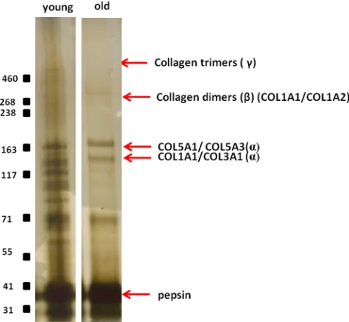
SDS-PAGE Comparative Analysis of Protein Extracts— One-dimensional-SDS-PAGE of the GdnHCl soluble protein extracts demonstrated differences in the intensity of the stain-ing between samples from normal young and old horses (Fig. 1). The decreased staining in old samples suggests that the extract-ability of proteins was reduced in older tendon. However, the soluble protein concentrations, corrected to wet weight of ten-don fascicle, did not decrease with increasing age (53! 1.2 !g/mg for young and 51!0.6!g/mg for old). The major pro-teins identified in each band using LC-MS/MS are indicated in Table 1. In addition, we undertook pepsin digestion of the insol-uble extract remaining after GdnHCl extraction to analyze non-soluble collagenous polypeptides (19). A number of addi-tional bands were evident in the pepsin digest of young tendon (Fig. 2).
[image:4.594.133.466.57.304.2]Protein Identification and Ontology—A total of 252 proteins were identified in combined samples from normal tendon; 230 with a significant Mascot score of&23.Supplemental Table 1
FIGURE 1.Silver-stained one-dimensional-SDS-PAGE of the guanidine-soluble protein extract of normal young (n!3) and old (n!3) tendon and diseased young (n!3) and old (n!2).Equal protein loading by volume (20!l per well) allowed a qualitative comparison of soluble tendon protein extracts. The most abundant protein bands are marked withred squares(1– 4) were excised from the gel and trypsin-digested, and the protein content of each single band was analyzed from peptides identified using LC-MS/MS. Theblack squarehighlights the additional bands evident in diseased young tendon only.
at UNIV OF NOTTINGHAM on April 16, 2015
http://www.jbc.org/
provides detailed information on the identification of peptides mapped to each protein and corresponding Mascot scores. When the mgf files for each trypsin-digested sample were ana-lyzed on an individual basis, there was significant variability in the number of proteins identified in normal young and old ten-don by Mascot; mean!S.E.; young 94.5!8.1 and old 58.6! 5.1 proteins;p"0.007.
For normal tendon the total dataset with a significant Mascot score was transformed to a non-redundant gene identifier list of the respective human homologues and then subjected to gene ontology using DAVID and analysis for protein networks by STRING. A total of 189 equivocal human gene names were used for bioinformatics analysis. These were classified according to their GO annotation as intermediate filament 15%,
[image:5.594.36.556.89.233.2]extracellu-lar matrix 19%, and keratin 12%. DAVID and STRING identi-fied two significant Kegg pathways from the data set; ECM receptor interaction and focal adhesion (Bonferroni adjustedp values of 2.44e$13 and 2.61e$10, respectively) (supplemental
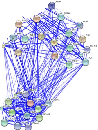
Table 2). STRING analyses resulted in a loose network of pro-teins containing two highly connected clusters around collagen fibril organization and ECM organization (Fig. 3).
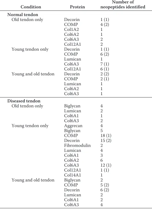
[image:5.594.40.289.260.490.2]Identification of ECM Fragmentation Patterns—A catalogue of age-related neopeptides was identified for COMP, decorin, lumican, collagen #-2(I), collagen #-2(VI), collagen #-3(VI), and collagen#-1(XII). These included those identified either in old normal tendon only, young only, and young and old tendon (Table 2).
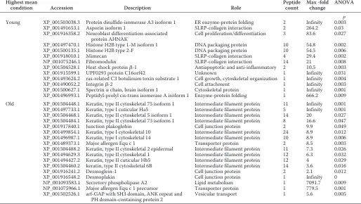
Label-free Relative Quantification—To compare relative pro-tein levels between normal young and old tendon, samples were processed for LC-MS/MS, and quantitative analysis was under-taken with ProgenesisTM. Principal component analysis of all the peptides identified revealed that the peptides clustered according to the age of donor, with a principal component of 83%. Levels of 34 proteins differed between young and old ten-don (25 with "2 peptides). 15 proteins were higher in young tendon (10 with"2 peptides), and 19 proteins were higher in old tendon (15 with"2 peptides) (Table 3). STRING analysis revealed the GO cellular component “intermediate filament” was significantly increased in old tendon (Bonferroni adjustedp values 3.7E$19). Interestingly, in young tendon the SLRP family proteins fibromodulin, mimecan (osteoglycin), and asporin were significantly increased. By contrast, in old tendon, levels of several cellular proteins were increased, including several cyto-skeletal keratins and gap junction proteins.
Western Blotting—Western blot analysis of normal young and old tendon fascicles confirmed the proteomic data by dem-onstrating a significant reduction in fibromodulin levels in older tendon (p'0.04; Student’sttest) (Fig. 4).
Differential Gene Expression—To investigate the increase in keratins in old normal tendon, RT-PCR was undertaken on an independent cohort of tendon from normal young and old donors using primer pairs for the genes KRT5 and KRT75. There was a significantincreaseinexpressionofboththesegenesinoldnormal tendon mirroring the protein expression changes (Fig. 5). TABLE 1
Proteins identified following in-gel trypsin digestion of bands 1– 4 of the soluble guanidine extract using LC-MS/MS
The table lists the most prominent proteins identified following significant peptide matches based on Mascot probability based scoring (p'0.05).
Gel
slice accessionProtein Protein description Proteinscore Proteinmass matchesProtein Protein matchessignificant EmPaia
Da
1 F6UW03 Collagen#-1(VI) chain 793 110,045 55 38 0.65
F7CGV8 Collagen#-2(VI) chain 680 110,009 53 34 0.75
F6QAT0 Collagen#-3(VI) chain 177 343,977 21 10 0.07
F6YR34 Thrombospondin-1 111 133,474 5 5 0.08
F7AQV3 Uncharacterized KIAA1211 protein 49 139,560 20 1 0.02
F6U4X2 #-Fetoprotein 41 70,131 5 2 0.02
2 F6U3D3 Cartilage oligomeric matrix protein 539 84,675 59 32 0.77
F7E0P3 Thrombospondin-4 69 98,508 11 4 0.08
F7CGV8 Collagen#-2(VI) chain 45 110,009 1 1 0.03
F6QAT0 Collagen#-1(VI) chain 35 110,045 2 1 0.03
3 F6RZ46 Prolargin 99 43,846 11 3 0.24
F6U4X2 #-Fetoprotein 45 70,131 4 3 0.04
A2Q126 Fibromodulin 35 43,407 7 2 0.16
4 O46542 Decorin 152 40,256 27 13 0.88
aExponentially modified protein abundance index (emPAI) approximates label-free relative quantification of proteins in a mixture based on protein coverage by peptide matches.
FIGURE 2.The entire guanidine-insoluble, pepsin-released material for each sample was resolved by Tris acetate, 3– 8% NuPAGE.The figure shows representative gels for young and old donors. Polypeptides corre-sponding to the pepsin-released domains of collagen I(#I) (COL1A1), col-lagen I(#2) (COL1A2), collagen V(#1) (COL5A1), collagen V(#3) (COL5A3), collagen III(#1) (COL3A1) and cross-linked collagen dimers and trimers are indicated.
at UNIV OF NOTTINGHAM on April 16, 2015
http://www.jbc.org/
Injured Tendon—Soluble protein content corrected to fasci-cle wet weight was 34.5!18.3!g/ml in young injured tendon and 31.4!12.4 in old injured tendon. SDS-PAGE analysis of guanidine-soluble proteins revealed a greater number of bands in injured tendon compared with normal (Fig. 1). A total of 278 proteins were identified in combined injured tendon samples; 250 had a significant Mascot score of&19. This was signifi-cantly greater than the number of proteins identified in normal tendon (p ' 0.01). Supplemental Table 1 provides detailed information on the identification of peptides mapped to each protein and the corresponding Mascot scores. In diseased ten-don 188!2 and 150!50 proteins were identified in young and old tendon, respectively. A large number of neopeptides were identified for proteoglycans and collagens in injured tendon, with many more neopeptides identified in young diseased than
in old diseased samples (Table 2,supplemental Table 3). PCA at both the peptide (principal component of 36%) and protein levels revealed separation between young and old samples. However, the young samples were more tightly clustered than the old samples. There were 26 proteins at significantly higher levels in young injured tendon (23 with"2 peptide) (Table 4). DAVID identified the term acetylation as significantly increased in this protein set (supplemental Table 4). However, STRING did not find any protein-protein interaction within this set.
DISCUSSION
[image:6.594.130.468.58.504.2]We have performed a comprehensive proteomic analysis of healthy tendon tissue, identifying age-related alterations to the proteins present within the tendon matrix. The results support
FIGURE 3.Protein-protein interaction map of soluble GdnHCl-extracted proteins in normal tendon.Proteins were input from the total dataset. Unconnected nodes and proteins not relating to the two clusters of matrix organizational proteins and collagens were removed to enable clarity of the interactome. The total cluster was built with STRING allowing for experimentally verified and predicted protein-protein interactions at high confidence levels (0.700). Two highly connected clusters were evident.
at UNIV OF NOTTINGHAM on April 16, 2015
http://www.jbc.org/
the hypothesis demonstrating distinct proteomic profiles in young and old tendon with decreased levels of several SLRPs and increases in intermediate filament proteins as a result of aging. In addition, a number of ECM protein fragments pro-duced by fragmentation of the original peptide by enzymatic cleavage between two amino acids, which we have termed “neo-peptides,” were identified in this study, and we propose these are related to specific cleavage sites. We have also assessed the proteomic profile of young and old injured tendon, demon-strating increased matrix fragmentation in disease and distinct proteomic profiles between age groups.
In both young and old normal samples, STRING analysis of the proteins present within the GndHCl-soluble extract revealed two connected clusters of proteins involved in colla-gen fibril and ECM organization as would be expected in ten-don tissue. The most abundant collagen identified in the Gnd-HCl-soluble extract was collagen type VI. Although few studies have investigated the role of collagen type VI in tendon, Izuet al.(40) showed that type VI collagen is localized to the pericel-lular region and is likely to play a role in collagen fibrillogenesis. Furthermore, it has been demonstrated that collagen VI mutant mice have abnormal collagen fibrils and exhibit muscle and
tendon defects similar to those seen in human muscular dystro-phy (41), suggesting that this minor collagen plays a crucial role in normal tendon function. A significant role for type VI colla-gen in tendon function is supported by these data.
Other proteins identified within the GndHCl-soluble extract include members of the thrombospondin family (COMP, thrombospondin-4 and -5) and several of the small SLRPs (decorin, fibromodulin, prolargin). Thrombospondins are known to regulate cell-matrix interactions, but their specific role in tendon has not been extensively studied. COMP is thought to catalyze collagen fibrillogenesis and stabilize the col-lagen network (42), and COMP levels have been correlated with tendon mechanical properties (43).
Decorin is the most abundant and the most studied of the SLRPs within tendon. However, its role is yet to be fully estab-lished. Both decorin and fibromodulin are involved in fibrillo-genesis (1), and decorin may also play a role in transfer of force between collagen fibrils (44), although this function is conten-tious (45). To the authors’ knowledge this is the first work to identify the presence of prolargin within tendon tissue. This class II SLRP is able to bind to type I collagen and is postulated to anchor basement membranes to the connective tissue (46).
Although there was no overall decrease in the concentration of soluble proteins extracted from normal tendon with increas-ing age as determined by the Bradford assay, silver stainincreas-ing of bands on one-dimensional gels of soluble protein extracts appeared to decrease with aging. Assessment of the normaliza-tion factor used by Progenesis during relative quantificanormaliza-tion revealed higher normalization factors for old samples even though a fixed amount of protein (based on Bradford assay results) was loaded. This suggests that the Bradford assay may have provided an incorrect estimation of sample protein con-tent, as reported previously (47), but the reasons for this are unclear. Taken together, these results suggest that in aged ten-don protein extractability was reduced, suggesting that the matrix in aged samples is more resistant to degradation, with more proteins remaining trapped within the insoluble portion of the matrix. There were also age-related differences in the pepsin-released portion of the samples, with more collagenous polypeptide bands evident in young samples (Fig. 2). These findings are supported by previous work which has shown that with aging partially degraded collagen accumulates within the matrix of the SDFT, which may be due to increased levels of glycation, rendering the matrix more resistant to degradation (14).
There were no alterations in the levels of the major matrix components with increasing age. This supports our previous studies which have shown that tendon water, collagen, and gly-cosaminoglycan content of the equine SDFT do not change with aging (14). However, there was a reduction in levels of several less-abundant proteins with increasing age in normal tendon, including several SLRPS (fibromodulin, mimecan, asporin). The age-related reduction in fibromodulin was fur-ther confirmed by Western blotting. These proteoglycans interact with collagen and have all been shown to regulate col-lagen fibrillogenesis and fibril diameter (1, 48–50). Heat shock protein$1, also known as heat shock protein 27, also decreased with aging. Heat shock proteins have apoptotic and anti-TABLE 2
Number of neopeptides identified in normal old tendon only, young tendon only, and young and old tendon
Neopeptides were identified by Mascot with a significant score more than once per donor and in"2 donors. Numbers in parentheses indicate number of peptides also identified in diseased tendon. For full details of neopeptide sequences, see supple-mentary information (supplemental Table 3).
Condition Protein neopeptides identifiedNumber of Normal tendon
Old tendon only Decorin 1 (1)
COMP 4 (2)
Col1A2 1
Col6A2 1
Col6A3 2
Col12A1 2
Young tendon only Decorin 1 (1)
COMP 6 (2)
Lumican 1
Col6A3 7 (1)
Col12A1 6 (1)
Young and old tendon Decorin 2 (2)
COMP 2 (1)
Lumican 1
Col6A2 1
Col6A3 1
Diseased tendon
Old tendon only Biglycan 4
Lumican 2
Col6A1 1
Col6A3 2
Young tendon only Aggrecan 4
Biglycan 5
COMP 18 (1)
Decorin 15 (2)
Fibromodulin 2
Lumican 4
Col6A1 3
Col6A2 6
Col6A3 12 (1)
Col12A1 1 (1)
Col14A1 1
Young and old tendon Biglycan 2
COMP 5 (2)
Decorin 6 (2)
Lumican 2
Col6A1 2
Col6A3 4
at UNIV OF NOTTINGHAM on April 16, 2015
http://www.jbc.org/
[image:7.594.41.288.125.465.2]inflammatory roles and have also been shown to increase in tendinopathy, where it is suggested they may play a role in ten-don healing (51). The reduction in levels of these proteins with aging may, therefore, affect maintenance and repair of matrix in old tendon and could contribute to the increased risk of tendon injury in aged individuals.
There were also alterations in levels of several cellular pro-teins with aging, with decreases in histones and integrins and increases in keratins and gap junction proteins with aging. Pre-vious studies have shown that cellularity within the SDFT does not change with aging (12), suggesting that these alterations may reflect an age-related change in cell phenotype. The major-ity of proteins that increased with age were keratins and gap junction proteins. Although some cuticular keratins were iden-tified, possibly due to contamination from hair during dissec-tion, the majority of keratins identified were cytoskeletal, and their increase was confirmed at the gene expression level. STRING analysis revealed these proteins belong to the GO cel-lular component intermediate filament. These filaments form cytoskeletal networks that are important in maintenance and regulation of cell tension, providing support for the plasma membrane during contact with cells and extracellular matrix (52). The increases in intermediate filament proteins observed with aging may indicate an increase in cell stiffness, which could result in an altered cell response to tensile loading in aged tendons. Although the specific roles of cytoskeletal keratins in tendon are not well understood, it has been previously demon-strated that keratin 1 and 10 are localized to the basement
membrane epithelium recently identified around tendon (53). This basement membrane is thought to regulate cell migration and maintain tendon functional integrity (53). A change in the levels of cytoskeletal components suggests differences in their mechanical behavior with aging.
In addition, a number of previously documented as well as novel neopeptides were identified in this study. It is likely that those found in both young and old normal samples could be due to normal ECM turnover. However, they may represent proteo-lytic cleavage occurring as a consequence of subclinical patho-logical degradation, or they could be neopeptides produced during tissue processing as cell death can release intracellular proteases. Protease activity during processing seems unlikely as attempts to mitigate this were made through the chilling and rapid post-mortem dissection of the limbs and snap-freezing of tissues.
It could be hypothesized that the neopeptides identified in normal young tendon alone represent ECM fragments pro-duced by normal tissue remodeling, which is altered with aging and injury. We identified the COMP neopeptide NTVMEC-DACGMQP2A in young tendon only. This cleavage pattern has been attributed to the activity of a disintegrin with thrombospondin motifs (ADAMTS)-5 in mouse.5 Con-versely, although a greater number of neopeptides were iden-tified in young than in old tendon, neopeptides present only in
[image:8.594.41.555.101.391.2]5P. Holden, personal communication. TABLE 3
A number of differentially abundant proteins were identified by ProgenesisTMLC-MS software between young and old tendon. All proteins with a>2-fold change in normalized abundance are shown
A value of infinity is given when no peptides for a given protein in the group other than the highest mean condition were identified.
Highest mean
condition Accession Description Role Peptidecount Max -foldchange ANOVA
p
Young XP_001503038.3 Protein disulfide-isomerase A3 isoform 1 ER enzyme-protein folding 2 Infinity 0.003
XP_001491653.1 Asporin isoform 1 SLRP-collagen interaction 2 264.2 0.03
XP_001916358.2 Neuroblast differentiation-associated
protein AHNAK Cell proliferation/differentiation 3 83.6 0.027
XP_001497470.1 Histone H2B type 1-M isoform 1 DNA packaging protein 10 54.8 0.002
XP_001500135.1 Histone H2B type 2-F DNA packaging protein 10 54.5 0.006
XP_001918010.1 Mimecan SLRP-collagen interaction 4 29.4 0.002
NP_001075246.1 Fibromodulin SLRP-collagen interaction 14 21 0.008
XP_001504528.1 Heat shock protein$-1 Antiapoptotic and anti-inflammatory 2 10.5 0.003
XP_001915599.1 UPF0293 protein C16orf42 Unknown 1 Infinity 0.031
XP_001493624.2 ras-related C3 botulinum toxin substrate 1 Cell growth, cytoskeletal organization 1 Infinity 0.004
XP_001490052.2 Integrin$-2 Cell attachment 1 Infinity 0.003
XP_001500627.1 Spectrin#chain, brain isoform 1 Cytoskeletal protein 1 Infinity 0.001
XP_001496993.1 Peptidyl-prolyl cis-trans isomerase A isoform 1 Enzyme-protein folding 1 666.2 0.009
Old XP_001504448.1 Keratin, type II cytoskeletal 75 isoform 1 Intermediate filament protein 11 Infinity 0.001
XP_001497733.1 Keratin, type I cuticular Ha5 Intermediate filament protein 5 Infinity 0.001
XP_001504468.1 Keratin, type II cytoskeletal 5 isoform 1 Intermediate filament protein 14 20 0.027
XP_001504484.1 Keratin, type II cytoskeletal 73 isoform 1 Intermediate filament protein 8 16.6 0.047
XP_001917840.1 Junction plakoglobin Cell junction protein 2 9.9 0.049
XP_001499854.1 Keratin, type I cytoskeletal 10 Intermediate filament protein 24 8.9 0.012
XP_001496987.1 Keratin, type I cytoskeletal 14 Intermediate filament protein 10 8.9 0.006
XP_001489373.1 Major allergen Equ c 1 Transporter protein 2 8.5 0.003
XP_001504488.2 Keratin, type II cytoskeletal 2 epidermal Intermediate filament protein 11 7.3 0.026
XP_001494629.3 Keratin, type II cytoskeletal 1 Intermediate filament protein 12 6.3 0.032
XP_001494427.2 Keratin, type II cuticular Hb5 Intermediate filament protein 12 4 0.029
XP_001504460.2 keratin, type II cytoskeletal 6B Intermediate filament protein 14 3.6 0.016
XP_001916241.2 Desmoglein-1 Cell junction protein 2 2.1 0.012
XP_001916548.2 Desmoplakin Cell junction protein 1 Infinity 0
NP_001093583.1 Secretory phospholipase A2 Lipid metabolism 1 7091.7 0.009
NP_001075966.1 Major allergen Equ c 1 precursor Transporter protein 1 779.5 0.001
XP_001502526.1 arf-GAP with SH3 domain, ANK repeat and
PH domain-containing protein 2 Vesicular transport 1 5.6 0.005
at UNIV OF NOTTINGHAM on April 16, 2015
http://www.jbc.org/
old tendon may represent important events in tendon aging and provide an insight into the underlying mechanisms that conse-quently increase the risk of tendon injury in aged individuals. Interestingly the COMP neopeptides F2CFSQENIIWANLR and C2PDGTPSPCHEK, identified in old healthy tendon only, have been recently identified within equine SDFT tissue (18). The neopeptide F2CFSQENIIWANLR was evident in sub-acute SDFT injury and also after IL-1$stimulation of macro-scopically normal equine SDFT explants. One proposed theory of aging is the “senescent secretory phenotype” (54). Accumu-lation of specific cells, which secrete increased amounts of cyto-kines, contributes to cell aging. The identification of this neo-peptide in both studies could indicate that there may be enhanced production of cytokines in tendon aging. This could provide a direct link between aging and inflammation similar to that proposed in cartilage (55).
In the study by Dakinet al., C2PDGTPSPCHEK was evident in both normal SDFT and in control explants maintained in culture for 24 h (11). Due to the wide age range used in their study, it would be interesting to undertake further work to assess if this neopeptide represents a specific aging biomarker and if so, at which age it appears.
A number of collagen neopeptides were also identified. Inter-estingly we previously proposed that an inability to remove par-tially degraded collagens from the tendon matrix may lead to reduced mechanical competence in aging tendon (14). The
col-lagen I, VI, and XII neopeptides identified here in only the old normal tissue could support this hypothesis. In young tendon a number of collagen VI and XII fragments were also evident. This could be explained by the role of collagen VI in fibrillogen-esis (40) and collagen XII in ECM organization (56) and repre-sent normal matrix turnover due to weaker ECM interactions of these collagens in young tendon. Indeed, in developing chick tendon it has been demonstrated that collagen VI is predomi-nantly located to the interfascicular matrix (56). This is inter-esting as our previous work has shown that the interfascicular matrix plays an important role in SDFT function but becomes stiffer in aged tendon (30, 57), which may be due to a reduced ability to turn over this matrix with aging.
It was not possible to directly compare the proteome of nor-mal and injured tendon, as these samples were analyzed at dif-ferent times. However, there are some clear differences between normal and injured states. Although the soluble pro-tein content was lower, a greater number of propro-teins were iden-tified in injured tendon compared with normal. This may be because soluble protein content was normalized to fascicle wet weight; it has previously been shown that tendinopathic tissue has a higher water content than normal tendon (58), which would result in a relatively lower protein content when normal-izing to wet weight. Furthermore, additional bands were visible in samples from injured tendon, which are likely to be due to increased matrix degradation and fragmentation (Fig. 1). An increase in cellularity may also contribute to the increase in the number of proteins identified in injured tendon. Many of the proteins identified in diseased tendon were cellular, and it is well established that cell numbers are increased in injured ten-don (59–61). Furthermore, a larger number of neopeptides were identified in injured tendon, indicating a greater degrada-tion of collagens and proteoglycans with disease.
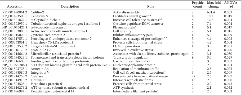
It is also apparent that the proteomic profile differs with age in injured tendon. A number of proteins were detected at higher levels in young compared with old diseased tendon. These include several cellular proteins, which have roles in pro-tection of cells from stress and synthesis and stabilization of matrix proteins (see Table 5). Furthermore, several of these proteins have been identified in developing tendon (25) and in artificially induced tendon lesions (24). The higher levels of these proteins in young diseased tendon may, therefore, repre-sent a healing response, which appears to be limited in old dis-eased tendon (63). A larger number of neopeptides was also identified in young injured tendon, suggesting a greater ability to degrade damaged regions of the matrix. This may further explain why aged tendons are more at risk of injury, as a failed healing response is likely to lead to the accumulation of microdamage and subsequent injury. However, it is unclear if this failed healing response is due to a decreased ability of ten-don cells to synthesize and degrade damaged regions of the matrix in aged tendons or whether the matrix is more resistant to degradation due to age-related glycation.
[image:9.594.40.288.58.370.2]There are several limitations to this study that need to be considered. The high levels of collagenous proteins in tendon mean that it is difficult to detect proteins present at a low abun-dance. Future studies could use hexapeptide peptide library protein normalization (62), which would allow identification of
FIGURE 4.Effect of age on fibromodulin levels in tendon fascicles.A, rep-resentative blots of fibromodulin and#-tubulin in samples from young and old horses.B, Western blot analysis of fibromodulin normalized to#-tubulin demonstrated a significant reduction in fibromodulin with age. The histo-gram represents mean pixel intensity!S.E.,n"3; *,p'0.05.
at UNIV OF NOTTINGHAM on April 16, 2015
http://www.jbc.org/
low abundance proteins. Alternatively, absolute protein quan-tification using QconCat technology could be used in an artifi-cially agedin vitromodel (20), which would allow the analysis of a greater number of samples. In addition, proteins present may not have been identified as they could not be extracted from the matrix, which is highly resistant to degradation. Furthermore, it is evident that protein extractability is altered with aging in normal tendon; therefore, some of the age-related alterations identified could be because the proteins were not extracted from the matrix. However, if this were the case it would be expected there would be a global decrease in protein levels with aging, which was not observed. It should also be considered that some of the proteins identified, particularly keratinous pro-teins, may be due to contamination from skin and hair. Care was taken during dissection to ensure minimal contamination, and as the majority of keratins identified were cytoskeletal rather than cutaneous, this is unlikely to be a major source of
contamination. Furthermore, we have confirmed the increased keratin levels with aging at the mRNA level.
CONCLUSIONS
[image:10.594.128.462.62.251.2]Although proteomic analysis is fast becoming a standard technique to study many soft tissues, few studies have attempted to use this technique to characterize tendon tissue. In the current study we have demonstrated age-related altera-tions in several proteins within normal tendon, with decreases in proteins that play a role in ECM organization and increases in cytoskeletal proteins. We have further demonstrated an altered proteomic profile in injured tendon, with significantly more proteins identified and a greater degree of matrix frag-mentation. We have also shown a decrease in levels of proteins associated with reduction of cell stress and increased matrix synthesis with aging in injured tendon. This study has increased
FIGURE 5.Gene expression of KRT5 and KRT75 in normal young and old tendon.Data are represented as 2 )Ct (2ˆ-DCT) compared with GAPDH. Histograms represent the means!S.E. of mean. *,p'0.05. Data were evaluated using Student’sttest after log transformation for normalization (n"7).
TABLE 4
A number of differentially abundant proteins were identified by ProgenesisTMLC-MS software between diseased young and old tendon
All proteins with a&2-fold change in normalized abundance are shown. All proteins were at higher levels in the young diseased tendon.
Accession Description Role Peptidecount Max foldchange ANOVA(p)
XP_001490861.2 Cofilin-2 Actin disassembly 2 631.4 0.003
XP_001493508.1 Tenascin-N Facilitates neural growtha 4 18.2 0.019
XP_001501829.1 #-Crystallin B chain Increase cell tolerance to stressa,b 8 12.7 0.004
XP_001503950.1 Tubulointerstitial nephritis antigen 1 isoform 1 Cysteine peptidase-ECM turnover 2 7.4 0.004
NP_001075421.1 #-Fetoprotein precursor Plasma proteina 1 5.1 0.017
XP_001503085.1 Actin, aortic smooth muscle isoform 1 Cell motility 20 5.1 0.015
XP_001915023.1 Cysteine-rich protein 2 Inhibits inflammatory pain 1 5.0 0.009
XP_001917554.1 Procollagen C-endopeptidase enhancer 2 Enhances cleavage of pro-collagena,b 3 4.4 0.003
XP_003363861.1 Heat shock 70-kDa protein 1 Protects cells from thermal stress 4 4.2 0.004
XP_001503538.2 Target of Nesh-SH3 isoform 4 ECM organisation 1 3.3 0.001
XP_001916170.2 protein SZT2 Involved in oxidative stress 5 3.2 0.020
XP_001915664.2 Microfibrillar-associated protein 5 Associates with elastic fibres, stabilizes procollagen 2 3.1 0.050
XP_001494523.1 Polymerase I and transcript release factor isoform Transcription regulation 9 3.0 0.029
XP_001916600.1 Insulin growth factor binding protein-6 Carrier protein for IGF-1 3 3.0 0.033
NP_001229384.1 SH3 domain binding glutamic acid-rich protein like 3 Nuclear/cytoplasmic protein 3 2.9 0.004
XP_001503725.1 Annexin A6 Regulation of membrane traffic 4 2.8 0.025
XP_001498580.3 Integrin#-V Cell-cell & cell-matrix interactionsa 3 2.6 0.009
XP_001914753.2 Catalase Prevents cells from oxidative damage 22 2.5 0.007
XP_001914918.2 Fibulin-2 Interacts with elastic fibres 13 2.3 0.030
XP_001504528.1 Heat shock protein$1 Protects cells from thermal stress 7 2.2 0.043
XP_001916279.2 ATP synthase subunit#, mitochondrial ATP synthesis 4 2.1 0.032
XP_001496987.1 Keratin, type I cytoskeletal 14 Intermediate filament proteina 3 2.0 0.002
aPreviously identified in developing tendon (25). bPreviously identified in healing tendon (24).
at UNIV OF NOTTINGHAM on April 16, 2015
http://www.jbc.org/
[image:10.594.44.553.313.515.2]the knowledge of molecular events associated with tendon deg-radation characteristic of aging and injury and identified pep-tides that may be useful as biomarkers of tendon injury. These findings suggest that maintenance and repair of tendon tissue may be reduced in aged individuals, resulting in an impaired healing response, and may help to explain why the risk of injury increases with aging.
Acknowledgment—We thank Dr Helen Birch for critical reading of this manuscript.
REFERENCES
1. Yoon, J. H., and Halper, J. (2005) Tendon proteoglycans: biochemistry and function.J. Musculoskelet. Neuronal Interact.5,22–34
2. Riley, G. (2008) Tendinopathy: from basic science to treatment.Nat. Clin. Pract. Rheumatol.4,82–89
3. Grant, T. M., Thompson, M. S., Urban, J., and Yu, J. (2013) Elastic fibres are broadly distributed in tendon and highly localized around tenocytes.J. Anat.222,573–579
4. Thorpe, C. T., Birch, H. L., Clegg, P. D., and Screen, H. R. (2013) The role of the non-collagenous matrix in tendon function.Int. J. Exp. Pathol.94,
248–259
5. Kastelic, J., Galeski, A., and Baer, E. (1978) The multicomposite structure of tendon.Connect Tissue Res.6,11–23
6. Kujala, U. M., Sarna, S., and Kaprio, J. (2005) Cumulative incidence of Achilles tendon rupture and tendinopathy in male former elite athletes. Clin. J. Sport Med.15,133–135
7. Clayton, R. A., and Court-Brown, C. M. (2008) The epidemiology of mus-culoskeletal tendinous and ligamentous injuries.Injury39,1338–1344 8. Birch, H. L., Bailey, A. J., and Goodship, A. E. (1998) Macroscopic
“degen-eration” of equine superficial digital flexor tendon is accompanied by a change in extracellular matrix composition.Equine Vet J.30,534–539 9. Tully, L. J., Murphy, A. M., Smith, R. K., Hulin-Curtis, S. L., Verheyen,
K. L., and Price, J. S. (2014) Polymorphisms in TNC and COL5A1 genes are associated with risk of superficial digital flexor tendinopathy in Na-tional Hunt Thoroughbred racehorses.Equine Vet J.46,289–293 10. September, A. V., Cook, J., Handley, C. J., van der Merwe, L., Schwellnus,
M. P., and Collins, M. (2009) Variants within the COL5A1 gene are asso-ciated with Achilles tendinopathy in two populations.Br. J. Sports Med.
43,357–365
11. Dakin, S. G., Dudhia, J., and Smith, R. K. (2014) Resolving an inflammatory concept: the importance of inflammation and resolution in tendinopathy. Vet. Immunol. Immunopathol.158,121–127
12. Birch, H. L., Bailey, J. V., Bailey, A. J., and Goodship, A. E. (1999) Age-related changes to the molecular and cellular components of equine flexor tendons.Equine Vet. J.31,391–396
13. Avery, N. C., and Bailey, A. J. (2005) Enzymic and non-enzymic cross-linking mechanisms in relation to turnover of collagen: relevance to aging and exercise.Scand. J. Med. Sci. Sports15,231–240
14. Thorpe, C. T., Streeter, I., Pinchbeck, G. L., Goodship, A. E., Clegg, P. D., and Birch, H. L. (2010) Aspartic acid racemization and collagen degrada-tion markers reveal an accumuladegrada-tion of damage in tendon collagen that is enhanced with aging.J. Biol. Chem.285,15674–15681
15. Riley, G. P., Harrall, R. L., Constant, C. R., Chard, M. D., Cawston, T. E., and Hazleman, B. L. (1994) Glycosaminoglycans of human rotator cuff tendons: changes with age and in chronic rotator cuff tendinitis.Ann. Rheum. Dis.53,367–376
16. Smith, R. K., Zunino, L., Webbon, P. M., and Heinegård, D. (1997) The distribution of cartilage oligomeric matrix protein (COMP) in tendon and its variation with tendon site, age, and load.Matrix Biol.16,255–271 17. Rousseau, J. C., and Delmas, P. D. (2007) Biological markers in
osteoar-thritis.Nat. Clin. Pract. Rheumatol.3,346–356
18. Dakin, S. G., Smith, R. K., Heinegård, D., Önnerfjord, P., Khabut, A., and Dudhia, J. (2014) Proteomic analysis of tendon extracellular matrix reveals disease stage-specific fragmentation and differential cleavage of COMP.
J. Biol. Chem.289,4919–4927
19. Wilson, R., Diseberg, A. F., Gordon, L., Zivkovic, S., Tatarczuch, L., Mackie, E. J., Gorman, J. J., and Bateman, J. F. (2010) Comprehensive profiling of cartilage extracellular matrix formation and maturation using sequential extraction and label-free quantitative proteomics.Mol. Cell. Proteomics9,1296–1313
20. Peffers, M. J., Beynon, R. J., and Clegg, P. D. (2013) Absolute quantification of the human osteoarthritic secretome.Int. J. Mol. Sci.14,20658–20681 21. Jiang, Y., Liu, H., Li, H., Wang, F., Cheng, K., Zhou, G., Zhang, W., Ye, M.,
Cao, Y., Liu, W., and Zou, H. (2011) A proteomic analysis of engineered tendon formation under dynamic mechanical loading in vitro. Biomateri-als32,4085–4095
22. Han, G.-Y., Park, S.-A., Kim, J.-H., Lee, E.-K., Kim, H.-J., Seo, Y.-K., Park, J.-K., and Kim, C.-W. (2011) Effects of vibration on the proteome expres-sion of anterior cruciate ligament cells.Exp. Biol. Med.236,783–789 23. Jielile, J., Jialili, A., Sabirhazi, G., Shawutali, N., Redati, D., Chen, J., Tang,
B., Bai, J., and Aldyarhan, K. (2011) Proteomic analysis of differential pro-tein expression of Achilles tendon in a rabbit model by two-dimensional polyacrylamide gel electrophoresis at 21 days postoperation. Appl. Biochem. Biotechnol.165,1092–1106
24. Jielile, J., Aibai, M., Sabirhazi, G., Shawutali, N., Tangkejie, W., Badelhan, A., Nuerduola, Y., Satewalede, T., Buranbai, D., Hunapia, B., Jialihasi, A., Bai, J. P., and Kizaibek, M. (2012) Active Achilles tendon kinesitherapy accelerates Achilles tendon repair by promoting neurite regeneration. Neural Regen. Res.7,2801–2810
25. Smith, S. M., Thomas, C. E., and Birk, D. E. (2012) Pericellular proteins of the developing mouse tendon: a proteomic analysis.Connect Tissue Res.
53,2–13
26. Kasashima, Y., Takahashi, T., Smith, R. K., Goodship, A. E., Kuwano, A., Ueno, T., and Hirano, S. (2004) Prevalence of superficial digital flexor tendonitis and suspensory desmitis in Japanese thoroughbred flat race-horses in 1999.Equine Vet. J.36,346–350
27. Innes, J. F., and Clegg, P. (2010) Comparative rheumatology: what can be learnt from naturally occurring musculoskeletal disorders in domestic animals?Rheumatology49,1030–1039
28. Lui, P. P., Maffulli, N., Rolf, C., and Smith, R. K. (2011) What are the validated animal models for tendinopathy?Scand J. Med. Sci. Sports21,
3–17
29. Ely, E. R., Avella, C. S., Price, J. S., Smith, R. K., Wood, J. L., and Verheyen, K. L. (2009) Descriptive epidemiology of fracture, tendon, and suspensory ligament injuries in National Hunt racehorses in training.Equine Vet. J.
41,372–378
30. Thorpe, C. T., Udeze, C. P., Birch, H. L., Clegg, P. D., and Screen, H. R. (2013) Capacity for sliding between tendon fascicles decreases with ageing in injury prone equine tendons: a possible mechanism for age-related tendinopathy?Eur. Cell Mater.25,48–60
31. Lichtwark, G. A., and Wilson, A. M. (2005)In vivomechanical properties of the human Achilles tendon during one-legged hopping.J. Exp. Biol.
208,4715–4725
32. Biewener, A. A. (1998) Muscle-tendon stresses and elastic energy storage during locomotion in the horse.Comp Biochem. Physiol B. Biochem. Mol. Biol.120,73–87
33. Legerlotz, K., Riley, G. P., and Screen, H. R. (2010) Specimen dimensions influence the measurement of material properties in tendon fascicles. J. Biomech.43,2274–2280
34. McLean, L., Hurst, J. L., Gaskell, C. J., Lewis, J. C., and Beynon, R. J. (2007) Characterization of cauxin in the urine of domestic and big cats.J. Chem. Ecol.33,1997–2009
35. Franceschini, A., Szklarczyk, D., Frankild, S., Kuhn, M., Simonovic, M., Roth, A., Lin, J., Minguez, P., Bork, P., von Mering, C., and Jensen, L. J. (2013) STRING v9.1: proteprotein interaction networks, with in-creased coverage and integration.Nucleic Acids Res.41,D808–D815 36. Peffers, M., Liu, X., and Clegg, P. (2013) Transcriptomic signatures in
cartilage ageing.Arthritis Res. Ther.15,R98
37. Birch, H. L., Worboys, S., Eissa, S., Jackson, B., Strassburg, S., and Clegg, P. D. (2008) Matrix metabolism rate differs in functionally distinct ten-dons.Matrix Biol.27,182–189
38. Dakin, S. G., Werling, D., Hibbert, A., Abayasekara, D. R., Young, N. J.,
at UNIV OF NOTTINGHAM on April 16, 2015
http://www.jbc.org/
Smith, R. K., and Dudhia, J. (2012) Macrophage subpopulations and the lipoxin A4 receptor implicate active inflammation during equine tendon repair.PLoS ONE7,e32333
39. Webbon, P. M. (1977) A post mortem study of equine digital flexor ten-dons.Equine Vet. J.9,61–67
40. Izu, Y., Ansorge, H. L., Zhang, G., Soslowsky, L. J., Bonaldo, P., Chu, M.-L., and Birk, D. E. (2011) Dysfunctional tendon collagen fibrillogenesis in collagen VI null mice.Matrix Biol.30,53–61
41. Pan, T.-C., Zhang, R.-Z., Markova, D., Arita, M., Zhang, Y., Bogdanovich, S., Khurana, T. S., Bönnemann, C. G., Birk, D. E., and Chu, M.-L. (2013) COL6A3 protein deficiency in mice leads to muscle and tendon defects similar to human collagen VI congenital muscular dystrophy.J. Biol. Chem.288,14320–14331
42. Halász, K., Kassner, A., Mörgelin, M., and Heinegård, D. (2007) COMP acts as a catalyst in collagen fibrillogenesis.J. Biol. Chem.282,31166–31173 43. Smith, R. K., Gerard, M., Dowling, B., Dart, A. J., Birch, H. L., and
Good-ship, A. E. (2002) Correlation of cartilage oligomeric matrix protein (COMP) levels in equine tendon with mechanical properties: a proposed role for COMP in determining function-specific mechanical characteris-tics of locomotor tendons.Equine Vet. J. Suppl.34,241–244
44. Redaelli, A., Vesentini, S., Soncini, M., Vena, P., Mantero, S., and Monte-vecchi, F. M. (2003) Possible role of decorin glycosaminoglycans in fibril to fibril force transfer in relative mature tendons: a computational study from molecular to microstructural level.J. Biomech.36,1555–1569 45. Provenzano, P. P., and Vanderby, R. (2006) Collagen fibril morphology and
organization: implications for force transmission in ligament and tendon. Matrix Biol.25,71–84
46. Bengtsson, E., Mörgelin, M., Sasaki, T., Timpl, R., Heinegård, D., and Asp-berg, A. (2002) The leucine-rich repeat protein prelp binds perlecan and collagens and may function as a basement membrane anchor. J. Biol. Chem.277,15061–15068
47. Weist, S., Eravci, M., Broedel, O., Fuxius, S., Eravci, S., and Baumgartner, A. (2008) Results and reliability of protein quantification for two-dimen-sional gel electrophoresis strongly depend on the type of protein sample and the method employed.Proteomics8,3389–3396
48. Kalamajski, S., Aspberg, A., Lindblom, K., Heinegård, D., and Oldberg, A. (2009) Asporin competes with decorin for collagen binding, binds cal-cium, and promotes osteoblast collagen mineralization.Biochem. J.423,
53–59
49. Kalamajski, S., and Oldberg, A. (2009) Homologous sequence in lumican and fibromodulin leucine-rich repeat 5–7 competes for collagen binding. J. Biol. Chem.284,534–539
50. Tasheva, E. S., Koester, A., Paulsen, A. Q., Garrett, A. S., Boyle, D. L.,
Davidson, H. J., Song, M., Fox, N., and Conrad, G. W. (2002) Mimecan/ osteoglycin-deficient mice have collagen fibril abnormalities.Mol. Vis.8,
407–415
51. Millar, N. L., and Murrell, G. A. (2012) Heat shock proteins in tendinopa-thy: novel molecular regulators.Mediators Inflamm.2012,436203 52. Lodish, H. B., A; Zipursky, SL et al. ( 2000)Molecular Cell Biology, 4th Ed.,
pp. 806–813, W. H. Freeman, New York
53. Taylor, S. H., Al-Youha, S., Van Agtmael, T., Lu, Y., Wong, J., Mc-Grouther, D. A., and Kadler, K. E. (2011) Tendon is covered by a basement membrane epithelium that is required for cell retention and the preven-tion of adhesion formapreven-tion.PLoS ONE6,e16337
54. Campisi, J. (2005) Senescent cells, tumor suppression, and organismal aging: good citizens, bad neighbors.Cell120,513–522
55. Loeser, R. F. (2010) Age-related changes in the musculoskeletal system and the development of osteoarthritis.Clin. Geriatr. Med.26,371–386 56. Zhang, G., Young, B. B., and Birk, D. E. (2003) Differential expression of
type XII collagen in developing chicken metatarsal tendons.J. Anat.202,
411–420
57. Thorpe, C. T., Udeze, C. P., Birch, H. L., Clegg, P. D., and Screen, H. R. (2012) Specialization of tendon mechanical properties results from inter-fascicular differences.J. R. Soc. Interface9,3108–3117
58. de Mos, M., van El, B., DeGroot, J., Jahr, H., van Schie, H. T., van Arkel, E. R., Tol, H., Heijboer, R., van Osch, G. J., and Verhaar, J. A. (2007) Achilles tendinosis: changes in biochemical composition and collagen turnover rate.Am. J. Sports Med.35,1549–1556
59. Södersten, F., Hultenby, K., Heinegård, D., Johnston, C., and Ekman, S. (2013) Immunolocalization of collagens (I and III) and cartilage oligo-meric matrix protein (COMP) in the normal and injured equine superfi-cial digital flexor tendon.Connect Tissue Res.54,62–69
60. Kobayashi, A., Sugisaka, M., Takehana, K., Yamaguchi, M., Eerdunchao-lu,Iwasa, E. K., and Abe, M. (1999) Morphological and histochemical anal-ysis of a case of superficial digital flexor tendon injury in the horse.J. Comp. Pathol.120,403–414
61. Patterson-Kane, J. C., and Firth, E. C. (2009) The pathobiology of exercise-induced superficial digital flexor tendon injury in thoroughbred race-horses.Vet. J.181,79–89
62. Dwivedi, R. C., Krokhin, O. V., Cortens, J. P., and Wilkins, J. A. (2010)) Assessment of the reproducibility of random hexapeptide peptide library-based protein normalization.J. Proteome Res.9,1144–1149
63. Dakin, S. G., Dudhia, J., Werling, N. J., Werling, D., Abayasekara, D. R., and Smith, R. K. (2012) Inflamm-Aging and Arachadonic Acid Metabolite Differences with Stage of Tendon Disease.PLoS ONE7,e48978
at UNIV OF NOTTINGHAM on April 16, 2015
http://www.jbc.org/
Hazel R. C. Screen and Peter D. Clegg
A. Collins, Robin Eong, Timothy K. J. Wei,
Mandy J. Peffers, Chavaunne T. Thorpe, John
Fragmentation
with Age- and Injury-specific Matrix
Changes in Tendon Matrix Composition,
doi: 10.1074/jbc.M114.566554 originally published online July 30, 20142014, 289:25867-25878. J. Biol. Chem.
10.1074/jbc.M114.566554
Access the most updated version of this article at doi:
JBC Affinity Sites.
Find articles, minireviews, Reflections and Classics on similar topics on the
Alerts:
When a correction for this article is posted
• When this article is cited •
to choose from all of JBC's e-mail alerts
Click here
Supplemental material:
http://www.jbc.org/content/suppl/2014/07/30/M114.566554.DC1.html
http://www.jbc.org/content/289/37/25867.full.html#ref-list-1
This article cites 62 references, 15 of which can be accessed free at
at UNIV OF NOTTINGHAM on April 16, 2015
http://www.jbc.org/