PERIY
US
The Tam
In p
DEPA
YAR COL
T
An IS
SING DIF
Dissert
milnadu D
Che
partial fulf
MASTER
PHA
Reg
ARTMENT
LLEGE OF
TIRUCHIR
A
O 9001: 2
FFERENT
tation subm
Dr.M.G.R.
ennai – 60
lfillment fo
R OF PHA
IN
ARMACEU
By
.No: 2612
T OF PHA
F PHARM
RAPPALL
APRIL - 20
008 Certif
POLYME
mitted to
.Medical U
00 032
or the degr
ARMACY
UTICS
10202
ARMACE
MACEUTI
LI – 620 0
Head i/c, Department of Pharmaceutics
Periyar College of Pharmaceutical Sciences
Tiruchirappalli – 620 021.
CERTIFICATE
This is to certify that the dissertation entitled “FORMULATION AND
EVALUATION OF TRANSDERMAL PATCH OF METHANOLIC
EXTRACT OF Acalypha indica Linn. USING DIFFERENT
POLYMERS” submitted by Mr. R. SIVABAL [Reg. No. 261210202] for
the award of the degree of “MASTER OF PHARMACY” is a
bonafide research work done by him in the Department of Pharmaceutics,
Periyar College of Pharmaceutical Sciences,Tiruchirappalli under my
guidance and direct supervision.
Place : Tiruchirappalli
Date :
Periyar College of Pharmaceutical Sciences
Tiruchirappalli – 620 021.
CERTIFICATE
This is to certify that the dissertation entitled “FORMULATION
AND EVALUATION OF TRANSDERMAL PATCH OF
METHANOLIC EXTRACT OF Acalypha indica Linn. USING
DIFFERENT POLYMERS” submitted by Mr. R. SIVABAL
[Reg. No. 261210202] for the award of the degree of “MASTER OF
PHARMACY” under The Tamilnadu Dr. M.G.R Medical University,
Chennai is a bonafide research work performed by her in the
Department of Pharmaceutics, Periyar College of Pharmaceutical Sciences,
Tiruchirappalli. The work was performed under the guidance and
supervision of Dr. K. ReetaVijaya Rani M. Pharm., Ph.D., Head i/c,
Department of Pharmaceutics, Periyar College of Pharmaceutical Sciences,
Tiruchirappalli.
This dissertation is submitted for acceptance as project for partial
fulfillment of the degree of “MASTER OF PHARMACY” in
Pharmaceutics
of The Tamilnadu Dr. M.G.R. Medical University, during
April 2014.
Place : Tiruchirappalli
Date :
Though words are seldom sufficient to express gratitude and feelings, it somehow gives me an opportunity to acknowledge those who helped me during the tenure of my study. The work of dissertation preparation was a daunting task and fascinating experience.
Foremost, I would like to express my sincere gratitude to my esteemed guide Dr. K. Reeta Vijaya Rani M.Pharm., Ph.D., Head i/c, Department of Pharmaceutics, Periyar College of Pharmaceutical Sciences, Tiruchirappalli for her patience, motivation, enthusiasm, immense knowledge and continuous support during this dissertation work. Her guidance helped me in all the time of research and writing the thesis. I have been extremely lucky to have a guide who cared so much about my work and who responded to my questions and queries so promptly. I could not have imagined having a better advisor and mentor for my present investigation.
I feel to owe my profound sense of gratitude and heartfelt thanks to Prof. Dr. R. Senthamarai, M.Pharm., Ph.D., Principal, Periyar College of
Pharmaceutical Sciences, Tiruchirappalli for rendering facilities, concern and motivation to complete my dissertation work.
I express my profound thanks to Prof. Dr. A. M. Ismail, M.Pharm., Ph.D., Vice Principal and Dean (Post graduate Studies), Periyar College of Pharmaceutical Sciences, Tiruchirappalli for his moral support, advice and guidance to complete my project work and have always propelled me to perform better.
My heartful and deep sense of gratitude to most respected Dr. K. Veeramani, M.A., B.L., Chairperson, Periyar College of Pharmaceutical
Sciences, Tiruchirappalli for providing all infra-structure facilities to carry out this work.
I extend my heartful thanks to all the Teaching, Non-Teaching and Library staff members of Periyar College of Pharmaceutical Sciences, Tiruchirappalli for their valuable support and timely help.
Not as words but from the depth I thank my Parents for giving me unconditional support and motivation to pursue my interest even it went beyond the boundaries.
I convey my thanks to everyone for their help in the completion of this research work successfully.
CONTENTS
CHAPTER TITLE PAGE NO
1. INTRODUCTION 1
2. LITERATURE SURVEY 44
3. AIM AND OBJECTIVE 49
4. PLAN OF WORK 50
5. PLANT AND EXCIPIENTS PROFILE 51
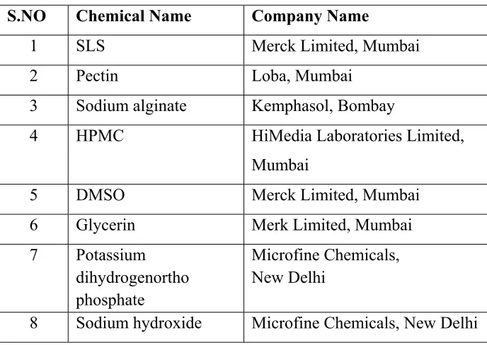
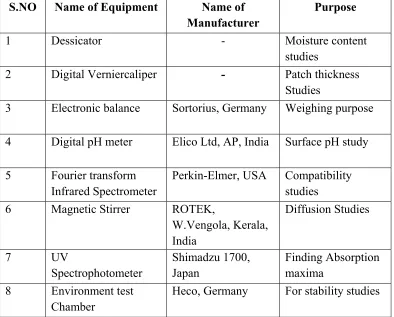
6. MATERIALS AND METHODS 69
7. RESULTS AND DISCUSSIONS 87
8. SUMMARY AND CONCLUSIONS 126
Table
No TITLE Page No.
1. List of Transdermal patches available in the market 32 2. List of chemicals used 69 3. List of equipment used 70 4. Phytochemical Tests 73 5. Stability Condition Chart 84 6. Standard curve of Methanolic Extract of
Acalypha indica Linn. 88
7. Phytochemical Test of Methanolic Extract of
Acalypha indica Linn. 89
8. Determination of Hygroscopic nature 90 9. FT-IR Spectral assignment Methanolic Extract of
Acalypha indica Linn. 91
10. FT-IR Spectral assignment of Pectin 92 11. FT-IR Spectral assignment of Sodium alginate 93 12. FT-IR Spectral assignment of HPMC 94 13. FT-IR Spectral assignment of (P2) Pectin
formulation 95
14. FT-IR spectral assignment of (H4) HPMC
formulation 96
15. FT-IR spectral assignment of(S7) Sodium alginate
formulation 97
16. Physicochemical evaluation of Methanolic Extract
of Acalypha indica Linn.Transdermal patch 98
17. Optimized formula of Methanolic Extract of
Acalypha indica Linn. Transdermal patch 99 18. Uniformity of weight 101 19. Thickness of patch 101
20. Drug content 101
21. Folding Endurance 102 22. Percentage Moisture uptake 102 23. Percentage Moisture content 102
24. Surface pH 103
25. Percent Elongation 103
26. Tensile Strength 103
27. In- vitro drug release profile of P2 104
28. In -vitro drug release profile of H4 105
29. In- vitro drug release profile of S7 107
30. Comparative in- vitro drug release profile 108
31. Ex- vivo drug release profile of H4 111
32. Release Kinetics 112
33. Stabilty study of Methanolic Extract of
Acalypha indica Linn. Transdermal Patch 121
34. Anti –Microbial activity 124
Table
No TITLE Page No.
1. Anatomy of the skin 12
2. Permeation through Subcutaneous route 14
3. Various approaches to enhance drug delivery
through the skin 17
4. Cross section view of Polymer Membrane
Permeation controlled TDDS 25
5. Cross section view of Polymer matrix
Diffusion Controlled TDDS 26
6. Cross section view of Drug Reservoir
Gradient-Controlled TDDS 27
7. Cross section view of Micro reservoir
Dissolution Controlled TDDS 28
8. Use of Transdermal Patch 30
9. Pie Diagram of Popularly Marketed Transdermal
patches 31
10. Life cycle of scabies mite 40
11. A. Adult female scabies and B. Adult
scabies mite 42
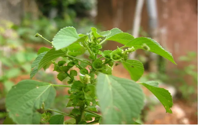
12. Leaves of Acalypha indica Linn. 52
13. Chemical structure of Pectin 56
14. Chemical structure of Sodium alginate 59
17.
Absorption maxima (λ max) of Methanolic
Extract of Acalypha indica Linn. 87
18. Standard curve of Methanolic Extract of
Acalypha indica Linn. 88
19. FT-IR spectrum of Methanolic Extract of
Acalypha indica Linn. 90
20. FT-IR Spectrum of Pectin 91
21. FT-IR Spectrum of Sodium alginate 92
22. FT-IR Spectrum of HPMC 93
23. FT-IR Spectrum of P2 formulation 94
24. FT-IR Spectrum of H4 formulation 95
25. FT-IR Spectrum of S7 formulation 96
26. Transdermal patch of P2 formulation 99
27. Transdermal patch of H4 formulation 100
28. Transdermal patch of S7 formulation 100
29. In- vitro release profile of P2 105
30. In-vitro release profile of H4 106
31. In- vitro release profile of S7 108
32. Comparative in -vitro release profile 109
35. Ex- vivo skin permeation study of H4
formulation 112
Release kinetics of P2 (Pectin) formulation
36. Zero order plot 113
37. First order plot 113
38. Higuchi plot 114
39. Korsemeyer peppas plot 114
Release kinetics of H4 (HPMC) formulation
40. Zero order plot 115
41. First order plot 115
42. Higuchi plot 116
43. Korsemeyer peppas plot 116
Release kinetics of S7 (Sodium alginate) formulation
44. Zero order plot 117
45. First order plot 117
46. Higuchi plot 118
47. Korsemeyer peppas plot 118
Ex-vivo Release Kinetic of H4
50 Higuchi plot 120
51 Korsemeyerpeppas plot 120
Fungi
52 Aspergillus niger 122
53 Candida albicans 122
Bacteria
54 Bacillus subtilis 122
55
Staphylococcus aureus 122
56 E.coli 123
57 Pseudomonas aeruginosa 123
58 Klebsiella pneumonia 123
o
C Degree Celsius
Cm2 Square centimeter SC Stratum corneum
HPMC Hydroxy Propy [Methyl cellulose
SLS Sodium Lauryl Sulphate TDDS Transdermal Drug Delivery System
FT-IR Fourier Transform Infrared Spectroscopy
e. g. Example
Sq.ft Square feet Rpm Revolution per minute
ml Milliliter nm Nanometer
RH Relative Humidity
hrs Hours
MU Moisture Uptake
SD Standard deviation
MC Moisture Content
IP Indian Pharmacopoeia
ME Methanolic Extract
MEA Methanolic Extract of Acalypha indica Linn.
kg Kilogram gm gram
SR Sustained release
PB Phosphate Buffer
Fig Figure
1. INTRODUCTION
1.1. Traditional System of Medicines
1, 2, 3India has a rich heritage of traditional medicine and the traditional health care system have been flourishing for many centuries. traditional medicine, defined by the WHO as "medical knowledge systems that developed over generations within various societies before the era of modern medicine, including the health practices, approaches, knowledge and beliefs incorporating plant, animal and mineral-based medicines, spiritual therapies, manual techniques and exercises, applied singularly or in combination to treat, diagnose and prevent illnesses or maintain well-being" is used globally and has rapidly growing economic importance. In developing countries, traditional medicine is often the only accessible and affordable treatment available. In Latin America, the WHO regional office for the Americas (AMRO/PAHO) reports that 71% of the population in China and 40% of the population in Columbia has used traditional medicine.
In many Asian countries traditional medicine is widely used, even though western medicine is often readily available. In Japan, 60-70% of allopathic doctors prescribe traditional medicines for their patients. In the US the number of visits to providers of Complementary Alternative Medicine (CAM, codified herbal medicine) now exceeds by far the number of visits to all primary care physicians.
Ayurveda
alternative medicine (CAM) in the western world, where several of its methods, such as the use of herbs, massage, and yoga, are applied on their own as a form of CAM treatment.
Ayurveda is the ancient (before 2500 B.C.) Indian system of health care involving a holistic view of man, his health, and illness. Ayurvedic treatment of a disease consists of salubrious use of drugs, diets, and certain practices. Medicinal preparation is invariably complex mixtures, based mostly on plant products. Around 1,250 plants are used in various ayurvedic preparations.
Many Indian medicinal plants have come under scientific scrutiny since the middle of the nineteenth century, although in a sporadic fashion.
The first significant contribution from ayurvedic material medica came with the isolation of the hypertensive alkaloids from the sarpagandha plant (Rauwolfia serpentina), valued in ayurveda for the treatment of hypertension, insomnia and insanity. This was the first important ancient-modern concordance in ayurvedic plants.
Use of these three measures is done in two ways. In one approach of treatment the three measures antagonize the disease by counteracting the etiological factors and various manifestations of the disease.
In the second approach the same three measures of medicine, diet and activity are targeted to exert effects similar to the etiological factors and manifestations of the disease process. These two types of therapeutic approaches are respectively known as Vipreeta and Vipreetarthkari treatments.
Siddha
Siddha system is one of the oldest systems of medicine in India. The term siddha means achievements and siddhars were saintly persons who achieved results in medicine. Eighteen Siddhars were said to have contributed towards the development of this medical system. Siddha literature is in tamiland it is practiced largely in tamil speaking part of India and abroad. The siddha system is largely therapeutic in nature.
The siddha system is capable of treating all types of disease other than emergency cases. In general, this system is effective in treating all types of skin problems particularly psoriasis, STD, urinary tract infections, diseases of liver and gastro intestinal tract, general debility, postpartum anemia, diarrhoea and general fevers.
Unani
Homoepathy
Homeopathy, founded by a German physician Samuel Hahnemann in 1790, is based on the idea that ‘like cures like’; that is substances that cause certain symptoms in a healthy person can also cure those same symptom in someone who is sick. This so called low of similar gives homeopathy its name ‘homeo’ for similar ‘pathy’ designating disease. In this experiment Hahnemann developed a method of ‘potentizing’ homeopathic remedies by diluting them in a water-alcohol solution and then vigorously shaking the mixtures.
The result convinced him that a high degree of dilution not only minimizes the side effects of the remedies but also simultaneously enhances their medical efficacy. Most Homeopathic remedies have undergone ‘proving’ or medical observation in which healthy individuals are given doses of undiluted homeopathic substances.
Mental, emotional, psychic and other details of the patients are most important. This leads the physician to a better understanding of which remedy will best suits a particular set of symptoms. Over the past 200 years, providing for almost 2,000 substances have been conducted.
International diversity
Traditional medicine practices have been adopted in different cultures and regions without the parallel advance of international standards and methods for evaluation.
National policy and regulation
Safety, effectiveness and quality
Scientific evidence from tests done to evaluate the safety and effectiveness of traditional medicine products and practices is limited. While evidence shows that acupuncture, some herbal medicines and some manual therapies (e.g. massage) are effective for specific conditions, further study of products and practices is needed. Requirements and methods for research and evaluation are complex. For example, it can be difficult to assess the quality of finished herbal products. The safety, effectiveness and quality of finished herbal medicine products depend on the quality of their source materials (which can include hundreds of natural constituents), and how elements are handled through production processes.
Knowledge and sustainability
The expanding herbal product market could drive over-harvesting of plants and threaten biodiversity. Poorly managed collection and cultivation practices could lead to the extinction of endangered plant species and the destruction of natural resources. Efforts to preserve both plant populations and knowledge on how to use them for medicinal purposes is needed to sustain traditional medicine.
Patient safety and use
Many people believe that because medicines are herbal (natural) or traditional they are safe (or carry no risk for harm). However, traditional medicines and practices can cause harmful, adverse reactions if the product or therapy is of poor quality, or it is taken in appropriately or in conjunction with other medicines. Increased patient awareness about safe usage is important, as well as more training, collaboration and communication among providers of traditional and other medicines.
1.2. Novel Drug Delivery System
4no therapeutic benefit at all. On the other hand, the very slow progress in the efficacy of the treatment of severe diseases, has suggested a growing need for a multidisciplinary approach to the delivery of therapeutics to targets in tissues. From this, new ideas on controlling the pharmacokinetics, pharmacodynamics, non-specific toxicity, immunogenicity, biorecognition, and efficacy of drugs were generated. These new strategies, often called drug delivery systems (DDS), are based on interdisciplinary approaches that combine polymer science, pharmaceutics, bioconjugate chemistry, and molecular biology.
To minimize drug degradation and loss, to prevent harmful side-effects and to increase drug bioavailability and the fraction of the drug accumulated in the required zone, various drug delivery and drug targeting systems are currently under development. Among drug carriers one can name soluble polymers, microparticles made of insoluble or biodegradable natural and synthetic polymers, microcapsules, cells, cell ghosts, lipoproteins,liposomes and micelles. The carriers can be made slowly degradable, stimuli-reactive (e.g., pH- or temperature-sensitive), and even targeted (e.g., by conjugating them with specific antibodies against certain characteristic components of the area of interest). Targeting is the ability to direct the drug-loaded system to the site of interest. Two major mechanisms can be distinguished for addressing the desired sites for drug release: (i) passive and (ii) active targeting. An example of passive targeting is the preferential accumulation of chemotherapeutic agents in solid tumors as a result of the enhanced vascular permeability of tumor tissues compared with healthy tissue. A strategy that could allow active targeting involves the surface fictionalization of drug carriers with ligands that are selectively recognized by receptors on the surface of the cells of interest. Since ligand–receptor interactions can be highly selective, this could allow a more precise targeting of the site of interest.
difference between a drug’s success and failure, as the choice of a drug is often influenced by the way the medicine is administered. Sustained (or continuous) release of a drug involves polymers that release the drug at a controlled rate due to diffusion out of the polymer or by degradation of the polymer over time. Pulsatile release is often the preferred method of drug delivery, as it closely mimics the way by which the body naturally produces hormones such as insulin. It is achieved by using drug-carrying polymers that respond to specific stimuli (e.g., exposure to light, changes in pH or temperature).
For over 20 years, researchers have appreciated the potential benefits of nanotechnologyin vast providing improvement in drug delivery and drug targeting.
Improving delivery techniques that minimize toxicity and improve efficacy offers great potential benefits to patients, and opens up new markets for pharmaceutical and drug delivery companies. Other approaches to drug delivery are focused on crossing particular physical barriers, such as the blood brain barrier, in order to better target the drug and improve its effectiveness; or on finding alternative and acceptable routes for the delivery of protein drugs other than via the gastro-intestinal tract, where degradation can occur.
1.2.1. Topical delivery
Topical delivery includes two basic types of product:
• External topical that are spread, sprayed, or otherwise dispersed on to cutaneous tissues to cover the affected area
• Internal topical that are applied to the mucous membrane orally, vaginally or on anorectal tissues for local activity
For the most part topical preparations are used for the localized effects at the site of their application by virtue of drug penetration into the underlying layers of skin or mucous membranes. Although some unintended drug absorption may occur, it is sub therapeutics quantities and generally of minor concern.
1.2.2. Advantages of Topical Drug Delivery Systems
• Avoidance of first pass metabolism
• Convenient and easy to apply
• Avoidance of the risks and inconveniences of intravenous therapy and of the varied conditions of absorption, like pH changes, presence of enzymes, gastric emptying time etc.,
• Achievement of efficacy with lower total daily dosage of drug by continuous drug input
• Avoids fluctuation in drug levels, inter- and intrapatient variations
• Ability to easily terminate the medications, when needed
• A relatively large area of application in comparison with buccal or nasal cavity
• Ability to deliver drug more selectively to a specific site
• Avoidance of gastro-intestinal incompatibility
1.2.3. Disadvantages of Topical Drug Delivery Systems
• Skin irritation of contact dermatitis may occur due to the drug and/or excipients
• Poor permeability of some drugs through the skin
• Can be used only for drugs which require very small plasma concentration for action
• Enzyme in epidermis may denature the drugs
1.3. Transdermal Drug Delivery Systems
5, 6The TDDS are defined as self-contained, discrete dosage forms which, when applied to the intact skin, deliver the drug(s), through the skin, at a controlled rate to the systemic circulation. Transdermal drug delivery is a viable administration route for potent, low-molecular weight therapeutic agents which cannot withstand the hostile environment of gastrointestinal tract and/or subject to considerable first-pass metabolism by the liver.
1.3.1. Advantages of TDDS:
• Transdermal medication delivers a steady infusion of a drug over an extended period of time. Adverse effects or therapeutic failures frequently associated with intermittent dosing can be avoided.
• Transdermal delivery can increase the therapeutic value of many drugs by avoiding specific problems associated with the drug e.g., gastro-intestinal irritation, low absorption, decomposition due to hepatic “first-pass” effect, formation of metabolites that cause side effects, short half-life necessitating frequent dosing etc.
• The simplified medication regimen leads to improved patient compliance and reduced inter-and intra-patient variability.
• Self-administration is possible with these systems.
• The drug input can be terminated at any point of time by removal of drug application from the skin surface.
• Substitute’s oral administration when route is unsuitable as in case of vomiting, diarrhoea and in unconscious patients.
• Provides utilization of drugs with short biological half-life, narrow therapeutic window.
• Avoids risk and inconveniences of intravenous therapy.
• Reduces the chances of over- or under dosing through the prolonged, preprogrammed delivery of drug at the required therapeutic rate.
1.3.2. Disadvantages of TDDS:
• The drug must have some desirable physicochemical properties for penetration through Stratum corneum (SC) and if the drug dosage required for therapeutic value is more than 10 mg/day, the transdermal delivery will be very difficult if not impossible. Daily doses of less than 5mg/day are preferred.
• Skin irritation or contact dermatitis due to the drug, excipients and penetration enhancers used to increase percutaneous absorption of the drug is another limitation.
• Clinical need is another area that has to be examined carefully before a decision is made to develop a transdermal product.
• The barrier function of the skin changes from one site to another on the same person, from person to person and with age.
• Poor permeability of drugs through the skin.
• Enzymes in epidermis may denature the drugs.
• Drugs that require high blood levels cannot be administered.
1.4.The Skin
7,8ci ch pr b
1
T 1 m th an bu With irculation ne hemical and rotects again lood pressur.4.1. Anat
The sk The three ma.4.2. Epider The e micrometer (c
he epidermis nd keratiniz undles of ke
a thickness etwork from d microbial a
nst harmful u re.
tomy of Sk
kin is a mult in layers thermis: epidermis is
cum) thick. s and is 25-3 zed the inter eratin fibres.
of only a m m the outside
attacks, acts ultraviolet ra
kin:
[image:31.612.160.481.273.475.2]ti-layered or e skin are epi
Fig No.
s the outerm The SC or n 30 layers of rior or these
millimeter, th e environmen s as a thermo ays of the su
rgan and has idermis, derm
1: Anatomy
most layer non-viable e f horny dead e cell layer
he skin sepa nt, serves as ostat in mai um and play
s anatomicall mis and hyp
of the Skin
of the skin epidermis is d cells, whic s is crisscro
arates the un s a barrier a intaining bod ys a role in th
ly many hist podermis .
n; it is appr the top, out ch are flatten
ossed with d
The dry composition of the horny layer is 75-85% protein; the bulk of the remainder of the substance is a complicated mixture of lipids, this combination of ceratnocytes with interspersed spidermal lipids form a water proof moisture barrier that minimizes trans epidermal water loss (TEWL) to keep moisture in the skin. It is approximately 10-20um thick and acts as protective membrane preventing water loss from the skin and limiting the entry of chemicals from the environment. Stratum corneum consists of lipids which are made up of ceramaides and neutral lipids such as free fatty acids free sterols and triglycerides, the remainder is made up of phospholipids, glycosphingolipids and cholesterol sulphate. Below the Stratum corneum are the other layers of the epiderm is the stratum lucidum, stratum granulosum, and stratum spinosum and stratum germinativum, together these layers constitute the viable epidermis.
1.4.3. Dermis:
The dermis is vascularized and the thickest of all the layers (3-5mm thick). It possesses sweat glands, hair follicles, nerve endings and lymph vessels and acts as the systemic absorption site for drugs. The dermis is located between the hypodermis and the epidermis. An average human skin is known to contain on the average 40-70 hair follicle and 200-300 sweat glads on each square cm of skin area.
1.4.4. Hypodermis:
The hypodermis, which is subcutaneous fat layer that functions as insulation and padding for the body. The hypodermis is the deepest section of the skin as shown in the (Fig No.1).
1 re to
1
in w ap th n S th th m re b in.4.5. Skin A The s egions two t o as skin app
.5. Pathwa
The p ntact epiderm which form ppendages o his pathway oted). As st Stratum corn he intercellu hrough the c must diffuse ecognized asFig No. 2: etween the ntercellular l
Appendages: kin is inters ypes of swe pendages.
ay of Tran
permeation o mis and throshunt path occupy only is usually co tated above, neum. Two p
lar lipid rou corneocytes at some p s the major d
Permeation corneocytes lipid matrix.
:
spersed with at glands ep
nsdermal P
of drugs thr ough the ski hways throu0.1% of the onsidered to , drug perm pathways thr ute between t
and the inte point throug determinate o
n routes thro (intercellula
h hair follicl picrine and a
Permeatio
rough the sk in appendag ugh the inte total huma o be small (w meation throu rough the in the corneocy ervening lipi
gh the inte of percutane
ough the Str ar route) an
le and assoc apocrine. Co
on:
kin includes es, ie., hair tact epiderm an skin surfa with only a f
ugh the skin ntact barrier ytes and the ids; that is, rcellular lip eous transpor
ratum corneu
nd (ii) acros
ciated sebac llectively th
s the diffusi follicles and mis. Howev face and the few exceptio n is usually r may be ide transcellular in both case pid matrix,
rt rate.
um: (i) via t
ss the corne
eous gland hese are refer
ion through d sweat glan ver, these s contribution ons having b
limited by entified (Fig r route cross es the perme which is n
1.5. Kinetics of Skin Permeation
9Transdermal permeation of a drug involves the following steps: 1. Sorption by Stratum corneum.
2. Penetration of drug through viable epidermis.
3. Uptake of the drug by the capillary network in the dermal papillary layer
This permeation can be possible only if the drug possesses certain physiochemical properties.
The rate of permeation across the skin (dQ/dt) is given by:
dt dQ
= Ps(cd – cr)---(1)
Where,
Cd – Concentration of the permeant in donor phase (Stratum corneum) Cr – Concentration of the permeant in receptor phase (systemic circulation). P2 – Overall permeability co-efficient of the skin.
Permeability coefficient is given by the relationship:
Ps = 2
2 2
h D K
=---(2)
Where,
Thus permeability coefficient (P2) may be constant if K d and h terms are constant under a given set of conditions. A constant rate of drug permeation is achieved if Cd>> C2
Then equation (1) becomes: t d dQ
= P2 Cd---(3)
The rate of skin permeation is constant provided the magnitude of C4 remains fairly constant throughout the course of skin permeation. For keeping C4 constant the drug should be released from the device at R4 (release rate of drug) which is either constant or greater than R1 (the rate takes up the drug) i.e. R1>> R2 Since R1>> R2, the drug concentration on the skin surface C4 is maintained at a level equal to or greater than the equilibrium solubility of the drug in the SC, C, i.e. Cd>> Cr
Therefore a maximum rate of skin permeation is obtained and is given by the equation:
(dQ/dt) m = P2 C2………..(4)
From the above equation it can be seen that the maximum rate of skin permeation depends upon the skin permeability coefficient P2 and the equilibrium solubility in the SC, C2. Thus skin permeation appears to be SC limited.
1.5.1. Enhancement of Transdermal Delivery:
A strategy to enhance the skin permeation in the present study is the use of permeation enhancers. One of the easiest approaches to enhance the permeation rate is the use of penetration enhancers. These are the substances added to pharmaceutical formulation in ordered to increase the membrane permeation or absorption rate of a co-administered drug.
1.5.2. Ideal characteristics of chemical penetration enhancers:
Ideally, penetration enhancers reversibly reduce the barrier resistance of the SC without damaging the viable cells. Some of the more desirable properties for penetration enhancers acting within the skin have been given as:
9 They should be non-toxic, non-irritating and non-allergenic
9 They would ideally work rapidly, and the activity and duration of effect should be both predictable and reproducible
9 They should have no pharmacological activity within the body i.e. should not bind to receptor sites
9 When removed from the skin, barrier properties should return both rapidly and fully
9 The penetration enhancers should be appropriate for formulation into diverse topical preparations, thus should be compatible with both excipients and drugs
9 They should be economicacally acceptable with an appropriate skin feel
1.5.3. Mechanism of Chemical Penetration Enhancement10
Penetration enhancers may act by one or more of three main mechanisms. 1. Disruption of the highly ordered structure of Stratum corneum lipid. 2. Interaction with intercellular protein.
3. Improved partition of the drug, co-enhancer or solvent into the Stratum coneum.
1.5.4. Different Classes of Permeation Enhancers are as follows: 1.5.4.1. Sulfoxides
Dimethylsulfoxide (DMSO) is an effective penetration enhancer that promotes permeation by reducing skin resistance to drug molecules or by promotion of drug partitioning from the dosage form. It has been postulated that DMSO denatures the intercellular structural proteins of the SC, or promotes lipid fluidity by disruption of the ordered structure of the lipid chains. Along with these, DMSO may alter the physical structure of the skin by elution of lipid, lipoprotein and nucleoprotein structures of the Stratum corneum.
1.5.4.2. Alcohols
1.5.4.3. Polyols
The activity of propylene glycol is thought to result from salvation of a keratin within the SC; the occupation of proteinaceous hydrogen bonding sites reducing drug-tissue binding and thus promoting permeation.
1.5.4.4. Alkanes
Long chain alkanes (C2-C16) have been shown to enhance skin permeability by nondestructive alteration of the SC barrier.
1.5.4.5. Fatty acids
Selective perturbation of the intercellular lipid bilayers in the SC appears to be the major mode of enhancing activity of the fatty acids.
1.5.4.6. Esters
Esters such as ethyl acetate are relatively polar, hydrogen bonding compounds that may enhance permeation in a similar manner to the sulphoxides and formamides by penetrating into the SC and increasing the lipid fluidity by disruption of lipid packing. 1.5.4.7. Amines and amides:
¾ Urea
Urea promotes transdermal permeation by facilitating hydration of the SC and by the formation of hydroplic diffusion channels within the barrier. Cyclical urea permeation enhancers are biodegradable, non-toxic molecules consisting of a polar parent moiety and a long chain alkyl ester group.
¾ Dimethyl acetamide and dimethyl formamide
¾ Pyrrolidones
Pyrrolidone and its derivatives are reported to interact with both keratin and with lipids in the skin. Azone is known to show significant accelerant effects at low concentrations for both hydrophilic and hydrophobic drugs and is one of the few enhancers that have been developed commercially. Differential scanning calorimetric studies have shown that azone affects lipid structures of the SC. In addition, azone is reported to decrease transition temperatures within lipid bilayers to induce formation of a liquid phase with a resultant increase in lipid fluidity.
1.5.4.8. Terpenes
Terpenes are found inessential oils, and are compounds comprising of only carbon, hydrogen and oxygen atoms, but which are not aromatic. Numerous terpenes have long been used as medicines as well as flavoring and fragrance agents. Terpenes are generally considered as less toxic and have less irritant effects compared to surfactants and other skin penetration enhacers, and some terpenes have been characterized as Generally Recognized As Safe (GRAS) by the US FDA. They have high percutaneous enhancement ability, reversible effect on the lipids of SC, minimal percutaneous irritancy at low concentrations (1-5%). Moreover, a variety of terpenes have been shown to increase percutaneos absorption of both hydrophilic and lipophilic drugs.11 Both the mono-and sesquiterpenes are known to increase percutaneous absorption of compounds by increasing diffusivity of the drug and/or by disruption of the intercellular lipid barrier. A further mechanism of activity that has been postulated is that the terpenoids increase electrical conductivity of tissues thereby opening polar pathways within the Stratum corneum.
1.5.4.9. Surface active agents:
Anionic surfactants may function by alteration of the barrier function of the SC as a result of removal of water soluble agents that act as plasticizers. Sodium lauryl sulphate has been implicated in reversible lipid modification with resultant disorganization of the SC and enhanced permeation. In addition, non-ionic surfactants are purported to be able to emulsify sebum, consequently altering partitioning potential of drugs in favour of enhanced permeation. The permeation enhancement generated by these compounds may be dependent on the ability of drug to partition between the free and bound or micellar form of the enhancer.
1.5.3.10. Cyclodextrins
Cyclodextrins are bio-compatible substances that can form inclusion complexes with lipophilic drugs with a resultant increase in their solubility, particularly in aqueous solutions. However, cyclodextrins alone were determined be to less effective as penetration enhancers than when combined with fatty acids and propylene glycol.
1.6. Basic Components of Transdermal Drug Delivery Systems
11 The components of transdermal devices include:1. Backing layer
2. Drug containing reservoir a. Polymer matrix b. Drug
c. Permeation enhancers d. Plasticizers
3. The release control layer 4. The adhesive
5. The peel strip. 1. Backing layer
The most commonly used backing materials are alupoly, polyester, polyethylene co-extruded films or metalized polyester laminated with polyethylene. The film can be either clear flesh colored or metalized. Backing membranes are flexible and provide a good bond to the drug reservoir prevent drug from leaving the dosage form through the top and accept printing. It protects the product during use on the skin.
2. Drug containing reservoir: A) Polymer Matrix:
The Polymer controls the release of the drug from the device. The development of transdermal systems requires judicious selection of a polymeric material or a series of polymers whose diffusive characteristic will be such that a desirable permeation rate of a specific drug or other bio-active agent can be obtained.
The polymer should meet the following requirements:
i) Molecular weight, glass transition temperature and chemical functionality of polymer must allow proper diffusion and release of the specific drug. ii) The polymer should not chemically react with the drug.
iii) The polymer and its degradation products must be non-toxic. iv) The polymer should not decompose on storage or use of the device. v) The polymer should be inexpensive.
vi) The polymer must be easy to manufacture and it should yield itself into the desired product and should allow incorporation of large quantities of active component without deterioration of its mechanical properties.
Polymers that can be used in the transdermal formulations are: a) Natural Polymers:
b) Synthetic Elastomers:
Polybutadieine, Hydrin rubber, Polysiloxane, Silicone rubber, Nitrile, Acrylonitrile, Butyl rubber, Styrenebutadieine rubber, Neoprene etc.
c) Synthetic Polymers:
Polyvinyl alcohol, Polyvinyl chloride, Polyethylene, Polypropylene, Polyacrylate, Polyamide, Polyurea, Polyvinylpyrrolidone, Poly methylmethacrylate, etc.,
B) Drug:
For successfully developing a TDDS, the drug should be chosen with great care. The following are some of the desirable properties of a drug for transdermal delivery. Physicochemical properties:
1. The drug should have a molecular weight less than approximately 1000 daltons. 2. The drug should have affinity for both lipophilic and hydrophilic phases.
3. The drug should have a low melting point. Biological properties:
1. The drug should be potent with a daily dose of the order of a few mg/day. 2. The halflife (tn) of the drug should be short.
3. The drug must not induce a cutaneous irritant or allergic response.
4. Drugs which degrade in the g.i.t or/are inactivated by hepatic first pass effect are suitable candidates for transdermal delivery.
5. Tolerance to the drug must not develop under the near zero order release profile of transdermal delivery.
C) Permeation Enhancers:
Discussed in detail in section 1.5. D) Plasticizers:
These are used to prevent the films from becoming brittle.12 An ideal plasticizer should possess the following properties.
1. Should not show any pharmacological action of its own. 2. Should be chemically and physically stable.
3. Should be compatible with the drug and the formulated components. 4. Should be colourless, colorless and tasteless.
5. Should be non-toxic, non-allergenic & non-irritant. 3.Release control layers:
The rate controlling membrane can be either a micro-porous or a non-porous polymeric membrane with defined drug permeability. The membrane can be constituted with any of the polymers discussed earlier.
4. Adhesives:
The fastening of all transdermal devices to the skin has so far been done by using a pressure sensitive adhesive which can be positioned on the face of the device or in the back of the device and extending peripherally. Both adhesive systems should fulfill the following criteria.
(i) Should adhere to the skin aggressively, should be easily removed. (ii) Should not leave an unwashable residue on the skin.
(iii) Should not irritate or sensitize the skin.
The face adhesive system should also fulfill the following criteria.
i) Physical and chemical compatibility with the drug, excipients and enhancers of the device of which it is a part.
ii) Permeation of drug should not be affected.
5. Peel Strip:
The peel strip prevents loss of drug that has migrated into adhesive layer during storage and also protects the finished device against contamination. Polyester foils and metalized laminates are the choices.
6. Packet:
Packet guards the patches against drug loss and contamination on storage. The patches are individually packed in heat sealed foil pouches.
1.7
. Approaches used in the Development of TDDS
12Several techniques have been successfully developed to provide a mechanism of rate control over the release and transdermal permeation of drugs. These are three types of devices currently available in the market. To be precise, there are two concepts in the design namely, the reservoir type and the matrix type.
1.7.1 Different technologies employed in the development of TDDS: I. Polymer Membrane Permeation controlled TDDS:
In the drug reservoir compartment the drug solids are dispersed homogeneously in a solid polymer matrix (e.g., poly isobutylene), suspended in an unleachable, viscous liquid medium (e.g., silicone fluid) to form a pase like suspension, or dissolved in a releasable solvent (e.g.,alkyl alcohol) to form a clear drug solution. On the external surface of the polymeric membrane a thin layer of drug compatible, hypoallergenic pressure sensitive adhesive polymer (e.g., silicone adhesive) may be applied to provide intimate contact of the TDDS with the skin surface. The intrinsic rate of drug release from this type of TDDS is defined by:
dt dQ
=
m 2 2/m 2 m m/r
m 2 l/r m/r
h D K h D K
D D K K
+ Cr ---Æ (5)
Where: C R – Drug concentration in the reservoir compartment.
Km/r& Ka/m -- Are the partition coefficients for the interfacial partitioning of drug from the reservoir to the membrane and from the membrane to the adhesive.
Dm& D2 --- Are the diffusion coefficients in the rate-controlling membrane and in the adhesive layer.
hm& ha -- are the thickness of rate-controlling membrane and adhesive layer.
II. Polymer matrix Diffusion-Controlled TDDS:
fo th o b p re d d W L C t
ormed is the hickness. Th
cclusive bas acking. In th atch to form elease from t
dt dQ
= ⎢
⎣ ⎡LC
2 p d
Where: Ld -- Drug lo Cp& Dp – So
– Time.
F
en molded in his drug re seplate in a his system th m a strip of a this polymer ⎥ ⎦ ⎤ t Dp p 1/2
----oading dose lubility and
[image:46.612.123.519.384.591.2]III.D
Fig No.6: Cro
nto medicate eservoir con
a compartm he adhesive adhesive rim r matrix drug
---initially disp diffusivity o
Drug Reserv
oss section vi
ed disks with ntaining poly ent, fabricat
polymer is a m surroundin
g dispersion
---Æ (6)
persed in the of the drug in
voir Gradie
iew of Drug R
h a defined ymer disk ted from a applied alon ng the medic type TDDS
e polymer m n the polyme
nt-Controll
Reservoir Gr
surface area is then mo drug imper ng the circum
cated disk. T is given by:
atrix. er matrix.
led TDDS:
radient-Cont
a and contro ounted onto
rmeable pla mference of The rate of d
dr al re d d d in th L d in an
The p rug loading long the dif elease from t
dt dQ = ) ( 2 2 a/r t h D K In thi iffuse increa n diffusional he multilam Ld(ha). This,
FigN
This t ispersion-typ
In this n an aqueou
nd then ho
polymer mat level varied ffusional pat this type of d
Ld(h2)---s Ld(h2)---syLd(h2)---stem the ases with tim l path as a re minate adhes , in theory sh
IV.M
No.7: Cross s
type of drug pe drug deliv s approach t us solution o
mogeneousl
trix drug dis in an increm th across the drug reservo
---e thickn---ess me, i.e., ha(t) esult of drug sive layers
hould yield a Microreservo
ection view o
system can very system the drug rese of a water-m ly dispersin
spersion typ mental mann e multilamin oir gradient c
----Æ (7)
of diffusion ). To comp g depletion d is also desi a more const oir Dissolut
of Microrese
be consider s.
ervoir is form miscible drug ng the drug
pe TDDS ca ner, forming nate adhesiv controlled TD
nal path thro pensate for th due to release igned to in tant drug rel tion Control
rvoir Dissolu
red a hybrid
med by first g solubilizer suspension
an be modifi a gradient o ve layers. Th
DDS can be
ough which his time dep e, the drug ncrease prop
ease profile. lled TDDS
ution Contro
of the reser
suspending r, e.g., poly n with cont
fied to have of drug reserv he rate of d expressed b drug molecu pendent incre loading leve portionally, . olled TDDS
rvoir and ma
solubility in a lipophilic polymer by high shear mechanical force to form thousands of unleachable microscopic drug reservoirs. This thermodynamically unstable dispersion is quickly stabilized by immediately cross linking the polymer chains in-situ, which produces a medicated polymer disk with a constant surface area and a fixed thickness. The rate of drug release from a microreservoir drug delivery system is defined by:
= dt dQ ) 7 ( 1 1 ) 1 ( m m 1 1 1 p 2 2 2 d p 2 r
p −−−−−−−−−−−>
⎥ ⎦ ⎤ ⎢ ⎣ ⎡ ⎟⎟ ⎠ ⎞ ⎜⎜ ⎝ ⎛ + − −
+ h k k
B S D BS AK h D h D AK D D Where:
A = a/b, a is the ratio of the drug concentration in the bulk of elution solution over the drug solubility in the same medium, and b is the ratio of the drug concentration at the outer edge of the polymer coating membrane over the drug solubility in the same polymer composition.
B = is the ratio of the drug concentration at the inner edge of the interfacial barrier over the drug solubility in the polymer matrix.
Kl, Km & Kp = are the partition co-efficients for the interfacial partitioning of drug from the liquid compartment to the polymer matrix, from the polymer matrix to the polymer coating membrane, and from the polymer coating membrane to the elution solution (or skin).
Dl, Dp & Ds – are the drug diffusivities in the liquid compartment, polymer coating membrane, and elution solution (or skin), respectively.
1.8
Use of Transdermal Patch
Fig No.8: Use of Transdermal Patch
It is important to use a different application site every day to avoid skin irritationSuggested rotation is :
Day 1 - Upper right arm Day 2 - Upper right chest Day 3 - Upper left chest Day 4 - Upper left arm, Then repeat from day 1.
1.8.1. Transdermal Patches Available in the Market13
Nowadays, the transdermal route has become one of the most successful and innovative focus for research in drug delivery with around 40% of the drug candidate being under clinical evaluation related to transdermal or dermal systems.
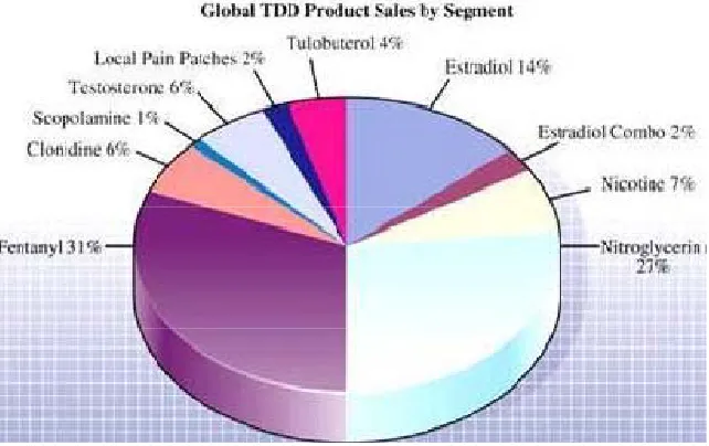
Statistics reveal a market of $ 12.7 billion in the year 2005 that is expected to increase to $ 21.5 billion in the year 2010 and $ 31.5 billion in the year 2015. The pie diagram shows the global transdermal product sales.
[image:50.612.161.482.161.363.2]
Fig No. 9: Pie diagram of popularly marketed Transdermal patch
Table No .1: List of Transdermal Patches available in The Market Brand Name Drug Manufacturer Indications
NicotinellR Nicotine Novartis Pharmacological
smoking cessation
MatrifenR Fentanyl Nycomed Pain relief patch
NeuproR Rigotine UCB and
Schwarx pharma
Early-state idiopathic parkinson’s disease
Alora Estradiol Thera Tech/
Procter and gamble
Postmenstrual syndrome
Nitrodisc Nitroglycerin Robert’s
Pharmaceuticals
Angina pectoris
Nuvelle TS Estrogen/
Progesterone
Ethical Holdings/
Schering
Hormone replacement therapy
Testoderm TTSR Testosterone Alza Hypogonadism in
males
OxytrolR Oxybutynin Watson Pharma Overactive bladder
Catapres TTSR Clonidine Alza/Boehinger
Ingelheim
Hypertension
Minitran Nitroglycerin 3M
Pharmaceuticals
Angina pectoris
Duragesic Fentanyl Alza/Jannssen
Pharmaceutical
Moderate/ severe pain
Emsam Selegiline Bristol-Myers
Squibb
Major depressive disorder
Neupro Rotigotine Schwarz
Pharma
Exelon Rivastigmine Novartis Dementia
Daytrana Methylphenidate Shire Attention deficit
hyperactivity disorder Synera Lidocaine/ tetracaine Endo Pharmaceuticals Local dermal Analgesia
Ionsys Fentanyl HCI
(iontophoresis)
Alza Acute postoperative
pain Scnoprep Lidocaine (ultrasound) Echo Therapeutics Local dermal Anesthesia
Oxytrol Oxybutynin Watson Pharma Overactive Bladder
Climara Pro Estradiol Bayer
Healthcare Pharmaceuticals
Menopausal symptoms
Ortho Evra Ethinyl estradiol/ norelgestromin Ortho-Mc Neil Pharmaceutical Contraception Nicoderm,Habitrol,Proste p
Nicotine Glaxo Smith
Kline, Novartis Consumer Health, Elan
Smoking cessation
Testoderm Testosterone Alza Testosterone
Deficiency Iontocaine Lidocaine/
epinephrine (iontophoresis)
Iomed Local dermal
1.9. Therapeutic Applications of TDDS13
¾ Hisetal, used in treatment of multiple sclerosis can be formulated in TDDS using oleic acid as permeation enhancer to achieve sufficient drug delivery.
¾ Diclofenac sodium, Celecoxib used as NSAID’s, formulated in TDDS can overcome gastric lesions associated with oral dose.
¾ Drugs used for long term dosing in chronic diseases like captopril, verapamil, terbutaline sulphate, pinacidil, propranolol which have short biological half life, considerable first pass metabolism can be formulated as TDDS to achieve prolonged steady state plasma concentration.
¾ Gel formulation with lipid disperse system of betahistine has potential for development of an efficient controlled release transdermal system.
¾ Enhancer and co-solvent can synergictically enhance the delivery of peptides like thyrotropin releasing hormone across human skin.
¾ Prazosin HCL in membrane controlled TDDS can deliver drug enough to maintain minimum effective concentration and can avoid hypotension associated with high initial oral dosing.
¾ TDDS of indomethacin in polyvinylpyrolidone polymer (acting as anti nucleating agent) can provide better anti-inflammatory activity and lower ulcer indices compared to oral administration.
¾ Diclofenac sodium, existing in anionic form at skin pH can be formulated as ion- pairs with oppositely charged enhancers to enhance transdermal delivery compared to non-ion paired forms.
¾ Iontophoresis can increase permeation rate of hydrophilic atenolol to a greater extent than permeation enhancer and overcome incomplete absorption on GIT.
¾ Nimesulide in sodium alginate transdermal gel can provide better analgesic and anti-inflammatory activity and avoid adverse effects associated with long term treatment with high oral dose.
¾ Bupropion HCl, an antidepressant drug can be converted to free base to increase lipophilicity and transdermal delivery and avoid release of fatal metabolites associated with oral dosing.
¾ Zidovudine, an anti-HIV drug, formulated in TDDS and overcome toxic effects associated with frequent higher oral dose.
¾ Levonorgestrel, a potent contraceptive agent, formulated as transdermal protransferosome gel can provide enhanced, prolonged and controlled delivery and overcome GI disturbances, weight gain, irregular bleeding, headache etc. associated with oral dosing.
¾ Polymerized rosin can be used ti design matrix type TDDS of Diltiazem HCl to prolong drugrelease and avoid variable and extensive first pass metabolism on oral dose.
¾ Ester prodrug of ketorolac can provide enhanced permeation whereas nanostructured lipid carrier can act as controlled release system and avoid gastric ulceration and renal failure associated with frequent long term oral dosing.
1.10. Limitations of Transdermal Delivery System
¾ Higher molecular weight candidates (>500Daltons) fail to penetrate the Stratum corneum.
¾ Drugs with very low or high partition coefficient fail to reach systemic circulation.
¾ High melting drugs are not suitable due to their low solubility both in water and fat.
¾ Possibility of local irritation at the site of patch application.
¾ A lag time associated with the delivery of the drug across the skin, resulting in a delay in onset of action.
¾ Variation of absorption rate based on site of application.
¾ Presence of skin diseases.
1.11. Evaluation of Transdermal Patches 14
Development of controlled release transdermal dosage form is a complex process involving extensive research. Transdermal patches have been developed to improve clinical efficacy of the drug and to enhance patient compliance by delivering smaller amount of drug at a predetermined rate. This makes evaluation studies even more important in order to ensure their desired performance and reproducibility under the specified environmental conditions.
These studies are predictive of transdermal dosage forms and can be classified into following types.
I. Physicochemical evaluation II. In-vitro evaluation
III. Ex-vivo evaluation.
Upon the success of physicochemical and in-vitro studies, in-vivo evaluations may be conducted.
I. Physicochemical Evaluation:
1. Thickness: The thickness of transdermal film is determined by screw gauge at different points of the film.15
2. Unformity of weight: Weight variation is studied by individually weighing 10 randomly selected patcvhes and calculating the average weight. The individual weight should not deviate significantly from the average weight.16
3. Drug content determination: An accurately weighed portion of film (about 100 mg) is dissolved in 100 ml. of suitable solvent in which drug is soluble and then the solution is shaken continuously for 24 h in shaker incubator. Then the whole solution is sonicated. After sonication and subsequent filtration, drug in solution is estimated spectrophotometrically by appropriate dilution.17
But if 3 patches have content in the range of 75% to 125%, then additional 20 patches are tested for drug content. If these 20 patches have range from 85% to 115% then the transdermal patches pass the test.
5. Moisture content: The prepared films are weighed individually and kept in a desiccators containing calcium chloride at room temperature for 24 h. The films are weighed again after a specified interval until they show a constant weight. Percent moisture content is calculated using following formula.18
% Moisture content = Initial weight – Final weight X 100
Final weight
6. Moisture Uptake: Weighed films are kept in a desiccator at room temperature for 24 h. These are then taken out and exposed to 84% relative humidity using saturated solution of potassium chloride in a desiccator until a constant weight is achieved. Percent moisture uptake is calculated as given below.18
% moisture uptake = Final weight – Initial weight X 100
Initial weight
7. Folding Endurance: Evaluation of folding endurance involves determining the folding capacity of the films subjected to frequent extreme conditions of folding. Folding endurance is determined by repeatedly folding the film at the same place until it break; the number of times the films could be folded at the same place without breaking is folding endurance value.18
Tensile strength = F/a.b(1=L/1)
F is the force required to break; a is width of film; b is thickness of film; L is length of film; 1 is elongation of film at break point.
II. The in-vitro evaluation 5,6
The objective of in-vitro research is often to find correlation between laboratory results and the transdermal absorption experienced by living subjects, so that, in-vivo experimentation may be curtailed.
The factors to be considered while selecting an in-vitro system include:
1. The rate limiting process: drug solubization or diffusion in the vehicle, partitioning from the vehicle, diffusion through the test membrane or partitioning and removal by the receptor phase.
2. The intrinsic diffusivity of the permanent and apparent diffusivity.
3. The predominating route of diffusion during the experiment and the relative extents of drug binding the metyabolism, occurring in the membrane, delivery and receptor phases.
4. The intrinsic barrier potential of the membrane and the effects that vehicle components may have on its retardative properties. Hydration of the membrane and the presence of penetration enhancers may be important here. Inter-specimen variability between membranes of the same type may markedly influence experimental results.
The various types of cells lused for carrying out in-vitro release studies are 1. Chien and Vilia cell
2. Franz diffusion cell
3. Keshary-chien diffusion cell
Usually Diffusion Cell Design contains two parts donor and received compartment:
All materials should be assessed for their ability to absorb or adsorb the test penetrant.
1. Donor Compartment is designed in such a way to achieve
Easy access to deliver the penetrants to the skin.
(37±1o)c of temperature.
Control of evaporation for volatile vehicles and penetrants. Membrane used in study should have following characteristics:
For the study of penetration kinetics, only porcine epithelium should be used.
For vehicle/device release studies, other barriers may be used.
The skin sample should ideally contain only SC.
A molecule of known penetration kinetics should be used prior to the test molecule, to assess barrier function.
Wherever applicable, metabolic viability of epidermis must be assessed. 2. Receptor Compartment
Either, flow-through or static.
(37±1o)c of temperature
Sufficient volume to maintain infinite sink conditions.
Stirred without obvious formation of boundary layers. 3. Receptor Fluid
Should not compromise barrier function.
Should be of favorable partitioning characteristics to receive the penetrant.
Capable of maintaining epidermal viability wherever necessary.
h th hu II su re m n T ra
1
p ag sc fa A vas airless mous here is as ye uman skin. II. Ex-vivo eAlthou ubjects, this esources are man. Consequ
Altho o general ag The various a
at, guinea pig
.12. SCAB
Scabie arasite that b ges. Howeve cabies. Hen acilities, hosst majority se, guinea pi et no animal
evaluation 5 ugh most r s desirable a required to uently, one m ough the ran greement as animal mode
g, rhesus mo
BIES
21es is a skin burrows into er, people w nce, outbrea stels and elde
of in-vitro ig and rabbit
skin that co
5,6
relevant dat approach is
conduct a sa must be pref ge of specie to the best o els those hav onkey, rabbi
disease cau o, resides an with weakene
aks of scab erly homes.
Fig No. 10:
experiment t too. Althou ompletely m
ta pertainin not always afe & meanin ferred to use
s employed or most pred ve been util t, dog etc.
used by a m d reproduce ed immunity bies have b
Life Cycle o
ts are condu ugh there exi mimics the pe
ng to TDDS s possible, s ngful percut
an in-vivo a in previous dictive mode lized for the
mite called Sa s in human y or the elde been reporte
f Scabies mit
ucted on an ists a numbe enetration ch
S are obtai since consid taneous abso animal mode work is very l for skin pe
in-vivo stud
arcoptesscab skin. It affec erly are mor ed in hospi
te
nimal skin er of similari haracteristic
ined in hum derable time orption study el.
y broad, ther enetration wo
dies are mou
biei, which cts people of
1.12.1 . Clinical features
Scabies manifests in two main ways: People infected with classical scabies will present with intense itchiness which is more severe at night or after a bath. The common affected areas are finger webs and skin folds of wrists, elbows, armpits, nipples, lower abdomen, external genitalia, buttocks and groins. Rashes, thread-like lesions or vesicles may be seen on the skin. The face and scalp are usually spared, except in infants, young children and immune-compromised persons.
A rare but severe type of scabies known as Norwegian or crusted scabies can occur in institutionalized people, particularly in those who are weak or mentally disabled. The skin lesions appear as marked scales and crusts. The nails may thicken, with debris in the nail bed. Face and scalp can also be affected. This type of scabies is highly contagious because an infected person may harbour thousands of mites, compared with 10-15 mites present in classical scabies.
1.12.2. Path physiologys of Scabies
22<