J
OURNAL OFV
IROLOGY,
0022-538X/97/$04.00
1
0
May 1997, p. 3972–3985
Vol. 71, No. 5
Copyright
q
1997, American Society for Microbiology
Purification of the Simian Virus 40 (SV40) T Antigen
DNA-Binding Domain and Characterization of Its
Interactions with the SV40 Origin
WOO S. JOO, XUELIAN LUO, DEBORAH DENIS, HENRY Y. KIM, GODFREY J. RAINEY,
CLYDE JONES, K. R. SREEKUMAR,
ANDPETER A. BULLOCK*
Department of Biochemistry, Tufts University School of Medicine, Boston, Massachusetts 02111
Received 7 May 1996/Accepted 18 February 1997
To better define protein-DNA interactions at a eukaryotic origin, the domain of simian virus 40 (SV40) large
T antigen that specifically interacts with the SV40 origin has been purified and its binding to DNA has been
characterized. Evidence is presented that the affinity of the purified T antigen DNA-binding domain for the
SV40 origin is comparable to that of the full-length T antigen. Furthermore, stable binding of the T antigen
DNA-binding domain to the SV40 origin requires pairs of pentanucleotide recognition sites separated by
approximately one turn of a DNA double helix and positioned in a head-to-head orientation. Although two
pairs of pentanucleotides are present in the SV40 origin, footprinting and band shift experiments indicate that
binding is limited to dimer formation on a single pair of pentanucleotides. Finally, it is demonstrated that the
T antigen DNA-binding domain interacts poorly with single-stranded DNA.
Initiation of DNA replication is a complicated process that
requires very precise protein-DNA interactions at replication
origins (1, 26). The replication of simian virus 40 (SV40) DNA
has served as a useful model system for studying initiation
events in mammalian cells (for reviews, see references 11, 34,
and 80). SV40 has a well-defined origin of replication (see
reference 18 and references cited therein) and encodes an
82-kDa protein, termed T antigen (T-ag), that is required for
initiation of DNA synthesis (82). SV40 large T-ag binds the
viral origin owing to major-groove interactions with GAGGC
pentanucleotide recognition sites (21, 35, 84). What is known
about SV40 large T-ag and its interactions with the SV40 origin
has been reviewed previously (5, 27, 28). It has been observed
that upon binding to the SV40 origin, T-ag oligomerizes to
form a double hexamer around the origin (14, 16, 46).
Assem-bly of T-ag hexamers around the SV40 origin induces
confor-mational changes (7, 17, 57) that enable subsequent DNA
unwinding events to occur (9, 15, 24, 89). DNA unwinding is
catalyzed by the inherent helicase activity of T-ag (30, 78).
Analyses of T-ag, including partial proteolysis (67, 71, 72,
91), expression of subfragments of T-ag as recombinant
pro-teins (2, 81), and genetic studies (2, 60, 81), have demonstrated
that T-ag is composed of several structural domains (for a
review, see reference 28). The best-defined and most
exten-sively studied domain of T-ag is the origin DNA-binding
do-main (T-ag-bd) (2, 49, 60, 71, 81), also termed the T-ag-obd
(44). Currently, there is some uncertainty regarding the exact
boundaries of the T-ag-bd; it has been mapped to amino acids
131 to 259 (2), 132 to 246 (49), and 147 to approximately 247
(42). Nevertheless, peptides from all three size classes are
known to be sufficient for sequence-specific binding to the
SV40 origin region (2, 33, 49, 72).
In addition to sequence-specific binding, mutation studies
have demonstrated that the T-ag-bd is involved in many
addi-tional activities catalyzed by T-ag. These activities include
pro-tein oligomerization (74), binding to single-stranded DNA (49,
75), interactions with cellular factors (31), and DNA
unwind-ing and helicase activities (74, 75, 92). In light of this
complex-ity, it is not surprising that the T-ag-bd is itself organized into
subdomains (73). The solution structure of the T-ag-bd region
between amino acids 131 and 260 (T-ag-bd131–260) has
pro-vided preliminary insights into these processes (44).
Previous studies of the T-ag-bd have, in general, been
con-ducted with peptides isolated via immunoprecipitation
tech-niques and not with purified protein preparations. Moreover,
certain properties of the T-ag-bd, such as the mechanism by
which it interacts with the SV40 origin and single-stranded
DNA, have been poorly characterized. Therefore, to provide
further insights into the biochemistry of the T-ag-bd, we have
overexpressed T-ag-bd131–260
in Escherichia coli. T-ag-bd131–260
has been purified to apparent homogeneity and used to
exam-ine its interaction with both double- and single-stranded DNA;
the results of these studies are presented herein.
MATERIALS AND METHODS
Commercial supplies of enzymes and oligonucleotides.Restriction endonucle-ases BamHI and EcoRI were purchased from Boehringer Mannheim Biochemi-cals. Both T4 polynucleotide kinase and HaeIII-digestedfx174 RF DNA were from Gibco-BRL. Thrombin was obtained from Haematologic Technologies Inc. Oligonucleotides were synthesized on an Applied Biosystems 394 DNA syn-thesizer at the protein chemistry facility at Tufts University, purified by electro-phoresis through 10% urea–polyacrylamide gels, and subsequently isolated as described previously (65).
Purification of T-ag.SV40 T-ag was prepared by using a baculovirus expres-sion vector containing the T-ag-encoding SV40 A gene (56) and isolated by immunoaffinity techniques using the PAb 419 monoclonal antibody as previously described (23, 70, 88). Purified T-ag was dialyzed against T-ag storage buffer (20 mM Tris-HCl [pH 8.0], 50 mM NaCl, 1 mM EDTA, 1 mM dithiothreitol [DTT], 0.1 mM phenylmethylsulfonyl fluoride, 0.2mg of leupeptin per ml, 0.2mg of antipain per ml, and 10% glycerol) and frozen at2708C until use.
Cloning DNA fragments containing T-ag-bd131–260and T-ag-bd112–260into pGEX1lT. (i) pGEX-T-ag-bd131–260.The region extending between SV40
nucle-otides 4427 and 4041 (encoding T-ag amino acids 131 to 259 [85]) were amplified with pSVLD as the template; this plasmid contains full-length SV40 DNA cloned into the BamHI site of pBR322 (51). Plasmid pSVLD was incubated with oli-gonucleotide 1 (59tcggatccAAGGTAGAAGACCCCAAGGA39) and oligonucle-otide 2 (59ccgaattcTGGATTAAAATCATGCTCCT39) in a PCR (53). (The boldfaced capital A in oligonucleotide 1 is position 4427, and the boldfaced capital T in oligonucleotide 2 is position 4041). Note that oligonucleotide 1 also contained a BamHI site (in lowercase boldfaced letters) and that oligonucleotide
* Corresponding author. Mailing address: Department of
Biochem-istry A703, Tufts University School of Medicine, 136 Harrison Ave.,
Boston, MA 02111. Phone: (617) 636-6874. Fax: (617) 636-6409.
E-mail: [email protected].
3972
on November 9, 2019 by guest
http://jvi.asm.org/
2 contained an EcoRI site (also in lowercase boldfaced letters). Thus, upon digestion of the PCR product with BamHI and EcoRI, DNA coding for T-ag-bd131–259could be readily ligated into BamHI and EcoRI-digested pGEX-1lT
(76). The first three nucleotides of the EcoRI site, GAA, encodes a glutamic acid residue. This is the normal residue at position 260 of T-ag; thus, upon ligation, the expression vector encoded T-ag amino acids 131 to 260. Moreover, during the construction of this molecule, a single nucleotide was spontaneously deleted in the codon following position 260; this resulted in a frameshift mutation that introduced a stop codon in what would have been codon 263.
(ii) pGEX-T-ag-bd112–260.The SV40 fragment containing nucleotides 4481 to
4041 (encoding T-ag amino acids 113 to 259) was amplified with pSVLD as the template. This plasmid was combined with oligonucleotide 3 (59cgggatccGATG ATGAGGCTACTGCTGACTCT39) and the previously described oligonucleo-tide 2 (59ccgaattcTGGATTAAAATCATGCTCCT39) in a PCR (53). (The bold-faced capital G in oligonucleotide 3 is position 4481, and the boldbold-faced capital T in oligonucleotide 2 is position 4041). Additional cloning procedures were per-formed as described above. However, in this vector, the normal pGEX-1lT stop codon, located six codons beyond position 260, is utilized. Furthermore, in purified T-ag-bd112–260, serine 112 is derived from the vector (see below).
After transformation into E. coli BL21 cells, the expression vectors pGEX-T-ag-bd131–260and pGEX-T-ag-bd112–260were isolated by standard methods (65).
Dideoxy sequencing reactions (66) were used to confirm the sequences of the constructs. Sequencing reactions (;90,000 cpm of35S-dATP/lane) were loaded
on 8% polyacrylamide gels containing 8 M urea; the gels were electrophoresed and processed as described previously (10).
Expression and purification of T-ag-bd131–260and T-ag-bd112–260.T-ag-bd131–260
was purified according to the method described by Smith and Johnson (76), adapted by Pharmacia and modified herein. Cultures of E. coli BL21 (200 ml) transformed with pGEX-T-ag-bd131–260were grown overnight at 378C in
Luria-Bertani medium containing ampicillin (50mg/ml). Cultures were subsequently diluted 1:10 into 2 liters of Luria-Bertani plus ampicillin and grown to an optical density at 600 nm of 0.8. Fusion protein expression was induced by adding isopropyl-b-D-thiogalactopyranoside (IPTG) to a final concentration of 0.1 mM;
cells were grown for an additional;6 h following IPTG addition. Bacteria were harvested by centrifugation and suspended in phosphate-buffered saline (PBS) (15 ml/liter of cells) containing 1 mM DTT and stored overnight at2708C. The cells were thawed, and lysozyme (Sigma), EDTA, phenylmethylsulfonyl fluoride, leupeptin, antipain, and EGTA were added to final concentrations of 1 mg/ml, 1 mM, 1 mM, 0.3 mg/ml, 0.3mg/ml, and 75mM, respectively. The cells were incubated at 48C for 10 min; Nonidet P-40 and MgCl2were then added to 1%
and 7 mM, respectively. The cells were sonicated (eight times, for 30 s each) in a Branson Sonifier (output 7, continuous pulse) and subsequently centrifuged at 30,0003g for 45 min in a Sorvall JA-20 rotor. To remove nucleic acids,
poly-ethyleneimine (average molecular weight, 750,000); Aldrich Chemical Company, Inc.) was added to the supernatant to a final concentration of 0.2% and the solution was mixed by repeated inversion. After a 5-min incubation at 48C, the sample was centrifuged at 30,0003g for 30 min in a Sorvall JA-20 rotor. Two
milliliters of a 50% slurry of glutathione-Sepharose 4B beads (Pharmacia) was then added to the polyethyleneimine-treated supernatant (;30 ml), and the solution was incubated for 30 min at 48C on a Clay Adams Nutator. The fusion protein-Sepharose bead complex was purified by centrifugation (1,0003g for 7
min) in a Beckman GS-6R centrifuge. The pellet was washed twice with wash buffer A (PBS [10 ml] supplemented with 1% Nonidet P-40, 1 mM DTT, and 7 mM MgCl2) and twice with 10 ml of wash buffer B (PBS containing 1 mM DTT
and 7 mM MgCl2). After the second wash,;2 ml of wash buffer B was left in the
tube containing the glutathione-Sepharose 4B beads complexed to the glutathi-one S-transferase (GST)–T-ag-bd131–260fusion protein. The sample was then
incubated with thrombin (;50mg) overnight at 48C on a Clay Adams Nutator. The sample was centrifuged (1,0003g) for 10 min in a microcentrifuge at 48C; the T-ag-bd131–260-containing supernatant was subsequently passed over a
Sephacryl S-100 sizing column (Pharmacia) equilibrated in column buffer (10 mM KPO4[pH 7.2], 100 mM KCl, 2 mM DTT, and 7 mM MgCl2). Fractions
containing T-ag-bd131–260were identified by sodium dodecyl sulfate
(SDS)-poly-acrylamide gel electrophoresis, pooled, and dialyzed against T-ag storage buffer (see above). This procedure resulted in;10 mg of purified T-ag-bd131–260per
liter of culture; protein concentrations were determined by the Bradford reagent (Bio-Rad), using bovine serum albumin as the standard. The T-ag-bd131–260
preparation appeared homogeneous; a single band migrating at the position expected for T-ag-bd131–260(15.4 kDa) was the only species present in an
SDS-polyacrylamide gel stained with Coomassie blue (data not presented). Finally, upon cleavage with thrombin, two additional amino acids, derived from the vector (Gly and Ser), are present at the amino terminus and two additional amino acids (Ser and Ser) are present at the carboxy terminus. Thus, the purified T-ag-bd131–260contains 134 amino acids.
T-ag-bd112–260(17.8 kDa) was overexpressed and purified by the procedure
developed to isolate T-ag-bd131–260. As a result of ligating the PCR fragment into
the pGEX-1lT vector, five extra amino acids (Phe, Ile, Val, Thr, and Asp) are present at the carboxy terminus of T-ag-bd112–260. Upon cleavage of the
gluta-thione–T-ag-bd112–260fusion protein with thrombin, two additional amino acids,
derived from the vector (Gly and Ser), are present at the amino terminus. Thus, the purified T-ag-bd112–260contains 155 amino acids.
Band shift assays.Double-stranded oligonucleotides were formed by incubat-ing complementary pairs of oligonucleotides in hybridization buffer as described previously (38). Both single-stranded and double-stranded oligonucleotides were labeled by standard kinase procedures (65).32P-labeled oligonucleotides were
electrophoresed in neutral 10% polyacrylamide gels, and the desired DNA frag-ments were removed; DNA was eluted in oligonucleotide extraction buffer (65). Following ethanol precipitation and an 80% ethanol wash, the labeled oligonu-cleotides were resuspended in deionized H2O (25 fmol/ml).
Band shift reactions (16, 45, 54), employing both the single-stranded and double-stranded oligonucleotides, were conducted under conditions that are essentially replication conditions (88). However, to minimize secondary-struc-ture formation, the single-stranded oligonucleotides were heated to;958C for 4 min prior to use. The reaction mixtures (20ml) contained 7 mM MgCl2, 0.5 mM
DTT, 4 mM ATP, 40 mM creatine phosphate (pH 7.6), 0.48mg of creatine phosphate kinase, 5mg of bovine serum albumin, 0.8mg of HaeIII-digested
fx174 RF DNA (;2.5 pmol; used as a nonspecific competitor),;50 fmol of either single-stranded or annealed double-stranded labeled oligonucleotide, and various amounts of either T-ag or T-ag-bd131–260(T-ag or T-ag-bd was the last
component added to the reaction). In general, after a 20-min incubation at 378C, glutaraldehyde (0.1% final concentration) was added and the reaction mixtures were further incubated for 5 min. Finally, the reactions were stopped by the addition of 5ml of 63loading dye II (15% Ficoll, 0.25% bromophenol blue, and 0.25% xylene cyanol) (65) to the reaction mixtures. Samples were then applied to 3.5 to 12% gradient polyacrylamide gels and electrophoresed in 0.53 Tris-borate-EDTA (TBE) for 2 h (;500 V, 20 mA, 10 W). The gels were dried, subjected to autoradiography, and subsequently placed in a PhosphorImager cassette. Quantitation of the band shifts was performed with a Molecular Dy-namics PhosphorImager.
Band shift assays with mixtures of T-ag-bd131–260and T-ag-bd112–260(6 pmol
total; mixed at ratios of 4:0, 3:1, 2:2, 1:3, and 0:4) were conducted as described above. However, the resolution between the band-shifted species was improved by employing 12% polyacrylamide gels (19:1 acrylamide-to-bisacrylamide ratio) that were run in 0.53TBE for 8.5 h (;520 V,;15 mA, 8 W).
1,10-Phenanthroline–copper footprinting.To prepare asymmetrically labeled substrates for footprinting, single-stranded oligonucleotides were labeled at their 59termini by standard methods (65). After the reactions were stopped, the kinase-labeled, single-stranded fragments were hybridized with their comple-mentary strands. The asymmetrically labeled duplex fragments were subse-quently isolated from 10% urea–polyacrylamide gels and purified as described previously (65). To provide adequate counts for footprinting, the previously described gel mobility shift assay was scaled up fivefold. Footprinting reactions were carried out as described by Kuwabara and Sigman (39). Briefly, after the samples were loaded on a 6% polyacrylamide gel and electrophoresed in 0.53
TBE for ;1.75 h (;370 V,;28 mA, 10 W), the gel was rinsed in 50 mM Tris-HCl, pH 8.0. DNA cleavage reactions were performed by soaking the gel in 1,10-phenanthroline (0.17 mM), CuSO4(38mM), and 3-mercaptopropionic acid
(5 mM) for 20 min at room temperature. The reaction was quenched by adding 2,9-dimethyl-1,10-phenanthroline (final concentration, 2.2 mM) for;2 min. The gel was rinsed in H2O, wrapped in plastic wrap, and subjected to
autoradiogra-phy for 1 h at room temperature. Acrylamide gel slices, containing either free DNA or protein-DNA complexes, were excised and eluted overnight at 378C in 550ml of elution buffer (0.3 M sodium acetate [pH 5.2], 0.2% SDS, 10 mM magnesium acetate, 10mg of proteinase K per ml). After removal of acrylamide gel fragments via centrifugation, DNA-containing solutions were subjected to extractions with phenol-isoamyl alcohol (25:24:1) and chloroform-isoamyl alcohol (24:1); DNA fragments were then ethanol precipitated. The DNA pellet was washed with 70% ethanol, dried, and resuspended at;10,000 cpm/ml in a solution consisting of 80% (vol/vol) formamide, 10 mM NaOH, 1 mM EDTA, 0.1% bromophenol blue, and 0.1% xylene cyanol. The samples (3
ml) were boiled for 3 min, applied to a 16% polyacrylamide gel, and electropho-resed in 13TBE at 55 W. Sequencing markers were obtained by conducting Maxam and Gilbert (48) G and G1A reactions on the appropriate asymmetri-cally labeled duplex DNA fragment.
RESULTS
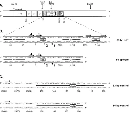
The SV40 origin region is diagrammed in Fig. 1A; this
re-gion contains two strong binding sites for T-ag (sites I and II).
Site I stimulates DNA replication both in vivo (see reference
22 for a review) and in vitro (41, 79); however, this site is not
required for DNA replication (21, 55). The main physiological
function of site I appears to be the autoregulation, via T-ag
binding, of early-gene transcription (see reference 28 for a
review). The sequences of T-ag binding site I and the SV40
core origin are presented in Fig. 1B; these sequences were
included in the oligonucleotide designated 83-bp ori
1. The
core origin consists of three functional domains: (i) the
AT-rich domain, (ii) a palindrome with four GAGGC recognition
on November 9, 2019 by guest
http://jvi.asm.org/
pentanucleotides (site II), and (iii) an imperfect inverted
re-peat (18, 57). Site II is the central region of the core origin; the
core origin is both necessary and sufficient for replication (see
references 18, 41, and 79 and references cited therein). Figure
1B also presents a second oligonucleotide, designated 64-bp
core, that contains sequences from just the core origin.
Se-quences from the region of DNA that served as a control in the
initial studies were included in the oligonucleotides designated
83-bp control and 64-bp control (Fig. 1C). The control
oligo-nucleotides have single GAGGC T-ag recognition sites (Fig.
1C).
Either in the presence or absence of DNA, SV40 T-ag is
known to oligomerize in solution (see reference 14 and
refer-ences cited therein). The domains of T-ag responsible for
oli-gomerization are not yet fully defined. Therefore, we tested
whether the purified T-ag-bd131–260
(see Materials and
Meth-ods) oligomerizes in solution in the absence of DNA. The line
widths from one-dimensional nuclear magnetic resonance
ex-periments indicated that T-ag-bd131–260
is a monomer even at
very high protein concentrations (
;
50 mg/ml) (43). Additional
analytical ultracentrifugation experiments, conducted with
pu-rified preparations of T-ag-bd131–260, also indicated that this
protein is a monomer in solution (25). Thus, while T-ag-bd131–
260
may be necessary for oligomerization (74), it does not
appear to be sufficient.
Interactions of T-ag and T-ag-bd
131–260with duplex DNA
from the SV40 origin.
Gel mobility shift assays were used to
test the ability of the purified T-ag-bd131–260
to distinguish
between origin- and non-origin-containing double-stranded
DNA (45, 54). Approximately 50 fmol of kinase-labeled DNA,
containing either the 83-bp ori
1or 83-bp control
oligonucle-otide, were incubated with T-ag or T-ag-bd131–260
as described
in Materials and Methods. (The sequence of the 83-bp ori
1 [image:3.612.100.516.70.436.2]oligonucleotide is shown in Fig. 1B, and the sequence of the
83-bp control oligonucleotide is presented in Fig. 1C). The
samples were cross-linked with glutaraldehyde and loaded on
gradient polyacrylamide gels. Inspection of Fig. 2 (lanes 2 to 4)
reveals that incubation of 1.5 to 6 pmol of T-ag with the 83-bp
FIG. 1. A map of the SV40 origin region and the oligonucleotide sequences used in this study (the map is based on sequences in pSV01DEP, a plasmid used to study SV40 replication 88). (A) The core origin, T-ag binding site I, the 21-bp repeats, and a partial copy of one of the 72-bp enhancer elements are indicated. Also indicated are the template positions from which the control oligonucleotides were derived (control; see below). SV40 nucleotides are numbered as described by Fiers (85). (B) Sequences comprising both the core origin and site I, and exclusively the core origin, are presented (85). These sequences were included in two oligonucleotides termed the 83-bp ori1and the 64-bp core oligonucleotides. The arrows depict the (GAGGC) pentanucleotide recognition sequences for T-ag; the
pentanucleotides were numbered as previously described (40). The locations of the adenine- and thymine-rich region (AT), site II, and the early palindrome regions (EP) are also depicted. (C) This figure presents the sequences of two oligonucleotides, termed the 83-bp control and 64-bp control oligonucleotides, that were used as controls in the band shift assays. The bars depict DNA derived from the SV40 enhancer. The positions of the single pentanucleotide T-ag binding sites are indicated by arrows. Finally, the numbers in parentheses are non-SV40 sequences and are labeled according to the pSV01DEP numbering system (88).
3974
JOO ET AL.
J. V
IROL.
on November 9, 2019 by guest
http://jvi.asm.org/
ori
1oligonucleotide resulted in two distinct species (the lower
species is more easily observed in subsequent figures [see Fig.
3 and 7]). These two species were previously characterized (14,
59, 87); the faster-migrating species consists of a single
hex-amer bound to the SV40 origin, while the more slowly
mi-grating species contains a double hexamer. In contrast to
re-sults with the 83-bp ori
1oligonucleotide, T-ag failed to
significantly shift the 83-bp control oligonucleotide (lanes 9 to
11). Analogous results were obtained in band shift reactions
conducted with the purified T-ag-bd131–260. When incubated
with the 83-bp ori
1oligonucleotide, T-ag-bd131–260
(1.5 to 6
pmol) induced the formation of one very distinct band shift
species (lanes 5 to 7). However, this species was not observed
when T-ag-bd131–260
was incubated with the 83-bp control
oli-gonucleotide (lanes 12 to 14). These studies demonstrate that
as with T-ag, T-ag-bd131–260
binds preferentially to the SV40
origin of replication.
Both in vivo and in vitro, replication of SV40 DNA depends
upon the specific binding of T-ag to site II within the SV40
core origin (55, 69, 79). Binding of T-ag to site II is known to
be regulated by ATP (6, 16, 20). However, assays based on
immunoaffinity techniques have indicated that certain
T-ag-bd-containing polypeptides purified from E. coli have difficulty
recognizing either site I or site II (33, 49, 81). Therefore, it was
of interest to determine whether the purified T-ag-bd131–260
interacted with isolated site II and site I and whether the
interaction with these sites was altered relative to the wild-type
SV40 origin.
The interaction of the purified T-ag-bd131–260
with an
oligo-nucleotide containing the SV40 core origin is presented in Fig.
3. (The sequences of the oligonucleotides designated 64-bp
core and 64-bp control are presented in Fig. 1B and C,
respec-tively.) As in the experiment shown in Fig. 2, T-ag (1.5 to 6
pmol) served as a positive control for binding to both the 64-bp
core (lanes 2 to 4) and 64-bp control (lanes 9 to 11)
oligonu-cleotides. It is apparent that although T-ag bound to the 64-bp
core oligonucleotide, it failed to significantly shift the 64-bp
control oligonucleotide. Analogous results were obtained in
band shift reactions conducted with T-ag-bd131–260. When
in-cubated with the 64-bp core oligonucleotide, T-ag-bd131–260
(1.5 to 6 pmol) induced the formation of one distinct species
(lanes 5 to 7). However, this species was not observed when
T-ag-bd131–260
was incubated with the 64-bp control
oligonu-cleotide (lanes 12 to 14). Thus, it is clear that T-ag-bd131–260
has a much higher affinity for the core origin than the control
DNA fragment. Furthermore, the interaction of T-ag-bd131–260
FIG. 2. Gel mobility shift assay used to compare the relative affinities of T-ag and T-ag-bd131–260for the SV40 wild-type origin. Lanes 1 and 8, control band
shift assays conducted with the 83-bp ori1and the 83-bp control
oligonucleo-tides, respectively, in the absence of protein; lanes 2 to 4, band shift assays conducted with the 83-bp ori1oligonucleotide and increasing concentrations of
T-ag (1.5 to 6 pmol); lanes 5 to 7, band shift assays conducted with the 83-bp ori1
oligonucleotide and increasing concentrations of T-ag-bd131–260(1.5 to 6 pmol);
lanes 9 to 11, band shift assays conducted with the 83-bp control oligonucleotide and increasing concentrations of T-ag (1.5 to 6 pmol); lanes 12 to 14, band shift assays conducted with the 83-bp control oligonucleotide and increasing concen-trations of T-ag-bd131–260(1.5 to 6 pmol). The input or free duplex DNA (F) is
indicated by the arrow. Single-stranded DNA, formed as a consequence of T-ag’s helicase activity (78), is visible at the bottom of this and subsequent gels. The protein-to-oligonucleotide ratios with 1.5, 3, and 6 pmol of T-ag (or T-ag-bd131–260)
and 50 fmol of oligonucleotide are 30:1, 60:1, and 120:1, respectively. Finally, formation of the T-ag-bd131–260-dependent band shift species required
glutaral-dehyde but did not require ATP or an ATP regenerating system (data not presented); this is the expected result, given that T-ag-bd131–260lacks the
ATP-binding domain present in T-ag (8).
FIG. 3. Gel mobility shift assay used to compare the relative affinities of T-ag and T-ag-bd131–260for the SV40 core origin. Lanes 1 and 8, control band shift
assay conducted with the 64-bp core and 64-bp control oligonucleotides, respec-tively, in the absence of protein; lanes 2 to 4, band shift assays conducted with the 64-bp core oligonucleotide and increasing concentrations of T-ag (1.5 to 6 pmol); lanes 5 to 7, band shift assays conducted with the 64-bp core oligonucleotide and increasing concentrations of T-ag-bd131–260(1.5 to 6 pmol); lanes 9 to 11, band
shift assays conducted with the 64-bp control oligonucleotide and increasing concentrations of T-ag (1.5 to 6 pmol); lanes 12 to 14, band shift assays con-ducted with the 64-bp control oligonucleotide and increasing concentrations of T-ag-bd131–260(1.5 to 6 pmol). The input or free DNA (F) is indicated by the
arrow.
on November 9, 2019 by guest
http://jvi.asm.org/
with the 64-bp core oligonucleotide (Fig. 3) was at least as
efficient as its interaction with the 83-bp ori
1oligonucleotide
(Fig. 2; quantitated in Table 1 [see below]). These experiments
indicate that T-ag-bd131–260
molecules bound to site I do not
stimulate the binding of T-ag-bd131–260
molecules to site II.
This observation is consistent with previous studies of T-ag
which indicated that there was little or no cooperativity
be-tween T-ag molecules bound to these two sites (7, 83). Finally,
the experiments presented in Fig. 2 and 3 were conducted after
a 20-min incubation, a time interval long enough to include
both early and late binding events (17). Therefore, a time
course experiment was conducted employing the 64-bp core
oligonucleotide. These studies indicated that the single
T-ag-bd131–260-dependent band shift species (Fig. 2 and 3) is the
product of protein-DNA interactions that take place soon (
#
1
min) after mixing (36) (data not presented).
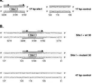
Gel mobility assays were also conducted to assess the ability
of both T-ag and T-ag-bd131–260
to interact with an
oligonucle-otide containing site I. Site I contains two high-affinity
GAGGC recognition sites arranged as direct repeats (64).
Ini-tially, a 17-bp oligonucleotide containing site I was used in gel
shift assays (Fig. 4A). However, when using the
oligonucleo-tide designated 17-bp site I, we failed to detect a band shift
signal with either T-ag or T-ag-bd131–260
(data not shown).
Therefore, the experiments were repeated with additional
oli-gonucleotides containing 15 bp of flanking sequence on either
side of site I (Fig. 4B). In one oligonucleotide, designated site
I
1
wt 30, the additional 30 bp were those normally
surround-ing site I in the SV40 origin. In the second oligonucleotide,
designated site I
1
mutant 30, the additional 30 bp were
arbi-trarily selected from the control oligonucleotide (SV40
resi-dues 91 to 105 and 123 to 137 [Fig. 1C]). Figure 5 presents data
on the interaction of T-ag and T-ag-bd131–260
with the
oligo-nucleotides designated site I
1
wt 30 and 47-bp control. It is
apparent from Fig. 5 that T-ag (3 and 6 pmol) interacted with
the oligonucleotide designated site I
1
wt 30 (lanes 2 and 3)
but not with the 47-bp control oligonucleotide (lanes 7 and 8).
In a similar manner, T-ag-bd131–260
(3 and 6 pmol) interacted
with the oligonucleotide designated site I
1
wt 30 (lanes 4 and
5) but not with the 47-bp control oligonucleotide (lanes 9 and
10). It is concluded that T-ag-bd131–260, like T-ag,
preferen-tially interacts with site I-containing oligonucleotides.
Further-more, since nearly identical results were obtained with the
oligonucleotide designated site I
1
mutant 30 (data not
shown), it is concluded that stable binding of either T-ag or
T-ag-bd131–260
to site I requires additional flanking sequences;
however, the exact sequence composition of the flanking
se-quences does not appear to be important. This observation is
consistent with previous studies of the interaction of T-ag with
site I (64). Finally, we are uncertain of the identities of the
three bands observed in the positive controls conducted with
T-ag (lanes 2 and 3); they may correspond to bound
mono-mers, dimono-mers, and trimers (47) or to higher oligomeric forms of
T-ag.
The results in Fig. 2, 3, and 5 were quantitated with a
phorImager, and the results are summarized in Table 1.
Phos-phorImager analyses of the data presented in Fig. 2, a measure
[image:5.612.155.465.76.354.2]of the relative affinities of T-ag and T-ag-bd131–260
for the
FIG. 4. Oligonucleotides used to compare the relative affinities of T-ag and T-ag-bd131–260for site I. (A) The 17-bp site I and 17-bp control oligonucleotides depicted
in this figure were used in initial experiments designed to measure the relative affinities of T-ag and T-ag-bd131–260for site I. (B) Since it was not possible to detect
a band shift with the initial set of site I oligonucleotides, a second set of site I-containing oligonucleotides was designed. One 47-bp oligonucleotide (designated site I1wt 30) contained site I (delineated by two parallel lines) flanked on either side by 15 bp of sequence normally found in SV40. The second 47-bp site I-containing oligonucleotide (designated site I1mutant 30) contained site I flanked on either side by 15 bp of sequence derived from the control oligonucleotide. Also depicted is the 47-bp control oligonucleotide that served as a negative control in these assays. The arrows depict two T-ag (GAGGC) pentanucleotide recognition sequences in site I.
3976
JOO ET AL.
J. V
IROL.
on November 9, 2019 by guest
http://jvi.asm.org/
83-bp ori
1oligonucleotide, are presented in the left-hand
col-umn. With 1.5, 3, and 6 pmol of T-ag, there was, respectively,
20-, 28-, and 9.7-fold greater interaction with the 83-bp ori
1oligonucleotide than with the 83-bp control oligonucleotide.
Similar analyses of T-ag-bd131–260
were difficult to perform
since it was not possible to detect a band shift with the 83-bp
control oligonucleotide (discussed below). PhosphorImager
analyses of the data presented in Fig. 3, a measure of the
relative affinities of T-ag and T-ag-bd131–260
for the core origin,
are also presented in Table 1 (middle column). With 1.5, 3, and
6 pmol of T-ag, there was 20-, 31-, and 27-fold greater
inter-action, respectively, with the 64-bp core oligonucleotide than
with the 64-bp control oligonucleotide. Furthermore, with 1.5,
3, and 6 pmol of T-ag-bd131–260, there was 23-, 59-, and 47-fold
greater interaction, respectively, with the 64-bp core
oligonu-cleotide than with the 64-bp control oligonuoligonu-cleotide.
Phospho-rImager analyses of the data presented in Fig. 5, a measure of the
relative affinity of T-ag and T-ag-bd131–260
for a fragment of
DNA containing site I, are presented in the right-hand column
of Table 1. With 3 and 6 pmol of T-ag, there was, respectively,
17- and 6-fold greater interaction with the oligonucleotide
designated site I
1
wt 30 than with the 47-bp control
oligonu-cleotide. As with the 83-bp control oligonucleotide,
quantita-tion of the preferential binding of T-ag-bd131–260
to the site
I-containing oligonucleotide was difficult, since it was not
pos-sible to detect a band shift with the 47-bp control
oligonucle-otide. Collectively, these analyses demonstrate that
T-ag-bd131–260
is at least as discriminating as T-ag in its
origin-specific DNA binding. Moreover, inspection of the data
presented in Table 1 indicates that on a molar basis, T-ag has
a slightly (two- to threefold) better ability to interact with DNA
from the SV40 origin than does T-ag-bd131–260. This may
re-flect a requirement for additional domains of T-ag for
high-affinity interactions with the SV40 origin.
It is interesting that T-ag-bd131–260
interacts very poorly with
the non-origin-containing DNA control fragments. This
obser-vation is consistent with studies by Lin et al. (42), who
con-cluded that a second region of T-ag, extending between
resi-dues 269 and 522, defines a region that is important for
nonspecific DNA binding. Since T-ag-bd131–260
lacks this
re-gion, this molecule would be expected to interact poorly with
non-origin-containing DNA. However, since mutations that
interfere with nonspecific binding have been described in
T-ag-bd (75) and T-T-ag-bd131–260
is capable of low, but detectable,
nonspecific DNA binding, T-ag-bd does have a role in
nonspe-cific binding. Finally, at high T-ag–to–DNA ratios, nonspenonspe-cific
binding may explain the drop in origin-specific binding
ob-served with the 83-bp ori
1and site I
1
wt 30 oligonucleotides.
FIG. 5. Gel mobility shift assay used to compare the relative affinities of T-ag and T-ag-bd131–260for site I-containing oligonucleotides. Lanes 1 and 6, control
band shift assays conducted in the absence of protein with the oligonucleotides designated site I1wt 30 and 47-bp control, respectively; lanes 2 and 3, band shift assays conducted with the oligonucleotide designated site I1wt 30 and either 3 or 6 pmol of T-ag; lanes 4 and 5, band shift assays conducted with the oligonu-cleotide designated site I1wt 30 and either 3 or 6 pmol of T-ag-bd131–260; lanes
7 and 8, band shift assays conducted with the 47-bp control oligonucleotide and either 3 or 6 pmol of T-ag; lanes 9 and 10, band shift assays conducted with the 47-bp control oligonucleotide and either 3 or 6 pmol of T-ag-bd131–260. The
[image:6.612.81.266.72.361.2]position of the 47-bp free DNA (F) is indicated by the arrow.
TABLE 1. Quantitation of relative affinities of T-ag and the T-ag-bd
131–260for binding to DNA fragments from the SV40 origin
aProtein (concn, in pmol)
% of total DNA and relative affinity with oligonucleotide:
ori1 Core I130 wt
ori1
fragment fragmentControl Relativeaffinity fragmentCore fragmentControl Relativeaffinity Ifragment130 wt fragmentControl Relativeaffinity
T-ag (1.5)
8.6
0.44
20
1.2
0.06
20
NT
bNT
NT
T-ag (3.0)
28
1.0
28
15
0.49
31
20
1.2
17
T-ag (6.0)
38
3.9
9.7
32
1.2
27
18
3.0
6.0
T-ag-bd (1.5)
1.3
;
0
2.3
0.10
23
NT
NT
NT
T-ag-bd (3.0)
5.3
;
0
9.4
0.16
59
14
;
0
T-ag-bd (6.0)
12
;
0
20
0.43
47
18
;
0
aSV40 origin DNA fragments tested include ori1(Fig. 1B), core (Fig. 1B), and site I flanked by 15 bp of wild-type DNA (I130 wt [Fig. 4B]). The results shown
in Fig. 2, 3, and 5 were quantitated with a Molecular Dynamics PhosphorImager, and the percentage of the total DNA in a given lane that is in a band-shifted species is given. For T-ag-bd131–260, this was a single distinct species; for T-ag, quantitation was conducted on all species above the presumptive hexamer species and included
DNA present in the wells. The relative affinity values represent the affinity of either T-ag or T-ag-bd131–260for the origin-containing fragment relative to the affinity
for the control fragment. As discussed in the text, there was very little indication of nonspecific binding of T-ag-bd131–260to control DNA fragments. Finally, the
protein-to-oligonucleotide ratios with 1.5, 3, and 6 pmol of T-ag (or T-ag-bd131–260) and 50 fmol of oligonucleotide were 30:1, 60:1, and 120:1, respectively. bNT, not tested.
on November 9, 2019 by guest
http://jvi.asm.org/
Interactions of T-ag and T-ag-bd
131–260with DNA
contain-ing mutant pentanucleotide bindcontain-ing sites.
The experiments
presented in Fig. 2 and 3 demonstrated that only a single,
rapidly formed species is detected in gel mobility shift assays
conducted with T-ag-bd131–260
and oligonucleotides containing
the SV40 origin. This single species could reflect complete
occupancy of the four pentanucleotide binding sites or binding
to a subset of these sites. To distinguish between these
possi-bilities, band shift assays were conducted with core origin
oli-gonucleotides containing various combinations of mutant
pen-tanucleotides.
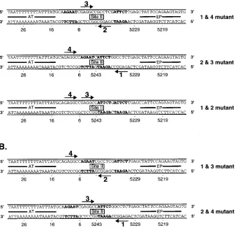
Previous experiments indicated that the two
pentanucleo-tides proximal to the early palindrome (EP) (pentanucleopentanucleo-tides
1 and 2; Fig. 1B) nucleate T-ag hexamer assembly (59).
Re-lated experiments indicated that pentanucleotide 1 is the
stron-gest binding site in the core origin palindrome (12, 40, 83). In
view of these observations, the band shift assays were initially
conducted with T-ag-bd131–260
and oligonucleotides containing
transition mutations in either pentanucleotide 1 or 2. It was
obvious from these experiments that relative to the 64-bp core
oligonucleotide there were few quantitative or qualitative
dif-ferences in the band shift experiments conducted with either
single pentanucleotide mutant (data not shown). Thus,
forma-tion of the distinct band shift species (Fig. 2 and 3) is not
dependent upon complete occupancy of all four
pentanucle-otide binding sites by T-ag-bd131–260. Therefore, to further
characterize the pentanucleotide requirements for binding to
the SV40 origin, oligonucleotides containing transition
muta-tions in particular pairs of pentanucleotide recognition sites
were designed (Fig. 6). A representative band shift assay
con-ducted with the pentanucleotide double mutants designated
1&4 and 1&3 is presented in Fig. 7. Lanes 1, 6, and 11 show
control reactions conducted in the absence of protein. Positive
controls are presented in lanes 2 to 5; they contain the products
of band shift reactions conducted with the 64-bp core
oligonu-cleotide (Fig. 1B) and either T-ag (lanes 2 and 3) or
T-ag-bd131–260
(lanes 4 and 5). Lanes 6 to 10 show reactions
con-ducted with the 1&4 pentanucleotide double mutant. It is clear
from lanes 9 and 10 that the 1&4 pentanucleotide double
mutant is inactive in band shift assays conducted with
T-ag-bd131–260. Lanes 11 to 15 contain the products of reactions
conducted with the 1&3 pentanucleotide double mutant. A
comparison of lanes 4 and 5 with lanes 14 and 15 demonstrates
that the 1&3 pentanucleotide double mutant is as good a
substrate for binding by the T-ag-bd131–260
as an
[image:7.612.140.481.82.408.2]oligonucleo-tide containing a wild-type copy of site II. Additional assays
demonstrated that the 1&4 pentanucleotide double mutant is a
member of a set of mutants (the inactive set of pentanucleotide
double mutants [Fig. 6A]) that were completely inactive in
band shift assays with T-ag-bd131–260. In contrast, the 1&3 and
FIG. 6. Sequences of the core origin oligonucleotides having transition mutations in particular pairs of pentanucleotide binding sites. (A) The inactive set of pentanucleotide double mutants is presented. The oligonucleotides are named after the mutated pair of pentanucleotide binding sites. The transition mutations are indicated with boldfaced letters. The arrows delineate the intact pair of pentanucleotides. (B) The active set of pentanucleotide double mutants is presented. The symbols used to designate the mutated and active pairs of pentanucleotides are the same as described for panel A. AT, adenine- and thymine-rich regions; EP, early palindrome regions.
3978
JOO ET AL.
J. V
IROL.
on November 9, 2019 by guest
http://jvi.asm.org/
2&4 pentanucleotide double mutants constitute a second set
(the active set of pentanucleotide double mutants [Fig. 6B])
that were quantitatively and qualitatively indistinguishable
from the wild-type site II-containing oligonucleotide in the
band shift assays conducted with this polypeptide.
We next addressed whether both pentanucleotide
recogni-tion sites in the active pentanucleotide double mutants (Fig.
6B) were required for stable binding. To conduct these assays,
oligonucleotides containing a third pentanucleotide mutation
were synthesized. For instance, with regard to the 2&4 double
mutant (Fig. 6B; pentanucleotides 1 and 3 intact), additional
pentanucleotide mutations in pentanucleotide 1 (the 1-2-4
tri-ple-mutant oligonucleotide) or pentanucleotide 3 (the 2-3-4
triple-mutant oligonucleotide) were synthesized and used in
band shift assays. These triple-pentanucleotide mutant
oligo-nucleotides were completely inactive in band shift assays (data
not shown). Thus, it is concluded that substrates containing
pairs of pentanucleotide recognition sites, positioned in a
head-to-head orientation and separated by approximately one
helical turn, are required for stable binding by T-ag-bd131–260.
Evidence that binding of T-ag-bd
131–260to the core origin is
limited to dimer formation.
The similar mobility of the
band-shifted species formed in reactions containing the 64-bp core
and active-set pentanucleotide double mutants (Fig. 7)
sug-gested that all four pentanucleotides in the 64-bp core
oligo-nucleotide were not occupied by T-ag-bd131–260. To test this
hypothesis, we employed the gel
retardation–1,10-phenanthro-line–copper ion footprinting procedure described by Kuwabara
and Sigman (39). This technique permits protein-DNA
com-plexes to be footprinted within the gel matrix, and the resulting
DNA fragments can then be resolved on a sequencing gel.
In one set of experiments, the 64-bp core, 1&3 mutant, and
2&4 mutant oligonucleotides were analyzed; these molecules
were asymmetrically labeled (see Materials and Methods) at
the 5
9
termini of the top strands (Fig. 1 and 6). Inspection of
Fig. 8 (lane 4) reveals that when an oligonucleotide containing
the core origin was used in the reaction, an
;
18-nucleotide
(nt) region was protected by T-ag-bd131–260. The protected
region initiates over pentanucleotide 3 and extends into a
re-gion containing sequences complementary to pentanucleotide
1. The footprint does not include any of the sequences that
comprise pentanucleotide 4. The footprint obtained when the
2&4 mutant (pentanucleotides 1 and 3 present [Fig. 6])
oligo-nucleotide was used in an identical assay is presented in lane 2.
It is clear that relative to the 64-bp core oligonucleotide, nearly
identical regions of site II are protected in the 2&4 mutant
oligonucleotide. Moreover, the DNA region containing the
transition mutations substituting for pentanucleotide 4 was not
protected in the 2&4 mutant oligonucleotide. The footprint
obtained when the 1&3 mutant (pentanucleotides 2 and 4
present) oligonucleotide was analyzed by the gel retardation–
1,10-phenanthroline–copper ion procedure is presented in lane
FIG. 7. A representative gel mobility assay used to assess the pentanucle-otide requirements for binding of T-ag-bd131–260to the SV40 core origin. Control
band shift assays were conducted in the absence of protein with the 64-bp core oligonucleotide (lane 1), the 1&4 mutant oligonucleotide (lane 6), and the 1&3 mutant oligonucleotide (lane 11). Additional control reactions included band shift assays conducted with the 64-bp core oligonucleotide and either T-ag (3 [lane 2] and 6 [lane 3] pmol) or T-ag-bd131–260(3 [lane 4] and 6 [lane 5] pmols).
Band shift assays conducted with the 1&4 mutant and T-ag (3 and 6 pmol, respectively) are presented in lanes 7 and 8. Band shift reactions conducted with the same oligonucleotide and T-ag-bd131–260(3 and 6 pmol, respectively) are
presented in lanes 9 and 10. Band shift assays conducted with the 1&3 mutant and T-ag (3 and 6 pmol, respectively) are presented in lanes 12 and 13. Band shift reactions conducted with the same oligonucleotide and T-ag-bd131–260(3 and 6
[image:8.612.66.292.69.349.2]pmol, respectively) are presented in lanes 14 and 15. As in previous figures, the position of free DNA (F) is indicated by the arrow.
FIG. 8. In situ footprinting of T-ag-bd131–260–core origin complexes by using
the nuclease activity of 1,10-phenanthroline–copper ion. Lanes 1, 3, and 5, control experiments conducted with free DNA isolated from samples containing the 2&4 mutant, 64-bp core, and 1&3 mutant oligonucleotides, respectively; lanes 2, 4, and 6, products of footprinting reactions conducted with the same oligonucleotides and T-ag-bd131–260. Size markers were generated by subjecting
the indicated oligonucleotides to the G and G1A sequencing reactions described by Maxam and Gilbert (48). Flanking each panel is a map of the relative positions of the early palindrome region (EP), pentanucleotides 1 to 4 (arrows), and the adenine- and thymine-rich region (AT). The arrows associated with the smaller arrowheads represent the complementary sequences of given pentanucleotides. The experiments presented in this figure were conducted at a 240:1 to-oligonucleotide ratio; however, similar results were obtained at a 120:1
protein-to-oligonucleotide ratio.
on November 9, 2019 by guest
http://jvi.asm.org/
[image:8.612.318.553.71.293.2]6. As with the previous two examples, the footprint extends
;
18 nt. However, in this instance, the footprint extends
be-tween pentanucleotides 2 and 4. Thus, T-ag-bd131–260
is
capa-ble of binding pentanucleotide 4, but it binds only when
pen-tanucleotides 1 and 3 are not present. Finally, control DNAs (i.e.,
free DNA obtained from reactions lacking T-ag-bd131–260) are
presented in lanes 1, 3, and 5. The incomplete cleavage of certain
regions within the control DNAs may be related to the
se-quence preferences exhibited by 1,10-phenanthroline–copper
(86).
These experiments were repeated with the same
oligonucle-otides which were asymmetrically labeled on the bottom strand
(data not presented). These experiments also revealed that the
footprint obtained with the 64-bp core oligonucleotide (
;
18
nt) was nearly identical to that obtained with the 2&4 mutant
oligonucleotide. Moreover, the footprint obtained with the
1&3 mutant (
;
18 nt) extended between pentanucleotides 2
and 4. In summary, these studies provided direct evidence that
T-ag-bd131–260
does not simultaneously bind all four
pen-tanucleotides. Moreover, they demonstrated that T-ag-bd131–
260
prefers to bind to pentanucleotides 1 and 3 but can bind
pentanucleotides 2 and 4 when pentanucleotides 1 and 3 are not
available.
The nearly identical footprints obtained with the 2&4
mu-tant oligonucleotide and the oligonucleotide containing the
core origin are most easily explained by dimer formation on
both DNA substrates. To test this hypothesis, a gel shift assay
was conducted to analyze the products formed in a mixing
experiment employing T-ag-bd131–260
and a T-ag-bd derivative
containing an additional 21 amino acids (T-ag-bd112–260) (see
Materials and Methods). Theoretically, mixing experiments
should generate a single intermediate species if a dimer is
formed between T-ag-bd131–260
and T-ag-bd112–260
(at a ratio
of 1:2:1; T-ag-bd131–260, T-ag-bd131–260-T-ag-bd112–260
interme-diates, T-ag-bd112–260, respectively). Alternatively, if trimers or
tetramers are assembled on the core origin, two or three
in-termediate species should form at a ratio of 1:3:3:1
(T-ag-bd131–260, T-ag-bd131–260-T-ag-bd112–260
intermediates,
T-ag-bd112–260, respectively) or 1:4:6:4:1 (T-ag-bd131–260, T-ag-bd131–
260
-T-ag-bd112–260
intermediates, T-ag-bd112–260, respectively).
Results from mixing experiments employing T-ag-bd131–260
and T-ag-bd112–260
are presented in Fig. 9. The distinct
T-ag-bd131–260-dependent band shift species is presented in lane 1,
while the slightly larger T-ag-bd112–260-dependent species is
shown in lane 5. The similar amounts of product formed in
these band shift reactions indicate that T-ag-bd112–260
and
T-ag-bd131–260
have similar affinities for the core origin. The products
formed when the band shift reactions were conducted with 3:1,
2:2, or 1:3 mixtures of T-ag-bd131–260
and T-ag-bd112–260,
re-spectively, are shown in lanes 2 to 4. A single novel species was
formed in these reactions whose mobility was roughly
interme-diate between those formed with the individual polypeptides. It
is concluded that T-ag-bd131–260
is binding as a dimer to two of
the four available pentanucleotides in the core origin.
Interactions of T-ag and T-ag-bd
131–260with single-stranded
DNA.
T-ag is known to bind single-stranded DNA in a
non-sequence-specific manner. Indeed, studies have indicated that
it has a higher affinity for single-stranded DNA than for
dou-ble-stranded DNA (3, 77). Moreover, previous experiments
indicated that T-ag-bd-containing polypeptides are capable of
binding single-stranded DNA (49). Therefore, we tested
whether T-ag-bd131–260
bound to single-stranded DNA (Fig.
10). In these assays, the top strands of the 83-bp ori
1and 83-bp
control oligonucleotides were used as single-stranded DNA
templates (Fig. 1). In Fig. 10, the reactions in lanes 1 to 9 were
conducted in the presence of competitor DNA (0.8
m
g/reac-tion); those in lanes 10 to 15 were conducted in the absence of
competitor DNA. As positive controls, band shift reactions
were performed with the 83-bp ori
1double-stranded
oligonu-cleotide and either T-ag (lane 2) or T-ag-bd131–260
(lane 3). It
is apparent that both the T-ag and the T-ag-bd131–260
prepa-rations used in these assays were active. As a negative control,
single-stranded DNA was incubated in the absence of protein
under replication conditions and the products were analyzed
(lanes 4, 7, 10, and 13). Inspection of lanes 4 and 10 revealed
that a protein-independent band is detected, owing to the
secondary structure of the 83-nt ori
1single-stranded
oligonu-cleotide. Products from band shift reactions containing T-ag (6
pmol) and the 83-nt ori
1single-stranded oligonucleotide are
shown in lanes 5 and 11; it is apparent that T-ag interacted with
this DNA. The interactions of T-ag with the single-stranded
83-nt control oligonucleotide (Fig. 1C) was also examined
(lanes 8 and 14). It is obvious that T-ag bound to the control
oligonucleotide and that the interaction of T-ag with
single-stranded DNA was not sequence specific. The interaction of
T-ag-bd131–260
(6 pmol) with the same single-stranded DNA
tem-plates was also examined. In contrast to T-ag, T-ag-bd131–260
failed to interact with single-stranded DNA from the upper
strand of the SV40 origin (lanes 6 and 12). Furthermore, there
was no indication of a band shift when T-ag-bd131–260
was
incubated with the single-stranded 83-nt control
oligonucleo-tide (lanes 9 and 15). Nearly identical results (not presented)
were obtained when these experiments were repeated with
origin-containing and non-origin-containing DNA from the
lower strand (Fig. 1). Collectively, these studies indicated that
under replication conditions, T-ag-bd131–260
is not sufficient for
a stable interaction with single-stranded DNA.
The reactions in lanes 1 to 9 were conducted in the presence
of competitor DNA (see Materials and Methods) that was
included in an effort to eliminate nonspecific protein-DNA
interactions. We were concerned that the competitor DNA
may have competed with the single-stranded DNA for binding
by T-ag-bd131–260. Therefore, the experiments were repeated in
FIG. 9. Evidence from a mixing experiment that T-ag-bd-containing polypep-tides form a dimer on the SV40 core origin. The T-ag-bd131–260and T-ag-bd112–260
polypeptides were mixed at various ratios with the 64-bp core oligonucleotide and analyzed via gel mobility assays. Lane 1, a band shift assay conducted with T-ag-bd131–260(6 pmol); lane 5, a band shift assay conducted with the
T-ag-bd112–260(6 pmol); lanes 2 to 4, band shift experiments conducted with 3:1, 2:2,
and 1:3 mixtures of the T-ag-bd131–260and T-ag-bd112–260polypeptides,
respec-tively (6 pmol of total protein [120:1 molar ratio of protein to oligonucleotide]). The location of a single novel intermediate species, the predicted outcome for dimer formation, is indicated. If trimers or tetramers were assembled on the core origin, two or three intermediate species should form, at ratios of 1:3:3:1 and 1:4:6:4:1, respectively. If it is argued that the intermediate species are not re-solved in this gel system, one would predict relative intensities of 1:6:1 for the trimer intermediate and 1:14:1 for the tetramer intermediate. PhosphorImager analyses of this and several related gels suggested that the intermediate is formed at the ratio expected for dimer formation (;1:2:1).
3980
JOO ET AL.
J. V
IROL.
on November 9, 2019 by guest
http://jvi.asm.org/
the absence of competitor DNA (lanes 10 to 15). It is apparent
that the inability of T-ag-bd131–260
to bind single-stranded
DNA is not a function of the presence of competitor DNA.
However, it is interesting that in the absence of competitor
DNA, T-ag forms a slower-mobility complex with both
single-stranded DNA substrates. This complex presumably reflects
the higher effective concentration of T-ag in the absence of
competitor DNA. These experiments indicate that T-ag can
oligomerize on single-stranded templates in a
sequence-inde-pendent manner.
DISCUSSION
Studies by Lin et al. (42), and those presented herein,
indi-cate that nonspecific DNA-binding events require a T-ag
do-main that is distinct from the T-ag-bd. Indeed, others have
reported that while the T-ag-bd contains the essential
origin-specific DNA-binding domain, additional regions of the
pro-tein are involved in stabilizing (2, 33, 49, 52, 72), or even
destabilizing (49, 81), the interaction of the T-ag-bd with the
SV40 origin. These observations raise the possibility that
pre-vious conclusions regarding the mechanism by which T-ag
in-teracts with the SV40 origin were based on composite signals
reflecting both nonspecific and specific DNA-binding events.
Therefore, to more clearly establish assembly events at a
eu-karyotic origin of replication, we initiated studies of the
inter-action of T-ag-bd131–260
with the SV40 origin.
With various DNA substrates, the sequence specificity of
purified T-ag-bd131–260
was analyzed. T-ag-bd131–260
is at least
as good as T-ag at distinguishing between an oligonucleotide
containing the entire SV40 origin (core and site I) and a
con-trol oligonucleotide of the same size. In related experiments, it
was demonstrated that purified T-ag-bd131–260
is at least as
good as T-ag at distinguishing between an oligonucleotide
con-taining the core origin and a control oligonucleotide.
There-fore, it is concluded that purified T-ag-bd131–260
is a good
[image:10.612.154.463.68.427.2]model polypeptide for studies of site-specific binding to the
SV40 origin.
FIG. 10. Gel mobility shift assay used to compare the relative affinities of T-ag and T-ag-bd131–260for single-stranded DNA. Lanes 1 to 3, control reactions
conducted with the 83-bp ori1oligonucleotide and either no protein (lane 1), T-ag (6 pmol) (lane 2), or T-ag-bd
131–260(6 pmol) (lane 3). The reaction mixtures in lanes
4 to 6 and 10 to 12 contained the single-stranded 83-nt ori1oligonucleotide from the upper strand of the SV40 origin (Fig. 1B); those in lanes 7 to 9 and 13 to 15
contained the single-stranded 83-nt control oligonucleotide from the upper strand of the control oligonucleotide (Fig. 1C). The reactions in lanes 4 to 9 were conducted in the presence of double-stranded competitor DNA; those in lanes 10 to 15 were performed in the absence of competitor DNA. The band shift assays in lanes 4, 7, 10, and 13 were conducted in the absence of protein. The band shift assays presented in lanes 5, 8, 11, and 14 were conducted in the presence of T-ag (6 pmol); those in lanes 6, 9, 12, and 15 were conducted in the presence of T-ag-bd131–260(6 pmol). The positions of free duplex DNA (F) and free single-stranded DNA (Fss) are
indicated. The unlabeled arrow indicates the position of a protein-independent, alternative DNA conformation adopted by the single-stranded 83-nt ori1
oligonu-cleotide during electrophoresis. Finally, the protein-to-oligonuoligonu-cleotide ratio with 6 pmol of T-ag (or T-ag-bd131–260) and 50 fmol of single-stranded oligonucleotide is
120:1.
on November 9, 2019 by guest
http://jvi.asm.org/
The interaction of T-ag-bd131–260
with site I was also
exam-ined. It is known that T-ag preferentially interacts with site I in
the absence of ATP (see reference 28 for a review).
Neverthe-less, when a 17-bp site I-containing oligonucleotide was used in
the band shift assay, we failed to detect a signal. When a larger
oligonucleotide was used, containing 15 bp on either side of
site I, a strong band shift was observed. The source of the
additional 15 bp on either side of site I was not a critical
determinant for detecting a band shift. Thus, under these
re-action conditions, a stable interre-action of T-ag-bd131–260
with
site I requires sequences flanking site I. It is interesting that the
yeast origin recognition complex failed to interact with an A
element oligonucleotide unless sequences adjacent to the A
element were included (61).
Having established that purified T-ag-bd131–260
is a good
model polypeptide for SV40 origin-specific binding, we next
addressed the pentanucleotide requirements for a stable
inter-action with T-ag-bd131–260. Previous studies demonstrated that
a single pentanucleotide is sufficient to bind and orient a T-ag
monomer (40, 83). Nuclear magnetic resonance experiments
were used to confirm that T-ag-bd131–260
binds to a single
GAGGC pentanucleotide, albeit at millimolar DNA
concen-trations (44). Nevertheless, oligonucleotides containing single
pentanucleotide binding sites (such as the 64-bp 1-2-4 and
2-3-4 triple pentanucleotide mutants) were inactive in band
shift assays conducted with T-ag-bd131–260. It should be noted
that the oligonucleotides used in these assays contained the
inverted-repeat region of the core origin. It has been reported
that this region constitutes a second, albeit low-affinity, binding
site for T-ag (57). In our assays, stable binding to the SV40
origin occurred only with those substrates containing at least
one pair of pentanucleotide recognition sites separated by
ap-proximately one turn of a double-stranded helix and positioned
in a head-to-head orientation (Fig. 7). These observations
sug-gest that binding of T-ag-bd131–260
monomers to suitably
ar-ranged pentanucleotides promotes dimer formation and that
dimerization stabilizes binding to the SV40 origin.
The intensity and relative position of the band shift species
generated with oligonucleotides containing either the 64-bp
core or active-set pentanucleotide mutants were nearly
identi-cal (Fig. 7). Given that the 64-bp core oligonucleotide has four
pentanucleotide binding sites and oligonucleotides for the
ac-tive set contain two pentanucleotides, this was a surprising
result. One possibility is that even with the 64-bp core
oligo-nucleotide, T-ag-bd131–260
binding may be limited to formation
of a single dimer. Support for this hypothesis was obtained with
the 1,10-phenanthroline–copper ion footprinting procedure
(39). These experiments revealed that T-ag-bd131–260
does not
[image:11.612.64.554.65.382.2]protect all four pentanucleotides in the 64-bp core
oligonucle-otide. They also demonstrated that the footprint obtained with
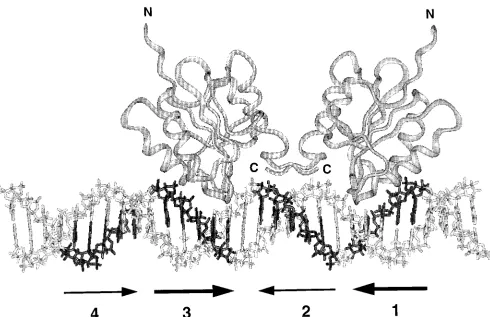
FIG. 11. A model of T-ag-bd131–260dimer formation on a 31-bp segment of B-DNA containing site II. The model was prepared by using the program INSIGHT
(Biosym, San Diego, Calif.) and depicts the interaction of two T-ag-bd131–260molecules with pentanucleotides 1 and 3. The N and C termini of the T-ag-bd131–260
molecules are indicated. Individual pentanucleotides in the B-DNA model are printed in boldface. The arrows below the figure are used to indicate the individual pentanucleotides; the active pairs of pentanucleotide recognition sites (1-3 and 2-4) are indicated by matching pairs of arrows. The costructure of T-ag-bd131–260bound
to a pentanucleotide has not been established. Therefore, to model this interaction, Arg-204 of T-ag-bd131–260, a critical residue for T-ag binding (75), was hydrogen
bonded to the third G in a given GAGGC sequence. Since individual molecules are free to rotate around this bond, the relative orientation of the individual T-ag-bd131–260molecules is arbitrary. Many alternative models were generated; this version was selected since there were minimal steric clashes and good protein-DNA
contacts.