0022-538X/86/010165-08$02.00/0
Copyright X 1986, American Society forMicrobiology
Mutation in the
Polyomavirus Genome That Activates the Properties
of
Large T Associated with Neoplastic
Transformation
CLAUDE ASSELIN,t JUDITVASS-MARENGO, ANDMARCELBASTIN*
Departmentof Microbiology, University ofSherbrooke, Sherbrooke, Quebec JIH 5N4, Canada
Received28May 1985/Accepted 30 September 1985
We haveconstructedapolyomavirusmutantgenomewhich exhibitsanincreasedimmortalizationpotential
whentransfected intoprimaryratembryo fibroblasts. The mutation isa30-base-pair deletion (nucleotides 1367
through 13%) thatinactivates the transformingpotential of middle T but activatessomeof theproperties of
large Tassociated withneoplastic transformation. Unlike the wild-type large T, themutantlarge Tcanfully
complementpolyoma middle T in thetumorigenicprocessin vivoaswellasin thetransformationof primary cells in vitro. Theactivity of themutant canbeexplained by its inabilitytoreplicate in cells and, hence, its inabilityto exertacytopathiceffectaftergenetransferathigh multiplicity. A recombinant which encodes the middle and smallTantigens, butnotthelargeT antigen,canalso elicitafully transformed phenotype when
introduced into primary rat fibroblasts. These results confirm previous observations from this laboratory indicating that two, and not three, viral gene functions are required forpolyomavirus-mediated oncogenic
transformation.
The early region of the polyomavirus genome encodes three distinct proteins, the large T(plt),middle T
(ppmt),
and small T (pst) antigens (see reference 31 for a review). Ofthese three proteins, thepmtantigen plays acentral role in
both oncogenic transformation in vitro (32) and
tumor-igenesis in vivo (3, 5).pmtalone iscapable of transforming established cells in culture (32), but, unlike the wild-type viral genome, it cannot efficiently transform primary cells (24) norinduce tumors when inoculated into newborn rats
(2). Thus, functions other than those expressed by thepmt
antigen may be required for the elaboration of all the
properties associated with neoplastic transformation. The role exerted bythe othertwoviralproteins is stillamatterof
controversy. Since therasoncogenecantransformprimary
rodent cells after cotransfer with polyomaplt, adenovirus ElA, or the myc oncogene (14, 26), one would expect
simultaneoustransfer of both thepmtandpltgenestoresult in the establishment of the fully transformed phenotype in vitro as well as in vivo. In agreement with this scheme, Rassoulzadegan et al. (25) have observed that cell lines establishedby transfer ofplt into primary fibroblasts canbe subsequently transformed by transfer of the pmt gene.
However, whenbothgenes weresimultaneously transferred
intocells, no stable transformant could be isolated by focus
formation. Combinations ofpmtandpstorpltandpstwere
similarly inefficient. Cuzin et al. (8) have reported that transformationof thefully normalratembryo fibroblastscan
only be achieved by the wild-type genomeorbya
combina-tion ofmodifiedgenomesthatallow simultaneous synthesis ofthethreeearly proteins.
Bycontrast,studies fromthis laboratory have shown that thepstproteincancomplementpmtin theabsenceof plt for
tumorinduction in animals (2). Similar complementary func-tionscanalsobe exertedby the simian virus 40 (SV40) large
T antigen as well as by one or several products of the
adenovirus ElA region (3). Thus, as for the in vitro trans-formation of primary cells, the in vivo steps in
poly-*Corresponding author.
t Present address: Institute for Cancer Research, Fox Chase, Philadelphia, PA 19111.
omavirus-mediatedtumorigenesis dependonadditional
cel-lular alterations beyond theacquisition of the polyomavirus transforming gene. As reported by this laboratory, such
alterationscanbeachievedby either polyomapstantigenor
otherearlygeneproducts from related DNAtumorviruses. Some of thesegenes,suchas SV40 large T (1, 6, 7, 23)and ElA(11),appeartobe associatedwith functions that confer
on primary cells the ability togrow indefinitely in culture.
However, although the polyoma plt gene is capable of
immortalizing primary cells (25), the combination of theplt andpmtgenesfailstoinducetumorsinnewbornrats(3). It
seems, therefore, that certain experimental conditions do notallow simultaneousexpression of both genesin vivo or
that plt is not sufficient to complement pmt in the tumorigenicprocess.
In this work, we have attempted to mutagenize the polyoma large T gene in such a way as to enable it to
complement pmt in tumorigenesis. We describe the
con-struction ofapltdeletionmutantwhich exhibitsanincreased
immortalization potential, and we show that the mutation enables plt to fully complement pmt not only in the tumorigenicprocessinvivo butalso in the transformation of
primary rat fibroblasts in vitro. These results demonstrate thattwo,andnotthree, viralgenefunctionsarerequired for
polyomavirus-mediated malignant transformation.
MATERIALS ANDMETHODS
Plasmids.pPyLT1 (Fig. 1) carries the polyomavirus large T
gene.This plasmidwasconstructed by deleting the largeT
protein intron from thepolyomavirus genome(35). pMT3 is arecombinantencoding only the middle T antigen (2). Itwas
obtained by deletingtwo HindIII fragments from pPyMT1 (32). bclO51 isa modifiedpolyomagenome expressing only
the middle and small T antigens (21). pPB21 carries the polyomavirus genome (A2 strain) in the BamHI site of
pBR322 (9). pSV2-neo is a plasmid expressing neo, a
dominant selective marker (28). pneo-LT1 (Fig. 1) was
constructed by inserting the polyoma large Tgene into the
BamHI site ofpSV2-neo. pMLT1 (Fig. 1) isahybrid plasmid
encodingindependently the polyomavirus largeane miiddleT
antigens.
165
on November 10, 2019 by guest
http://jvi.asm.org/
FIG. 1. Structure of recombinant plasmids. pPyLT1 carries the polyomavirus large T gene (pli) inserted in the BamHI site of
pAT153. The hatchedarearepresentsthe large Tcodingsequence.
Thepositionof the deleted intron is indicated. Thearrowshowsthe
position of the 30-bp deletion in pLT97. pLT3 was derived from
pPyLT1 by removing twoHindlll fragments (polyoma nucleotide 1656 to the HindlIl site in pAT153). This plasmid encodes the N-terminal portion of large T antigen (366 residues). pneo-LT1 carries both polyoma large T (plt) and neo. This plasmid was
constructed by insertingpltinto theBamHI site of pSV2-neo(28).
Tolink pLT3 tothe neogene, the plasmid wascleaved by BamHI
plusEcoRI (nucleotide 1560) and inserted between the BamHI and
EcoRI sites of pSV2-neo. pMLT1 is a hybrid plasmid encoding
independently polyoma large T antigen (MIA)and middle Tantigen (13).Theposition of the deleted intron is indicatedonbothgenes.
The arrow shows the position of the 30-bp deletion in the large T gene. All plasmids confer ampicillin resistance. Abbreviations: B,
BamHI;Ss, SstI; H, Hindlll; Pv, PvuII;ori, origin.
Mutagenesis in the polyomavirus large T gene. pPyLT1
DNAwas partially digestedwith SstI (pPyLT1 containstwo
SstI sitesatnucleotides 1373and 4341;Fig. 1), andthelinear full-length plasmid was isolated by agarose gel
electropho-resis. About 2 pLg of DNA wasadded toapreheated (30°C)
reaction mixture (50
Rl)
containing 20 mM Tris hydrochlo-ride (pH 8.0), 600 mM NaCI, 12.5 mM CaCI2, 12.5 mM MgCl2 and 1 U of BAL 31 (Bethesda ResearchLaborato-ries). The solution was incubated for 15 s to4 min at 30°C.
Under these conditions, BAL 31 removed about 80 base
pairs (bp) per min from either end of the molecule. The
reaction was terminated by addition of 3
IlI
of EDTA (0.2M), 3
RI
of EGTA [ethylene glycol-bis(,B-aminoethyl ether)-N,N,N',-N'-tetraacetic acid; 0.1 M], and 50 pl ofphenol at0°C.After phenolextraction,theDNAwasprecipitated with
ethanol, ligated at 20°C for 2 to 3 h with T4 DNA ligase
(10-fold excess), and used to transform Escherichia coli HB101 toampicillinresistance. Apreliminary determination
ofthesizes and locations ofthe deletionswascarriedoutby
digestion with BamHI and SstI. Recombinants lacking the
SstI site at nucleotide 1373 were analyzed further by
map-ping withHpaII, Hinfl, andAluI.
Cells, transfections, and growth assays. Fischer rat
em-bryos (15 daysold) were washed three times in Trissaline, minced with dissecting scissors,trypsinized (20 min at37°C), and sieved through a gauze mesh toisolate singlecells. The cells were collected in 30 to 50 ml of fetal calf serum, centrifuged at 1,800 rpm for 10 min, andplatedat adensity of 3 x 10⁶ cells per 60-mm dish in Dulbecco modifiedEagle medium supplemented with 10% fetal calf serum. The me-dium was changed after 16 to 20 hof culture at37°C (30to 50% confluence), and 4 h later, the cells weretransfectedby the calcium phosphate-DNA coprecipitation procedure (4) with modifications (33). The DNA to be transfected (8 ,ug) was mixed withhigh-molecular-weight calf thymus DNA (10 ,ug) as carrier. After 20 hof exposure to DNA, the cellswere passaged and replated at 20 to30% confluence. After 18h, G418 was added to the medium at a concentration of400
p,g/ml.
The medium plus drug was changed every 5 days. Colonies werefirst detected after 7 to 10days in the selective medium, and 2 to 3weeks later, independent colonieswere picked,transferred to15-mmLinbromicroplates,and grown at least once in medium containing G418(400 ,ug/ml). Most of the cells that underwent senescence did not sustainmore than one to two divisions after this first passage. The cell lines that were either immortalized or oncogenically trans-formed were subcultured at a 1:5 dilution each time they reached confluence and maintained inG418-free medium.Tumorigenicity assays. Recombinant DNAs wereinjected subcutaneously (2p.gof DNA in 50
RI
ofphosphate-buffered saline) into the neck of 1-day-old Fischer rats as described previously (5). To evaluate the tumorigenicity of trans-formed celllines, cellswere harvestedbytrypsinization,
and the indicated number of washed cells was injected into youngadult Fischer rats and nude mice as described previ-ously (2).DNA replication assay. CsClgradient-purified, supercoiled plasmid DNAs were transfected into mouse 3T6 cells by a modification of the DEAE-dextran transfection technique (17, 30). At 72 h posttransfection,
low-molecular-weight
plasmid DNA was isolatedby the Hirtextraction procedure (10). Portions of the DNA samples were sequentially di-gested with BclI and DpnI restriction endonucleases, and subjected to electrophoresis through 1.0% (wt/vol) agarose gels. The DNAfragments were transferred tonitrocellulose filters (27) and hybridized to a [32P]-labeled RNA probe aspreviously described (8a).
RESULTS
Isolation of plt mutants. We wished todetermine whether the failure of pPyLT1 to complement pmt in
tumorigenesis
was due to a lack of activity of the pltantigen
or to acytopathic effect exerted by the
protein,
as hadpreviously
been observed in transformation of
primary
embryo
fibro-blasts (14, 18). Wepostulated thattocomplementpmtinan
invivo assay, plt should lose itstoxicitywhile
retaining
most of itsimmortalizingactivity. Initialexperiments
werecarried outwithaclonedfragmentof thepit
gene,termedpLT3,
that allowed synthesis ofthe N-proximal half of the pltantigen
implicated in alteration ofserumdependency and immortal-ization (25). Althoughasimilar construction had been shown tocomplement EJ rasin the transformation ofprimary
cells (14), injection of both pLT3 and pMT3(truncated
pltplus
pmt) failed to induce tumors in newborn rats(Table 1).
We then attempted to determine whether pLT3 had retained all of theimmortalizing potentialofpPyLT1,theintactpltgene.To this end, we used a simple immortalization assay to
detect the ability of recombinant
plasmids
tomodify
the growthof rat embryo fibroblasts. Theplasmids
were linkedon November 10, 2019 by guest
http://jvi.asm.org/
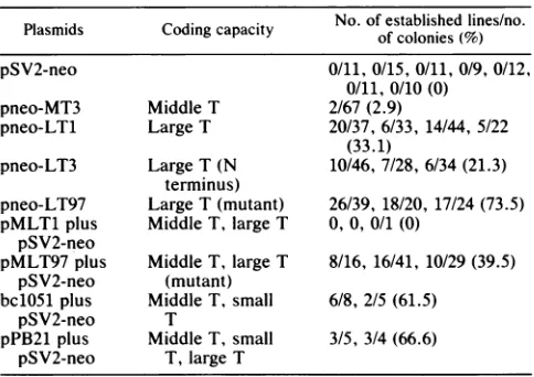
[image:2.612.57.300.72.313.2]topSV2-neo (Fig. 1) and introduced into primary cells by the calcium phosphate transfection procedure (33). Transfection with pneo-LT1 and pneo-LT3 gave rise to continuous cell lines withfrequencies of 33.1 and 21.3%, respectively (Table 2). Thus, although the truncated plt antigen was able to
immortalize primary cells, it didso withafrequency
signif-icantly lower (P - 0.056) than that observed with the intact
protein. For this reason,weattemptedto generatedeletions in plt that did notreduce its immortalizing potential. Muta-genesis with BAL 31 yielded recombinants with deletions of various lengths around the SstI site at nucleotide 1373. Detailsonthesemutantsand theirbiological properties have been presented elsewhere (1). One of the mutants, desig-nated pLT97, immortalized with high efficiency (Table 2). The colonies obtained with this mutant reached the fifth transfer in culture sooner than the others. However, like
other established celllines, they appeared normal withaflat,
regular morphology and exhibitedcontactinhibition. Several representative cell lines were examined by immunofluores-cence staining withapolyvalent antitumor serum. The cell
lines established by transfection with either pneo-LT1 or
pneo-LT97 exhibited a strong nuclear fluorescence charac-teristic ofplt antigen (datanot shown). Those immortalized with pneo-LT3 showed virtually no fluorescence (not
shown).
Complementation assays. To determine the activity of
pLT97 in tumorigenesis, we constructed plasmid pMLT97
(Fig. 1), which independently encoded pmt and the plt deletion mutant. Surprisingly, injection of this plasmid into newborn rats induced more tumors than did wild-type genomic DNAs that encoded the three polyoma early
pro-teins (Table 1). Thus, the 30-bp deletionnotonly conferred
to plt a greater ability to immortalize primary embryo fibroblasts but also enabled thegene tocomplementpmtin thetumorigenicprocess.This resultpromptedustoexamine theproperties of thepltmutantinatransformationassayof
primary cells. pMLT97 was introduced into primary rat
embryo fibroblasts together with the neo marker, and the
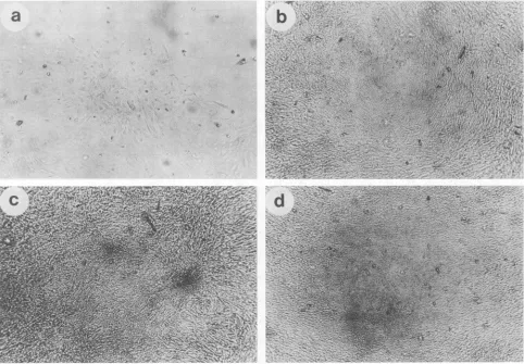
cells were selected for growth in G418. Of the resultant G418-resistant colonies, about 5% resembled oncogenically transformed cells. The cellsovergrewthe flatmonolayer and
reached high densities in the plates to produce dense foci characteristic of transformed cells (Fig. 2). When 2 x 10⁵
cells derived from two such foci were inoculatedinto nude mice and Fischer rats,they did notproduce tumors (Table
TABLE 1. Tumorigenicityof recombinantplasmids Incidence of
Plasmid Coding capacity tumorsin
newborn rats'(%)
pMT3 Middle T 0/58(0)
pMLT1 MiddleT, largeT 0/39(0)
pMLT3 MiddleT, largetT(N terminus) 0/24(0) pMLT97 MiddleT, largeT(mutant) 5/7(71.4)
bc1051 MiddleT, small T 8/20(40.0)
pPB21 MiddleT, small T, largeT 18/37(48.6) pPB97 SmallT, middleT, largeT(mutants) 0/16(0)
aFischer rats(1 day old)wereinoculated in theneck with 2 p.gof DNA in 50 Jilofphosphate-buffered saline. The DNAs werelinearized asfollows: pMT3 and bc1051werecleavedbyHindIII;pMLT1 and pMLT3werecleaved bySalI; pMLT97wascleavedbyClaI; pPB97wascleavedby BamHI; and pPB21 wascleaved by HindIII, BamHI,orHindIII plus BamHI. Animals wereobserved for 4 months for the development oftumors. Incidence of tumorsis shownasthe numberofratsinwhichtumorswerefound/the number
[image:3.612.316.558.84.255.2]ofratsthatweretested.
TABLE 2. Properties of recombinant plasmids No.of established lines/no.
Plasmids
Coding capacity ofcolonies(%)pSV2-neo 0/11, 0/15, 0/11, 0/9, 0/12,
0/11, 0/10 (0)
pneo-MT3 Middle T 2/67(2.9)
pneo-LT1 Large T 20/37,6/33, 14/44, 5/22 (33.1)
pneo-LT3 Large T (N 10/46,7/28, 6/34(21.3) terminus)
pneo-LT97 Large T (mutant) 26/39, 18/20, 17/24 (73.5) pMLT1 plus Middle T, largeT 0,0, 0/1 (0)
pSV2-neo
pMLT97 plus Middle T, large T 8/16, 16/41, 10/29 (39.5) pSV2-neo (mutant)
bc1051 plus MiddleT, small 6/8, 2/5 (61.5)
pSV2-neo T
pPB21 plus MiddleT, small 3/5, 3/4 (66.6) pSV2-neo T, large T
aNinefociwereobservedin confluent cultures transfected with pMLT97 in the absence ofneo selection. Some foci were also observed in cultures transfected with pMLT1 and bc1051; however, these couldnotbeestablished in culture.
bColonies of cells resistanttoG418werepicked3 to 4weeks afterselection andtransferred into Linbromicroplates. The cellsweresubculturedat a1:5
dilution eachtimethey reached confluence. Immortalization efficiencieswere
evaluated by counting the percentage of cell lines reaching five or more
passages 7 to 8 weeks after the first transfer. A zero indicates that no
G418-resistant colonywasobtainedin theexperiment.
[image:3.612.315.557.455.636.2]3). This may indicate that cells transfected with the combi-nation of pmt pluspltexhibitedonly a partially transformed phenotype. It could also be that the tumorigenicity defect was a result of the selection procedure. Two different observations support this hypothesis. First, transfection of primary cells with wild-type polyomavirus DNA in the
TABLE 3. Phenotypic properties of celllines"
Tumor
DNA incidence Growth Cellular
transfected Cellline inagarb morphology Nude Fischer aab mrhlg
mice rats
pneo-MT3 2-7 0/4 - Flat
2-12 0/4 - Flat
pneo-LT97 LT97neo-1 0/4 - Flatc
pMLT97 plus MLT97-7neo 0/4 0/12 - Flat pSV2-neo MLT97-3neo 0/4 0/12 + Transformed pMLT97 MLT97-12 4/4d ++ + Transformed
MLT97-13 + + Transformed
bc1051plus 1051-l0neo 0/4 - Flat
pSV2-neo 1051-llneo 0/4 0/10 - Flat 1051-22neo 12/12 1/12 + Transformed
1051-7neo 4/4d 4/10 + Transformed
aThe celllines wereisolated as indicated in footnote b of Table 2. Unless otherwiseindicated,2x 105cells wereinjected subcutaneouslyas a
suspen-sion in50-100 ±J ofphosphate-buffered saline. Tumors appeared within 3 weeks in nude mice and within 2 months inFischerrats.
bCells (105)weregrown in Dulbeccomodified Eagle medium containing 10% fetal calf serum and 0.34% (wt/vol) agarose (Difco). Microscopically
visible colonies were counted after 14 days at 37°C. Symbols:-,no growth; +,1to103colonies; ++,103 to104colonies;+ ++, more than104colonies.
Allcell lines establishedby pneo-LT97wereflat.
dOnly 5 x 10⁴ cells were injected.
on November 10, 2019 by guest
http://jvi.asm.org/
a
#
t
FIG. 2. Morphology of primary rat embryo fibroblasts transfected with recombinant plasmids, as observed through an inverted microscope. (a) pSV2-neo. Colony ofG418-resistant cells withnormal morphology. The cells stopped growing after a few divisions. (b) pMLT1. Rarecolony ofG418-resistant cells with altered morphology. The cells could not be established into cell lines. (c) bc1051. (d) pMLT97.About5% oftheG418-resistantcolonies had an alteredmorphology.Supercoiled plasmid DNA (6p.g)wascotransfectedwith 2 ,ug ofpSV2-neo. Magnification, x100.
presence of pSV2-neo yielded several G418-resistant colo-nies(three of fourcolonies)thatexhibitedthe samepartially
transformed phenotype (datanotshown). Second,
introduc-tionofpMLT97into primaryratembryofibroblastswithout
a selection marker produced foci of oncogenically
trans-formed cells (Table 2), some of which grew rapidly as did
tumors upon transplantation into nude mice (Table 3) or
Fischerrats (data not shown). Transfection of pMLT1 with
pSV2-neo also led to the formation of dense colonies (Fig.
2). However, in this case, the total numberofcolonies was
greatly reduced(1versus86),presumablybecauseofatoxic
effect ofthelargeTantigen. Thistoxicityof largeThasbeen
observed in several instances (14, 18) and could very well
explainwhy simultaneous transferofboth pmt andplt genes
failed toinducetumorsin vivo(Table 1)aswell as transfor-mation ofprimary cells in vitro (25) (Table2).
Cuzin et al. (8) reported that bc1051, a recombinant
encodingonly the middle and smallTproteins (21), didnot
transform rat embryo fibroblasts. This result seemed
para-doxical
becauseexperimentsfrom thislaboratoryhad shown that thesame recombinant induced tumorsin newbornrats with virtually the efficiency ofwild-type genomic DNA (2).Wefound,in agreement with Cuzinetal. (8), thatno stably
transformed linecould be isolated by focusformation after
transfer ofbc1051 intoprimaryratembryofibroblasts(Table 2). However, under the conditions oftransfection with the
neomarker,bc1051produceddensecoloniesoftransformed
cells (Fig. 2). Most of the cell lines derived from these
colonies exhibited a partially transformed phenotype. The
cells appeared large and flat and resembled immortalized
cells. By contrast, some cell lines displayed an irregular
growth pattern in culture and had a morphology
character-istic offully transformed cells. Unlike the flat cells, these cells grew insoft agar andweretumorigenicwheninoculated into nude miceor Fischerrats (Table3).
Replication of plasmidDNAs. Itwas shownby Landetal. (14) that the plt gene strongly inhibited establishment of
cotransfectedgenes, whichmightbe due tothepresenceof
thepolyomareplicationorigin together withthegene forthe entirelargeTantigen.Thismightallow thelargeTantigento
trigger repeated rounds of viral DNA replication and, in
turn, create a cytopathiceffect. We wishedto see whether
thecapability of pLT97tocomplementpmtin both
transfor-mation andtumorigenesiscould beexplained bytheinability of the mutant protein to trigger viral DNA replication. To this end, we transfected permissive mouse 3T6 cells by a
modification of the DEAE-dextran procedure (17, 30) and assessed the capacity of both pPyLT1 and its deletion mutantpLT97toreplicatein thesecells. The
low-molecular-weight DNA wasextracted 72 h aftertransfection, and the
stateofmethylationof the recombinant DNAwasmeasured asanindirect assayofreplication bydigestionwith
DpnI
and10,
ik
z.3t.
on November 10, 2019 by guest
http://jvi.asm.org/
[image:4.612.68.550.78.413.2]N
A%. \%
qA
*0
49
Ik
4.. *
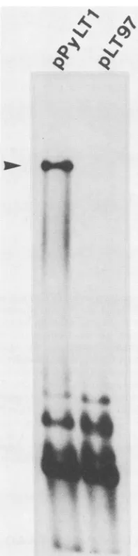
FIG. 3. ReplicationofpPyLT1and its deletion mutantpLT97in
mouse3T6 cells. Plasmid DNA(1 ,ug)wastransfected intoca. 5 x
105cells in 60-mm petri dishes by the DEAE-dextran procedure. Replication of plasmid DNAs was measured by isolating low-molecular-weight DNA and cleaving it with Bcll and DpnI as
described in the text. The replicated DNA is resistant to DpnI cleavage and appears as a band which comigrates with a linear marker of pPyLT1. The position of the marker is shown by an
arrowhead. The input unreplicated DNA appears as a smear of low-molecular-weight fragmentsatthe bottom of theautoradiogram.
BcllfollowedbySouthern blotting hybridization (19, 20). In thisassay,thereplicated DpnI-resistantDNAappearedas a
single band that comigrated with linear recombinant DNA
(BclIcleaves the large T plasmid only once). Any
unrepli-cated DNA appeared as numerous fragments of
low-molecular-weight at the bottom of the autoradiogram
be-causethelargeTplasmidscontainedmanysitesofcleavage
for DpnI. The results ofthe analysis are shown in Fig. 3. Unlike the control plasmid pPyLT1, the deletion mutant pLT97 was not abletoreplicate inpermissive cells.
Transfer of the mutation into thepmt gene. The precise
location of the deletion in pLT97 was determined byDNA
sequencing. We found that the plt gene had an in-frame deletion of 30 bp between nucleotides 1366 and 1397 (Fig. 4a). This deletion removed 10 amino acids from the plt
protein. However, inapolyomagenomeencodingthethree early proteins, thesame deletion would affect the structure
of the pmt protein as well. To determine what effect the
mutation would haveonmiddleT,wetransferred theformer into two different plasmids carrying the pmt gene and evaluated the transformation properties of the mutant
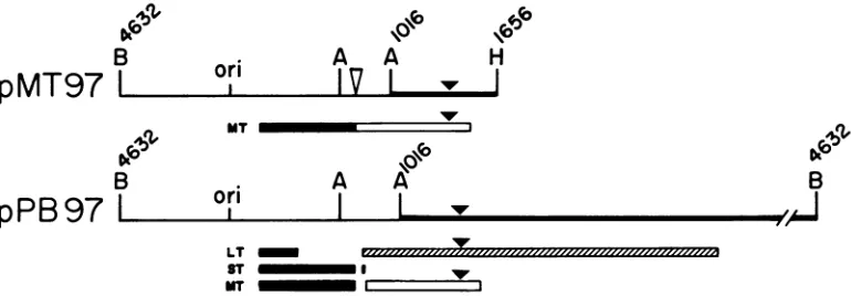
genomes.Inafirstconstruction, pLT97wascleavedbyAvaI and HindIII, and the AvaI-HindIII fragment spanning the deletion was substituted for the corresponding fragment in pMT3, a recombinant capable ofencoding exclusively the pmt protein (32). The resulting plasmid, pMT97 (Fig. 5), contained the pmt antigen-coding sequence with a 30-bp deletion affecting the protein as illustrated in Fig. 4b.
Al-though pMT3, the wild-typepmtgene, is abletotransform FR3T3 cells in culture (2), its deletion mutant pMT97 was
inactive (data not shown). By exchanging appropriate
re-strictionfragments (Fig. 5),wealso transferred themutation
intopPB21, aplasmid carrying the entirepolyomagenome
(9) and capable of expressing the three polyoma early proteins.The deletion inpPB21affected both the middle and largeTantigens (Fig. 4b)butnotthe small Tantigen.Mutant pPB97 can therefore be considered as a mit mutant, i.e.,a
mutantaffected in bothpmtandpltgenefunctions. Although pPB21 was highly tumorigenic in newborn rats, inoculation of its deletion mutantpPB97failed to induce tumors(Table 1). Taken together, these results indicate that the 30-bp deletion severely impaired or inactivated the transforming andtumorigenic potential of thepmtgene.
DISCUSSION
We have shown that only two of the three viral gene
functions arerequiredfor thepolyomavirus-mediated trans-formation of primary cells. One is conferred by the pmt
antigen, the transforming protein, whereas the second one can be conferred by either small T or large T. This is in agreementwithprevious studiesfrom thislaboratory show-ingthatpmtrequiresthepresenceof eitherpolyomasmall T
oranimmortalizinggenefromarelated DNAtumorvirusto induce tumors in newborn rats (3). These results can be interpreted byamultistep processofcarcinogenesis: afirst step inwhich cellsarepreventedfrom senescing
(immortal-ization), and a second one involving the acquisition of phenotypes characteristic of malignant cells (completion). Thus, some viral and cellular transforming genes can be classified operationally into two complementation groups,
eachgroupdefiningadifferent steprequiredfor
transforma-tion ofprimarycells invitro(14, 22, 26),aswellasinduction oftumors in vivo(3). Although polyoma small T is
opera-tionallyverysimilartotheimmortalizinggenes,itcannotbe implicated in establishment and immortalization functions (25). This suggests that all the genes that complement the transforming functions of pmt do not create identical changes in cellular behavior but may, on the contrary,
interact with differentcellulartargets. Polyomavirus has,in fact, developed three distinct oncogenes, and it has been reportedthatafunctionof the small Tproteinisnecessaryin
additiontoand inconjunctionwiththefunctions of thelarge and middle Tproteinsfortransformation ofprimarycells(8). The resultspresented here showthat this isnotthe case.
Transfection of thepolyomagenomeswith theneomarker yieldedcell lines thatdisplayed onlyapartially transformed
phenotype. For several reasons, we believe that this is due
to the neo selection rather than to the lack ofoncogenic potentialof thecombination ofgenestransfected.First,both combinations of pmt plus pst and pmt plus pLT97 are
on November 10, 2019 by guest
http://jvi.asm.org/
[image:5.612.124.223.73.470.2]170 ASSELIN ET AL.
G
C
G A T C
_
= ..w
_ w :.
i
* ...
w i.
j; .
C ,..
v ..
iiSlE SF "'
._ *.::. _
.
*
.
.. _ ..w * ,....
,
,ii - i..W..: ..
w ^ .X....
:w *
-_ , ....
=T w
_ o.
- %
_
_ .
i.
*_:
-F X- s..r
-::6>.
-:>
:s
lA
C
T
CA
CA
G
A
G
CT
G
1366
T 1397
A
A
G
A
FIG. 4. (a) Autoradiogramof pLT97 subjectedtochemical deg-radation andelectrophoresis. pLT97wasdigested byAccI,andthe
fragment spanning thesequencesbetween nucleotides 368 and 1500 was isolated by agarose gel electrophoresis. After labeling with [cx-32P]ddATP and terminal transferase, the DNA wascleaved by
AvaI, and the AvaI-AccI fragment (nucleotides 1016 through 1500)
wasisolated bypolyacrylamide gel electrophoresis and sequenced
by the method of Maxam and Gilbert (16). The products were
separatedon8% polyacrylamide gels. (b)Sequence of the region of polyoma DNA (5' strand) including the deletion found in pLT97 and predicted modifications in the sequence oflarge T antigen. After
transferof the deletion in pMT3 (Fig. 5), thestructureof middle T antigen is affectedas indicated. In pPB97, agenomeencoding the
threeearly proteins (Fig.5), the deletion affects thestructureof both middle andlarge T antigens.
b 1367 deletion 1396
CT G CT C TC AG AG C TC AT T C AATGC A A CG C CA CC TA AG largeT cys ser gin ser ser phe asn ala thr pro pro Iys middleT aGI leu org ala his ser met gin arg his Ieu
tumorigenic when inoculated into newbornrats, which can
be considered the most stringent and rigorous criterion of oncogenictransformation (5). Second, foci of oncogenically transformed cells, obtained by transfecting primaryrat
em-bryo fibroblasts with pMLT97 in the absence ofneo
selec-tion, arehighly tumorigenic when transplanted into animals.
Third, transfection of primary cells with wild-type genomic DNA in the presence of pSV2-neo yields G418-resistant
colonies thatusually exhibitthe same partially transformed
phenotype. Thus, cells selected for G418 resistanceexhibita
phenotype indistinguishable from that of immortalized cell lines onthe basis of several criteria. Suchaphenotype can
be conferred by the combination of pmt plus pst and, to
some extend, bypmtalone (1). Thesegenes, however, are
not usually considered to be immortalizing genes. Similar
results have beenobtained with theHa-ras-1oncogene (29).
Furthermore, the adenovirus Ela gene, which is usually
considered to be an immortalizing gene (14, 26), has been shown to behave as a second-step conversion gene in
immortalized Chinese hamster cells (29). These results show thatsomeoncogenescanhavebothstep 1(immortalization) andstep2(conversion) transforming potential, dependingon
theassay system chosen. They also suggest that classifica-tionofoncogenesintodifferentcomplementationgroupson
the basis of their transformation properties may be more
artificial thanreal.
As to the role of large T, our work shows that this
oncogeneis sufficienttocomplementpmtintransformation,
even when both genes are transferred simultaneously into cells. However, the full-sized plt appearsto be very ineffi-cient in the process. Land et al. (14) could not study the activity ofthe intact protein because ofa toxic effect after genetransferat high multiplicity by the calcium phosphate transfection method. This toxicity was not observed when
westudied theeffect oflargeT inimmortalization,but itwas
apparentwhenpMLT1 (pmt plusplt)wascotransfectedwith pSV2-neoin transformation assays. It isinteresting tonote thattumorsinducedbyinoculating polyomavirusoritsDNA
into animalsinvariablycontain deletions or rearrangements in the distalportionof thepolyoma early region (5, 12). Loss
orinterruptionofviralsequences codingforthe C-terminal portion of the large T antigen is well documented (12, 15), and it has been postulatedthat a selection operatesin vivo against cells producing a functional large T antigen and
a
J. VIROL.
on November 10, 2019 by guest
http://jvi.asm.org/
[image:6.612.91.514.75.505.2]B
pMT97
LB
pPB97
Lori
,\lb
A
A
IV
I
Hi
H
MT
or
A
AB
I L
LT
ST I
UT
FIG. 5. Diagram showing how the30bp deletionwasintroduced intopM l , arecombinantencodingexclusively the middleTantigen, and pPB21, a recombinant encoding the three early proteins. pMT97 was obtained by replacing the Aval-HindIII fragment of pMT3 (nucleotides 1016through1656)by thecorresponding fragment from pLT97(thick lineonthemap).TheBamHI-HindIII fragment is inserted between the BamHI and Hindlll sites of pAT153. Theplasmid encodes amiddleTantigen withadeletion of10amino acidsnearthe C terminus. pPB97 was obtained by replacingtheAvaI-HindIII fragment of pPB21 (nucleotides 1016through4632) with the corresponding fragment frompLT97(thick lineonthemap). The genome isclonedintheBamHI site ofpBR322.Itencodes thewild-type smallTantigen, the middleTantigenfrompMT97,and thelargeTantigen frompLT97. Thecoding regions forthe Tantigensareindicated. Shownarethe sequencestranslatedin frame1 (),frame2(1MI72),andframe3(LII).The30-bp deletion isrepresented byanarrowhead.Abbreviations: B, BamHI; A,AvaI; H,HindlIl; ori, origin; MT, middle T; ST,smallT; LT, largeT.
consequently free viral genomes (15). One would expect the tumors to produce, in addition to the middle T antigen, a modified large T antigen equivalent to mutant pLT97 or, more likely, the small T antigen. The latter, which is sufficient to complement middle T in tumorigenesis, is
in-deedfoundin tumors inducedbypolyomavirus DNA(2, 5).
Truncated forms of large T antigen have been found in
transformed cells (13) but not in tumors (12), and it is not
clearwhether they have any role toplay in transformation (34). The large T fragments arise very likely as a conse-quence of deletions and rearrangements affecting the distal half of the earlygeneregion whichprecludes thesynthesis of
largeTantigen(5, 12) but not thatofthemiddle and small T
antigens.Thedeletionin pLT97 may have occurred in avery
particular region ofthe large T gene; i.e., removal of a few
amino acids from someparts of theproteinmay result in an
activation of itsbiological properties. Although theactivity
of the mutant is likely due to its inability to replicate and,
hence, its inability to exert a toxic effect, its efficiency in
both immortalization and complementation ofpmt is
remi-niscent of proto-oncogene activation. However,because the
deletion occurs in the region coding for the C terminus of
middle T antigen, it inactivates the transforming and
tumorigenic functions ofthe genome. For this reason,
mu-tations analogous to pPB97 are unlikely to be generated in
vivo.
ACKNOWLEDGMENTS
We thank C. Bergeron and J. Toutant for excellent technical assistance,A.Ratiarson for helpingwith cellcultures, and C. Gelinas for fruitful discussions.
Thisresearch wassupported bygrantsfromtheMedical Research Council ofCanada and the National CancerInstitute ofCanada. C.A. is a research student from the National Cancer Institute of Canada. J.V.-M. issupported bya Biotechnology TrainingCenter AwardfromtheMedical ResearchCouncilof Canada.
LITERATURE CITED
1. Asselin, C., and M. Bastin. 1985.Sequencesfrom polyomavirus and simian virus 40 large T genes capable of immortalizing primaryratembryo fibroblasts. J. Virol. 56:958-968.
2. Asselin, C., C. Gelinas, and M.Bastin. 1983. Role of the three
polyomavirusearly proteinsintumorigenesis. Mol.Cell. Biol. 3:1451-1459.
3. Asselin, C., C. Gelinas, P. E. Branton, and M. Bastin. 1984. Polyoma middle T antigen requires cooperation from another gene to express the malignant phenotype in vivo. Mol. Cell. Biol. 4:755-760.
4. Bacchetti,S., and F. L. Graham. 1977. Transfer of the gene for thymidine kinase tothymidine kinase-deficienthuman cells by purified herpes simplexviral DNA.Proc. Natl.Acad.Sci. USA 74:1590-1594.
5. Bouchard, L., C. Gelinas, C. Asselin, and M. Bastin. 1984. Tumorigenic activity of polyoma virus and SV40 DNAs in newborn rodents.Virology 135:53-64.
6. Clayton, C.E., D. Murphy, M.Lovett,and P. W.J. Rigby. 1982. Afragment oftheSV40largeT-antigengenetransforms.Nature (London)299:59-61.
7. Colby, W. W., and T. Shenk. 1982. Fragments of the simian virus40transforming gene facilitate transformation ofrat em-bryocells. Proc. Natl. Acad. Sci. USA 79:5189-5193. 8. Cuzin, F.,M.Rassoulzadegan,and L.Lemieux.1984.Multigenic
control oftumorigenesis: threedistinct oncogenesarerequired for transformation ofratembryofibroblastsby polyoma virus, p.109-116. InG. F. VandeWoude,A.J.Levine,W.C. Topp, and J. D. Watson(ed.), Cancercells: oncogenes andviralgenes. ColdSpring Harbor Laboratory, Cold Spring Harbor,N.Y. 8a.Gelinas, C.,and M. Bastin. 1985. Malignant transformation of
rat cells by the polyomavirus middle T gene. Virology 146:233-245.
9. Gelinas, C., L. Bouchard, and M. Bastin. 1981. Tumorigenic activity of cloned polyoma virus DNA in newborn rats. Experientia37:1074-1075.
10. Hirt, B. 1967. Selective extraction of polyoma DNA from infectedmousecellcultures. J.Mol. Biol. 26:365-369. 11. Houweling, A., P. J.vandenElsen,and A.J.vander Eb.1980.
Partial transformation ofprimaryratcellsbytheleftmost 4.5% fragment ofadenovirus 5 DNA. Virology105:537-550. 12. Israel, M. A., D. F. Vanderryn, M. L. Meltzer, and M. A.
Martin. 1980. Characterization of polyoma viral DNA se-quencesin polyoma-induced hamstertumorcell lines. J. Biol. Chem. 255:3798-3805.
13. Ito, Y.,and N.Spurr.1980.PolyomavirusTantigensexpressed intransformedcells: significanceof middle T antigenin trans-formation. Cold Spring Harbor Symp. Quant. Biol. 44:149-157.
14. Land, H.,L. F.Parada, andR.A.Weinberg. 1983.Tumorigenic conversion ofprimary embryo fibroblasts requiresat leasttwo cooperatingoncogenes. Nature(London)304:596-602.
on November 10, 2019 by guest
http://jvi.asm.org/
[image:7.612.118.503.78.212.2]15. Lania, L., A. Hayday, and M. Fried. 1981. Loss of functional large T-antigen andfree viral genomes from cells transformed in vitro by polyoma virus after passage in vivo as tumor cells. J. Virol. 39:422-431.
16. Maxam, A. M., and W. Gilbert. 1977. A new method for sequencing DNA. Proc. Natl. Acad. Sci. USA 74:560-564. 17. McCutchan, J. H., and J. Pagano. 1968. Enhancement of the
infectivity of simian virus 40 deoxyribonucleic acid with diethylaminoethyl-dextran. J. Natl. Cancer Inst. 41:351-357. 18. Mougneau, E., L. Lemieux, M. Rassoulzadegan, and F. Cuzin.
1984.Biological activitiesof v-myc and rearranged c-myc onco-genes in ratfibroblast cells in culture. Proc. Natl. Acad. Sci. USA 81:5758-5762.
19. Muller, W. J., C. R. Mueller, A.-M. Mes, and J. A. Hassell. 1983. Polyomavirusorigin forDNAreplication comprises mul-tiple genetic elements. J. Virol. 47:586-599.
20. Muller, W. J., M. A. Naujokas, and J. A. Hassell. 1983. Polyomavirus-plasmid recombinants capable of replicating have an enhanced transforming potential. Mol. Cell. Biol. 3:1670-1674.
21. Nilsson, S. V., and G. Magnusson. 1983. T-antigenexpression by polyoma mutants with modified RNA splicing. EMBO J. 2:2095-2101.
22. Parada, L. F., H. Land, R. A. Weinberg,D.Wolf,and V. Rotter. 1984.Cooperation between gene encoding p53 tumourantigen and ras in cellular transformation. Nature (London) 312: 649-651.
23. Petit, C. A., M.Gardes,andJ. Feunteun. 1983. Immortalization of rodent embryo fibroblasts by SV40 is maintained by the A gene. Virology 127:74-82.
24. Rassoulzadegan, M., A. Cowie, A. Carr, N. Glaichenhaus, R. Kamen, and F. Cuzin. 1982. The roles of individual polyoma virus early proteins in oncogenictransformation. Nature (Lon-don) 300:713-718.
25. Rassoulzadegan, M., Z. Naghashfar, A. Cowie, A. Carr, M. Grisoni,R.Kamen,and F.Cuzin. 1983. Expression of the large
T protein of polyoma virus promotes the establishment in culture of "normal" rodent fibroblast cell lines. Proc. Natl. Acad. Sci. USA 80:4354-4358.
26. Ruley, E. 1983. Adenovirus early region 1A enables viral and cellulartransforming genes to transform primary cells in cul-ture. Nature (London)304:602-606.
27. Southern, E. M. 1975. Detection of specific sequences among DNAfragments separated by gelelectrophoresis. J. Mol. Biol. 98:503-517.
28. Southern, P. J., and P. Berg. 1982.Transformation of mamma-lian cells to antibiotic resistance with a bacterial gene under control of the SV40early region promoter.J.Mol.Appl.Genet. 1:327-341.
29. Spandidos, D.A.,and N. M. Wilkie. 1984. Malignant transfor-mation of early passage rodent cellsbyasingle mutated human oncogene. Nature(London) 310:469-475.
30. Sussman, D. J., and G. Milman. 1984. Short-term, high-efficiency expression of transfected DNA. Mol. Cell. Biol. 4:1641-1643.
31. Tooze, J. (ed.). 1981.DNAtumorviruses. ColdSpring Harbor Laboratory, Cold Spring Harbor, N.Y.
32. Treisman, R., U. Novak, J. Favaloro, and R. Kamen. 1981. Transformation ofratcells byanalteredpolyoma virus genome expressing only the middle-T protein. Nature (London) 292:595-600.
33. Wigler, M., A. Pellicer, S. Silverstein, and R. Axel. 1978. Biochemical transfer ofsingle-copyeucaryotic genesusing total cellularDNA as donor. Cell 14:725-731.
34. Winberry, L. K., C. J. Stewart, B. S. Schaffhausen, and M. Fluck. 1985.Transformationby polyomats-a mutants. I. Char-acterization of the transformed phenotype. Virology 144:433-447.
35. Zhu, Z., G. M.Veldman, A. Cowie, A.Carr,B.Schaffhausen, and R.Kamen. 1984. Construction and functional characteriza-tionofpolyomavirus genomesthatseparately encode thethree earlyproteins. J.Virol.51:170-180.

![FIG. 4.AvaI,fragmentbyantigenthreepredictedtransferpolyomawaswasradationmiddleseparated[cx-32P]ddATP the (a) Autoradiogram of pLT97 subjected to chemical deg- and electrophoresis](https://thumb-us.123doks.com/thumbv2/123dok_us/1390487.92191/6.612.91.514.75.505/fig-avai-fragmentbyantigenthreepredictedtransferpolyomawaswasradationmiddleseparated-ddatp-autoradiogram-subjected-chemical-electrophoresis.webp)