Int. J. Electrochem. Sci., 9 (2014) 4374 - 4383
International Journal of
ELECTROCHEMICAL
SCIENCE
www.electrochemsci.org
Highly Selective Determination of Pioglitazone in Urine and
Pharmaceutical Formulations by Novel PVC-Membrane sensors
Amr L. Saber1,2,* and Reem K. Shah2 1
Department of Chemistry, Faculty of Science, University of Zagazig, Zagazig, Egypt 2
Department of Chemistry, Faculty of Applied Science, University of Umm Al Qura, Makkah, Saudi Arabia
*
E-mail: [email protected]
Received: 3 April 2014 / Accepted: 29 April 2014 / Published: 19 May 2014
Novel simple, selective and sensitive poly(vinyl chloride) membrane sensors are developed for the determination of pioglitazone in biological samples (urine) and pharmaceutical preparations. Potentiometric measurements were based on iodobismuthite-drug ion-pair as novel electroactive materials incorporating a plasticized PVC membrane with o-nitrophenyl octyl ether or dioctyl phthalate. Each sensor was conditioned for at least two days in 0.1 M drug solution before use. It exhibited fast and stable Nernstian response for pioglitazone over the concentration range of 1.0x10⁻⁷– 1.0x10⁻² M, pH range of 3.0–7.0 pioglitazone sensors. Results with an average recovery not more than 100.4% and a mean standard deviation less than 1.0% of the nominal were obtained for the two sensors. The sensors showed reasonable selectivity towards investigated drugs in presence of many cations.
Keywords: Novel potentiometric sensors, Ion pairs, Biological samples, Pharmaceutical analysis, Pioglitazone hydrochloride.
1. INTRODUCTION
H O
O
N S
O N
.HCl
Scheme 1. Chemical structure of pioglitazone hydrochloride
The assay of the drug in pure and dosage forms is, as far as we know, not official in any pharmacopoeia, and therefore requires much more investigation. The different analytical methods that have been reported for determining pioglitazone hydrochloride in tablets [2-9] by HPLC and also quantitative determination of pioglitazone in human serum by direct-injection HPLC mass spectrometry and its application to a bioequivalence study has been reported [10]. Yamashita determined pioglitazone and its metabolites in human serum and urine [11] and also Zhang and Lakings reported an assay method for pioglitazone alone in dog plasma [12]. LC|MS [13-16], TLC [17], HPTLC [18-20]. In the literature, only few spectrphotometric [21-25], and separation of drug enantiomers by capillary electrophoresis [26-28] methods have been reported. Drug determination is one of the important tools for drug quality control and the development of potentiometric ion-selective electrodes (ISEs) is an area of interest. When compared with other analytical methodologies, ion selective electrodes are simple, relatively inexpensive, robust, durable and ideal for their use in field environments. Some other advantages involve that they can be used very rapidly, are invaluable tools for continuous monitoring, they measure the activity rather than the concentration and are not affected by turbidity or sample colour. It is well known that ISE are one of the few techniques that can measure both positive and negative ions depending on the nature of the ionophore.
Therefore, focus of the present study was to develop an accurate, precise and robust potentiometric method for the determination of pioglitazone hydrochloride in tablets and urine samples. Electrochemical performance characteristics of the proposed sensors were evaluated according to the IUPAC recommendation guidelines [29] using a standard PG stock solution. One sensor or potentiometric method for determination of pioglitazone has been reported in the literature to date [30]. This study also includes development of a new potentiometric sensor based on reaction between sodium iodobismuthite (Na2BiI5.4H2O) and pioglitazone hydrochloride to form ion pair complex as a novel electroactive material incorporating in poly(vinylchloride) matrix membrane plasticized with either o-nitrophenyl octyl ether or dioctyl phthalate for determination of pioglitazone. The sensors offer a new, fast and accurate method for routine quality control analysis of pioglitazone in various pharmaceutical preparations.
2. MATERIAL AND METHODS
2.1. Materials
a Milli-Q ultrapure water purification system (Millipore, Bedford, MA, USA). Bismuth(III) iodide, sodium iodide and tetrahydrofuran (THF) were obtained from Aldrich Chemical Co. (Milwaukee, USA). PVC (Breon S 110/0P) was obtained from BP Chemicals International (Barry, UK), o-nitrophenyl octyl ether (o-NPOE) was purchased from Fluka (Buchs, Switzerland) and dioctyl phthalate (DOP) from BDH (Poole, England).
2.2. Instrumentation
Electrochemical measurements were made at room temperature (25±1 o
C) with a PTI-15 digital pH meter using iodobismuthite-pioglitazone membrane sensor in conjunction with a double-junction Ag-AgCl reference electrode (Orion Model 90-02) containing 10% (w/v) potassium nitrate in outer compartment. A glass Ag-AgCl combination electrode (consort, S 210 B BB5) was used for pH measurements. The ISE internal reference solution was a silver-silver chloride in 0.1 M pioglitazone solution.
2.3. Pioglitazone PVC Membrane Sensor
Pioglitazone sensor was assembled as described previously [31,32]. In a representative example, the ionophore is made by adding 5 mL of 0.06 M BiI3 (bismuth(III) iodide) to 10 mL of 0.06 M NaI (sodium iodide) to form sodium iodobismuthite on heating (2 NaI + BiI3 → Na2[BiI5]). Then 36 mL of 0.05 M concentration pioglitazone is added, forming a solid ion-pair complex that precipitates. The resulting precipitate was filtered, washed with cold water, allowed to dry at room temperature and grounded to fine powder. By way of further example, 40 mg of the iodobismuthite-pioglitazone precipitate just described was combined with 360 mg of ortho-nitrophenyl octyl ether (or 360 mg of dioctyl phthalate) and 170 mg of poly (vinyl chloride) to form a plasticized polymer membrane suitable for use in an ion selective membrane (ISE) in a potentiometric sensor. The sensor was conditioned by soaking in 0.1 M pioglitazone solution for at least two days before use and was stored in the same solution when not in use.
2.4. Sensor Calibration
2.5. Selectivity Coefficient
Potentiometric selectivity coefficients KPG.,B were evaluated using the separate solution method in which the potential of a cell comprising the membrane electrode and a reference electrode is measured with two separate solutions, one containing the PG ion of the activity aPG (but no B) and the other containing the interfering ion B of the same activity aPG = aB (but no PG). Here the measured potential values are expressed by ED and EB, respectively. Different interfering anions of a concentration of 1×10⁻²
mol l-1 at pH 5 are utilized and the results are obtained using the equation: PG
B PG PG
B pot
B
PG., a
Z Z 1 S
E E k
log ( )log
where
pot
B PG.,k is the potentiometric selectivity coefficient, S the slope of the calibration plot, aPG the activity of pioglitazone, and ZPG and ZB are the charges on PG and the interfering anion, respectively.
2.6. Conditioning of membranes and EMF measurements
The prepared membranes were equilibrated for 2 days in different concentrations of outer (1.0×10⁻² to 1.0×10⁻⁷ mol l-1) with side by side inner solution of different concentration range (1.0×10⁻¹ to 1.0×10⁻³ mol l-1) pioglitazone solution. The potentials were measured by varying the concentration of pioglitazone in test solution in the range of 1.0×10⁻⁷ to 1.0×10⁻² mol l−1. The standard pioglitazone solutions were obtained by the gradual dilution of 0.1 mol l−1 pioglitazone stock solution. The best results were obtained when the concentration of inner electrolyte was 10−2 mol l−1.
All the EMF measurements were carried out with the following assembly:
Ag–AgCl/ internal solution (1.0×10⁻² mol l−1 pioglitazone)/PVC membrane/sample solution/ Ag-AgCl reference electrode. PTI-15 digital pH meter was used for potential measurements at 25±1 ◦C.
2.7. Determination of Pioglitazone in Pharmaceutical Preparation
Five tablets of Actos drug (30 mg PG/Tablet) were weighed and the average weight was calculated. The tablets were crushed to furnish a homogeneous powder and a quantity equivalent to one tablet (0.2442 g) which contains 30 mg of (PG) was weighed in a 100-mL volumetric flask, dissolved in methanol, and filtered through 0.45-mm (HVLP, Germany) membrane filter. The pH was adjusted to (5.0) then the potential of the sensor was measured and compared with the calibration curve. A standard additions technique was also used to confirm the accuracy and precisions.
2.8. In Vitro Determination of Pioglitazone in Urine
sensor was immersed in conjunction with the single junction reference electrode in these solutions. The membrane sensor was washed with water between measurements. The emf produced for each solution was measured by the proposed electrodes then the concentration of pioglitazone hydrochloride was determined from the corresponding regression equations.
[image:5.596.140.490.202.459.2]3. RESULTS AND DISCUSSION
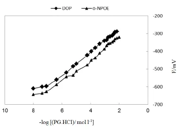
Figure 1. Potentiometric response of pioglitazone PVC matrix membrane sensors.
Plasticized PVC membrane sensor incorporating iodobismuth-drug ion pair was prepared with suitable solvent mediators and electrochemically evaluated as membrane sensors for pioglitazone under static mode of operations according to IUPAC recommendations [29]. The membrane was prepared using a casting solution of the composition 7 : 63 : 30 wt % of ion pair, o-nitrophenyl octyl ether or dioctyl phthalate and PVC, respectively. The two plasticizers have different dielectric constants [34]. The sensors were soaked in drug solution and tested as drug sensors. Table 1 summarizes the potentiometric response characteristics of the sensors. It was found that the drug sensors plasticized with the two different plasticizers have almost the same characteristics. The sensors show Nernstian response over the concentration range of 1.0 × 10⁻⁸
- 1.0 × 10⁻² M with cationic slopes of 58.98 and 60.40 mV decade-1 for o-NPOE and DOP based sensor, respectively. The detection limit is 6.0 × 10⁻⁸ M and 7.0 × 10⁻⁸
M with o-NPOE and DOP, respectively. Least squares analysis of the data give the relationships:
Table 1. Performance characteristics of pioglitazone PVC matrix membrane sensors
Parameter Value
o-Nitrophenyloctylether Dioctylphthalate
Slope (mV decade-1) 58.98 0.2 60.40 0.2
Intercept (mV) 159.42 0.2 205.97 0.2
Correlation coefficient (r) 0.994 0.988
Linear range (mol l-1) 1.0×10⁻⁷ - 1.0×10⁻² 1.0×10⁻⁷ - 1.0×10⁻²
Lower limit of detection (mol l-1) 6 × 10⁻⁸ 7 × 10⁻⁸
Working pH range 3 - 7 3 -7
Response time for 10-3 mol l-1 (s) 8 10
Life span (week) 8 10
Accuracy (%) 100.2 99.4
Repeatability (CVw(%)) 0.5 0.6
Between-day-variability (CVb(%)) 0.8 0.7
Standard deviation (%) 0.91 0.78
The sensors displayed constant and stable potential readings within 0.2 mV from day to day and the calibration slope did not change by more than 1.0 mV decade-1 over a period of 8 weeks for o-NPOE and 10 weeks for DOP sensors.
3.1. Effect of pH
[image:6.596.195.417.418.697.2]
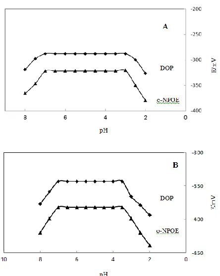
Influence of pH on the potentiometric response of the sensor was studied [36] using 10-3 and 10-2 M of pioglitazone solutions (Fig. 2). From pH-potential profiles, it is evident that the potential readings are constant over the pH range 3.0 - 7.0. Within this acidic range, PG is completely soluble, dissociated and sensed as a monovalent charged ion. At pH values lower than 3.0, the potential readings decreased due to interference by H+ ions. At higher pH values (>7.0), progressive precipitation of the drug was observed.
3.2.Effect of foreign Ions
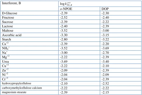
[image:7.596.56.541.408.735.2]The potentiometric response of Iodobismuth-PG-PVC membrane sensor was tested in the presence of several inorganic cations. Potentiometric selectivity coefficient KPG,B was used to evaluate the degree of interference. The data given in Table 2 were obtained using the separate solutions method [33] at 10-2 M pioglitazone solution. It is clear that the sensor is highly selective for PG ions compared with some common cations. Pharmaceutical excipients and diluents (e.g., glucose, maltose, manitol, starch, talc powder and magnesium stearate) at concentration as high as 400-fold molar excess over pioglitazone did not interfere. The o-NPOE based sensor was generally more selective than the DOP based sensor.
Table 2. Potentiometric selectivity coefficients
kPG,potB of pioglitazone PVC matrix membrane sensorsInterferent, B log pot
B PG., k
o-NPOE DOP
D-Glucose -2.39 -2.30
Fructose -2.52 -2.40
Sucrose -2.39 -2.22
Lactose -2.40 -2.39
Maltose -3.52 -3.00
Ascorbic acid -3.30 -3.15
Starch -2.80 -3.22
Ca+2 -2.39 -2.20
NH4+ -3.52 -3.69
Na+ -3.00 -2.70
Mg+2 -2.22 -2.39
Urea -3.69 -3.40
Cu+2 -2.22 -2.10
Zn+2 -2.09 -2.39
Ni+2 -2.04 -2.09
Cr+3 -2.04 -2.39
hydroxypropylcellulose -2.10 -2.52 carboxymethylcellulose calcium -2.22 -2.22
magnesium stearate -2.39 -2.15
3.3. Determination of pioglitazone
The evaluation of the pioglitazone-PVC membrane sensor for the determination of pioglitazone was assessed by determining 3.9 µg - 3.9 mg mL-1 standard PG solutions using the calibration graph method. The results obtained showed an average recovery of 99.8 % and a mean standard deviation of 0.85 % (n=5) using both solvent mediators based sensors. Pioglitazone (as antidiabetes) in different pharmaceutical preparations was also determined (Table 3). Average recoveries of 100.1 % and 99.9 % of the nominal and mean standard deviation of 0.73 % and 0.72 % for the o-NPOE and DOP based sensors were obtained, respectively.
Table 3. Determination of pioglitazone in pharmaceutical preparations using pioglitazone PVC matrix membrane sensors
Trade name Nominal content
(mg/tab.)
Recovery*, (%)SD
o-NOPE DOP HPLC method+
Actos 15 99.8 0.70 100.2 0.79 100.3 0.62
Actos 30 100.1 0.88 99.7 0. 82 99.8 0.41
Actos 45 100.3 0.61 99.8 0.58 99.8 0.58
*
Average of 5 measurements +
Ref. 35.
3.4. Application to urine samples
Table 4. Determination of pioglitazone hydrochloride in spiked human urine by the proposed electrodes
Concentration (M) Recovery %a of pioglitazone hydrochloride
o-NPOE DOP HPLC method+
1 x 10⁻² 1 x 10⁻³
100.22±0.3 100.17±0.3
99.8±0.3 99.9±0.1 100.23±0.3 100.1±0.1 a
Average of three determinations. +
Ref. 35.
4. CONCLUSION
The present work involves the preparation of new sensors using novel ionphore for the determination of PG.HCl drug. The new electrodes were characterized and optimized with respect to the main experimental parameters affecting the electrode performance, including composition, pH, response time and interference. The electrodes showed good selectivity for pioglitazone hydrochloride (PG.HCl) with respect to some inorganic cations, sugars, cellulose derivatives, magnesium stearate and ascorbic acid. The developed electrodes have been used successfully for the determination of pioglitazone hydrochloride in pharmaceutical preparation and urine using calibration methods. The obtained results were in good agreement with those obtained using the HPLC method. According to the results obtained, the potentiometric sensors can be applied successfully for the routine analysis of this drug.
References
1. Physician,s Desk Reference®, 54 ed., Medical Economics Company, Inc., Montivale, NJ, P.3088(2000).
2. R.T. Sane, S.N. Menon, S. Inamdar, M. Mote, and G. Gundi, Chromatographia, 59 (2004)453. 3. Z. Minbo, and Z. Yeping, Zhongguo Yiyao Gongye Zazhi, 34 (2003) 407.
4. A. Jedlička, J. Klimeš, and T. Grafnetterova, Pharmazie, 59 (2004) 182.
5. B.L. Kolte, B.B. Raut, A.A. Deo, M.A. Bagool, and D.B. Shinde, J.Chromatogr. Sci., 42 (2004) 31.
6. T. Radhakrishna, R.D. Sreenivas, and R.G. Om, J. Pharma. Biomed. Anal., 29 (2002) 607. 7. Y. Kenji, M. Hiromi, O. Teruaki, and M. Michio, J. Chromator. B:, 677 (1996) 146. 8. J. Dai, W. Jin, and Y. Liu, Yaowu Fenxi Zazhi, 21 (2001) 39.
9. W. Z. Zhong, and M. G. Williams, J. Pharm. and Biomed. Anal., 14 (1996) 473.
10. Y.J. Xue, K.C. Turner, J.B. Meeker, P. Janice, A. Mark, and U. Stere, J. Chromatogr. B:, 795 (2003) 226.
11. K. Yamashita, H. Murakami, T. Okuda, M. Motohashi, J. Chromatogr., B:, 677 (1996) 146. 12. W. Z. Zhong, and D. B. Lakings, J. Chromatogr., 490 (1989) 385.
13. T. M. Baughman, R. A. Graham, K. Wells-Knecht, I. S. Silver, L. O. Tyler, M. Wells-Knecht, and Z. Zhao, Bio. Fate Chem., 33 (2005) 738.
14. L. J. Zhongping, J. Weihua, D. K. Daksha, and S. Linyee, J. Pharm. and Biomed. Anal., 33 (2003) 108.
15. E. N. M. Ho, K. C. H. Yiu, T. S. M. Wan, B. D. Stewart, and K. L. Watkins, J. Chromatogr., B: Analytical Technologies in the Biomedical and Life Sciences, 811 (2004) 73.
16. M. Thevis, H. Geyer, and W. Schaenzer, Rapid Commun Mass Spectrom, 19 (2005) 936.
17. Y. Kiyota, T. Kondo, Y. Maeshiba, A. Hashimoto, K. Yamashita, Y. Yoshimura, M. Motohashi, and S. Tanayama, Arzneimittel-Forschung, 47 (1997) 28.
18. Z. Shen, J. R. Reed, M. Creighton, D. Q. Liu, Y. S. Tang, D. F. Hora, W. Feeney, J. Szewczyk, R. Bakhtiar, R. B. Franklin, and S. H. Vincent, Xenobiotica, 33 (2003) 509.
19. H. Wong, Y. Ozalp, and A. Lainesse, Arzneimittel-Forschung, 54 (2004) 624.
20. A. Gumieniczek, H. Hopkala, and A. Berecka, J. Liq. Chrom. & Rel. Tech., 27 (2004) 2070. 21. R. T. S. Menon, S. Inamdar, M. Mote, and A. Menezes, J. Planar Chromatogr.--Modern TLC, 17
(2004) 156.
23. J. R. Colca, W. G. McDonald, D. J. Waldon, J. W. Leone, J. M. Lull, C. A. Bannow, E. T. Lund, and W. R. Mathews, Am. J. Physiol., 286 (2004) E260.
24. G. D. Sankar, R. J. M. Kumar, and M. V. V. N. Reddy, Asian J. Chem., 16 (2004) 539.
25. M. B. Shankar, V. D. Modi, D. A. Shah, K. K. Bhatt, R. S. Mehta, M. Geetha, and B. J. A. R. Patel, J. AOAC Intern., 88 (2005)1172.
26. E. Tahmasebi, Y. Yamini, and A. Saleh, J. Chromatogr. B:, 877 (2009) 1929 27. B. Jamali, G. C. Theill, and L. L. Sørensen, J. Chromatogr A:, 1049 (2004) 187
28. B. Du, L. Pang, Y. Yang, G. Shen, and Z. Zhang, J. Chromatogr. B Analyt. Technol. Biomed. Life Sci. 952 (2014) 143.
29. IUPAC recommendation: Pure and Appl. Chem. 67 (1995) 1723.
30. G. A. E. Mostafa, and A. Al-Majed, J. Pharm. and Biomed. Anal., 48 (2008) 61. 31. A. Craggs, G.J. Moody, J.D.R. Thomas, J. Chem. Educ. 51 (1974) 544.
32. M.A.F. Elmosallamy, G.J. Moody, J.D.R. Thomas, and S.S.M. Hassan, Anal. Lett. 20 (1987) 1555.
33. A. Craggs, G.J. Moody, and J.D.R. Thomas, J. Chem. Educ. 51 (1974) 549.
34. R.1. Kumar, A.K. Pandey, M.K. Sharma, L.V. Panicker, S. Sodaye, G. Suresh, S.V. Ramagiri, J.R. Bellare, and A. Goswami, J Phys Chem B. 115 (2011) 5856.
35. P. Sripalakit, P. Neamhom, and A. Saraphanchotiwitthaya, J. Chromatogr. B Analyt. Technol. Biomed. Life Sci., 843 (2006) 164.
36. A. L. Saber, Electroanalysis J. 25 (2013) 2707.