Phosphate Based Glasses: A Perspective
Ensanya Ali Abou Neel
*a, David Mark Pickup
b, Sabeel Padinhara
Valappil
a, Robert John Newport
band
Jonathan Campbell Knowles
+a5
The general trend in biomaterials is to use and employ materials that play an active ro le in tissue regeneration rather than passive and inert materials. Therefore, understanding how a material interacts with the surrounding environments, including cells and tissue fluid, allows material design to be tailored so that implants can be constru cted to promote a specific biological response,
10
helping them better perform their function. This class of materials has been described as the “Third Generation” of biomaterials. Phosphate based glasses fall into this category and it has been shown that the properties of these glasses can be tuned via their composition according to the desired end application. These glasses can be prepared as melt quenched or sol -gel bulk form suitable for potential hard tissue engineering applications and as vehicles for antimicrobial agents.
15
They can also be prepared as fibres suitable for soft tissue engineering applications such as those involving muscle, ligaments, and tendon, where, like the fibres, the tissue has a high degree of anisotropy.
1. Introduction
Recently, interest in soft and hard tissue engineering for
20
improved tissue regeneration has fuelled the need for novel biodegradable materials having a specific and controllable bioactivity [1]. Bioactive glasses, silicate based in particular, have been the subject of intense interest for the last three decades as materials for tissue regeneration applications. In
25
vivo, when they exposed to physiological fluids, they form a surface apatite layer; this layer has the capacity to bond to collagen synthesised by connective tissue cells such as osteoblasts [2]. One commercially available bioactive glass is Bioglass® which has a composition known as 45S5
30
corresponding to 45.0 wt% SiO2, 24.5 wt% CaO, 24.5 wt%
Na2O and 6.0 wt% P2O5 [3-5]. Today, the 45S5 composition
is used as a benchmark by which the performance of new silicate based bioactive glasses is measured. Such glasses have shown great success in many clinical applications
35
especially in dental and orthopaedic fields. However, there are questions raised related to the long term effect of silica [6] and the slow degradation of these glasses, often taking 1 to 2 years to disappear from the body [7, 8]. Because of these limitations, the search for new materials for the repair of bone
40
defects has continued and has led to the emergence of phosphate based glasses as potential alternatives.
Phosphate based glasses in the CaO-Na2O-P2O5 system
have unique dissolution properties in aqueous based fluids. Degradation rates can be varied from hours to several weeks
45
by changing the glass composition. Moreover, all the constituents of these glasses are elements naturally inside the body and therefore can be excreted by the normal physiological processes. Furthermore, these glasses can be synthesised to include dopants that are able to induce a
50
specific biological function and enhance biocompatibility [9 -12].
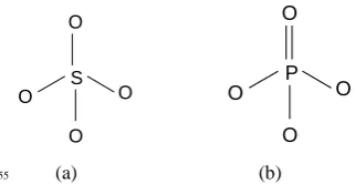
S O O
O
O
P O O
O
O
(a) (b)
[image:1.595.305.466.336.419.2]55
Figure 1: Silicate (a) and phosphate (b) tetrahedra.
This review starts with a general description of phosphate glasses, highlighting their differences with their silicate based counterparts and explores what these glasses can offer in terms of biomedical applications. Particular focus is placed
60
on phosphate glass chemistry, terminology and structure. Next we discuss the processing techniques used to prepare these glasses. In the case of the melt-quenched prepared glasses, both monoliths and glass fibres are described. Recent developments in using sol-gel methods to prepare phosphate
65
glasses for biomedical applications are reviewed. The article concludes with a discussion of the future of phosphate based glasses as biomaterials and highlights possible avenues of potential application.
2. Comparison of Phosphate and Silicate Based
70Glasses
Silica (SiO2) is a classic network forming oxide. It is
common in nature in both crystalline and glassy forms because of the strong affinity of silicon towards oxygen, and the natural abundance of these two elements. The basic unit
75
P O O
O O
Q3 tetrahedron Q2 tetrahedron
P O O
O O
3
-P O O
O O
2
-P O O
O O
1
-Q1
tetrahedron Q0 tetrahedron
consequence phosphates are common in nature. In common with silicate based glasses, the building block of phosphate glasses is a tetrahedral unit. However, as can be seen in Figure 1, the PO4 unit is quite different from the SiO4 unit. This is
because phosphorus nominally has a charge of 5+ whereas
5
silicon has a charge of 4+. Therefore when SiO4 tetrahedra
form a network they can share all four of their oxygen atoms
to give the stoichiometry SiO4/2 or SiO2 which is charge
balanced (assuming a charge of 2- on the oxygen). In contrast, when forming an analogous charge balanced 3D
10
binary oxide, phosphorus can only share three out of its four
oxygens which gives the stoichiometry of PO(1+3/2) or P2O5.
In the case of the P2O5, the oxygen atoms that are not shared
between phosphate tetrahedral share their two unpaired electrons with the P5+ ions to form a terminal double bond
15
(Figure 1b) [13].
The fact that phosphate anions contain at least one terminal oxygen limits the connectivity of phosphate based glasses relative to their silicate based counterparts. Therefore, in general the rigidity, which is related to the interatomic forces,
20
is less in phosphate glasses compared to silicate glasses. Moreover, when mixed with metal oxides, phosphate glasses contains fewer cross-links but a higher number of terminal oxygen atoms than silica glasses of the same metal oxide content. These two structural properties result in more
25
flexibility in the orientation of PO4 tetrahedra [13].
Therefore, the range of glass formation in binary phosphate glass in systems is wider than in the analogous silicate based system, even in the presence of alkali or alkali earths.
Pure vitreous silica is thermally and chemically stable. The
30
addition of modifying oxides such as Na2O, K2O, MgO, and
CaO, which are not part of the glass network and disrupt it resulting in terminal Si–O bonds, produces less stable glasses
[14-15]. Pure vitreous phosphorus pentoxide (P2O5), on the
other hand, is chemically unstable with regard to hydrolysis of
35
the P–O–P bonding by atmospheric moisture; in this case the addition of metal oxides improves its stability because P–O-Mn+ (where M = metal cation) bonds are generally more stable towards atmospheric hydrolysis [16].
From a technical standpoint of pure melt quenched,
40
vitreous silica is only used in very specialized applications due to the very high melting temperatures involved in its manufacture. The addition of modifying oxides which reduce network connectivity significantly reduce the melting temperature required. Phosphate based glasses, however, can
45
be prepared at relatively low temperatures [6].
These two types of glass also differ in their aqueous dissolution mechanisms and in the stability of resultant anionic species. Dissolved silicate species can easily be polymerised or repolymerised to form species with no
50
resemblance to the original glass structure, whereas phosphate chains and rings are quite stable in aqueous solution [17]. Due to their high melting temperatures, silica based bioactive glasses are difficult to draw into fibers and the
addition of metal oxides such as Na2O and CaO to lower the
55
melting temperature can adversely affect the glass bioactivity by increasing its tendency towards crystallization. The of use sol-gel chemistry to overcome the problems of fiber drawing
[image:2.595.301.543.64.124.2]60
Figure 2: Nomenclature and representation of PO4 tetrahedra 65
with different polymerizations [22].
is still in its infancy [18]. In contrast, phosphate glasses
containing more than 45 mol % P2O5 can easily be drawn into
fibers [10]. From a structural viewpoint, the fibre drawing ability or spinnability is related to the ability of the longer
70
phosphate chains to entangle with other chains. Such interactions allow continuous filaments to be formed instead of clusters or droplets. Milberg and Daly [19] proposed that in perfectly oriented fibres, all chain axes are parallel to the fibre axis, and the rotational disorder of the chains around
75
their own axes corresponds to the cylindrical symmetry of the fibre. Moreover, Murgatroyd [20] suggested that the drawing operation preferentially selects the strong P-O-P bonds and the continuity of fibres is related to the ability of these bonds to be aligned along the long axis of the fibre. Whereas, weak
80
P-O-P bonds can be extended for a short distance along the strong bonds, but are not able to form continuous fibres [20]. Compared to silicate glasses, phosphate glasses have poor chemical durability and the poor durability limits their use in technological applications. However, phosphate glasses are
85
preferred in other applications such as release of oligo -elements in soils. Moreover the solubility of phosphate species in Bioglass is responsible for the nucleation and apatite layer formation that is considered to be the main factor responsible for the bioactivity of Bioglass [21].
90
3. Terminology and Chemistry of
Phosphate-Based Glasses
As discussed in the previous section, the PO43- tetrahedron is
the basic building block of structure the phosphate based glasses. Phosphate tetrahedra are classified by the number of
95
oxygen atoms they share with other phosphate tetrahedra. An oxygen atom shared in this way is usually referred to as bridging oxygen, abbreviated to BO hereafter. The various types of phosphate tetrahedra that result from this
classification are labeled using Qi, where i refers to the
100
number of BOs and ranges from 0 to 3. For example, Q3 PO
43-tetrahedra share three covalently bonded BOs with
neighbouring PO43-tetrahedra as found in vitreous P2O5. In
terms of the structure of the phosphate network, Q3 moieties
are known as a branching units and since they have a O/P
105
ratio of 2.5 (i.e. PO(1+3/2)) they have neutral charge. Q2
tetrahedra possess two BOs which results in an O/P ratio of 3
and a net charge of 1-. Structurally, Q2 units can be
considered as PO3- middle groups in phosphate chains. Q2
tetrahedra have one BO and hence an O/P ratio of 3.5 and a
110
net charge of 2-. Structurally Q1 units can be considered as
representing P2O7
dimmers or as terminating groups at the
end of phosphate chains. Q0 represents an isolated (PO4)
3-tetrahedron (i.e. O/P = 4) with no BOs to neighbouring
tetrahedra; (PO4)
groups are also known as orthophosphate
units [22-24]. The various Q species discussed above are illustrated in Figure 2.
The structure of vitreous P2O5 consists of only Q3
phosphate tetrahedra that form a three dimensional network; the addition of modifying metal oxides, however, results in
5
the “depolymerisation” of this network via the cleavage of P-O-P bonds with the creation of negatively charged NBOs at the expense of BOs. The negatively charged NBOs charge balance the metal cations and coordinate them such that they achieve their preferred coordination number [13, 23, 25, 26].
10
The depolymerisation model proposed by Kirkpatrick and Brow [23] predicts that the dominant Qi species changes
according to Q3 → Q2 → Q1 → Q0 as the amount of
modifying oxide increases. Increasing the amount of metal oxide results in an increase in the overall O/P ratio and this
15
determines the average number of BO per phosphate
tetrahedron; from this one can predict the dominant Qi
species.
3.1. Ultraphosphate Glass Structure
Ultraphosphate glasses are those with compositions in the
20
range (M2/vO)x(P2O5)1-x, where 0 ≤ x ≤ 0.5 and v is the
valance of the metal cation. The ultraphosphate composition can also be described in terms of the atomic ratio of oxygen phosphorus, i.e. 3.0 ≤O/P ≤ 2.5. Glasses with compositions in this range are expected to have network structures dominated
25
by Q2 and Q3 species. Structural studies using 31P NMR have
confirmed this to be the case and verified that the
concentrations of Q2 and Q3 groups can be calculated from
Van Wazer‟s chemically simple depolymerisation model [27].
The Q2 and Q3 tetrahedra appear to be randomly linked, at
30
least in alkali ultraphosphate glasses. In glasses with a high
P2O5 concentration (greater than 75-80 mol%), the phosphate
network resembles that of vitreous P2O5. With increasing
modifier content, there is a loss of the extended -range order
associated with vitreous P2O5, as the concentration of Q
2 35
species. The composition at which this transition occurs depends on the preferred coordination number and valence of the modifying cation since it relates to the relative concentrations of NBOs and cations. Hoppe [13] has postulated that at low cation concentrations, both NBOs of the
40
Q2 units coordinate to the same cation; whereas at higher
cation concentrations, each NBO can coordinate a separate cation resulting in a structural relaxation.
3.2. Metaphosphate Glass Structure
Metaphosphate glass have the composition
45
(M2/vO)0.5(P2O5)0.5 and an O/P ratio of 3. Their structures
consist of infinitely long chains and/or rings of Q2 units [24].
These phosphate chains are linked through bonds between the terminal NBOs and the modifying cations. Structural studies have revealed that the properties of metaphosphate glasses are
50
less dependant on the nature of the P O P that form the chains than on the P O Me bonding between chains. In general, as the field strength of the modifying cation increases, there is an increase in the rigidity of the metaphosphate network and an associated increase in glass
55
transition temperature [23].
3.3. Polyphosphate Glass Structure
Polyphosphate glasses have a composition compositions in the range (M2/vO)x(P2O5)1-x, where x > 0.5, and an O/P >3.
The structure of polyphosphate glasses is based on Q2 chains
60
terminated by Q1 units. At the pyrophosphate composition (x
= 0.667), the structure is dominated by phosphate dimers, i.e.
two Q1 units sharing a bridging oxygen atom. At x = 0.75,
only isolated orthophosphate Q0 units are present [24].
Polyphosphate glasses are often more durable than their
65
metaphosphate counterparts due to the reduction of the more
readily hydrolyzed Q2 units in the structure [23].
4. Melt Quenched Phosphate Based Bulk Glasses
Most phosphate based glasses are prepared by melt-quenching methods. A mixture of oxide precursors is melted in a furnace
70
at temperatures of over 1000°C; the exact temperature used depends on the final composition of the glass. Once a homogeneous melt has been achieved, the glass is formed by casting different shapes such as rods and plates. To remove residual stress, the melts are normally cooled quickly through
75
the glass transition temperature (Tg) and then the cooled very slowly to room temperature in an annealing step.
4.1. Technological Applications
Phosphate based glasses have been used in a wide range of technological applications such as sensors, solid-state
80
batteries, laser devices, and air tight seals for metals with a high coefficient of thermal expansion [28]. Phosphate glasses have also been developed for achromatic optical elements due to their low dispersion and relatively high refractive indices. Iron-containing phosphate glasses have found uses as matrices
85
for vitrifying nuclear waste products [29-31]. Their capacity for high waste loading, low processing temperature and high chemical durability offer significant advantages compared to most silicate and borosilicate glasses. They are reported to
maintain their high chemical durability even after
90
devitrification of waste forms [32].
4.2. Medical Applications
4.2.1Controlled Release Glasses (CRG)
Controlled release glasses are a class of materials that completely dissolve in aqueous media leaving no solid
95
residues. Their degradation is an erosion controlled process that follows zero order release over the life of the material, i.e. the release rate is constant and independent of time and concentration [33]. They can be produced in different forms such as powder, granules, fibre, cloth, tubes, and monoliths of
100
various shapes [34]. These glasses have been under development in the Standard Telecomminucation Laboratories since the early 1970s [35]. They find application in many different areas such as feeding of bacteria, controlling parasite infection in water canals, veterinary use or even treatment of
105
infections in humans. Some examples of these uses are described below.
Polyphosphate glass provides a source of phosphate ions that can support the growth of recombinant Escherichia Coli to a density 40 % higher than that obtained with typical
fermentation media. The high solubility of polyphosphates together with the absence of precipitate formation when mixed with the fermentation media are key benefits for such applications [36].
Soluble phosphate glasses containing such as copper,
5
cobalt, and selenium, designed for oral administration in form of a rumen bolus to ruminant animals for the treatment of trace element deficiencies, are manufactured under the trade name of Cosecure® [37-40].
Copper releasing phosphate glasses have been used as
10
molluscicides to control the snail hosts of schistosomiasis. Glass composition and physical form can be tailored in a reproducible manner to suit the chemistry of the water body being treated. Moreover, most of the released Cu is in non-toxic or weakly non-toxic forms such as copper polyphosphate
15
complexes which acts as secondary releasing agents [38-40]. Silver releasing phosphate glasses are used clinically to combat long term infection in indwelling catheters. A cartridge with silver containing glass is inserted in line between the catheter and urine collection bag. This cartridge
20
treats the urine as it flows through it from the bladder to the collection bag. The silver ions released are found to inhibit bacterial proliferation [34] and have the potential to be used in the treatment of vesicoureteral flux and urinary incontinence [41].
25
4.2. 2. Hard Tissue Engineering Applications
Biodegradable scaffolds, which are eventually replaced by the natural tissue, are desirable constructs for tissue engineering
applications. CaO-Na2O-P2O5 glasses have properties that
lend them to use as hard tissue substitutes or as substrates for
30
synthetic orthopaedic graft materials. Compositionally they are similar to the inorganic component of bone [42]. Furthermore, fluoride-doped phosphate glasses have been developed that play an active role in stabilising the apatite layer [42]. Phosphate glasses can also be doped with a variety
35
of metal oxides to modify their physical properties [43, 44]. It is reported that the ionic environment caused by the leaching of ions from these glasses during their degradation has an impact on the biological response of cells [45]. For
example, Ca2+ ions have been implicated in stimulating
40
osteoblast-like cell proliferation and differentiation and phosphate ions act as extracellular „pool‟ responsible for the release of Cbfa-1, an important bone marker, from bone cells [45].
For hard tissue engineering applications, a number of glass
45
systems have been developed by additions of various metal oxides such as Fe2O3, Al2O3, ZnO, and TiO2 into the parent
glass. Comprehensive studies have been reported that give an overview of the correlation between the basic glass structure and the biocompatibility.
50
4.2.2.1. Binary System (s)
Binary sodium phosphate glasses (Na2PO4H-NaPO4H2),
developed by Gough et al. [46, 47], demonstrated a minimal level of macrophage activation evident from low amounts of peroxide and interleukin-1β release. Moreover, early primary
55
craniofacial osteoblast attachment and spreading was also observed on those glasses. Upon long term culture (28 days),
the craniofacial osteoblasts exhibited cytoskeletal
characteristics and a level of collagen synthesis similar to those of the positive control. The biocompatibility of these
60
glasses was related to their degradation and the ions released. However, it was difficult for cells to attach to such highly soluble glasses because the labile surface prevented the formation of a physical anchorage.
4.2.2.2. Ternary System (s) 65
Uo et al. [48] developed the P2O5-CaO-Na2O ternary glass
system and assessed the cytocompatibility by direct contact cytotoxicity assay using dental pulp cells. Their study
reported that the samples containing 50 mol% P2O5 were not
very cytotoxic. The cytotoxicity was found to decrease with
70
increasing P2O5 content due to a change in pH from neutral at
50 mol % to acidic at 60 mol% or more. Thus, the authors related the cytotoxicity to the glass degradation, the associated pH changes and ionic concentration in the media. Franks et al. [49] studied the same ternary glass system in
75
the composition range (P2O5)45(CaO)x(Na2O)55-x, where x
was between 8 and 40 mol %. Initial work focused on the degradation and ion release of these glasses; the results suggested that the interaction of the Ca2+ ions with the glass network controlled glass degradation, and an inverse
80
relationship existed between calcium oxide content and the degradation rate. The biological response of this glass system was tested by Salih et al. [6] to assess their suitability for potential bone regeneration applications. Two human osteoblast cell lines, MG63 and HOS (TE85) were incubated
85
in the glass extracts with different concentrations (neat, 1:4, 1:16, 1:64 dilution) for two and five days. MTT assay was used to study cell growth, and ELISA was used to measure the expression of antigens such as bone sialoprotein, osteonectin, and fibronectin which play a vital role in bone metabolism
90
and integrity. The results showed that the glasses with lower solubility enhanced bone cell growth and antigen expression at all tested dilutions. The highly soluble glasses, however, significantly reduced cell proliferation, and down-regulated antigen expression especially with neat and 1:4 dilutions at
95
five days. The authors suggested that these results were related to ions released from the glass during degradation and the resultant pH changes. They also suggested that with less
soluble glasses, greater amounts of Ca2+ ions are released,
which is known to have an essential role in cell activation
100
mechanisms affecting both cell growth and function. However, with highly soluble glasses, a sharp increase in pH
associated with high release rates of Na+ and phosphate ions
(PO4
3-) may have a deleterious effect on cells.
Due to the high degradation rate and unfavorable cellular
105
response associated with high sodium content glasses, Franks
studied the effect of replacing the Na2O with K2O [42]. For
this new study, glasses of composition
(P2O5)45(CaO)x(K2O)55-x, where x is between 16 and 32 mol
% were used. Glass with CaO content outside this range was
110
difficult to prepare due to its crystallisation upon casting. It
was observed that the P2O5-CaO-K2O system dissolved at a
higher rate than the analogous P2O5-CaO-Na2O system and
hence no biocompatibility study was conducted on this glass system.
115
exposure to phosphate based glass of two typical cellular components of a hard/soft tissue interface, periodontal ligament/mandible and patellar tendon/tibia. Human oral osteoblasts, oral fibroblasts and hand flexor tendon fibroblasts were co-cultured on glasses with different degradation rates
5
((P2O5)45(CaO)x(Na2O)55-x where x = 30-48). Quantitative
and morphological assessment of cell adhesion and proliferation for all cell types was recorded. Immunolabelling was also used to establish phenotypying of both osteoblasts and fibroblasts. The results showed that glass discs with less
10
than 40 mol% CaO support little or no cell adhesion and survival. This behaviour was related to the high solubility of the surface layer of these glasses; therefore, it is difficult for cells to attach to a labile surface and to form a physical anchorage as observed by Gough et al., [46, 47]. The authors
15
concluded that ternary glass compositions with high CaO content (46 and 48 mol%) support high numbers of adherent and viable cells as indicated by DNA content, and also maintain cellular function as indicated by phenotypic gene expression up to 7 days.
20
From these studies, it is clear that the glass degradation, and the associated ion release, and pH change of the
surrounding environments, are factors affecting
biocompatibility. Therefore, additions of metal oxides known to affect the chemical durability of phosphate glass to the
25
P2O5-CaO-Na2O system are expected to have an effect on
biocompatibility. The quaternary systems that result from such additions are discussed in the next section.
4.2.2.3. Quaternary Systems/Dopants
Knowles et al., [51] synthesized quaternary glasses of
30
composition (P2O5)45(CaO)x(Na2O)55-x-y(K2O)y, where y =
20, 24, 28 or 32 and x = 0-25, in order to study the affect of
substituting Na+ ions with larger K+ ions. The aqueous
degradation of this system was affected by both CaO and K2O
content. An anomaly in degradation was observed at high
35
CaO content, where weight gain was observed prior to weight
loss. The MTT assay showed that the K+ had a positive effect
on cell proliferation only at high content, 20 mol % K2O,
regardless of the associated increase in degradation.
In order to understand the effect of changing the radius of
40
the divalent cation in a quaternary system, Franks et al. [52]
substituted Ca2+ ions with smaller Mg2+ ions in
(P2O5)45(CaO)32-x(Na2O)23(MgO)x, where x = 0-22,
glasses. This study concentrated on the overall degradation characteristics of the glasses and the effect of released ions on
45
cell proliferation. The results showed that degradation of the glass as a function of time changed from exponential to linear with decreasing CaO content. This emphasised the influential role of CaO on the degradation process. The degradation rate
was decreased by substitution of CaO with MgO despite Mg2+
50
having the same valence as Ca2+. The MTT assay was used to
assess the effect of different dilutions of glass extracts on the proliferation of human osteoblasts (MG63) for two and five days. The results were normalised to the control cells incubated in normal medium. The result showed that glasses
55
with little or no MgO showed a slight decrease in cell proliferation only after two days; however, after five days all glass compositions tested showed equal or greater cell
proliferation than the control.
Salih et al. [53] added zinc oxide to PBG with the aim of
60
promoting osteoblast cell adhesion and improving the potential for use in bone tissue engineering applications. The
compositions investigated were (P2O5
)50(CaO)40-x(Na2O)10(ZnO)x, where x = 0-20. Attachment of
osteoblast-like cells was assessed morphologically by scanning electron
65
microscopy and the effect of the glass extract (neat and 10% diluted) on cell proliferation over periods of up to 7 days was determined by cyquant assay. The results showed that after 24 hours of culture, the cells attached to all glass compositions but still maintained round morphology
70
suggesting lack of spreading on the glass surfaces. Moreover, cell proliferation increased with increasing ZnO content up to 5 mol%, but never reached levels exhibited by cells grown on the positive control.
Abou Neel et al. [54, 55] prepared bulk quaternary glasses
75
containing TiO2 by conventional melt quenching methods. The aim was to test the hypothesis that the combination of
Ti4+ and Ca2+ ions would further improve the biological
response of phosphate glasses. The glass compositions studied were (P2O5)50(CaO)30(Na2O)20-x(TiO)x, where x = 1, 3
80
and 5. MG63 cell proliferation, gene expression, and bioactivity were the focus of this study. Cell proliferation and gene expression (Core binding protein factor alpha 1 (Cbfa1), alkaline phosphatase (ALP), Collagen type I alpha subunit I (COLIAI), and Osteonectin (Sparc)) were reproducibly
85
enhanced on the surfaces of the Ti4+containing glasses,
particularly those with 3 and 5 mol% TiO2. The authors
suggested that this enhancement may be associated with the lower degradation of these compositions which help maintain pH at a level favoured by osteoblasts. It was also suggested
90
that the release of Ti4+ ions may have a beneficial effect on
bioactivity.
Of the three compositions of Ti-doped phosphate based
glasses investigated, the 5 mol% TiO2 glass induced the most
favourable cellular response [54]. As a follow-up study, Abou
95
Neel et al. [56] replaced some of the CaO with in this glass with ZnO (1, 3 and 5 mol%) in an attempt to further improve the biological properties. This work concentrated of the effect of ZnO additions on the thermal properties, degradation, ion release, surface and biological properties. The results showed
100
that the addition of ZnO was effective in controlling the bulk, and surface properties of the glass. Glasses containing both TiO2 and ZnO demonstrated similar high viability of MG63
cells up to 7 days to both the 5 mol% TiO2 parent glass with
and the positive control, Thermanox®. This cell proliferation
105
was correlated with the release of beneficial Ca2+, P, Ti4+
and Zn2+ ions at suitable level coupled with an increase in
surface hydrophilicity. The hydrophilicity is thought to be associated with enhanced protein adsorption and adhesion of anchorage dependant cells such as osteoblast, fibroblast and
110
endothelial cells on the surface of biomaterials [57].
4.2.3. Antimicrobial Delivery Devices
Phosphate glasses offer potential alternatives to the current methods available for the treatment of infections since they can be used as localised antibacterial delivery systems via the
5
10
Figure 3: AFM image of silver free glass surface coated with
S. aureus biofilm.
inclusion of ions known for their antibacterial effects such as copper, silver, and gallium. Such materials could therefore be
15
placed at a site of infection, with the aim of releasing antibacterial ions as the glass degrades, which may be useful in wound healing applications. Their use could be extended to the prevention of implant or biomaterial related infections, which are one of the main causes of revision surgery, and to
20
augment or replace the current prophylaxis of systemically administered antibiotics [58].
Antimicrobial glass systems incorporating either Cu2+ or
Ag+ ions were successfully prepared by Mulligan et al. [59,
60] for potential applications in the treatment of oral
25
infections. The aim was to develop glass devices that could be placed at the site of an infection such as in a periodontal pocket to treat the infection with the antimicrobial ions released as the glass degrades. Both reports focused on glass
systems with a fixed P2O5 concentration of 45 mol %, and
30
concentrations of the antibacterial ions, Cu2+ or Ag+, of 0, 1,
5, 10 and 15 mol %. For each system, the calcium oxide to
sodium oxide (CaO/Na2O) ratio was varied to give the same
degradation rate over all compositions. Consequently, the overall effect on bacteria was due to the presence or absence
35
of antibacterial ions and their concentrations. The effect of
both glass systems on the viability of a Streptococcus Sanguis
biofilm using constant depth film fermenter (CDFF) was evaluated in a simulated oral environment using the glass sample containing no antimicrobial and HA (hydroxyapatite)
40
as controls. The results demonstrated that after 24 h, there was a significant reduction in viable counts of bacteria compared to the controls, which was attributed to the release of antimicrobial ions. This reduction was correlated with the concentration of antimicrobial ions in the glass. Despite
45
recovery of the bacterial counts after 48 h, they were significantly lower than those of the controls and remained relatively constant between 48 h and eight days. Two possible reasons were proposed for this recovery: firstly, the formation of a sacrificial layer of dead bacterial cells that acts as a
50
barrier against further penetration of the antimicrobial ions into the biofilm; secondly, the differentiation of bacteria into another phenotype that was resistant. The results also showed
that Ag+ ions display more potent antimicrobial activity than
to Cu2+ ions.
55
[image:6.595.61.200.52.182.2]Further work on antimicrobial phosphate glasses was carried out by Ahmed et al. [61] who investigated glasses with a relatively higher phosphate content. The compositions
Figure 4: A cross- sectional view of the S. aureus biofilm on 20% silver doped PBGs. Viable (green) and non-viable (red) 60
bacteria (62).
studied were (P2O5)50(CaO)30(Na2O)20-x(Ag2O)x where x =
0-15. Disc diffusion assay was used to screen the antibacterial activity of these glasses against various
micro-organisms including Staphylococcus aureus [Figure 3],
65
Escherichia coli, Bacillus cereus, Pseudomonas aeruginosa,
methicilline resistant Staphylococcus aureus, and Candida
albicans. The results showed that the phosphate glasses
containing 3 and 5 mol% Ag2O were more effective than the
remaining compositions in the inhibition of bacterial growth.
70
Overall it was concluded that the glass with 3 mol% Ag2O
was of optimal composition to mount a potent antibacterial effect against the test micro-organisms since it was
bactericidal against Staphylococcus aureus, Escherichia coli,
and significantly reduced the growth of Candida albicans.
75
These findings were correlated with the excellent long term release of Ag ions from that composition into the surrounding medium.
Further study of silver containing phosphate glasses (10, 15
and 20 mol%) was performed by Valappil et al. [62] who
80
tested their effect on the formation of the highly resistant S.
aureus biofilms. Silver ions were found to reduce the growth
of S. aureus biofilms. Variations in bactericidal activity were
correlated with glass degradation rates which varied between
0.42 and1.22 µg.mm-2. h-1 depending on composition. Due to
85
the antibacterial action of the Ag+ ions, a dead layer,
approximately 20µm thick, of non-viable bacterial cells was formed on the glass surface [Figure 4]; this layer was covered by a top layer of viable cells. The antibacterial effect of these glasses was attributed to the silver ions being present in the
90
most potent +1 oxidation state; confirmation of this was provided by Ag K-edge XANES (X-ray absorption near-edge structure) measurements.
As well as silver and copper, gallium was also investigated as a dopant for phosphate glasses because of its antibacterial
95
activity [63]. Novel quaternary gallium-doped phosphate
based glasses ((P2O5)45(CaO)16(Na2O)39-x(Ga2O3)x where x
= 1, 3 and 5) were synthesized, and their bactericidal activities
tested against both Gram negative (Escherichia coli and
Pseudomonas aeruginosa) and Gram positive (Staphylococcus
100
aureus, methicillin-resistant Staphylococcus aureus, and
Clostridium difficile) bacteria. The report confirmed that the
controlled delivery of Ga3+ ions from the glass containing 1
mol% Ga2O3 was sufficient to mount a potent bactericidal
effect and demonstrated the potential of these glasses as a new
105
therapeutic agent for pathogenic bacteria including the super
bugs MRSA and C. difficile.
5. Melt Quenched Phosphate Based Glass Fibres
Glass fibres have potential applications in the engineering of soft tissues such as muscle and ligament due to a combination
110
(b) (a)
10 m 10 m 10 m
[image:7.595.58.289.61.112.2](a) (b)
Figure 5:Phosphate glass fibres drawn with increasing drum speeds, which results in decreasing fibre diameter; (a) 35 5, (b) 25 3, (c) 16 2, and 11 1 m.
the glass fibres can act as a template with muscle cells
5
growing along their long axis and forming myotubes; in particular, the three dimensional mesh arrangements have proven to be the best configuration for supporting cell attachment and proliferation [10, 64]. Glass fibre scaffolds with open mesh morphology allow for the diffusion of
10
nutrient and waste products in and out of the construct, and permit the ingrowth of vasculatures and hence the tissue. They also provide the necessary structural support without compromising porosity [65-67]. Recently, it has been suggested that glass fibres could also act as a nerve conduit,
15
since they can provide a guide for cell orientation, proliferation and growth [68, 69].
Phosphate glass fibres are conventionally fabricated by drawing from a high temperature melt. Typically fragments of the starting glass are remelted and fibres drawn from the
20
melt onto a rotating collecting drum [66, 70]. Adjustment of the melt temperature is necessary to obtain a suitable viscosity for fibre drawing, since it is not feasible glasses with low melt viscosities to be drawn into fibres [10]. Additionally, the melt temperature should be above the glass crystallisation
25
temperature; otherwise, fibre drawing becomes difficult [71] or the bioactivity of the glasses is reduced [72].
During the drawing process, the fibre diameter can easily be controlled by adjusting the drum speed: higher drum speed results in smaller fibre diameter as shown in Figure 5. From a
30
biological standpoint, it has been reported that the fibre diameter has an effect on cell orientation [68]. It was found that as long as the fibre diameter is comparable to the size of the cell body, the cells will orientate along the long axis of fibre rather than around its circumference. Cells tend to wrap
35
around the smaller diameter fibres, but in presence of less curvature, they can grow either perpendicular or parallel to the long axis. In such case, the fibres act as a contact gu ide, i.e. guide the cell growth; this is most useful for nerve regeneration since neuronal cells can be guided from both
40
ends of the injured nerve in the right direction.
Fibre spacing (within a mess construct) can also be adjusted by changing the speed of the rotating drum. As previously mentioned, the inter-fibre spacing has an effect of the cell proliferation with the number of cells increasing as this
45
spacing decreases [69]. A small fibre spacing makes it easy for cells to cross the gap between these fibres which reduces cell compaction of and prolongs proliferation. Moreover, a small inter-fibre spacing also increases the surface area for cells to attach and then proliferate.
50
One of the milestones in tissue engineering has been the development of 3D scaffolds that guide cells to form functional tissue. Tissue-engineered constructs that contain a controlled spatial distribution of cells and growth factors, as
Figure 6: MPCs muscle cells (a) attached on iron phosphate 55
glass fibres (BrdU staining was used to test the ability of attached MPCs to undergo replication.), and (b) fused and form multinucleate myotubes [Desmin, a cytoplasmic marker of all skeletal muscle cells, stained green, while Myogenin, a nuclear marker of differentiation, stained, and the nuclei 60
stained blue] [10].
Figure 7: SEM images showing the tubular structures formed from glass fibres after 18 months of degradation; (a) 3 and (b) 5 mol% Fe2O3 containing glass fibres
respectively [75]. 65
well as engineered scaffold materials with a well-defined microstructure, can now be fabricated [73]. Laying out phosphate glass fibres into two or three dimensional scaffolds has potential in this regard since such constructs have been shown to support cell attachment and proliferation [64].
70
5.1. Fibres for Potential Soft Tissue Engineering Applications
A recent study of a 3D Phosphate glass fibre construct made
from fibres with a composition of
(P2O5)62.9(Al2O3)21.9(ZnO)15.2 demonstrated that it could
support the proliferation and differentiation of human
75
masseter muscle-derived cell cultures [64]. Parameters such as cell density, glass fibre configuration, growth factors and extracellular components were shown to be key factors in determining how well these glass fibres performed as an experimental scaffold material for engineered muscle tissue
80
[64].
Phosphate glass fibres containing 5-22.5 wt % Fe2O3 have
been used as reinforcing agents in the development of
bioabsorbable composites designed for orthopedic
applications. A cortical plug method was used to test the
85
biocompatibility of these glasses; the results showed that no inflammation was observed over periods of up to five weeks [74].
Ahmed et al. [10] prepared and characterised iron containing glass fibres with different of composition
90
(P2O5)50(CaO)30(Na2O)20-x(Fe2O3)x where x = 1-5. A
dramatic improvement in immortal muscle precursor cell line
attachment was observed for fibres with 4 and 5 mol % Fe2O3.
The high cell density achieved is illustrated in Figure 6 [10].
[image:7.595.323.533.248.328.2]glass fibres-collagen scaffold showing (a) a cluster of channels left in the matrix as the fibres degrades, and (b) close up of a typical channel [76].
In another study, it was that fibres containing 3 mol % Fe2O3
are compatible with both primary human osteoblasts and
5
fibroblasts, supporting a clear proliferation pattern, and permitting an even growth morphology [50].
Furthermore, Fe2O3 doped phosphate glass fibres have an
intriguing ability of to form capillary-like channels as they degrade in aqueous media. Figure 7 shows images of these
10
tubes viewed under an electron microscope. The degradation process which these glass fibres undergo is a combination of surface hydration and internal hydrolysis. Initially, the hydration of the outer surface of the fibres forms a protective barrier against degradation. Over longer periods, bulk
15
degradation takes place by hydrolysis of the long Q2 chains
into shorter phosphate chains with Q1 and Q0 units dominating
the structure of the resultant channels as evidenced from both FTIR and Raman spectroscopy [75].
Further to the previous findings, studies incorporating
20
phosphate glass fibres containing 3 mol % Fe2O3 have also been used as templates for the in situ formation of uni -directionally aligned channels in 3D dense collagen scaffolds [76]. Assessment of diffusion through these scaffolds was made by recording the movement of micro-bubble agents
25
through the construct using ultrasound and SEM imaging. The free movement of the coated micro-bubble agents confirmed that the channels were continuous in nature and 30-40 m in diameter (approximately the same fibre diameter) as shown in Figure 8. Moreover, this construct maintained
30
excellent viability of human oral fibroblasts after 24 hours in culture, and the cells showed tightly packed spindle shaped appearance forming a three dimensional network; spreading over both the collagen matrix and the glass fibres with no preference for either of them.
35
Phosphate glass fibres with varying amounts of copper oxide, 0, 1, 5 and 10 mol% CuO, were produced for potential use in wound healing applications. The effect of two fibre diameters on short term (3 hours) attachment and killing against Staphylococcus epidermidis were investigated, and
40
related to their rate of dissolution in deionised water and copper ion release. The results showed that there was a significant decrease in the rate of degradation both with increasing CuO content and increasing in fibre diameter. Over six hours, the amount of copper ions released increased
45
with both increasing CuO content, and decreasing fibre diameter (i.e. increasing surface area to volume ratio). A decrease in the number of viable Staphylococci was observed both attached to the CuO containing fibres and in the surrounding environment [11].
50
6. Low Temperature Sol-gel Synthesis of
Phosphate Based Glasses
6.1 Introduction to Sol-Gel Methods
The sol-gel process is a low temperature wet-chemical technique for the fabrication of oxide materials. The process
55
starts with a chemical solution that reacts to produce colloidal particles; this solution is known as the sol. Typical precursors are inorganic alkoxides and metal chlorides, which undergo hydrolysis and polycondensation reactions to form a colloid. The sol evolves via further condensation reactions towards the
60
formation of an inorganic network containing a liquid phase; this is known as the gel. Growth of an inorganic network occurs via the formation of M-O-M and M-OH-M bridges (where M is an electropositive element, typically Si, Ti, Al or Zr) which generates polymeric species throughout the solvent
65
medium. A drying step serves to remove the liquid phase from the gel thus forming a porous material. Thermal treatment (often referred to as calcination) can be used to promote further polycondensation, leading to consolidation and densification of the material‟s structure. One of the main
70
advantages of the sol-gel process is versatility: the precursor sol can be either deposited on a substrate to form a film (e.g. by dip-coating or spin-coating), cast into a suitable container with the desired shape (e.g. to obtain a monolithic ceramics, glasses, fibers, membranes, aerogels), or used to synthesize
75
ultra-fine powders (e.g. microspheres, nanospheres) [77, 78]. The low temperature of the sol-gel process is generally below the crystallisation temperature of oxide materials which allows for the production or novel glasses and amorphous materials. This and the availability of suitable precursors has
80
led to the publication of over 5000 papers concerning sol-gel derived silica based materials over the last 25 years. Many for these papers concern biomaterials and are a direct result of the discovery that silicate based bioactive glasses can be prepared by gel methods [79]. The great potential of
sol-85
gel chemistry in this regard is that the low temperature nature of the synthesis permits the inclusion of biologically active molecules that could not survive the high temperatures necessary in the preparation of glass biomaterials by melt quenching methods. To date, proteins [80, 81], antibiotics [82]
90
and chemotherapy agents [83, 84] have all been successfully incorporated into silica based glass biomaterials using sol -gel chemistry: the porous nature of these materials allows controlled release of the biologically active molecules over long periods of time, thus offering the potential of sustained
95
in-situ therapy. Furthermore, biocompatible polymers can also be included in the synthesis to produce materials with improved mechanical properties for use as tissue engineering scaffolds [85].
In contrast to the volume of work on silica based sol-gel
100
biomaterials, there is much less on phosphate based biomaterials prepared by the same methods. Of the studies published so far, most concern the sol-gel preparation of powders or coatings of hydroxyapatite [86-89] or other crystalline calcium phosphates [90-92]. The examples
105
6.2 Sol-Gel Synthesis of Phosphate Based Glasses
The reason for the dearth of literature examples of the sol-gel preparation of phosphate based glasses is that it is significantly more demanding than the preparation of silicate glasses by the same methods. The problem is finding the right
5
phosphorus precursor: the hydrolysis of alkyl phosphates is very slow under sol-gel conditions and phosphate anions (e.g.
PO43-) tend to form precipitates rather than network structures
based upon P O P bonding [95]. As a solution to this
problem, Livage et al. [95] suggested using PO(OH)3-x(OR)x
10
(where R = alkyl group) precursors, obtained via the
dissolution of P2O5 in alcohols. Using this method, they
prepared some Ti-phosphates from clear sols with a P/Ti ratio of 1. Lee et al. also successfully reacted PO(OH)3-x(OR)x (R = ethyl or butyl, x = 1 or 2) with alkoxides of lithium, sodium,
15
silicon, potassium and zinc to form a multicomponent glass
[96]. The suitability of PO(OH)3-x(OR)x compounds as sol-gel
precursors was further illustrated by a study by Noda et al. [97]. This study focused on the effect of phosphorus sources on the synthesis of KTiOPO4 thin films by the sol-gel
20
method. Triethylphosphate (OP(OEt)3), phosphorus pentoxide
(P2O5),di-n-butylphosphate ((nBuO)2PO(OH)),
ethylphosphonate ((EtO)P(OH)2), methylphosphonate
((MeO)P(OH)2), and trimethylphosphonate ((P(OMe)3) were
all tested as starting phosphorus compounds. The results
25
indicated that phosphorus compounds with hydroxy groups reacted with the Ti alkoxides to form Ti-O-P bonds, which prevented the undesirable evaporation of phosphorus compounds during heat treatment.
Some of the less favourable precursors phosphate have also
30
successfully been employed in the synthesis of amorphous phosphate based materials. Makino et al. [98] prepared
Mg0.5Ti2(PO4)3 gels from Mg(CH3COO)2·4H2O and
NH4H2PO4 in aqueous solution and
C4H9O[Ti(OC4H9)2O]4C4H9 in ethanol. Their samples
35
remained amorphous even after heating to 500 oC. Tang et al.
[99] prepared TiO2-P2O5 glasses with potential photonics
application from the reaction of triethylphosphate with titanium isopropoxide.
`Over the last five years, nearly all the work on sol-gel
40
prepared phosphate based glasses has involved one of two synthetic routes, which have now emerged as almost the standard procedures. The first of these is the, previously
described, reaction of a 1:1 molar mixture of OP(OH)2(OR)
and OP(OH)(OR)2 with reactive metal alkoxides and the
45
second is the reaction of aluminium lactate with an aqu eous phosphate solution. The former method has recently been
used to prepare (TiO2)0.5(P2O5)0.5 gels and glasses which
have potential applications in humidity sensors and photonics, respectively [100]. The latter method has been successfully
50
used to prepare clear, monolithic Al2O3-P2O5 and Na2
O-Al2O3-P2O5 gels by reacting aluminium lactate with
phosphoric acid solution or sodium polyphosphate solution, respectively [101, 102]. Such glasses are useful in a number of applications including as catalysts and catalyst supports,
55
laser devices, solid-state batteries, and hermetic seals to materials with high thermal expansion coefficients. This sol-gel synthesis has recently been used to extend the glass
forming region of the B2O3-Al2O3-P2O5 system [103] and to
prepare aluminium fluoride phosphate glasses [104].
60
6.3 Phosphate Based Sol-Gel Biomaterials
The first sol-gel phosphate based glasses specifically aimed at biomedical applications were synthesized by Carta et al.
[105]. CaO-Na2O-P2O5 glasses were prepared by reacting
mono- and di-substituted ethylphosphate with alkoxides of the
65
calcium and sodium in ethylene glycol. This method, however, has significant disadvantages in that relatively high temperatures are required to remove the ethylene glycol solvent from the gels and the resultant glasses do not exhibit significant porosity, possibly as a result of the necessary heat
70
treatment. Recently, a new sol-gel route to phosphate-based materials that produces glassy gels at lower temperatures than previously reported has been developed [106]. Samples were prepared by the reaction of a 1:1 molar mixture of mono- and di-substituted n-butylphosphate with sodium methoxide and
75
calcium methoxyethoxide in an alcohol solvent mix. Sructural characterisation of the samples was carried out using a combination of thermal analysis, FTIR, 31P solid state NMR and high-energy XRD. The results demonstrated that
hydrated (CaO)0.3(Na2O)0.2(P2O5)0.5 samples with structures
80
comparable to their melt-quenched counterparts could be prepared with a maximum processing temperature in the range 200-250 °C. The main structural difference between the melt-quenched and the sol-gel samples was that the latter were partially hydrated. The results also suggest that the reactive
85
nature of the sodium methoxide and calcium methoxyethoxide helps promote P-O-P linkages during the sol-gel reation. Furthermore, it was shown that this method can be used to produce porous foams, which have potential applications as tissue engineering scaffolds.
90
In a related study, CaO-TiO2-P2O5 glasses for potential
biomedical applications were prepared by a similar sol-gel method [107]. The structure of samples were characterised using high enrgy X-ray diffraction [107] and Ti K-edge XANES [108].
95
The recent developments described above encourage further exploitation of sol-gel chemistry in the preparation of phosphate based biomaterials. In particular, it should now be possible to include biologically active molecules that are not stable to high temperature, such as proteins, antibiotic and
100
other drugs, in the synthesis. Bioactive polymers could also be included to improve the mechanical properties of the resultant materials, thus providing materials with improved properties for use in tissue engineering constructs. Finally, there exists the potential to coat biomedical devices with a
105
sol-gel derived antimicrobial coating via the inclusion of biocidal metal ions such as Ag+ and Ga3+.
7. Outlook for Phosphate Based Glasses
Zinc phosphate glasses could be potentially be used for the treatment of chronic inflammatory diseases such as Crohn‟s
110
disease and rheumatoid arthritis, which are both characterised
by decreased Zn2+ levels in the blood.
periodontal ligament cells in the treatment of advanced periodontitis.
Phosphate based glass fibres with antimicrobial properties could be prepared in a mesh form for use as a wound dressing for the treatment of sever burns, leg ulcers, pressure sores,
5
and infected surgical wounds, providing both protection against the ingress of micro-organisms and releasing
antimicrobial ions (e.g. Cu2+, Ag+ and Ga3+) as they dissolve
to help combat infection. Such meshes would be used on a temporary bases with the highly degradable nature of the
10
fibres is benefiting the release of antimicrobial agents. These fibrescould also be incorporated into bone cements used in the fixation of orthopaedic devices such as replacement hips. The antibacterial ions released from the bone cement into the tissue surrounding the replacement device could help control
15
the number of bacteria left in the operative wound.
The ability of phosphate glass fibres to form microtubes as they degrade formation through the degradation of these could potentially be applied to a number of areas including drug delivery and cell transportation, e.g. to act as a conduit during
20
nerve healing by transporting nerve cells. Moreover, they could be used in combination with either natural or synthetic polymers to help the in-growth of vascularisation and the diffusion of nutrient and waste through 3D scaffolds for soft and hard tissue engineering: e.g., the engineering of muscle,
25
ligament, tendon, or bone. It would also be possible to fabricate one construct containing fibres with different degradation rates so that the rapidly degrading fibres could
provide in situ channels for the rapid growth of blood vessels,
and the more stable fibres could aid the alignment of cells to
30
form the proposed tissue.
Recent developments in phosphate based sol-gel chemistry, now mean that biologically active molecules that are not stable to high temperature, such as proteins, antibiotic and other drugs, can be included in the synthesis. The resultant
35
materials have potential to be used in devices to target the delivery of such molecules in the human body and provide
controlled, sustained in-situ therapy. Bioactive polymers
could also be included in the synthesis to produce materials with improved mechanical properties for use in tissue
40
engineering constructs. Finally, there exists the potential to coat biomedical devices with a sol-gel derived antimicrobial
coating via the inclusion of biocidal metal ions such as Ag+
and Ga3+.
Acknowledgments
45The authors would like to acknowledge the financial support from the EPSRC.
Notes and references
aDivision of Biomaterials and Tissue Engineering, UCL Eastman Dental
Institute, 256 Gray’s Inn Road, London, WC1X 8LD. Fax +44 (0) 207
50
915 1227. Tel: +44(0) 207 915 1189.
+ Email:[email protected].
*Email:[email protected]..
bSchool of Physical Sciences, University of Kent, Canterbury, Kent, CT2 7NH. Fax: +44 (0)1227 827558. Tel: +44 (0)1227 827887.
55
Email: [email protected]
1. LL. Hench, JM. Polak. Science 2002, 295 (5557), 1014.
2. LL. Hench, JK. West. Life Chem Rep., 1996, 13,187.
3. P. Saravanapavan, JR. Jones, RS. Pryce, LL Hench. J Biomed Mater Res., 2003, 66, 110.
60
4. JE Gough, I Nottingher, LL Hench. J Biomed Mater Res., 2004, 66A, 640.
5. RM. Day, AR. Boccaccini. J Biomed Mater Res., 2005, 73A (1), 73. 6. V. Salih, K. Franks, M. James, GW. Hastings, JC. Knowles. Journal of
Material Science: Materials in Medicine, 2000, 11, 615.
65
7. ES. Tadjoedin, GL. de Lange, DM. Lyaruu, L. Kuiper, EH. Burger. Clin. Oral Implants Res., 2002, 13 (4), 428.
8. ES. Tadjoedin, GL. de Lange, PJ. Holzmann, L. Kulper, EH. Burger. Clin. Oral Implants Res., 2000, 11(4),334.
9. J C. Knowles. J Mater Chem., 2003, 13, 2395.
70
10. I. Ahmed, CA. Collins, M. Lewis, I. Olsen, JC. Knowles. Biomaterials, 2004, 25, 3223.
11.EA. Abou Neel, I. Ahmed, J. Pratten, SN. Nazhat, JC. Knowles. Biomaterials, 2005, 26, 2247.
12.EA. Abou Neel, I. Ahmed, JJ. Blaker, A. Bismarck, AR. Boccaccini,
75
MP. Lewis, SN. Nazhat, JC. Knowles. Acta Biomaterialia, 2005, 1, 553.
13.U. Hoppe. Journal of Non-Crystalline Solids, 1996, 195, 138. 14.WH. Zachariasen. American Chemical Society, 1932, 54(3), 3841. 15.GN Greaves, W. Smith, E. Giulotto, E. Pantos. Journal of
Non-80
Crystalline Solids, 1997, 222, 13.
16.BS. Bae, MC. Weinberg. J. Am. Ceram. Soc., 1991, 74(12), 3039. 17.BC. Sales, LA. Boatner, JO. Ramey. Journal of Non-Crystalline
Solids, 2000, 263&264, 155.
18.D C. Clupper, JE. Gough, PM. Embanga, I. Notingher, LL. Hench.
85
Journal of Material Science: Material in Medicine, 2004, 15, 803. 19.ME. Milberg, MC. Daly. The Journal of Chemical Physics. 1963, 39(11), 2966.
20.JB Murgatroyd. Journal of the Society of Glass Technology, 1948, 32, 291.
90
21.F. Delahaye, L. Montagne, G. Palavit, JC. Touray, P. Baillif, Journal of Non-Crystalline Solids 1998, 242, 25.
22.RJ. Kirkpatrick, RK. Brow, Solid State Nuclear Magnetic Resonance, 1995, 5, 9.
23.RK. Brow. Journal of Non-Crystalline Solids, 2000, 263 & 264, 1.
95
24.G. Walter, J. Vogel, U. Hoppe, P. Hartmann. Journal of Non-Crystalline Solids, 2001, 296, 212.
25.G. Walter, U. Hoppe, T. Baade, R. Kranold, D. Stachel. Journal of Non-Crystalline Solids, 1997, 217, 299.
26.U. Hoppe, G. Walter, R. Kranold, D. Stachel. Journal of
Non-100
Crystalline Solids, 2000, 263&264, 29.
27.JR. Van Wazer., Interscience, New York, vol 1,1985.
28.J. Schneider, SL. Oliveira, LAO. Nunes, F. Bonk, H. Panepucci. Inorg Chem., 2005, 44; 423.
29.GK. Marasinghe, M. Karabulut, CS. Ray, DE. Day, DK. Shuh, PG.
105
Allen, ML. Saboungi, M. Grimsditch, D. Haeffner. Journal of Non-Crystalline Solids, 2000, 263&264, 146.
30.MG. Mesko, DE. Day. Journal of Nuclear Materials, 1999, 273; 27. 31.X. Fang, CS. Ray, GK. Marasinghe, DE. Day. Journal of Non-Crystalline Solids 2000, 263&264, 292.
110
32.CA. Ray, X. Fang, GK. Karabulut, DE. Day. Journal of Non-Crystalline Solids, 1999, 249, 1.
33.H. Gao, T. Tan, D. Wang, Journal of Controlled Release 2004, 96, 21. 34.T. Gilchrist, DM. Healy, C. Drake. Biomaterials 1991, 12, 76. 35.CF. Drake. Consultation on Immunomodulation. 1985, Bellagio, Italy,
115
16-18 April.
36.C. Curless, J. Baclaski, R. Sachdev. Biotechnol Prog, 1996, 12(1), 22. 37.SB. Telfer, DV. Illingworth, PJB. Anderson, G. Zervas, G. Carlos. Biochemical Society Transactions1985, 13, 529.
38.WM. Allen, CF. Drake, M. Tripp. TEMA 1984, 5, 29th June-4th July,
120
Aberdeen.
39.WM. Allen, BF. Sansom, CF. Drake, PR. Moore. In Y. Ruchebusch, P-L. Toutain, GD. Korltz. Proceedings from the 2nd European Association for Veterinary Pharmacology and Toxicology, 1983, Toulouse, 13-17th September, MTP Press Limited , Boston, p.183.
125
![Figure 2: Nomenclature and representation of POwith different polymerizations [22]. is still in its infancy [18]](https://thumb-us.123doks.com/thumbv2/123dok_us/8059759.225653/2.595.301.543.64.124/figure-nomenclature-representation-powith-different-polymerizations-infancy.webp)


![Report on the application of the rules on state aid to the steel industry [1991]. SEC (92) 2438 final, 23 December 1992](data:image/gif;base64,R0lGODlhAQABAIAAAP///wAAACH5BAEAAAAALAAAAAABAAEAAAICRAEAOw==)